Case Report Hyperemesis Gravidarum in Undiagnosed Gitelman’s Syndrome Maria Czarina Acelajado, R. Michael Culpepper, and Wilburn D. Bolton III Department of Medicine, University of South Alabama, Mobile, AL 36617, USA Correspondence should be addressed to Maria Czarina Acelajado; [email protected] Received 25 May 2016; Accepted 5 July 2016 Academic Editor: Raoul Orvieto Copyright © 2016 Maria Czarina Acelajado et al. is is an open access article distributed under the Creative Commons Attribution License, which permits unrestricted use, distribution, and reproduction in any medium, provided the original work is properly cited. Introduction. Gitelman’s syndrome (GS) is an autosomal recessive inherited defect in the thiazide-sensitive sodium-chloride cotransporter (NCCT) in the renal distal convoluted tubule. Physiologic changes of pregnancy promote renal potassium wasting, but serum potassium levels are kept in the physiologic range by increased levels of progesterone, which resist kaliuresis. In the presence of GS, this compensatory mechanism is easily overwhelmed, resulting in profound hypokalemia. We present a case of an 18-year-old primigravida with undiagnosed GS who presented with hyperemesis gravidarum in her 7th week of pregnancy. is report adds to the limited experience with GS in pregnancy as reported in literature and provides additional information on medical management that leads to successful maternal and fetal outcomes. 1. Introduction Gitelman’s syndrome (GS) is an autosomal recessive inherited defect in the thiazide-sensitive sodium-chloride cotrans- porter (NCCT) in the distal convoluted tubule. Defec- tive sodium-chloride absorption at this site causes urinary wasting of sodium, chloride, potassium, and magnesium, resulting in hypokalemic metabolic alkalosis with normal or reduced blood pressure [1]. Most commonly, this defect arises from inactivating mutations in the solute carrier family 12, member 3, SLC12A3 gene localized on the 16q13 chromosome, which encodes the cotransporter. As many as 200 distinct NCCT mutants related to Gitelman’s syndrome have been identified to date [2–4]. While considered to be a rare disease, it is one of the more common inherited renal tubular disorders. e estimated prevalence of GS is 1 in 40,000 and typically presents aſter 6 years of age with some cases presenting in adulthood [5]. e clinical picture of GS varies and some patients may be asymptomatic owing to heterozygosity. Studies attempt- ing to identify a genotype-phenotype correlation have not been successful, given that there are multiple mutations and very few patients. In general, increasing numbers of mutations result in more severe disease, and male patients appear to be more markedly affected than female patients, who may experience later onset and milder symptoms [6]. Maintaining electrolyte levels in pregnant patients with GS can be quite difficult. Potassium homeostasis typically remains normal in pregnancy despite multiple physiologic changes that promote renal potassium wasting: volume expansion, increased renal blood flow, increased glomerular filtration rate (GFR), and activation of the renin-angiotensin- aldosterone axis. Serum potassium levels are kept at phys- iologic levels by increased levels of progesterone, which resist kaliuresis. is compensatory mechanism can be easily overwhelmed by maternal conditions such as gastrointesti- nal disorders, alcoholism, malnutrition, diabetes mellitus, and tubulointerstitial disorders and can lead to maternal hypokalemia. Nausea and vomiting in pregnancy, which are very common (occurring in about 70% of pregnant women [7]), can lead to severe hypokalemia in a patient with GS. Its extreme form, hyperemesis gravidarum, seen in 0.3 to 2% of all pregnancies, can lead to ketonuria, marked electrolyte abnormalities, and encephalopathy [7], even with- out concomitant GS. We present a case of an 18-year-old primigravida with hyperemesis gravidarum complicated by profound hypokalemia and hypomagnesemia that persisted even aſter vomiting episodes ceased, and that was refractory to parenteral electrolyte supplementation. Hindawi Publishing Corporation Case Reports in Medicine Volume 2016, Article ID 2407607, 4 pages http://dx.doi.org/10.1155/2016/2407607

Welcome message from author
This document is posted to help you gain knowledge. Please leave a comment to let me know what you think about it! Share it to your friends and learn new things together.
Transcript
Case ReportHyperemesis Gravidarum in Undiagnosed Gitelman’s Syndrome
Maria Czarina Acelajado, R. Michael Culpepper, and Wilburn D. Bolton III
Department of Medicine, University of South Alabama, Mobile, AL 36617, USA
Correspondence should be addressed to Maria Czarina Acelajado; [email protected]
Received 25 May 2016; Accepted 5 July 2016
Academic Editor: Raoul Orvieto
Copyright © 2016 Maria Czarina Acelajado et al.This is an open access article distributed under theCreativeCommonsAttributionLicense, which permits unrestricted use, distribution, and reproduction in anymedium, provided the originalwork is properly cited.
Introduction. Gitelman’s syndrome (GS) is an autosomal recessive inherited defect in the thiazide-sensitive sodium-chloridecotransporter (NCCT) in the renal distal convoluted tubule. Physiologic changes of pregnancy promote renal potassium wasting,but serum potassium levels are kept in the physiologic range by increased levels of progesterone, which resist kaliuresis. In thepresence of GS, this compensatory mechanism is easily overwhelmed, resulting in profound hypokalemia. We present a case of an18-year-old primigravida with undiagnosed GS who presented with hyperemesis gravidarum in her 7th week of pregnancy. Thisreport adds to the limited experience withGS in pregnancy as reported in literature and provides additional information onmedicalmanagement that leads to successful maternal and fetal outcomes.
1. Introduction
Gitelman’s syndrome (GS) is an autosomal recessive inheriteddefect in the thiazide-sensitive sodium-chloride cotrans-porter (NCCT) in the distal convoluted tubule. Defec-tive sodium-chloride absorption at this site causes urinarywasting of sodium, chloride, potassium, and magnesium,resulting in hypokalemic metabolic alkalosis with normal orreduced blood pressure [1].
Most commonly, this defect arises from inactivatingmutations in the solute carrier family 12, member 3, SLC12A3gene localized on the 16q13 chromosome, which encodes thecotransporter. Asmany as 200 distinctNCCTmutants relatedto Gitelman’s syndrome have been identified to date [2–4].While considered to be a rare disease, it is one of the morecommon inherited renal tubular disorders. The estimatedprevalence of GS is 1 in 40,000 and typically presents after6 years of age with some cases presenting in adulthood [5].
The clinical picture of GS varies and some patients maybe asymptomatic owing to heterozygosity. Studies attempt-ing to identify a genotype-phenotype correlation have notbeen successful, given that there are multiple mutationsand very few patients. In general, increasing numbers ofmutations result in more severe disease, and male patientsappear to be more markedly affected than female patients,
who may experience later onset and milder symptoms[6].
Maintaining electrolyte levels in pregnant patients withGS can be quite difficult. Potassium homeostasis typicallyremains normal in pregnancy despite multiple physiologicchanges that promote renal potassium wasting: volumeexpansion, increased renal blood flow, increased glomerularfiltration rate (GFR), and activation of the renin-angiotensin-aldosterone axis. Serum potassium levels are kept at phys-iologic levels by increased levels of progesterone, whichresist kaliuresis. This compensatory mechanism can be easilyoverwhelmed by maternal conditions such as gastrointesti-nal disorders, alcoholism, malnutrition, diabetes mellitus,and tubulointerstitial disorders and can lead to maternalhypokalemia. Nausea and vomiting in pregnancy, whichare very common (occurring in about 70% of pregnantwomen [7]), can lead to severe hypokalemia in a patientwith GS. Its extreme form, hyperemesis gravidarum, seen in0.3 to 2% of all pregnancies, can lead to ketonuria, markedelectrolyte abnormalities, and encephalopathy [7], even with-out concomitant GS. We present a case of an 18-year-oldprimigravida with hyperemesis gravidarum complicated byprofound hypokalemia and hypomagnesemia that persistedeven after vomiting episodes ceased, and that was refractoryto parenteral electrolyte supplementation.
Hindawi Publishing CorporationCase Reports in MedicineVolume 2016, Article ID 2407607, 4 pageshttp://dx.doi.org/10.1155/2016/2407607
2 Case Reports in Medicine
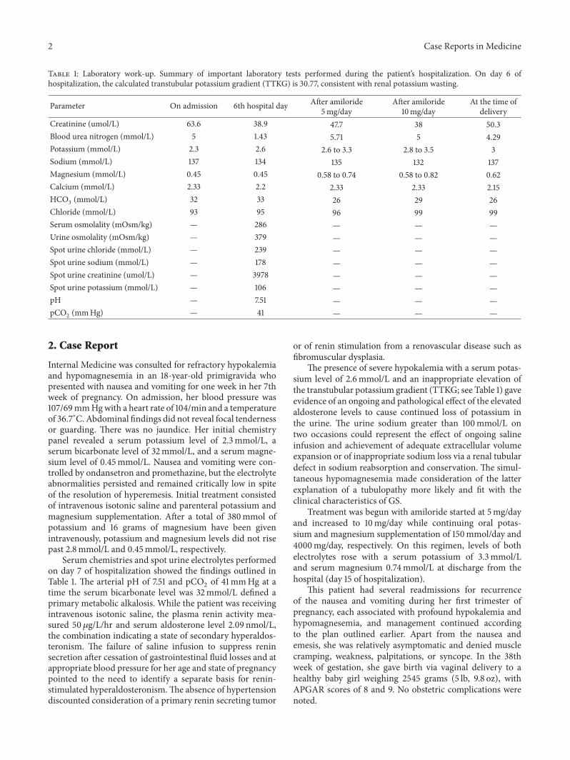
Table 1: Laboratory work-up. Summary of important laboratory tests performed during the patient’s hospitalization. On day 6 ofhospitalization, the calculated transtubular potassium gradient (TTKG) is 30.77, consistent with renal potassium wasting.
Parameter On admission 6th hospital day After amiloride5mg/day
After amiloride10mg/day
At the time ofdelivery
Creatinine (umol/L) 63.6 38.9 47.7 38 50.3Blood urea nitrogen (mmol/L) 5 1.43 5.71 5 4.29Potassium (mmol/L) 2.3 2.6 2.6 to 3.3 2.8 to 3.5 3Sodium (mmol/L) 137 134 135 132 137Magnesium (mmol/L) 0.45 0.45 0.58 to 0.74 0.58 to 0.82 0.62Calcium (mmol/L) 2.33 2.2 2.33 2.33 2.15HCO
3(mmol/L) 32 33 26 29 26
Chloride (mmol/L) 93 95 96 99 99Serum osmolality (mOsm/kg) — 286 — — —Urine osmolality (mOsm/kg) — 379 — — —Spot urine chloride (mmol/L) — 239 — — —Spot urine sodium (mmol/L) — 178 — — —Spot urine creatinine (umol/L) — 3978 — — —Spot urine potassium (mmol/L) — 106 — — —pH — 7.51 — — —pCO2(mmHg) — 41 — — —
2. Case Report
Internal Medicine was consulted for refractory hypokalemiaand hypomagnesemia in an 18-year-old primigravida whopresented with nausea and vomiting for one week in her 7thweek of pregnancy. On admission, her blood pressure was107/69mmHgwith a heart rate of 104/min and a temperatureof 36.7∘C. Abdominal findings did not reveal focal tendernessor guarding. There was no jaundice. Her initial chemistrypanel revealed a serum potassium level of 2.3mmol/L, aserum bicarbonate level of 32mmol/L, and a serum magne-sium level of 0.45mmol/L. Nausea and vomiting were con-trolled by ondansetron and promethazine, but the electrolyteabnormalities persisted and remained critically low in spiteof the resolution of hyperemesis. Initial treatment consistedof intravenous isotonic saline and parenteral potassium andmagnesium supplementation. After a total of 380mmol ofpotassium and 16 grams of magnesium have been givenintravenously, potassium and magnesium levels did not risepast 2.8mmol/L and 0.45mmol/L, respectively.
Serum chemistries and spot urine electrolytes performedon day 7 of hospitalization showed the findings outlined inTable 1. The arterial pH of 7.51 and pCO
2of 41mmHg at a
time the serum bicarbonate level was 32mmol/L defined aprimary metabolic alkalosis. While the patient was receivingintravenous isotonic saline, the plasma renin activity mea-sured 50 𝜇g/L/hr and serum aldosterone level 2.09 nmol/L,the combination indicating a state of secondary hyperaldos-teronism. The failure of saline infusion to suppress reninsecretion after cessation of gastrointestinal fluid losses and atappropriate blood pressure for her age and state of pregnancypointed to the need to identify a separate basis for renin-stimulated hyperaldosteronism.The absence of hypertensiondiscounted consideration of a primary renin secreting tumor
or of renin stimulation from a renovascular disease such asfibromuscular dysplasia.
The presence of severe hypokalemia with a serum potas-sium level of 2.6mmol/L and an inappropriate elevation ofthe transtubular potassium gradient (TTKG; see Table 1) gaveevidence of an ongoing and pathological effect of the elevatedaldosterone levels to cause continued loss of potassium inthe urine. The urine sodium greater than 100mmol/L ontwo occasions could represent the effect of ongoing salineinfusion and achievement of adequate extracellular volumeexpansion or of inappropriate sodium loss via a renal tubulardefect in sodium reabsorption and conservation. The simul-taneous hypomagnesemia made consideration of the latterexplanation of a tubulopathy more likely and fit with theclinical characteristics of GS.
Treatment was begun with amiloride started at 5mg/dayand increased to 10mg/day while continuing oral potas-sium and magnesium supplementation of 150mmol/day and4000mg/day, respectively. On this regimen, levels of bothelectrolytes rose with a serum potassium of 3.3mmol/Land serum magnesium 0.74mmol/L at discharge from thehospital (day 15 of hospitalization).
This patient had several readmissions for recurrenceof the nausea and vomiting during her first trimester ofpregnancy, each associated with profound hypokalemia andhypomagnesemia, and management continued accordingto the plan outlined earlier. Apart from the nausea andemesis, she was relatively asymptomatic and denied musclecramping, weakness, palpitations, or syncope. In the 38thweek of gestation, she gave birth via vaginal delivery to ahealthy baby girl weighing 2545 grams (5 lb, 9.8 oz), withAPGAR scores of 8 and 9. No obstetric complications werenoted.
Case Reports in Medicine 3
3. Discussion
Gitelman’s syndrome (GS) is an inherited hypokalemicsalt-losing tubulopathy with secondary hyperaldosteronismthat affects the thiazide-inhibitable sodium-chloride cotrans-porter (NCCT) of the distal tubule, making its presenta-tion similar to that of treatment with thiazide diuretics.Defects in the NCCT lead to increased delivery of sodiumto the distal tubule, causing increased sodium reabsorptionin exchange for potassium and hydrogen ions, potentiallycausing hypokalemia and metabolic alkalosis. It is importantto differentiate GS from classic Bartter’s syndrome which isalso characterized by hypokalemia, renal potassium wasting,and activation of the renin-angiotensin-aldosterone axis[8]. Unlike Bartter’s syndrome which affects the Na-K-2Cltransporter of the thick ascending limb and is manifest ininfancy, patients with GS develop hypomagnesemia fromincreased urinary magnesium losses and have decreasedurinary calcium excretion. Gitelman’s syndrome typicallyhas a good prognosis and a milder course than Bartter’sand typically presents later in childhood or even in earlyadulthood.
During normal pregnancy, there is renal tubular lossof potassium and magnesium; this urinary potassium wast-ing occurs due to increased aldosterone levels and othersubstances with mineralocorticoid activity. In spite of this,serum potassium levels are kept at physiologic range becauseincreased levels of progesterone resist kaliuresis. In a patientwith GS, this mechanism may not fully compensate for theincreased urinary losses of magnesium and potassium [9].Gestational emesis and fetal demands for these electrolytescan further aggravate the clinical picture, as seen in thispatient.
The required supplemental magnesium and potassiumduring the pregnancy in a patient with GS can be quitesubstantial. McCarthy and colleagues reported that a patientwith GS required a total of 3,680mmol of potassiumand 940mmol of magnesium supplementation given intra-venously during the course of her pregnancy [10]. Theseestimates are reflected in the requirements in this patientof 80–160mmol of potassium and 4 grams (150mMol) ofmagnesium daily needed to make modest improvements inher electrolyte deficits.
It appears from the experience of others that the goal ofmanagement is not to achieve normal levels of potassiumand magnesium; rather, it is to achieve levels at which thepatient is relatively asymptomatic. Basu et al. described apatient with known GS who had three successful normalpregnancies in spite of low potassium and magnesium levels,with the potassium level during pregnancy remaining at 2.8–3.1mmol/L [11]. Likewise, Talaulikar and Falk reported acase in a pregnant patient with GS, where the potassiumand magnesium requirements increased sixfold during thepregnancy and normal levels of both electrolytes were notachieved despite intravenous supplementation [12]. In bothcases, therewas no adverse impact on the course of pregnancyor fetal outcome.
The cornerstone of therapy is oral supplementation ofpotassium and magnesium, and intravenous therapy may be
considered if the patient cannot tolerate oral intake. Kwan andFalk described their experience in treating a pregnant patientwith GS using just oral supplementation (potassium chloride24 g/day and magnesium aspartate 16 g/day) and reportedthat their patient did not suffer from any adverse outcome[13]. Magnesium and potassium levels weremaintained at thelower end of normal range.
The use of mineralocorticoid antagonists should bereserved for highly refractory cases, as its administrationduring pregnancy has been controversial for safety reasons.Although there is limited experience with the use of spirono-lactone or eplerenone in pregnancy, available data suggeststhese drugs are safe [14, 15]. Concern for decreased viril-ization in male infants of mothers who took spironolactoneduring pregnancy has been raised but was not the case in apatient described by deArriba and colleagues [15]. Amiloride,which is a potassium-sparing diuretic that acts as a directantagonist of the epithelial sodium channel (ENaC) in thedistal convoluted tubules, has also been successfully used inpregnancy without report of any adverse outcomes [16].
The most common fetal complication seen in the casereports we reviewed was oligohydramnios, which could berelated to the salt wasting seen in GS and the inability tomaintain extracellular fluid volume [9, 10, 17]. McCarthyand colleagues recommend that in addition to monitoringserum electrolyte levels, determining maternal weight andvolume status is imperative as well [10]. de Haan et al. alsodescribed prolonged labor that could be attributed to lowserum potassium levels [9]. Women with GS have had bothuneventful vaginal and caesarean deliveries and GS does notseem to predispose to the risk of premature delivery. In allreports, the infant did well after delivery.
To our knowledge, this is the first report of GS diagnosedduring pregnancy complicated by hyperemesis gravidarum.The clue to the diagnosis was the difficulty in correctingmagnesium and potassium levels in spite of massive amountsof intravenous and oral supplementation despite resolutionof emesis and restoration of extracellular fluid volume withsaline. While this patient was relatively asymptomatic lead-ing up to her pregnancy, the diagnosis was unmasked byintractable vomiting episodes associated with the hyper-emesis gravidarum. Her presentation highlights the mildercourse of GS compared to Bartter’s and the possibility thatthe diagnosis may become evident only in early adulthood.Further, based on our experience, the use of amiloride duringpregnancy was not associated with any adverse effects on themother or the fetus.
4. Conclusion
GS is a rare inherited condition of the distal convolutedtubule that leads to hypokalemia and metabolic alkalosis. Innormal pregnancy, there is an increased tendency towardskaliuresis, which is countered by excess progesterone. Theoccurrence of increased gastrointestinal losses as seen inthe patient we described may aggravate the hypokalemiaand hypomagnesemia already found in patients with GS.In an undiagnosed patient, the workup should include
4 Case Reports in Medicine
measurement of urine electrolytes, arterial blood gas, andplasma renin and aldosterone levels. The goals of treatmentare to keep the patient normovolemic and asymptomatic,even if normal levels of potassium and magnesium arenot attained. The use of mineralocorticoid antagonists andamiloride has been described and has not been associatedwith adverse effects on the mother and the fetus. There islimited experience reported in literature on GS in pregnancy.Available data suggest that GS is not associated with adverseeffects on the pregnancy outcome.
Competing Interests
The authors certify that they have no conflicts of interest inthe subject matter or materials discussed in this paper.
References
[1] H. J. Gitelman, J. B. Graham, and L. G. Welt, “A new familialdisorder characterized by hypokalemia and hypomagnesemia,”Transactions of the Association of American Physicians, vol. 79,pp. 221–235, 1966.
[2] D. B. Simon, C. Nelson-Williams, M. J. Bia et al., “Gitelman’svariant of Bartter’s syndrome, inherited hypokalaemic alkalosis,is caused by mutations in the thiazide-sensitive Na-Cl cotrans-porter,” Nature Genetics, vol. 12, no. 1, pp. 24–30, 1996.
[3] M.-H. Tseng, S.-S. Yang, Y.-J. Hsu et al., “Genotype, phenotype,and follow-up in Taiwanese patients with salt-losing tubu-lopathy associated with SLC12A3 mutation,” Journal of ClinicalEndocrinology and Metabolism, vol. 97, no. 8, pp. E1478–E1482,2012.
[4] S.-H. Lin, J.-C. Shiang, C.-C. Huang, S.-S. Yang, Y.-J. Hsu,and C.-J. Cheng, “Phenotype and genotype analysis in Chinesepatients with Gitelman’s syndrome,” The Journal of ClinicalEndocrinology&Metabolism, vol. 90, no. 5, pp. 2500–2507, 2005.
[5] U. I. Scholl and R. P. Lifton, “Molecular genetics of Gitelman’sand Bartter’s syndromes and their implications for bloodpressure variation,” Genetic Diseases of the Kidney, vol. 13, pp.229–247, 2009.
[6] E. Riveira-Munoz, Q. Chang, N. Godefroid et al., “Transcrip-tional and functional analyses of SLC12A3mutations: new cluesfor the pathogenesis of gitelman syndrome,” Journal of theAmerican Society of Nephrology, vol. 18, no. 4, pp. 1271–1283,2007.
[7] M. Hod, R. Orvieto, B. Kaplan, S. Friedman, and J. Ovadia,“Hyperemesis gravidarum. A review,”The Journal of Reproduc-tive Medicine, vol. 39, no. 8, pp. 605–612, 1994.
[8] I. Amirlak and K. P. Dawson, “Bartter syndrome: an overview,”Quarterly Journal of Medicine, vol. 93, no. 4, pp. 207–215, 2000.
[9] J. de Haan, T. Geers, and A. Berghout, “Gitelman syndrome inpregnancy,” International Journal of Gynecology and Obstetrics,vol. 103, no. 1, pp. 69–71, 2008.
[10] F. P. McCarthy, C. N. Magee, W. D. Plant, and L. C. Kenny,“Gitelman’s syndrome in pregnancy: case report and review ofthe literature,” Nephrology Dialysis Transplantation, vol. 25, no.4, pp. 1338–1340, 2010.
[11] A. Basu, R. D. S. Dillon, R. Taylor, J. M. Davison, and S.M.Mar-shall, “Is normalisation of serum potassium and magnesiumalways necessary in Gitelman syndrome for a successful obstet-ric outcome?” BJOG: An International Journal of Obstetrics andGynaecology, vol. 111, no. 6, pp. 630–634, 2004.
[12] G. S. Talaulikar and M. C. Falk, “Outcome of pregnancy ina patient with Gitelman syndrome: a case report,” NephronPhysiology, vol. 101, pp. 35–38, 2005.
[13] T. K. Kwan and M. C. Falk, “Second pregnancy outcome in apatient with Gitelman syndrome without the use of parenteralelectrolyte supplementation,”Australian and New Zealand Jour-nal of Obstetrics and Gynaecology, vol. 51, no. 1, pp. 94–95, 2011.
[14] A. Morton, B. Panitz, and A. Bush, “Eplerenone for gitelmansyndrome in pregnancy,”Nephrology, vol. 16, no. 3, pp. 349–350,2011.
[15] G. deArriba,M. Sanchez-Heras, andM.A. Basterrechea, “Gitel-man syndrome during pregnancy: a therapeutic challenge,”Archives of Gynecology and Obstetrics, vol. 280, no. 5, pp. 807–809, 2009.
[16] O. D. Almeida Jr. and J. A. Spinnato, “Maternal Bartter’s syn-drome and pregnancy,” American Journal of Obstetrics andGynecology, vol. 160, no. 5, pp. 1225–1226, 1989.
[17] G. Daskalakis, S. Marinopoulos, A. Mousiolis, S. Mesogitis, N.Papantoniou, andA.Antsaklis, “Gitelman syndrome-associatedsevere hypokalemia and hypomagnesemia: case report andreview of the literature,” Journal of Maternal-Fetal and NeonatalMedicine, vol. 23, no. 11, pp. 1301–1304, 2010.

Submit your manuscripts athttp://www.hindawi.com
Stem CellsInternational
Hindawi Publishing Corporationhttp://www.hindawi.com Volume 2014
Hindawi Publishing Corporationhttp://www.hindawi.com Volume 2014
MEDIATORSINFLAMMATION
of
Hindawi Publishing Corporationhttp://www.hindawi.com Volume 2014
Behavioural Neurology
EndocrinologyInternational Journal of
Hindawi Publishing Corporationhttp://www.hindawi.com Volume 2014
Hindawi Publishing Corporationhttp://www.hindawi.com Volume 2014
Disease Markers
Hindawi Publishing Corporationhttp://www.hindawi.com Volume 2014
BioMed Research International
OncologyJournal of
Hindawi Publishing Corporationhttp://www.hindawi.com Volume 2014
Hindawi Publishing Corporationhttp://www.hindawi.com Volume 2014
Oxidative Medicine and Cellular Longevity
Hindawi Publishing Corporationhttp://www.hindawi.com Volume 2014
PPAR Research
The Scientific World JournalHindawi Publishing Corporation http://www.hindawi.com Volume 2014
Immunology ResearchHindawi Publishing Corporationhttp://www.hindawi.com Volume 2014
Journal of
ObesityJournal of
Hindawi Publishing Corporationhttp://www.hindawi.com Volume 2014
Hindawi Publishing Corporationhttp://www.hindawi.com Volume 2014
Computational and Mathematical Methods in Medicine
OphthalmologyJournal of
Hindawi Publishing Corporationhttp://www.hindawi.com Volume 2014
Diabetes ResearchJournal of
Hindawi Publishing Corporationhttp://www.hindawi.com Volume 2014
Hindawi Publishing Corporationhttp://www.hindawi.com Volume 2014
Research and TreatmentAIDS
Hindawi Publishing Corporationhttp://www.hindawi.com Volume 2014
Gastroenterology Research and Practice
Hindawi Publishing Corporationhttp://www.hindawi.com Volume 2014
Parkinson’s Disease
Evidence-Based Complementary and Alternative Medicine
Volume 2014Hindawi Publishing Corporationhttp://www.hindawi.com
Related Documents