6 TECHNIQUE • MAY 2012 Hill Hastings II MD Clinical Professor Orthopedic Surgery Indiana University, Indiana Hand to Shoulder Center Consultant USA Gymnastics ANATOMY OF THE ELBOW The elbow joint consists of 3 parts that provide for movement between the humerus, ulna, and radius. The end of the hu- merus has two joint surfaces: the trochlea that forms a hinge joint with the ulna, and the capitellum that provides for both flexion/extension and rotation of the radius (Figure 1). The joint surfaces are covered with articular cartilage which is a smooth, almost frictionless surface that is tough enough to provide some shock absorp- tion but also durable. WHAT IS OSTEOCHONDROSIS DESSICANS (OCD)? OCD affects the cartilage surface of the capitellum and the bone support just underneath. Usually there is collapse of the underlying bony support that then leads to a crack or separation in the cartilage surface (Figure 2 – MR arthrogram of unstable OCD lesion). Over time the section of the cartilage surface involved may become detached, float around in the joint, and at times get caught between joint surfaces. In late cases it can damage the opposing joint surface of the radial head. OCD is common in adolescent baseball players and also in female gymnasts. WHAT CAUSES OCD? While the exact cause is not clear, in part, all cases involve repetitive trauma. The capitellum and radius support more than half of the forces across the elbow. While their elbows are still developing, Gymnasts repetitively stress their elbows with extremely high forces required to support their body weight. This affects both the smooth cartilage surface and also the underlying bone. Small fractures can occur in the capitellum leading to collapse and loss of blood supply to the affected bone. When the bone under the cartilage collapses it no longer supports the cartilage properly and predisposes to its fissuring or breakdown. The cartilage on the outside of the capitellum has been found to be softer than that more centrally located, potentially predisposing it to break down. During growth, blood does not cross the growth plate at the end of the humerus so it has to enter in the back and front of the capitellum. This leads to a “watershed” area between blood channels that has less predictable blood supply and perhaps impaired healing capacity. The basic predisposition towards developing OCD relates to the rate of repetitive trauma and damage to the joint and the rate and capacity to repair damage. No genetic predisposition has been found. SYMPTOMS OF OCD Most athletes do not recall a specific injury. The elbow may stiffen over time and lose the ability to fully straighten. Most patients will have soreness after training that improves with rest but recurs again with use. In advanced cases, the elbow may swell or lock up. This occurs when a partially or completely detached fragment gets stuck between joint surfaces. CAPITELLAR OSTEOC (OCD) IN FIGURE 2: MR ARTHROGRAM OF UNSTABLE OCD LESION FIGURE 1

Welcome message from author
This document is posted to help you gain knowledge. Please leave a comment to let me know what you think about it! Share it to your friends and learn new things together.
Transcript
6 T E C H N I Q U E • M A Y 2 0 1 2
Hill Hastings II MDClinical Professor Orthopedic SurgeryIndiana University, Indiana Hand to Shoulder CenterConsultant USA Gymnastics
ANATOMY OF THE ELBOWThe elbow joint consists of 3 parts that provide for movement between the humerus, ulna, and radius. The end of the hu-merus has two joint surfaces: the trochlea that forms a hinge joint with the ulna, and the capitellum that provides for both flexion/extension and rotation of the radius (Figure 1).
The joint surfaces are covered with articular cartilage which is a smooth, almost frictionless surface that is tough enough to provide some shock absorp-tion but also durable.
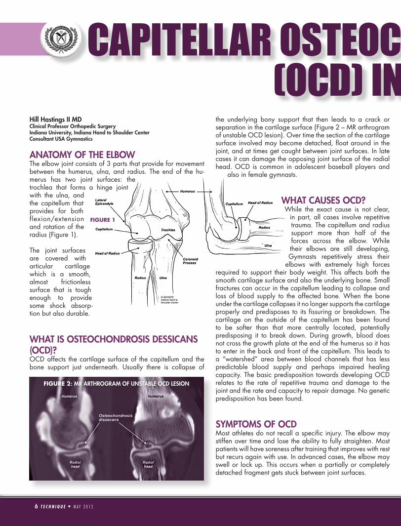
WHAT IS OSTEOCHONDROSIS DESSICANS (OCD)?OCD affects the cartilage surface of the capitellum and the bone support just underneath. Usually there is collapse of
the underlying bony support that then leads to a crack or separation in the cartilage surface (Figure 2 – MR arthrogram of unstable OCD lesion). Over time the section of the cartilage surface involved may become detached, float around in the joint, and at times get caught between joint surfaces. In late cases it can damage the opposing joint surface of the radial head. OCD is common in adolescent baseball players and
also in female gymnasts.
WHAT CAUSES OCD?While the exact cause is not clear,
in part, all cases involve repetitive trauma. The capitellum and radius support more than half of the forces across the elbow. While their elbows are still developing, Gymnasts repetitively stress their
elbows with extremely high forces required to support their body weight. This affects both the smooth cartilage surface and also the underlying bone. Small fractures can occur in the capitellum leading to collapse and loss of blood supply to the affected bone. When the bone under the cartilage collapses it no longer supports the cartilage properly and predisposes to its fissuring or breakdown. The cartilage on the outside of the capitellum has been found to be softer than that more centrally located, potentially predisposing it to break down. During growth, blood does not cross the growth plate at the end of the humerus so it has to enter in the back and front of the capitellum. This leads to a “watershed” area between blood channels that has less predictable blood supply and perhaps impaired healing capacity. The basic predisposition towards developing OCD relates to the rate of repetitive trauma and damage to the joint and the rate and capacity to repair damage. No genetic predisposition has been found.
SYMPTOMS OF OCDMost athletes do not recall a specific injury. The elbow may stiffen over time and lose the ability to fully straighten. Most patients will have soreness after training that improves with rest but recurs again with use. In advanced cases, the elbow may swell or lock up. This occurs when a partially or completely detached fragment gets stuck between joint surfaces.
CAPITELLAR OSTEOCHONDROSIS DISSECANS (OCD) IN GYMNASTS
FIGURE 2: MR ARTHROGRAM OF UNSTABLE OCD LESION
FIGURE 1
HOW IS IT DIAGNOSED?There often is mild tenderness and subtle loss of elbow motion. Stressing the elbow during motion may elicit a grinding or rough sensation. X-rays are essential and usually require special views (Radiocapitellar View). Magnetic resonance imaging (MRI) is the most sensitive test to evaluate the cartilage surfaces and Computed Tomagraphy (CT) the most sensitive for underlying bone fragmentation. At times these studies are performed with dye injected into the elbow joint to help detect cracks in the cartilage surface. (Figure 2) I am currently evaluating injured gymnasts also with a special vascular MRI study (MR angiography) to evaluate the blood supply of the affected area. Since our studies have found OCD to affect both elbows 40% of the time, the opposite elbow, even when asymptomatic, should also be evaluated.
HOW IS IT TREATED?NON-SURGICALAthletes should refrain from axial loading of the extremity, including bars, vault, and floor training. One-handed skills with the opposite arm are permitted when the other side is not affected. Conditioning, balance, and tumbling skills are encouraged. Since the condition is not inflammatory in origin, non-steroidal anti-inflammatory medicines (ie. ibuprofen) are not usually helpful. Therapy is used when there is swelling or loss of motion.
There is some evidence that lesions in young athletes can heal when diagnosed early and protected; yet most studies have reported poor results with nonsurgical management. OCD cartilage lesions in baseball athletes often have some bone attached that can allow for the fragment to be fixed. I have found that lesions in gymnasts do not have significant bone attached. Reattachment is not possible. Furthermore, many lesions initially assessed to be stable by MRI have been found to be unstable when viewed carefully by arthroscopy.
When MRI suggests an intact normal cartilage surface and the underlying bone has not collapsed, non-operative treatment is advised. The arm is rested and MRIs repeated at 3 months and 6 months. If there are no progressive signs of healing, surgery is recommended.
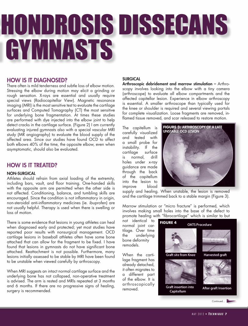
SURGICALArthroscopic debridement and marrow stimulation – Arthro-scopy involves looking into the elbow with a tiny camera (arthroscope) to evaluate all elbow compartments and the affected capitellar lesion. Experience in elbow arthroscopy is essential. A smaller arthroscope than typically used for the knee or shoulder is required and several viewing portals for complete visualization. Loose fragments are removed, in-flamed tissue removed, and scar released to restore motion.
The capitellum is carefully visualized and tested with a small probe for instability. If the cartilage surface is normal, drill holes under x-ray guidance are made through the back of the capitellum into the lesion to improve blood supply and healing. When unstable, the lesion is removed and the cartilage trimmed back to a stable margin (Figure 3).
Marrow stimulation or “micro fracture” is performed, which involves making small holes into the base of the defect to promote healing with “fibrocartilage” which is similar to but not identical to normal joint car-tilage. Over time the underlying bone deformityremodels.
When the carti-lage fragment has already detached, it often migrates to a different part of the elbow. It is arthroscopically removed.
CAPITELLAR OSTEOCHONDROSIS DISSECANS (OCD) IN GYMNASTS
FIGURE 3: ARTHROSCOPY OF A LATE UNSTABLE OCD LESION
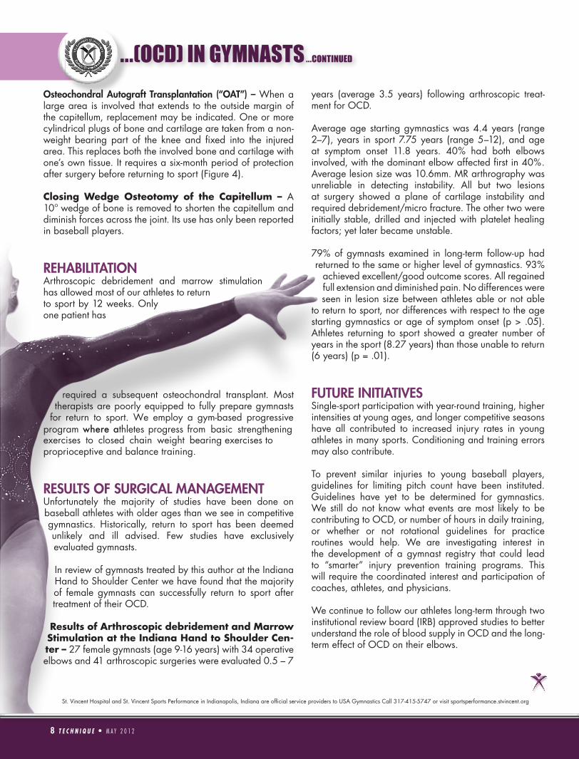
FIGURE 4
M A Y 2 0 1 2 • T EC H N I Q U E 7
Continued...
8 T E C H N I Q U E • M A Y 2 0 1 2
Osteochondral Autograft Transplantation (“OAT”) – When a large area is involved that extends to the outside margin of the capitellum, replacement may be indicated. One or more cylindrical plugs of bone and cartilage are taken from a non-weight bearing part of the knee and fixed into the injured area. This replaces both the involved bone and cartilage with one’s own tissue. It requires a six-month period of protection after surgery before returning to sport (Figure 4).
Closing Wedge Osteotomy of the Capitellum – A 10º wedge of bone is removed to shorten the capitellum and diminish forces across the joint. Its use has only been reported in baseball players.
REHABILITATIONArthroscopic debridement and marrow stimulation has allowed most of our athletes to return to sport by 12 weeks. Only one patient has
required a subsequent osteochondral transplant. Most therapists are poorly equipped to fully prepare gymnasts
for return to sport. We employ a gym-based progressive program where athletes progress from basic strengthening exercises to closed chain weight bearing exercises to proprioceptive and balance training.
RESULTS OF SURGICAL MANAGEMENT Unfortunately the majority of studies have been done on baseball athletes with older ages than we see in competitive gymnastics. Historically, return to sport has been deemed unlikely and ill advised. Few studies have exclusively evaluated gymnasts.
In review of gymnasts treated by this author at the Indiana Hand to Shoulder Center we have found that the majority of female gymnasts can successfully return to sport after treatment of their OCD.
Results of Arthroscopic debridement and Marrow Stimulation at the Indiana Hand to Shoulder Cen-ter – 27 female gymnasts (age 9-16 years) with 34 operative elbows and 41 arthroscopic surgeries were evaluated 0.5 – 7
years (average 3.5 years) following arthroscopic treat-ment for OCD.
Average age starting gymnastics was 4.4 years (range 2–7), years in sport 7.75 years (range 5–12), and age at symptom onset 11.8 years. 40% had both elbows involved, with the dominant elbow affected first in 40%. Average lesion size was 10.6mm. MR arthrography was unreliable in detecting instability. All but two lesions at surgery showed a plane of cartilage instability and required debridement/micro fracture. The other two were initially stable, drilled and injected with platelet healing factors; yet later became unstable.
79% of gymnasts examined in long-term follow-up had returned to the same or higher level of gymnastics. 93%
achieved excellent/good outcome scores. All regained full extension and diminished pain. No differences were seen in lesion size between athletes able or not able
to return to sport, nor differences with respect to the age starting gymnastics or age of symptom onset (p > .05). Athletes returning to sport showed a greater number of years in the sport (8.27 years) than those unable to return (6 years) (p = .01).
FUTURE INITIATIVESSingle-sport participation with year-round training, higher intensities at young ages, and longer competitive seasons have all contributed to increased injury rates in young athletes in many sports. Conditioning and training errors may also contribute.
To prevent similar injuries to young baseball players, guidelines for limiting pitch count have been instituted. Guidelines have yet to be determined for gymnastics. We still do not know what events are most likely to be contributing to OCD, or number of hours in daily training, or whether or not rotational guidelines for practice routines would help. We are investigating interest in the development of a gymnast registry that could lead to “smarter” injury prevention training programs. This will require the coordinated interest and participation of coaches, athletes, and physicians.
We continue to follow our athletes long-term through two institutional review board (IRB) approved studies to better understand the role of blood supply in OCD and the long-term effect of OCD on their elbows.
...(OCD) IN GYMNASTS ...CONTINUED
St. Vincent Hospital and St. Vincent Sports Performance in Indianapolis, Indiana are official service providers to USA Gymnastics Call 317-415-5747 or visit sportsperformance.stvincent.org
8 T E C H N I Q U E • M A Y 2 0 1 2
Related Documents