http://www.lef.org/ Cancer Adjuvant Therapy ■ Preventing and Controlling Cancer ■ Importance of Scheduled Blood Tests ■ Complementary Therapies ■ Other Factors Affecting Patient Outcome ■ Stress and Cancer ■ Summary The good news is that many of the 4 million people being treated for cancer in America will survive the disease and go on to live full and productive lives. While the numbers that survive are far too low (about 44%), many of the more than 1500 daily cancer deaths occur because patients and their families are unaware of the depth of the resources currently available. Unfortunately, some die avowing they would never resort to natural medicine, while others are interested but lack the expertise to implement the program to their best advantage. Regrettably, some turn to alternative care fairly late in the course of the disease process, weakening the probability of recovery. Mainstream medicine (relying upon surgery, chemotherapy, and radiation) may initially appear successful, but the indications of the disease process are less often addressed. Conventional cancer treatments are not for those individuals who are frail in body or spirit. For the past 30 years, cancer therapies have experienced tremendous setbacks because of an associated toxic response, resulting in significant numbers of treatment-induced deaths rather than disease-induced fatalities. Awareness regarding historic numbers of unsuccessful outcomes has forced patients to look for alternatives to bolster survival odds. Many who use alternative therapies report doing so without their oncologist's knowledge, fearful of criticism or rejection by a physician (Richardson et al. 2000). The University of Texas M.D. Anderson Cancer Center (Houston) found that 99.3% of patients had heard of complementary medicine, and 68.7% of patients reported having used at least one unconventional therapy (Richardson et al. 2000). About 75% of the patients surveyed, however, yearned for more information concerning complementary medicine and about one-half of those participating in the survey wanted the information to come from their physician. Until most recently, major medical schools granted only a few hours to nutritional education out of the hundreds of academic hours required to complete medical school. The exclusion began when Abraham Flexner (commissioned to correct inequities occurring in medical schools) penned the Flexner Report of 1910. His contribution, entitled Medical Education in the United States and Canada, closed smaller medical schools and forced those that survived to adopt a uniform curriculum that excluded nutritional courses. Thus, some physicians emerged from medical schools, scoffing at the concept of nutrition influencing health or overcoming disease. Sir William Osler (1849-1919), chief physician at Johns Hopkins's School of Medicine, drilled into students that medical research must be validated and replicated to be good medicine. This led to controlled experiments (as randomized, controlled trials) that became the backbone of mainstream medicine. Nutritional protocols often used multiple nutrients, a difficult model to apply in clinical trials. Testing a single nutraceutical denied the patient full support of nutritional pharmacology, an injustice when treating a seriously ill patient. In addition, trials are expensive to conduct and early natural healers (by and large) did not represent an affluent subset of society. But, ever so slowly, the medical scene is being revolutionized. According to the American College for Advancement in Medicine, physicians (in many cases) are showing eagerness to learn more about natural medicine and how to best implement it into their practice (Corbin-Winslow et al. 2002). Scientists, teaching at nutritional seminars, report attendees are often medical doctors, a vast departure from years past. PREVENTING AND CONTROLLING CANCER While some individuals will be reading this protocol looking for help managing a malignancy, others will be focusing upon prevention and recurrence. The alphabetical list that follows provides quick guidelines for structuring a program, highlighting major nutrients in the prevention and treatment of cancer.

Welcome message from author
This document is posted to help you gain knowledge. Please leave a comment to let me know what you think about it! Share it to your friends and learn new things together.
Transcript
http://www.lef.org/
Cancer Adjuvant Therapy
■ Preventing and Controlling Cancer ■ Importance of Scheduled Blood Tests ■ Complementary Therapies ■ Other Factors Affecting Patient Outcome ■ Stress and Cancer ■ Summary
The good news is that many of the 4 million people being treated for cancer in America will survive the disease and go on to live full and productive lives.
While the numbers that survive are far too low (about 44%), many of the more than 1500 daily cancer deaths occur because patients and their families are unaware of the depth of the resources currently available. Unfortunately, some die avowing they would never resort to natural medicine, while others are interested but lack the expertise to implement the program to their best advantage. Regrettably, some turn to alternative care fairly late in the course of the disease process, weakening the probability of recovery.
Mainstream medicine (relying upon surgery, chemotherapy, and radiation) may initially appear successful, but the indications of the disease process are less often addressed. Conventional cancer treatments are not for those individuals who are frail in body or spirit. For the past 30 years, cancer therapies have experienced tremendous setbacks because of an associated toxic response, resulting in significant numbers of treatment-induced deaths rather than disease-induced fatalities. Awareness regarding historic numbers of unsuccessful outcomes has forced patients to look for alternatives to bolster survival odds. Many who use alternative therapies report doing so without their oncologist's knowledge, fearful of criticism or rejection by a physician (Richardson et al. 2000).
The University of Texas M.D. Anderson Cancer Center (Houston) found that 99.3% of patients had heard of complementary medicine, and 68.7% of patients reported having used at least one unconventional therapy (Richardson et al. 2000). About 75% of the patients surveyed, however, yearned for more information concerning complementary medicine and about one-half of those participating in the survey wanted the information to come from their physician.
Until most recently, major medical schools granted only a few hours to nutritional education out of the hundreds of academic hours required to complete medical school. The exclusion began when Abraham Flexner (commissioned to correct inequities occurring in medical schools) penned the Flexner Report of 1910. His contribution, entitled Medical Education in the United States and Canada, closed smaller medical schools and forced those that survived to adopt a uniform curriculum that excluded nutritional courses. Thus, some physicians emerged from medical schools, scoffing at the concept of nutrition influencing health or overcoming disease.
Sir William Osler (1849-1919), chief physician at Johns Hopkins's School of Medicine, drilled into students that medical research must be validated and replicated to be good medicine. This led to controlled experiments (as randomized, controlled trials) that became the backbone of mainstream medicine. Nutritional protocols often used multiple nutrients, a difficult model to apply in clinical trials. Testing a single nutraceutical denied the patient full support of nutritional pharmacology, an injustice when treating a seriously ill patient. In addition, trials are expensive to conduct and early natural healers (by and large) did not represent an affluent subset of society.
But, ever so slowly, the medical scene is being revolutionized. According to the American College for Advancement in Medicine, physicians (in many cases) are showing eagerness to learn more about natural medicine and how to best implement it into their practice (Corbin-Winslow et al. 2002). Scientists, teaching at nutritional seminars, report attendees are often medical doctors, a vast departure from years past.
PREVENTING AND CONTROLLING CANCER
While some individuals will be reading this protocol looking for help managing a malignancy, others will be focusing upon prevention and recurrence. The alphabetical list that follows provides quick guidelines for structuring a program, highlighting major nutrients in the prevention and treatment of cancer.
These recommendations should not be implemented individually in aggressive cancers without careful consultation of the remainder of the material. Cancer patients (and physicians) should be deliberate about reading the entirety of this protocol in order to avoid missing information that could prove to be lifesaving. Note: It is important that the reader also consult the protocols entitled Cancer Treatment: The Critical Factors and Cancer: Should Patients Take Dietary Supplements?
The dosages required for treating cancer (which are considerably larger than those required for prevention) can change the effects that a nutrient has on the body. The risk is multidirectional. Overdosing or underdosing, as well as a lack of patient awareness regarding the full potential of natural pharmaceuticals, hampers recovery.
THE CRITICAL IMPORTANCE OF SCHEDULED BLOOD TESTS
It is important to measure the successes or losses in regard to treatment-associated tumor response. Evaluating tumor markers in the blood or tumor imagery provides a basis for calculating regression of the disease. In addition, tumor markers provide direction for introducing other therapies if failures are evidenced.
It is also important to evaluate the effectiveness of immune-boosting therapies and guard against anemia and therapeutic toxicities. At a minimum, a monthly complete blood chemistry (CBC) test that includes assessment of hematocrit, hemoglobin, and liver and kidney function should be done in all cancer patients undergoing treatment.
An immune cell test should be performed bimonthly, measuring total blood count, CD4 (T-helper), CD4/CD8 (T-helper-to-T-suppressor) ratio, and NK (natural killer) cell activity. Also consider tests measuring cortisol levels (Cortisol am and pm) and HCG (human chorionic gonadotropin), a hormone that may be elevated 10-12 years prior to a diagnosis of cancer. For information regarding test availability call (800) 208-3444.
COMPLEMENTARY THERAPIES
■ Alpha-Lipoic Acid ■ Arginine ■ Carotenoids ■ Cimetidine ■ Clodronate ■ Coenzyme Q10 and Statin Drugs ■ Conjugated Linoleic Acid ■ Cyclooxygenase-2 Inhibitors ■ Berberine Containing Herbs ■ Feverfew ■ Ginger ■ Green Tea ■ Curcumin ■ Dimethyl Sulfoxide ■ Essential Fatty Acids ■ Garlic ■ Glutamine
Table 1: Type of Cancers and the Tumor Marker Used for AssessmentType of Cancer Tumor Marker Blood TestOvarian cancer CA 125, CK-BBProstate cancer PSA, PAP, prolactin, testosterone
Breast cancer CA 27.29, CEA, alkaline phosphatase, and prolactin (or CA 15-3 rather than the CA 27.29)
Colon, rectum, liver, stomach, and other organ cancers CEA, CA 19-9, AFP, TPS, and GGTP
Pancreatic cancer CA 19.9, CEA, and GGTPLeukemia, lymphoma, and Hodgkin's disease LDH, CBC with differential, immune cell differentiation and leukemia profile
■ Inositol hexaphosphate ■ Lactoferrin ■ Melatonin ■ Modified Citrus Pectin ■ N-acetyl-cysteine ■ Resveratrol ■ Selenium ■ Silibinin ■ Soy ■ Theanine ■ Thymus Extract ■ Vitamin A ■ Vitamin C ■ Vitamin D ■ Vitamin E ■ Vitamin K
When describing the various complementary cancer therapies, it is not possible to endorse one supplement, hormone, or drug over another. We have provided as much evidence as space allows so that patients and their physicians can evaluate what approach may be suited for the individual situation.
A great deal of effort has been made to identify therapies that are substantiated in published scientific literature or that provide a cancer patient with the opportunity to experiment with cutting-edge treatment strategies. The focus of our effort has been to identify potentially lifesaving therapies that have been overlooked by mainstream oncology. We also attempt to discuss both positive and negative studies when applicable.
The Life Extension Foundation can assume no responsibility for outcome, apart from a self-assigned duty to stay abreast of the most promising of therapies and to share the data with members. No warranties (expressed or implied) accompany the material; neither is the information intended to replace medical advice. As always, each reader is urged to consult professional help for medical problems, especially those involving cancer. All supplements, drugs, and hormones are listed alphabetically and not in order of importance.
Alpha-Lipoic Acid--is a powerful antioxidant that regulates gene expression and preserves hearing during cisplatin therapy Lester Packer, Ph.D. (scientist and professor at the Berkeley Laboratory of the University of California), refers to lipoic acid as the most powerful of all the antioxidants; in fact, Packer says that if he were to invent an ideal antioxidant, it would closely resemble lipoic acid (Packer et al. 1999). Alpha-lipoic acid claims anticarcinogenic credits because it independently scavenges free radicals, including the hydroxyl radical (a free radical involved in all stages of the cancer process and linked to an increase in the likelihood of metastasis).
Lipoic acid increases the efficacy of other antioxidants, regenerating vitamins C and E, coenzyme Q10, and glutathione for continued service. In fact, lipoic acid boosts the levels of glutathione by 30-70%, particularly in the lungs, liver, and kidney cells of laboratory animals injected with the antioxidant. In addition, glutathione tempers the synthesis of damaging cytokines and adhesion molecules by influencing the activity of nuclear factor kappa B (NF-kB), a transcription factor (Exner et al. 2000). Note: A great deal of material relating to NF-kB is presented in the protocol Cancer Treatment: The Critical Factors.
Lipoic acid can down-regulate genes that accelerate cancer without inducing toxicity. So responsive are cancer cells that laboratory-induced cancers literally soak up lipoic acid, a saturation that increased the lifespan of rats with aggressive cancer by 25% (Karpov et al. 1977).
Alpha-lipoic acid was preferentially toxic to leukemia cells lines (Jurkat and CCRF-CEM cells). The selective toxicity of lipoic acid to Jurkat cells was credited (in part) to the antioxidant’s ability to induce apoptosis. Lipoic acid activated (by nearly 100%) an enzyme (caspase) that kills leukemia cells (Pack et al. 2002). Other researchers showed that lipoic acid acted as a potentiator, amplifying the anti-leukemic effects of vitamin D. It is speculated that lipoic acid delivers much of its advantage by inhibiting NF-kB and the appearance of damaging cytokines (Sokoloski et al. 1997; Zhang et al. 2001). Finding that lipoic acid can differentiate between normal and leukemic cells charts new courses in treatment strategies to slow or overcome the disease (Packer et al. 1999).
As with all antioxidants, the appropriateness of using lipoic acid with chemotherapy arises. Animal studies indicate that alpha-lipoic acid decreased side effects associated with cyclophosphamide and vincristine (chemotherapeutic agents) but did not
hamper drug effectiveness (Berger et al. 1983). More recently, a combination of alpha-lipoic acid and doxorubicin resulted in a marginally significant increase in survival of leukemic mice (Dovinova et al. 1999). Nonetheless, the definitive answer regarding coupling antioxidants with conventional cancer therapy is complex. Factors, such as type of malignancy, as well as the nature of the cytotoxic chemical and even the time of day the agents are administered, appear to influence outcome (please consult the protocol Cancer: Should Patients Take Dietary Supplements to learn more about the advisability of antioxidant therapy during conventional treatments).
To its credit, lipoic acid appears able to counter the hearing loss and deafness that often accompanies cisplatin therapy. Depreciated hearing occurs as free radicals, produced as a result of treatment, plunder the inner ear; lipoic acid preserves glutathione levels and thus prevents deafness in rats (Rybak et al. 1999).
A suggested lipoic acid dosage for healthy individuals is from 150-300 mg a day. Degenerative diseases usually require larger dosages (sometimes as much as 500 mg 3 times a day).
Arginine Various scientists have attempted to describe the complex role of arginine in cancer biology and treatment. L-arginine is the common substrate for two enzymes, arginase and nitric oxide synthase. Arginase converts L-arginine to L-ornithine, a pathway that can increase cell proliferation. Nitric oxide synthase converts L-arginine to nitric oxide, a conversion process with uncertain effects regarding cancer.
A positive study conducted by a team of German researchers showed that arginine contributed significantly to immune function by increasing levels of white blood cells. Scottish scientists added that dietary supplementation with arginine in breast cancer patients enhanced NK cell activity and lymphokine cytotoxicity (Brittenden et al. 1994). (Lymphokines are chemical factors produced and released by T-lymphocytes that attract macrophages to a site of infection or inflammation in preparation for attack.) Various researchers have shown that increasing arginine increases neutrophils (white blood cells that remove bacteria, cellular debris, and solid particles), significantly upgrading host defense (Muhling et al. 2002).
Apart from enhancing immune function, arginine increases a number of amino acids, creating the possibility of an amino acid imbalance. Oversupplying some amino acids while undersupplying others is thought to destabilize the tumor. All cells, both healthy and diseased, have amino acid requirements; if not met, the cell is significantly disabled (Muhling et al. 2002). Amino acid manipulation has been applied in oncology for decades with varying degrees of success.
Interesting studies have emerged regarding arginine or arginine analogs in cancer treatment. For example, infusions of arginine significantly reduced the incidence of liver and lung metastasis in laboratory mice. Earlier research found that supplemental arginine altered the number of tumor-infiltrating lymphocytes in human colorectal cancer, offering important implications for new strategies in cancer treatment (Heys et al. 1997). Though many factors are involved (including appropriate dosages), Japanese researchers found that arginine induced apoptosis in pancreatic (AR4-2J) cells, inhibiting cell proliferation (Motoo et al. 2000).
The two faces of arginine, however, cloud dosing with confidence. The role of nitric oxide (NO), a molecule synthesized from arginine, remains controversial and poorly understood. While a few reports indicate that the presence of NO in tumor cells or their microenvironment is detrimental to tumor-cell survival, and subsequently their metastatic potential, a large body of data suggests that NO actually promotes tumor progression. Illustrative of its fickleness, NO was recently identified as a downstream regulator of prolactin, an inhibitor of apoptosis. However, arginine stimulated proliferation of prolactin-dependent Nb2 lymphoma cells in laboratory rats (Dodd et al. 2000). In addition, NO production (by murine mammary adenocarcinoma cells) promoted tumorcell invasiveness. Whereas, introducing NO inhibitors resulted in an antitumor, antimetastatic profile (Orucevic et al. 1999).
Ambiguity and nonconformity reduce arginine's role at the present time to adjunctive support with either traditional cancer treatment or fish oil supplementation. A heartening report regarding arginine, fish oil, and doxorubicin therapy appears in this protocol in the section devoted to Essential Fatty Acids (Ogilvie et al. 2000). Nonetheless, the diverse biological properties of L-arginine demand further careful studies, clarifying chemopreventive advantages and endangerments (Szende et al. 2000).
Cancer Adjuvant Therapy
Carotenoids--have antioxidant activity, inhibit cellular proliferation, and offer protection against numerous types of malignancies Carotenoids, acting as immune enhancers and free-radical scavengers, are important substances in oncology. When using carotenoids for antioxidant and cancer protection, it appears wise to use mixed carotenoids, that is, alpha-carotene, lycopene, zeaxanthin, canthaxanthin, beta-crytoxanthine, and lutein rather than emphasizing only beta-carotene.
The following are illustrative of the worth of mixed carotenoids:
■ Lycopene offers targeted protection against cancers arising in the prostate (Kucuk et al. 2001), pancreas (Burney et al. 1989), digestive tract (De Stefani 2000), and colon (Nair et al. 2001).
■ The American Journal of Clinical Nutrition added that individuals seeking broad-spectrum colon protection should also include lutein-rich foods in their diet (spinach, broccoli, lettuce, tomatoes, oranges, carrots, celery, and greens) (Slattery et al. 2000).
■ Canthaxanthin, a less well-known carotenoid, was shown to induce apoptosis and inhibit cell growth in both WiDR colon adenocarcinoma and SK-MEL-2 melanoma cells (Palozza et al. 1998).
■ Researchers showed that the risk of breast cancer approximately doubled (2.21-fold) among subjects with blood levels of beta-carotene in the lowest quartile, compared with those in the highest quartile. The risk of breast cancer associated with low levels of other carotenoids was similar, that is, a 2.08-fold increased risk if lutein is deficient and a 1.68-fold greater risk if beta-cryptoxanthin is lacking (Toniolo et al. 2001). A Swedish study found that menopausal status has an impact on the protection delivered by carotenoids. Analysis showed that lycopene was associated with decreased breast cancer risk in postmenopausal women, but in premenopausal women, lutein offered greater protection (Hulten et al. 2001).
■ Leukoplakia (an often precancerous condition marked by white thickened patches on the mucous membranes of the cheeks, gums, or tongue) is responsive to spirulina, a source of proteins, carotenoids, and other micronutrients (Sankaranarayanan et al. 1995). An inverse relationship between beta-carotene and thyroid carcinoma was observed in both papillary and follicular carcinomas (D'Avanzo et al. 1997). A high dietary intake of beta-carotene appears a protective (though modest) factor for the development of ovarian cancer (Huncharek et al. 2001).
■ Lastly, Japanese researchers showed that all the carotenoids inhibited hepatic (liver) invasion, probably through antioxidant properties (Kozuki et al. 2000).
Men who consume 10 or more servings of tomato products per week reduce their risk of prostate cancer by about 35%. The American Chemical Society in August 2001 reported that 32 (largely African-American) patients diagnosed with prostate cancer and awaiting radical prostatectomy were placed on diets that included tomato sauce, providing 30 mg a day of lycopene. After 3 weeks, mean serum prostate specific antigen (PSA) concentrations fell by 17.5%, oxidative burden by 21.3%, DNA damage by 40%, while programmed cell death increased threefold in cancer cells (Holzman 2002). Part of lycopene's protection involves the ability of carotenoids to counteract the proliferation of cancer cells induced by insulin-like growth factors (Agarwal et al. 2000a).
Beta-carotene exhibited a radio-protective effect among 709 children exposed to radiation inflicted by the Chernobyl nuclear accident. For example, the Chernobyl accident showed that irradiation increases the susceptibility of lipids to oxidative damage and that natural beta-carotene may act as an in vivo lipophilic antioxidant or radio-protective agent (Ben-Amotz et al. 1998). Therefore, using beta-carotene following radiotherapy may reduce the tissue damage caused during treatment.
Beta-carotene, perhaps the most controversial of the family of carotenoids, has come under attack several times in the past few years. For example, smokers who received synthetic beta-carotene (as a prophylactic) in the CARET study had a higher rate of lung cancer and death than smokers not supplemented. In fact, the study was terminated by the National Cancer Institute (NCI) because of the widespread discrepancy between the two groups. The CARET study is not new, but because it still concerns beta-carotene users, we will attempt to explain the unexpected results of the study.
Dr. Packer described the subjects as "walking time bombs." Many were victims of asbestos exposure or heavy smoking. The form of beta-carotene selected for the study (synthetic versus natural) was also cited as another possible explanation for the negative outcome.
Dr. Leo Galland, M.D. (practitioner and director of the Foundation of Integrated Medicine, New York City), also explains that high-dose beta-carotene (25,000 IU a day) administered to smokers results in a particular pattern of metabolism (Galland 2000). The process is orchestrated as cytochrome p450 enzymes (Phase I detoxification system) are summoned into action by tars in cigarette smoke. As beta-carotene is acted on by cytochrome p450, oxidized end products are formed, as well as toxic derivatives.
Simultaneously, vitamins C and A, as well as glutathione, are depleted, severing antioxidant protection. This sequence can damage DNA and increase the likelihood of lung cancer, particularly in an environment with initially high oxidative stress, a profile common to smokers. Without full spectrum antioxidant support, the single dose of beta-carotene produces an oxidative environment rather than one of protection. (Comment: As one free radical is neutralized by an antioxidant, another oxidant may be formed. It is well established that vitamin C can serve as a pro-oxidant through the formation of ascorbyl radicals. It is also known that this radical is quenched by vitamin E to yield a tocopheryl radical, which in turn is reduced by the conversion of glutathione to glutathione disulfide. Thus, the full spectrum of antioxidants is preferable, rather than emphasizing single antioxidants.)
Beta-carotene is largely considered nontoxic even at high doses; for example, some nonconventional cancer therapies recommend large amounts of carrot juice. One large glass of carrot juice can contain 100,000-200,000 IU of provitamin A or carotene. The problem with carrot juice is that it is loaded with fructose (sugar). Cancer cells feed on sugar, and drinking carrot juice may induce an insulin spike that could potentially fuel cancer cell propagation.
Cancer patients should consider natural beta-carotene supplements in lieu of carrot juice. Suggested phytonutrient dosages are from 9-20 mg of sulphoraphane, 10-30 mg a day of lycopene, and 15-40 mg of lutein, along with a mixed carotenoid blend that includes alpha- and beta-carotene. A product called PhytoFood Powder provides potent amounts of sulphoraphane, while carotenoid extracts are available in a variety of encapsulated preparations. Note: What Should the Cancer Patient Eat, appearing later in this protocol, contains a discussion regarding the value of sulphoraphanes in the diet.
Cimetidine (Tagamet) Histamine (H2) receptor antagonists (such as cimetidine) became popular in the late 1970s to treat gastrointestinal ulcers and other benign conditions of the stomach, esophagus, and duodenum. In 1985, the Life Extension Foundation announced that cimetidine had merit as a cancer adjunct. Since then, many studies have been published encouraging the use of cimetidine as a means of disabling tumors and expanding survival rates (Tonnesen et al.1988; Yoshimatsuk et al. 2003).
Ways through which cimetidine impacts cancer involves a three-pronged mechanism including (1) inhibition of cancer cell proliferation, (2) stimulation of lymphocyte activity by inhibition of T-cell suppressor function, and (3) inhibition of histamine's activity as a growth factor (Siegers et al. 1999).
In a Japanese study, a total of 64 colorectal cancer patients (who had earlier undergone surgery) were evaluated for the effects of cimetidine on survival and disease recurrence. The cimetidine arm of the study received 800 mg a day of cimetidine along with 200 mg a day of the chemotherapy drug 5-fluorouracil (5-FU); the control group received only 5-FU. The treatment was initiated 2 weeks following surgery and terminated 1 year later. Strikingly beneficial effects were noted: The 10-year survival rate for patients treated with cimetidine/5-FU was 84.6%, whereas that of the control group (5-FU alone) was only 49.8% (Matsumoto et al. 2002).
The effect of cimetidine on a particularly aggressive form of colon cancer (Dukes grade C) was investigated. The cumulative 10-year survival rate of the cimetidine-treated group was consistently 84.6%, whereas that of the control group was only 23.1%. (Less virulent cancers (Dukes A or B) responded less well to cimetidine treatment) (Matsumoto et al. 2002).
Cimetidine treatment is particularly effective in patients whose tumors express higher levels of Lewis A and Lewis X antigens (i.e., breast and pancreatic cancers, as well as about 70% of colon cancers). Lewis A and Lewis X antigens are cell surface ligands that adhere to a molecule in the blood vessels called E-selectin. (Ligand comes from the Latin word ligare, meaning that which binds.)
The adhesion of the cancer cell to vascular endothelial cells expressing E-selectin is a key step in invasion and metastasis. Cimetidine improved patient outcome presumably by inhibiting the expression of E-selectin, thus abolishing the binding site for continued cancer growth and metastasis. The 10-year cumulative survival rate of the cimetidine group displaying Lewis antigens was 95.5%, whereas the control group was only 35.1% (Matsumoto et al. 2002). Comment: Patients are well-advised to undergo Lewis antigen determinations for optimal therapy and a more favorable outcome. Contact Impath Laboratories at 521 West 57 Street, New York, NY 10019, Telephone: (800) 447-8881, for information regarding testing.
Researchers recently unearthed another mechanism through which cimetidine offers cancer protection. Cimetidine enhanced cell-mediated immunity by improving suppressed dendritic cell function (Kubota et al. 2002). Dendritic cells capture foreign invaders and carry the antigen to lymph nodes and spleen. The "hand-delivered" antigen shows the immune system exactly what it has to fight. A more in-depth explanation regarding dendritic cells appears in a separate protocol entitled Cancer Vaccines.
The growth inhibitory effects of cimetidine were assessed on five cell lines derived from human brain tumors of different tissue types and grades of malignancy. Each cell line was treated with cimetidine 24 hours before analysis. Cimetidine significantly inhibited cell proliferation in three of five cell lines, which indicates the apparent dependence of these cells on histamine stimulation (Finn et al. 1996).
Because we do not wish the reader to interpret positive material as a universal ameliorant for all cancers, the following findings are noted:
■ Fred Hutchinson Cancer Research Center researchers explored whether cimetidine exerted a cancer-preventive effect on prostate and breast cancers by tracking 48,512 individuals from 1977-1995. Unfortunately, the study concluded that cimetidine did not influence the risk of female breast cancers; in addition, the researchers concluded that there was little evidence to support the previously hypothesized preventive effect of cimetidine on the risk of prostate cancers (Rossing et al. 2000).
■ In multiple myeloma patients, cimetidine reduced by about 30% the bioavailability of melphalan (Alkeran), the standard treatment for the disease (Sviland et al. 1987).
■ A total of 132 male rats were evaluated for immune status after ingesting cimetidine to forestall a diagnosis of gastric cancer. In the cimetidine-fed group, 19 of 48 developed cancer, versus 12 of 43 in the control group. The Norwegian researchers concluded that cimetidine had no significant immune-modulating effects on the development of gastric cancer in rodents (Hortemo et al. 1999).
While cimetidine is not efficacious in cancer prevention, it shows efficacy in treating certain cancers. A suggested cimetidine dosage for cancer patients is 800 mg (taken at night). Do not supplement with cimetidine without physician awareness; the drug can interact with several medications (such as digoxin, theophylline, phenytoin, warfarin, and lidocaine), increasing or decreasing drug potency.
Clodronate--is a bisphosphonate that inhibits cell proliferation and the threat of metastasis Clodronate reduced the incidence and number of metastasis in bone and viscera (organs enclosed in the abdominal, thoracic, or pelvic cavity) in high-risk breast cancer patients by 50% (Diel et al. 1998; also see Journal Club on the Web).
Between 1990 and 1995, 302 patients (median age 51 years) with primary breast cancer and tumor cells in the bone marrow (the presence of which is a risk factor for the development of distant metastasis) were randomly assigned to receive 1600 mg a day of oral clodronate for 2 years or standard follow-up without clodronate supplementation (Diel et al. 1998).
At the conclusion of the trial, bone metastases were detected in 12 (8%) of the clodronate group versus 25 (17%) of the control group. The mean number of bony metastases per patient was 3.1 in the clodronate group versus 6.3 in the nontreated group. Visceral metastasis was observed in 13 (8%) versus 27 (19%) of controls; 6 patients (4%) died in the clodronate group, compared to 22 (15%) in the untreated group. Researchers concluded that clodronate opposed metastasis by altering the binding capacities of adhesion molecules on tumors and bone cells. Women with existing metastatic breast cancer (who added bisphosphonates to their regimen) reported less bone pain and fewer fractures with treatment.
The bisphosphonates (particularly zoledronic acid) appear to be effective against the skeletal complications of multiple myeloma, reducing vertebral fractures and pain. In the early phase of metastasis to bone, tumor cells activate osteoclasts, cells that break down and resorb bony tissue. This favors tumor growth, as growth factors are released when bone is degraded. Bisphosphonates inhibit the development of monocytes into osteoclasts (cells that digest and remove bone) and promote osteoclast death.
In addition, bisphosphonates restrain the production of bone-resorbing cytokines such as interleukin-6, an inflammatory marker for myeloma prognosis. Lastly, bisphosphonates directly affect myeloma by inducing apoptosis of malignant plasma cells. The biochemical effects of zoledronic acid continued for as long as 8 weeks after a single administration (Berenson 2001), but myeloma mortality was not decreased by bisphosphonates (Djulbegovic et al. 2001; Fromique et al. 2000). Typically, a synergism (a cooperative effort) exists between bisphosphonates and cytotoxic agents, increasing chemotherapy's effectiveness.
The standard dose for treating cancer is 800 mg of clodronate taken twice daily, although double this dosage has been used safely. Breast cancer patients may consider a 3- to 5-year regimen of clodronate or other bisphosphonate therapy. Blood tests to measure serum calcium levels and kidney function are required 10 days after beginning clodronate and every 1-2 months thereafter. Persons who are pregnant or who have severe renal insufficiency requiring dialysis should avoid clodronate.
Note: Newer bisphosphonate drugs such as Zometa, Actonel, Fosamax, and Aredia, more potent than clodronate, are now FDA approved and readily available in the United States and covered by most health insurance plans. Prophylactic bisphosphonate therapy is highly recommended for cancers with a propensity to metastasize to bone, such as prostate and breast cancers. Most cancer patients should be on bisphosphonate therapy since any amount of bone breakdown releases growth factors that fuel cancer cell growth. Refer to Cancer Treatment: The Critical Factors for more information about bisphosphonate drugs approved in the United States.
Coenzyme Q10 and Statin Drugs Statins, a class of cholesterol-lowering drugs, have been shown to inhibit the activity of ras oncogenes. ras oncogenes are
involved in the regulation of cell growth, modulating the signals that govern the cancer cell cycle. Mutations in genes encoding Ras proteins have been closely associated with unregulated cell proliferation, a hallmark of cancer (refer to the protocol Cancer Treatment: The Critical Factors to read more about Ras oncogenes).
A number of studies have shown the value of statin drugs in a cancer regimen, and the benefit escalates when a statin is combined with a nonsteroidal anti-inflammatory drug (NSAID). People who regularly used NSAIDs lowered their risk of colon cancer by as much as 50%; when lovastatin was added to a cyclo-oxygenase 2 (COX-2) inhibitor, the rate of cell death of three colon cancer cell lines increased up to five-fold (Agarwal et al. 1999).
The statin’s mode of operation, however, raises concern. Statin drugs reduce cholesterol synthesis in the liver by inhibiting the activity of 3-hydroxy-3-methylglutaryl coenzyme A (HMG-CoA) reductase. HMG-CoA reductase is required for the conversion of HMG-CoA to mevalonic acid, a step in cholesterol synthesis (Folkers et al. 1990). Inhibiting HMG-CoA reductase results in lower amounts of cholesterol being produced. Disruption of the cascade also interferes with the synthesis of coenzyme Q10 (CoQ10), creating a potential tradeoff regarding advantages and disadvantages gathered from statin usage (Folkers et al 1990; Hattersley 1994).
The impact upon CoQ10 levels when taking statin drugs can be significant. For example, patients taking CoQ10, who later started lovastatin, lowered their CoQ10 levels by 44-75%. The problems associated with drug-related suppression of CoQ10 escalate when age-associated decline in serum CoQ10 levels are also present. A CoQ10 deficiency of 25% is linked with illness in animals and a deficit of 75% with death (Hattersley 1996; Bliznakov et al. 1988). Administering adequate amounts of CoQ10 with a statin drug allows the cancer patient the value of the drug without the risks imposed by depletion of the coenzyme.
Cancer Adjuvant Therapy
In 1997 the Life Extension Foundation suggested that cancer patients ask their oncologist to consider lovastatin (80 mg a day) as adjunct therapy. The recommendation was based on scientific studies indicating lovastatin interfered with the cancer cell cycle and appeared to encourage cell death (apoptosis) (Dimitroulakos et al. 2001). Lovastatin, sold under the name Mevacor, is a fat-soluble statin drug, as are Zocor and Lipitor. Water-soluble statin drugs such as Pravachol may not work as effectively against cancer as the fat-soluble varieties, although one study showed Pravachol induced significant benefits to a group of primary liver cancer patients (Wang et al. 2000).
One of the concerns associated with low levels of CoQ10 is an increased risk of developing cancer. CoQ10 has been reported to be effective in inhibiting the progression of cancers and metastasis, even in patients for whom all conventional treatment failed (Folkers et al. 1993; Lockwood et al. 1995). CoQ10, acting as a nonspecific stimulant to the immune system, increases blood levels of T-lymphocytes and improves the T4-T8 lymphocyte ratio (Folkers et al. 1991). Contrast this with the energy loss and immune suppression associated with conventional cancer therapies.
Dr. Karl Folkers, a pioneer in CoQ10 exploration, reported that in a study of blood levels of CoQ10 in 116 breast cancer patients, 23.1% had blood levels of CoQ10 below 0.5 mcg/mL. The incidence of breast cancer cases with levels below 0.6 mcg/mL was 38.5%, higher percentages than observed in healthy women. A subsequent study reported in the Journal of Clinical Pharmacology and Therapeutics showed a statistically significant relationship between the level of CoQ10 deficiency and breast cancer prognosis (Folkers et al. 1997; Joliet et al. 1998).
Molecular Aspects of Medicine reported the results of an 18-month study conducted in Denmark involving 32 breast cancer patients (Lockwood et al. 1994). The patients had complicated medical profiles, that is, some had involvement in axillary lymph nodes and others had distant metastasis. The patients all received antioxidant therapy, consisting of vitamins C, E, and beta-carotene, select minerals and trace minerals, along with essential fatty acids, and 90 mg of CoQ10 a day. Their treatment was an integrated approach that also included surgery, radiation therapy, and chemotherapy. The survival rate during the 18-month study was 100%; a follow-up evaluation at the 24-month interval indicated all participants were still alive, although the expected deaths were four at 18 months and six at 24 months. All 32 of the enrollees in the study reported improvement in quality of life, stabilization of weight, a withdrawal from pain medications, and no signs of further distant metastases; six of the 32 patients showed apparent partial remissions.
Patients (n = 15) with myeloma showed a mean CoQ10 blood level of 0.67 ± 0.17 mcg/mL. The incidence of a CoQ10 blood level below 0.7 mcg/dL was 53.3%, which is higher than the 24.5% found among a group of nonmyeloma patients (Folkers et al. 1997). Individuals with bloodborne tumors are often saddened with the scarcity of nutritional material relevant to their type of cancer. When links are found, patients and physicians should take special note. The full clinical implication of this finding remains to be explored.
Patients, with and without cancer, report a decrease in the incidence of infection while taking CoQ10 (Bliznakov et al. 1970). This is particularly important to the cancer patient, who often faces additional challenges because of a suppressed immune system. Another extremely important characteristic of CoQ10 is its antioxidant potential, stabilizing cell membranes and preserving cellular integrity (Ernster et al. 1993).
One of the most potent chemicals used in cancer chemotherapy treatment is Adriamycin (doxorubicin). A significant consequence of this drug is cardiac damage, especially in older patients with established heart disease. Italian researcher Dr. Mario Ghione discovered a depletion of CoQ10 in the diseased hearts of animals after long-term Adriamycin administration. When CoQ10 was given to a group of mice before Adriamycin therapy, 80-86% survived; a control group (receiving Adriamycin but without CoQ10) had only a 36-42% survival rate (Bertazzaoli et al. 1977; Cortes et al. 1978).
Dosage suggestions are 90-390 mg a day of CoQ10, taken with some fat to enhance absorption. The American Journal of Health-System Pharmacy reported that liver enzymes could become elevated when taking 300 mg of CoQ10 a day for extended periods of time (Pepping 1999). Also, Folia Microbiologica reported that mice injected with human small cell lung cancer cells and then given high doses of CoQ10 had a diminished response to radiation therapy compared to the non-supplemented group (Lund et al. 1998). Note: Refer to the Cancer Chemotherapy and/or Cancer Radiation protocols along with Cancer: Should Patients Take Dietary Supplements to read about the appropriateness of supplementing with CoQ10 during chemotherapy or radiation therapy.Food sources of CoQ10 include mackerel, salmon, and sardines along with beef, peanuts, and spinach.
Conjugated Linoleic Acid (CLA)--is a trace fatty acid that inhibits tumor formation and metastasis, suppresses arachidonic acid, and encourages apoptosis Researchers at the Roswell Park Cancer Institute (Buffalo, NY) showed that CLA, derived mainly from dairy products, reduced the incidence of breast cancer (Ip et al. 1999). Animal experiments showed that only 50% of rats feeding on CLA butter developed
mammary tumors when exposed to high doses of known carcinogens, compared to 93% of the rats deprived CLA. This research demonstrated for the first time that CLA in foods is biologically active and that a food can offer significant protection against cancer (Cornell News 1999).
Anticancer Research published supporting data that CLA (in both test tube and animal models) demonstrates strong antitumor activity. Particularly gratifying effects were observed regarding inhibition of growth and metastatic spread of transplantable mammary tumors in severely immune deficient mice. The mice were fed CLA for 2 weeks prior to inoculation with human breast adenocarcinoma cells (107 MDA-MB468) and throughout the trial. CLA completely abolished the spread of breast cancer cells to the lungs, blood, and bone marrow. These results indicate that CLA blocks the local growth and spread of human breast cancer via mechanisms independent of the immune system (Visonneau et al. 1997; Banni et al. 1999; Ipet al. 1999).
The effects of CLA and beta-carotene were assessed on white blood cell (lymphocyte) and macrophage function. CLA alone increased lymphocyte numbers and their cell killing ability. Conversely, CLA inhibited interleukin-2 production (a desirable cytokine) and suppressed the ability of macrophages to destroy foreign material. When given together, CLA and beta-carotene interacted in an additive manner to increase lymphocyte production and their cytotoxicity. In addition, beta-carotene was able to overcome the inhibitory action of CLA on the phagocytic activity of macrophages (Chew et al. 1997).
Note: The Melanoma Center at the University of Pittsburgh Cancer Institute showed a potential role for histamine in cancer immunotherapy. A Phase II trial of IL-2 versus IL-2 and histamine in patients with metastatic melanoma demonstrated a trend toward a superior survival benefit from IL-2 and histamine for all patients enrolled and a statistically significant survival benefit for patients with hepatic metastasis (Agarwala et al. 2001).
The effect of three different diets on the local growth and metastatic potential of human prostatic carcinoma cells (DU-145) in severely immune-deficient mice was studied. Animals were fed either a standard diet or diets supplemented with 1% linoleic acid (LA) or 1% CLA for 2 weeks prior to inoculation with cancer cells and throughout the 14-week study. Mice receiving the LA-supplemented diet displayed significantly higher body weight, lower food intake, and increased local tumor load as compared to the other two groups of mice. Mice fed the CLA-supplemented diet exhibited not only smaller local tumors, but also a significant reduction in lung metastasis (Cesano et al. 1998). It was estimated that CLA inhibited the formation of premalignant lesions by approximately 50%, while increasing apoptosis in diseased cells (Ip et al. 2000).
CLA, in a dose-related fashion, has an ability to suppress arachidonic acid (AA). Since AA produces inflammatory mediators that can promote cancer at initiation and progression, CLA's ability to stifle AA elevates its status as a chemopreventive (Miller et al. 2001; Urquhart et al. 2002).
In 1996, the Life Extension Foundation was in the forefront, recommendingCLA; after evaluating the results of numerous studies, the Foundation presented the promising anticarcinogenic nature of CLA to members. Relatively small doses (3-4 grams of CLA) are effective. For example, young female rats (still maturing) fed 0.8% of their diet from CLA achieved long-term protection against breast cancer. The dose of 0.8% correlates positively to the recommended daily dosage of 3-4 grams endorsed by the Foundation. A dose of six 1000-mg CLA capsules (76%) each day is suggested for cancer patients, pregnant and lactating women should avoid CLA.
Cyclooxygenase-2 (COX-2) Inhibitors (Naturally Occurring) Note: The following compendium drawn (in part) from Beyond Aspirin (Newmark et al. 2000) underscores herbs that inhibit COX-2, an enzyme intricately involved in the cancer process. Natural compounds usually have many mechanisms of action; thus, the protective mechanisms common to the herb often extend beyond enzyme inhibition and are described herein. Because of the synergism of herbs, combinations are often of greater value than a single herb. The COX-2-cancer connection is thoroughly discussed in the protocol Cancer Treatment: The Critical Factors.
Berberine--Containing Herbs (Goldenseal, Barberry, Goldthread, and Oregon Grape) Berberine, strong and bitter in taste and found in various herbs, delivers anti-inflammatory properties via COX-2 inhibition (Fukuda et al. 1999). Kaempferol, a constituent of berberine, is a strikingly active inhibitor of COX-2 activity (Chen et al. 1999; Newmark et al. 2000). Berberine is unique, having the ability to inhibit COX-2 activity without involving the beneficial COX-1 enzyme. Berberine, perhaps by impacting the production of cyclooxygenase, influences the development of cancers at various sites:
■ Berberine is effective against bladder cancers (Chung et al. 1999). ■ Berberine suppressed colon carcinogenesis and inhibited COX-2 without COX-1 inhibition. The COX-2 enzyme is
abundantly expressed in colon cancer cells and plays a role in tumorigenesis. The berberine-COX-2 connection appears to best explain the mechanism of berberine's anti-inflammatory and antitumor-promoting effects (Fukuda et al. 1999, Newmark et al. 2000).
■ Berberine-induced apoptosis in human leukemia cells (Kuo et al. 1995).
■ Berberine inhibited the development of skin tumors (Kitagawa et al. 1986). ■ Berberine has potent antitumor activity against human and rat malignant brain tumors (Zhang et al. 1990). Studies using
goldenseal, which contains the alkaloid berberine, showed average cancer kill rate of 91% in rats, over twice that seen in BCNU (a standard chemotherapy agent for brain tumors). Rat studies used 10 mg/kg of berberine.
A suggested dose is three 250-mg capsules of goldenseal each day. The preparation should be standardized to provide 5% hydrastine. Various respected herbalists suggest that goldenseal should be cycled (rotated with other herbals) rather than routinely administered. Goldenseal contains the alkaloids berberine, hydrastine, and canadine.
Feverfew (Tanacetum parthenium) The anti-inflammatory traits of Feverfew have an ability to inhibit the COX-2 enzyme (Hwang et al. 1996). According to Newmark et al. (2000), feverfew contains a lactone, or chemical compound called parthenolide. Parthenolide, in turn, contains a variant of methylene-gamma-lactone (MGL) that interacts with macrophages. The white blood cell-lactone interaction suppresses a critical protein process, a repression that ultimately inhibits the COX-2 enzyme. In addition, feverfew contains apigenin (a flavonoid) and melatonin, both COX-2 inhibitors (Murch et al. 1997).
Researchers at Children's Hospital Medical Center (Cincinnati, Ohio) explained another of parthenolide's anti-inflammatory traits: its ability to inhibit NF-kB, the predecessor of a number of potentially damaging cytokines (Sheehan et al. 2002). Recall that as inflammation is reduced the risks of many degenerative diseases decrease as well (turn to the protocol entitled Cancer Treatment: The Critical Factors to read about the cytokine/cancer connection).
In addition, feverfew inhibits 5-lipoxygenase, an enzyme that metabolizes AA. A byproduct of this metabolism (hydroxy-eicosatetraenoic acid or HETE) feeds cancer cells and promotes angiogenesis, the development of new blood vessels. Agents that inhibit the production of lipoxygenase should be of particular interest to individuals taking COX-2 inhibitors; as the COX-2 enzyme is inhibited, 5-lipoxygenase enzymes become activated (Pizzorno 2001).
A suggested dosage is 1-2 capsules of feverfew a day, standardized to contain 600 mcg of parthenolide. Pregnant and lactating women should avoid feverfew, as well as those showing allergic sensitivities.
Ginger (Zingiber officinalis) From the scores of biologically active components contained in ginger, some are specific for inhibiting COX-2 and others for inhibiting 5-lipoxygenase, enzymes responsible for the formation of pro-inflammatory agents (prostaglandin E2 and leukotriene B4) from AA. Ginger safely modulates COX-2 activity but also brings balance to COX-1 (an enzyme responsible for gastric mucosal integrity) in a manner vastly superior to synthetic NSAIDs (Newmark et al. 2000; Reiter et al. 2001).
As COX-2 and 5-lipoxygenase are repressed, two distinct metabolic pathways are inhibited, one leading to the synthesis of prostaglandins and the other leading to the production of HETEs. Prostaglandin E2 (PGE2) (produced from COX-2-arachidonic acid interactions) promotes cellular proliferation, and 5-HETE is considered indispensable fuel for tumor growth (prostate in particular).
It has been speculated that therapeutic dosages of ginger inhibit PGE2 by up to 56%. As ginger slows down 5-lipoxygenase and 5-HETE production, cell death is stimulated in both hormone responsive and nonresponsive human prostate cancer cells (Suekawa et al. 1986; Ghosh et al. 1998). Leukotrienes, produced by lipoxygenase, are considered 1000 times more reactive than histamine. Ginger has more 5-lipoxygenase inhibitors than any other botanical source (Newmark et al. 2000).
Ginger may also be useful in overcoming nausea that accompanies chemotherapy and toxicity associated with the breakdown products of cancerous tissue. James Duke, Ph.D., distinguished botanist and author, has high regard for ginger, adding that it has a major advantage over other antiemetics because of its safety profile. Ginger's antioxidant activity adds another plus to a booming list of anticancer credits. A suggested dosage is 2 grams of ginger a day.
Green Tea Salicylic acid, the main anti-inflammatory component of aspirin, is a naturally occurring compound found in green tea, having COX-2 inhibiting qualities. The polyphenols and flavonoids contained in green tea are also COX-2 inhibitors (Noreen et al. 1998).
Mayo Clinic researchers showed that green tea consumption inhibited cancer growth (Paschka et al. 1998). They identified the green tea polyphenol EGCG (epigallocatechin gallate) as the most potent inhibitor of cancer cell proliferation. Japanese researchers pinpointed the types of cancer most responsive to green tea (breast, esophageal, liver, lung, skin, and stomach) by surveying cancer-free individuals who consumed 4-6 cups of green tea a day.
The odds ratio of stomach cancer decreased to 0.69 with a high intake of green tea (7 cups or more a day) (Inoue et al. 1998). Another study conducted in Yangzhong (a region in China with a high incidence of chronic gastritis and gastric cancer) showed the amount and duration of green tea consumption governed the rate of stomach cancer. Frequent long-term green tea drinkers had approximately 50% less risk of developing gastric cancer compared to individuals consuming little or no tea (Setiawan et al. 2001). Green tea reduces the damaging effects of nitrites in the acidic environment of the stomach with greater efficiency than vitamin C.
The growth of non-Hodgkin's lymphoma cells, transplanted in mice, was reduced by 50% when green tea was a part of the animal's diet. Cyclophosphamide, a chemotherapeutic drug, administered at the maximum tolerable dose, was unable to replicate similar benefits (Bertolini et al. 2000). Part of green tea's anticancer profile includes an antimutagenic factor that helps DNA replicate accurately (Uhlenbruck et al. 1998).
PGE2 is thought to stimulate tumor promotion in precancerous and cancerous cells (August et al. 1999; Bertolini et al. 2000). Of 14 subjects, 10 (71%) demonstrated a response to green tea, as evidenced by at least a 50% inhibition of PGE2 in rectal mucosa.
EGCG appears to normalize the cell growth cycle and prompt apoptosis in cancer cells by inhibiting NF-kB, a growth vehicle cancer cells use to escape cell regulatory control (Ahmad et al. 2000). EGCG strongly and directly inhibits telomerase, an enzyme (normally dormant from birth) that delivers immortal status to cancer cells (Naasani et al. 1998).
Cigarette smokers who drink green tea have a 45% lower risk of lung cancer compared to non-tea drinkers. Even though Japan has one of the highest numbers of smokers in the world, they have one of the lowest rates of lung cancer of any developed nation, a protection thought to be delivered by green tea.
The number of anticarcinogens, antioxidants, and anti-proliferative agents found in green tea (carotenoids, chlorophyll, polysaccharides, vitamins C and E, and numerous flavonoids) explains why some researchers advocate using a broad-spectrum extract, replicating the plant's total constituents. Considering the vastness of green tea’s anti-cancer effects, incorporating green tea into the diet 5-10 cups a day (or five 350-mg capsules three times a day of a 95% polyphenol extract) would appear to be wise for individuals concerned with cancer.
Cancer Adjuvant Therapy
Curcumin Worldwide clinical trials have chiseled out a definite place for curcumin in oncology. Among them are New York Presbyterian Hospital and the Weill Medical College, which reported that curcumin, a curcuminoid found in turmeric, directly inhibited the COX-2 enzyme (Zhang et al. 1999). So excited are various oncologists regarding curcumin that the potent anti-inflammatory has been classed as a potential third generation cancer chemopreventive agent.
Curcumin inhibited thromboxane A2 (TxA2), a highly unstable, biologically active compound created by COX from AA (Shah et al. 1999; Newmark et al. 2000). Unless controlled, TxA2 promotes tumor endothelial cell migration (metastasis) and angiogenesis. By inhibiting TxA2, curcumin reduces the tumor's blood supply and lessens the threat of metastasis (Arbiser et al. 1998; Nie et al. 2000). Curcumin is effective at inhibiting 5-lipoxygenase and subsequently HETE, a survival factor for prostate, breast, and pancreatic cancers (Ghosh et al. 1998; Ding et al. 1999; Newmark et al. 2000; Li et al. 2001).
The following list illustrates the depth of curcumin's defenses against cancer:
■ Colon: Curcumin inhibited chemically induced carcinogenesis in the colon when administered at different stages of the cancer process. Laboratory rats, administered curcumin during either initiation or late in the premalignant phase, had a lesser incidence and fewer numbers of invasive malignant colon tumors (Kawamori et al. 1999). Also, by inhibiting COX-2-arachidonic acid interactions, curcumin suppresses prostaglandins responsible for inflammatory processes (Plummer et al. 1999). Chronic inflammation has for decades been regarded as a cause of colon cancer (Konig et al. 1976).
■ Antioxidant activity: Curcumin inhibits or possibly even reverses oxidative damage by scavenging and neutralizing free radicals. By defusing the hydroxyl and superoxide radicals and breaking oxidative chain reactions, curcumin protects DNA with greater efficiency than lipoic acid, vitamin E, or beta-carotene (Ruby et al. 1995; Ahsan et al. 1999; Li et al. 2001).
■ Breast cancer: Curcumin inhibits the growth of multiple breast cancer cell lines (Inano et al. 1999), particularly those that result from exposure to environmental estrogens such as chemicals and pesticides (Verma et al. 1998). Also, curcumin, estrogen, and estrogen mimickers gain entry into the cell through the aryl hydrocarbon receptor. Because curcumin competes for entry, it can crowd out damaging materials (Ciolino et al. 1998). According to researchers, curcumin blends well with other cancer inhibitors. For example, a curcumin-isoflavonoid combination suppressed the growth of estrogen receptor-positive cancer cells up to 95% (Verma et al. 1998).
■ Oral tumors: Curcumin inhibits oral squamous cell carcinoma more effectively than either genistein or quercetin (Ellatar et al. 2000). Only cisplatin, a platinum-based chemotherapy drug, was more effective.
■ Skin tumors: Curcumin inhibits skin tumors. When applied topically, curcumin reduces skin inflammation and inhibits local swelling (Huang et al. 1997).
■ Prostate cancer: Curcumin was able to decrease the proliferative potential of androgen-independent prostate cancer cells--and cells of other androgen-dependent cancers--largely by encouraging apoptosis. Moreover, a significant decrease in microvessel density, the sustaining blood supply of a tumor, was also observed (Dorai et al. 2001).
■ Leukemia: Curcumin-induced apoptotic cell death in promyelocytic leukemia HL-60 cells at concentrations as low as 3.5 mcg/mL (Kuo et al. 1996).
■ Protein kinase C (PKC) and epidermal growth factors (EGF): Curcumin was proclaimed "potentially useful" in developing anti-proliferative strategies to control tumor growth by suppressing the activity of protein kinase C (PKC) (Korutla et al. 1995). As the activity of PKC is slowed down, tumor proliferation is halted (Lin et al. 1997). PKC transmits signals from the epidermal growth factor receptor (EGF-R), a cycle that ultimately encourages the growth of tumors. Conversely, cancers awaiting EGF stimulation are dealt a severe blow if this pathway is severed. Curcumin blocked the activation of EGF by 90%.
■ p53 potentiator: Curcumin increases expression of healthy nuclear p53 protein in human basal cell carcinomas, hepatomas, and leukemia cell lines (Jee et al. 1998). Turn to the protocol Cancer: Gene Therapies, Stem Cells, Telomeres, and Cytokines to read more about tumor suppressor genes.
■ Tumor necrosis factor-alpha (TNF-alpha): Researchers at the University of Kentucky showed that TNF-alpha acts as a catalyst in cytokine production, stimulating interleukin-6 (IL-6) and -8 (IL-8) and activating NF-kB (Blanchard et al. 2001). Curcumin inhibits TNF-alpha, thus blocking TNF-alpha, NF-kB pathways, and the emergence of pro-inflammatory cytokines (Xu et al. 1997-1998; Li et al. 2001; Literat et al. 2001). To read more about proinflammatory cytokines, turn to the protocol Cancer: Gene Therapies, Stem Cells, Telomeres and Cytokines.
■ Helicobactor pylori: Exposure of gastric epithelial cells to the ulcer-causing bacterium H. pylori (considered a potential gastric and pancreatic carcinogen) induces secretion of IL-8. IL-8 plays a pivotal role in the development of cancer. The more virulent H. pylori, the greater the production of IL-8. H. pylori strains that fail to induce IL-8 secretion do not activate NF-kB, while all IL-8 inducing strains activate the transcription factor. Curcumin is capable of inhibiting NF-kB and completely suppressing IL-8. By restraining essential players in the development of H. pylori, curcumin diminishes the risks of both gastric and pancreatic cancer (Munzenmaier et al. 1997; Stolzenberg-Solomon et al. 2001).
Although the benefits of curcumin are impressive, curcumin is poorly assimilated. This means that while the digestive tract and liver profit, the remainder of the body may be denied benefit. Administering 2000 mg of curcumin showed that very little reached the bloodstream. This dilemma is amendable by adding a small amount of piperine (a component of black pepper) to curcumin, increasing bioavailability by 2000% (Shoba et al. 1998). However, it is possible that piperine in combination with prescription drugs could increase the bioavailability of the drug. Therefore, it is recommended that curcumin (containing piperine) be taken 2 hours apart from prescription medications.
Super Curcumin dosage: Healthy people typically take 900 mg of curcumin each day. Cancer patients often take as much as four 900-mg capsules 3 times a day for a 6- to 12-month period, reducing the dosage thereafter. Individuals with biliary tract obstruction should avoid curcumin because it enhances biliary flow from the liver. High doses of curcumin should not be taken on an empty stomach to protect against gastric irritation.
Note: The question ultimately arises as to whether curcumin is appropriate during chemotherapy. A recent study from the University of North Carolina (Chapel Hill) showed that curcumin reduced the effectiveness of chemotherapy in breast cancer patients by inhibiting reactive oxygen species (Somasundaram et al. 2002). Please refer to the protocols Cancer: Should Patients Take Dietary Supplements? and Cancer Chemotherapy to read more about this study and the advisability of taking curcumin during conventional treatment.
Dimethyl Sulfoxide (DMSO) In August 1995, Dr. Julian Whitaker, M.D., relayed his own experience with DMSO, when a basal cell carcinoma (about the size of a dime) appeared on his ear. A dermatologist recommended surgical removal of the cancerous portion and a skin graft replacement. Instead, Dr. Whitaker made a paste from shark cartilage, vitamin C, and DMSO and applied the mixture to the lesion daily. Within 3.5 weeks, the basal cell had completely disappeared. Stanley Jacob, M.D., professor at the Oregon Health Sciences University (Portland) suspected DMSO was the hero, although Dr. Whitaker has confidence in the full formula (Whitaker 1995).
The Sealy Center for Molecular Sciences reported that DMSO, administered either before or 15 minutes after TNF-alpha, blocked 80% of NF-kB. By suppressing TNF-alpha and NF-kB, DMSO broke an inflammatory cascade that otherwise terminates in an onslaught of potentially damaging cytokines (Vlahopoulos et al. 1999).
DMSO is an excellent transporter of other therapies into cancerous cells. In fact, many offshore cancer clinics consider it the standard for all patients who are undergoing various therapies.
Essential Fatty Acids (EFAs)-- block arachidonic acid, inhibit COX-2 enzyme, regulate cell division and inhibit adhesion, prevent cachexia, potentiate traditional cancer therapies, and suppress the activity of pro-inflammatory cytokines As a result of the current fat phobia, over 80% of Americans consume inadequate amounts of essential fatty acids (especially omega-3 fatty acids). Physicians report that this scarcity is contributing to epidemic proportions of degenerative diseases, including cancer (Murray et al. 1996). The omega-6 to omega-3 fatty acid ratio typically seen may be as high as 20:1, whereas the optimal ratio may be nearer 1:1 (Mercola 2002a). EFAs, although not manufactured by the body, perform vital functions that prevent and control cancer.
■ As enzymes metabolize AA, the byproducts of the metabolism fuel the cancer process (Comprehensive Cancer Care 2001). Oxidized AA is, in fact, considered a primary initiator of cancer (Newmark et al. 2000). One gram of omega-3 fatty acids blocks 10 grams of AA (Pizzorno 2001).
■ The COX-2 enzyme (interacting with AA) can cause excess production of PGE2, promoting cancer cell growth. Eicosapentaenoic acid (EPA) and docosahexaenoic acid (DHA) (derived from alpha-linolenic acid or fish oil) are effective COX-2 inhibitors (Ringbom et al. 2001).
■ Fish oil is the most documented supplement to suppress (up to 90%) a cascade of damaging cytokines, including TNF-alpha and IL-1 (James et al. 2000). It should be noted that psychological stress induces the production of pro-inflammatory cytokines, such as TNF-alpha, IL-6, and IL-10. Increasing omega-3 fatty acids lessened the pro-inflammatory response to psychological stress (Maes et al. 2000). For information regarding a blood test to obtain a cytokine profile, call (800) 208-3444.
■ Women with high levels of alpha-linolenic acid in breast tissue have a 60% lower risk of breast cancer compared to women with low levels (Klein et al. 2000; Maillard et al. 2002). Jeffrey Bland, esteemed scientist and teacher, reported a supportive study involving 500 (C3H) mice prone to breast cancer. The mice were divided into 10 groups of 50 animals and evaluated regarding the impact of various dietary oils on the occurrence of cancer. One-tenth of the animals received standard chow and served as a control group; another group received standard chow plus benzanthracene, a carcinogen. The other eight groups received isocaloric diets along with the cancer inducer; the variable was the type of fat (not the amount) fed in conjunction with the chow. Eight oils were evaluated: tallow, fish, corn, primrose, safflower, linseed oils, and two others. At
the conclusion of the study, eight of the 10 groups (400 animals) were dead with mammary cancer. The 100 survivors were animals fed omega-3 rich oils. The study was repeated using different types of oils and varying amounts of the cancer inducer. The end results werethe same. Researchers postulated that the advantage of omega-3 fatty acid was the oil's ability to reduce inflammatory mediators, those signaling tumor progression and metastasis (Cameron et al. 1989).
■ Epidemiologic and experimental studies suggest that oils rich in omega-3 fatty acids lessen the risk of colon cancer. A relatively small fraction of alpha-linolenic-rich perilla oil (25% of total dietary fat) provided an appreciable beneficial effect in reducing cancer risk (Narisawa et al. 1994).
■ Low EFA status results in a lack of oncogene control with a shift toward cell proliferation (Pizzorno 2001). EFAs also regulate the adhesiveness of cancer cells, including cell-cell and cell-matrix adhesions (Jiang 1998).
■ Fatty acids, particularly EPA, inhibited the growth of three human pancreatic cancer cell lines (MIA PaCA-2, PANC-1, and CFPAC), suggesting therapeutic benefit to pancreatic cancer patients (Falconer et al. 1994).
■ Omega-3 fatty acids prevent cachexia (the muscle wasting and weight loss that occurs in some cancer patients irrespective of proper nutritional intake). Controlling the symptoms common to cachexia (anorexia, abnormal macronutrient metabolism, and fatigue) improves quality of life and extends periods of remission (Bruera 2003).
■ Researchers found DHA and EPA cytotoxic to myeloma cells in vitro (Sravan et al. 1997). Individuals who regularly consume fish and cruciferous vegetables appear to lessen their risk of developing multiple myeloma (Brown et al. 2001).
Thirty-two dogs with Stage III lymphoma and their response to a dietary and chemotherapeutic regime were evaluated. All of the animals were fed identical diets, but they received varying types of oils. For example, one group received menhaden fish oil (rich in omega-3 fatty acid) and arginine, while the control group received soybean oil (Ogilvie et al. 2000). The animals also received doxorubicin every 3 weeks.
As DHA and EPA levels increased in the test group, the animals experienced longer disease-free intervals and subsequently increased survival time. Dogs receiving the supplemented diet lived about 700 days; animals receiving the soybean oil lived only about 400 days. The time until relapse was also significant: 425 days in the treatment group versus 275 days in the control group. Note: Since fish oil increases the effectiveness of chemotherapeutic agents, the animals receiving the menhaden oil realized an additional advantage over the soybean-treated animals (Hardman et al. 2001).
Suggested dosages for various EFAs: Take six 1000-mg capsules a day of perilla oil, which provide 550-620 mg of alpha-linolenic. Flaxseed oil, 1000-mg softgels, is a rich source of omega-3 fatty acids. Take 7 softgels a day. A preventive dose of a fish oil concentrate called Mega EPA is 4 capsules a day (2800 mg of EPA/DHA). Cancer patients often use 8-12 Mega EPA softgels daily along with 4 Mega GLA softgels to balance the high amount of omega-3 being consumed in the fish oil. Another option for cancer patients is 8 capsules a day of Super GLA/DHA, providing a highly concentrated amount of DHA, GLA, and a moderate amount of EPA. Higher dosages should be physician supervised.
Garlic (Allium sativum)--is inhibitory to a number of malignancies, minimizes damage imposed by known carcinogens, and boosts the immune system No plant has the medicinal history, spanning as many cultures, of garlic. Garlic, in fact, appears to be the quintessential medicine/food, having influence on simplistic diseases from common colds to degenerative diseases. For centuries the Chinese have used garlic-containing herbal formulas to treat tumors, but scientists were challenged to find the mechanism that rendered it efficacious.
Among those dedicated to validating garlic is Dr. Benjamin Lau, M.D., Ph.D. Dr. Lau, focusing upon cancer biology and immunology, was motivated by an epidemiological study reported by the People's Republic of China. The study compared two large populations in the Shandong Province: Cangshan Country and Qixia Country (Mei et al. 1982). Residents of Cangshan County experienced the lowest death rate due to stomach cancer (three per 100,000), regularly consuming about 20 grams of garlic a day; the people of Qixia had a 13-fold higher stomach cancer death rate, eating garlic only rarely. It appears that lowering nitrite concentrations may be the protective mechanism resulting in fewer numbers of gastric cancers. Jhinzou Liu, Ph.D., a Chinese biochemist, found garlic "much more effective than vitamin C" in keeping nitrosamines, potentially carcinogenic compounds, from forming.
Garlic's anticarcinogenic effects are not restricted to gastric malignances.
■ Garlic (administered intralesionally to mice) was significantly more effective than BCG (bacillus Calmette-Guerin), a weakened form of the tuberculosis bacilli, in treating bladder cancer (Lau et al. 1986).
■ Garlic extract reduced the incidence of breast cancer (in mice) by 70-90% (Langer 1991). ■ Diallyl disulfide, a sulfur compound, induced cell death (apoptosis) in non small cell lung cancer cells (Hong et al. 2000);
Diallyl sulfide, a component of garlic oil, inhibited liver carcinogenicity following carcinogenic exposure (Hayes et al. 1987); S-allyl cysteine, (a derivative of aged garlic extract), inhibited human neuroblastoma cell growth in vitro (Welch et al. 1992); allixin, one of the compounds of aged garlic extract, inhibited the development of skin cancer (Nishino et al. 1990). Diallyl sulfide was highly inhibitory during the initiation phase of esophageal cancer (Wargovich et al. 1992).
■ S-allyl cysteine (SAC) inhibited proliferation and cell growth of nine human and murine melanoma cell lines, producing positive results without side effects (Takeyama et al. 1993). Of equal importance, garlic modulated major cell differentiation markers of melanoma. As the cell shows distinguishable characteristics (differentiation), it eventually loses its uncontrollable propensity to divide.
■ S-allyl cysteine and diallyl sulfide reduced colonic damage and the incidence and frequency of colon tumors if administered 3 hours prior to each carcinogenic injection. Colonic damage was inhibited by 36% and 47% respectively (Sumiyoshi et al. 1990). Michael Wargovish, M.D. (Houston), claims that diallyl sulfide is one of the most active chemopreventive agents known.
S-allyl cysteine (SAC) appears to be able to overcome the adverse side effects (heart and liver damage) associated with the chemotherapeutic agent doxorubicin. Doxorubicin resulted in a 58% mortality rate among laboratory mice; SAC reduced doxorubicin-induced mortality to 30% (Mostafa et al. 2000). Weight loss, typical with doxorubicin, was reduced from 13% to 9% with SAC.
Certain garlic constituents possess antioxidant properties, while other constituents act as oxidants. The latter case is strikingly demonstrated when human hemoglobin is mixed with extracts from fresh garlic and from dried raw garlic powder products. The hemoglobin-garlic extract mixtures turn dark, and their spectra reveal the oxidation of hemoglobin to methemoglobin. Contrarily, extracts from aged garlic do not cause oxidative changes.
Cancer Adjuvant Therapy
When t-butylhydroperoxide, a free-radical generator and oxidant, is used to oxidize red blood cells, it results in rupturing of the cells and darkening of the hemoglobin. An extract of aged garlic, added to the red blood cell suspension prior to the addition of the oxidant, minimized oxidation and cell rupture (Lin 1989). Since many cancer therapies produce free radicals in an attempt to kill cancer cells, researchers concluded that garlic could offer significant protection against treatment-induced tissue damage. Comment: Please consult the protocol Cancer: Should Patients Take Dietary Supplements to read about the appropriateness of antioxidant therapy during conventional cancer treatment.
Another benefit of garlic to the cancer patients is its effect on enhancing immune function. Here are a few of the numerous studies relating to garlic's effect on immune cells:
■ Garlic stimulates proliferation of lymphocytes, those cells comprising 25% of total white blood cells that carry out the principal responsibilities of the immune system (Colic et al. 2000).
■ Garlic quickens macrophage phagocytosis, a process by which microorganisms and cellular debris are engulfed and destroyed (Lau et al. 1991).
■ Fraction 4 (F4), a protein isolated from aged garlic extract, enhanced the cytotoxicity of human lymphocytes. Although F4 alone increased cytotoxicity, the effect was amplified when F4 was combined with suboptimal doses of interleukin-2. F4 is an efficient immune potentiator and may be used for immune therapy (Morioka et al. 1993).
T-helper/T-suppressor ratios converted to normal among a small group of AIDS patients supplementing with garlic. Thrombocytopenia (a reduction in platelet count) is often therapy-resistant in individuals with AIDS. Yet, platelet numbers have been reported to increase, sometimes greater than 100,000, during 4 months of garlic supplementation. Although AIDS is not cancer, this feared disease has forced researchers and clinicians to look closely at the immune system (Abdullah et al. 1989).
Research suggests that garlic preparations are not equal in pharmacologic value. While raw garlic juice, heated garlic juice, dehydrated garlic powder, and aged garlic extract all significantly enhanced natural killer cell numbers and activity, only aged garlic extract and heated garlic juice inhibited the growth of tumor cells in mice (Kasuga et al. 2001).
Dr. Abdullah evaluated the percentage of tumor kill using raw and aged (Kyolic brand) garlic. Raw garlic killed 139% of tumor cells compared to an untreated group, while Kyolic killed 159% (Abdullah et al. 1988). Note: Defining the most efficacious type of garlic is confounding. Some physicians and clinicians report greater gains from odorous garlic supplementation. If garlic is part of your nutritional program, experiment with different varieties, assessing both subjective and objective improvements. It is highly possible that different metabolic types respond differently to various forms of garlic.
A good source of supplemental garlic is PureGar Caps. PureGar Caps contain the highest available potency (9 mg) of the active allicin compound, deemed by some as the yardstick for measuring the worth of garlic. Use 4 capsules, 2-4 times daily, with meals. If Kyolic aged garlic is the selection, use one 1000-mg caplet daily with meals. PureGar can cause a temporary gastric burning and pungent odor, whereas Kyolic aged garlic extract is free of these effects. No serious side effects have been reported.
Evaluating hundreds of garlic users, however, it should be noted that garlic thins the blood, and for some individuals (particularly those using anticoagulants) it is essential to abstain from or to watchfully monitor supplementation coagulation status.
Therapeutic factors contained in garlic include magnesium, selenium, 17 amino acids, 33 sulfur compounds, and vitamins B1, A, and C, as well as germanium. Germanium has been shown to induce production of interferon, enhance natural killer cell activity, and activate macrophage activity in experimental animals (Aso 1985).
Glutamine--increases NK cell activity, decreases PGE2 synthesis, inhibits tumor growth, stabilizes weight loss, and reduces incidence of stomatitis and infection Tumors typically have high concentrations of glutamine; thus, physicians have been reluctant to add supplemental glutamine to a cancer protocol. However, oral glutamine (1 gram per kg of body weight a day administered to rats) upregulated tissue glutathione (a powerful antioxidant) by 25% and increased natural killer cell activity 2.5-fold. PGE2 synthesis (a pro-inflammatory prostaglandin that fuels tumor growth) decreased and tumors were inhibited by 40% (Klimberg et al. 1996a).
When glutamine accompanied either chemotherapy or radiotherapy, it protected the host and actually increased the selectivity of therapy for the tumor. This was evidenced among a group of rats (receiving either methotrexate, cyclophosphamide, or cisplatin) whose tumor reduction nearly doubled with glutamine supplementation (Klimberg et al. 1992, 1996b).
Researchers also observed that glutamine decreased progression of tumor formation in rats implanted with mammary tumors,
suggesting oral glutamine may be useful as a chemopreventive in breast cancer (Feng et al. 1997). Oral glutamine maintained lymphocyte numbers and protected the gut of esophageal cancer patients during radio/chemotherapies (Yoshida et al. 1998).
Glutamine typically stabilizes weight loss by preserving intestinal function and allowing better nutrient absorption. Subsequently, glutamine prolongs survival by slowing down catabolicwasting, a disorder characterized by weight loss, diminished muscle mass, and loss of body fat. Fewer incidences of stomatitis (a generalized inflammation of the oral mucosa) and bouts of infection help reduce the number of days spent in a hospital (Anderson et al. 1998). Harvard University research showed that glutamine supplementation decreased medical expenses of leukemia patients undergoing bone marrow transplants by $21,095 per patient (MacBurney et al. 1994). (The retail cost of glutamine is $10.00 per day.)
A suggested glutamine dosage is 2 or more grams a day taken on an empty stomach. Glutamine is regarded as nontoxic, but cancer patients contemplating higher dosages should do so only after consulting with a health care provider.
Inositol hexaphosphate (IP-6)--activates natural killer cells, promotes differentiation, supports p53 activity, and normalizes the cell cycle by modifying signal transduction pathways IP-6, a promising anticancer compound sold as a nutritional supplement, is a combination of inositol (a B vitamin) and phytic acid, also known as inositol hexaphosphate. According to Dr. A. Shamsuddin, M.D., Ph.D., who introduced IP-6 after more than 15 years of research, it works by enhancing the body's ability to defend itself against cancer, making it of equal importance as either a cancer preventive or therapeutic agent.
Inositol hexaphosphate is a sugar, very much like glucose, except it has six phosphates attached to its molecules. Every animal and plant species tested had varying levels of IP-6, but the highest amounts were found in rice, about 2% by weight: 100 grams of rice provide approximately 2 grams of IP-6, but even that amount is not readily available. Since the body is dependent upon digestive enzymes to break it down, only a meager amount is actually absorbed from foodstuffs. Thus, IP-6 in encapsulated or bulk forms should be of special interest to cancer patients and those desiring protection against cancer.
The following chemotherapeutic properties are assigned to the immune modulator:
■ IP-6 activates natural killer cells, cells that work without antibody participation (Baten et al. 1989). ■ IP-6 decreases cellular proliferation (Sakamoto et al. 1993; Shamsuddin et al. 1989b). Illustrative of its potential, IP-6
reduced large intestinal cancer (by regulating cell proliferation) in F344 rats even when the treatment was begun 5 months after carcinogenic induction (Shamsuddin et al. 1989a).
■ IP-6 promotes differentiation (“normalization”) of cancer cells, that is, an unspecialized, atypical cell structure assumes the likeness of the tissue of origin, indicating the virulence of the malignancy is waning (Yang et al. 1995). IP-6 was shown to inhibit growth and induce differentiation in HT-29 human colon cancer cells, making it valuable as an adjunctive treatment in colon cancer. IP-6 also strongly inhibited growth and induced differentiation in human prostate cancer cells (PC-3) in both in vitro and in vivo studies (Shamsuddin et al. 1995).
■ IP-6 has been effective against every cancer cell tested (Shamsuddin et al. 1997; Grases et al. 2002). ■ After inducing cancer in laboratory animals, IP-6 administered either orally or by injection at the site of the tumor, or
intraperitoneally, resulted in tumors two-thirds smaller than the controls. As tumors reduced in size, survival rate increased (Shamsuddin et al. 1989a).
■ IP-6 increases expression of the tumor suppressor gene p53 by up to 17-fold. p53 acts on cells under stress, such as those with DNA damage, reducing proliferation and encouraging apoptosis. When cancer arises, a mutation in p53 is commonly involved. Lastly, since loss of p53 function increases cancer cells' resistance to chemotherapeutic agents, the stimulating action of IP-6 on p53 makes it an attractive adjuvant chemotherapeutic agent (Shamsuddin et al. 1997; Saied et al. 1998).
Toxicity studies (dating back to 1958) showed that a daily dose of 9 grams of IP-6 for 3 years resulted in side effects, including lesser incidences of kidney stones and fatty liver, as well as lower cholesterol levels. It is important to note that IP-6 does not kill cancer cells, as most anticancer agents do; thus, hair loss and immune suppression do not occur. A suggested dosage of 1-3 grams a day is adequate for most individuals. For those requiring larger doses, a powder is available (1 scoop twice daily is equivalent to 16 capsules, supplying about 6.4 grams of IP-6).
Lactoferrin--is immunoregulatory, inhibits angiogenesis, and binds iron Perhaps one of the most promising therapeutic uses of lactoferrin, a milk protein with bacteriostatic properties, may be as a nontoxic, anticancer agent. Lactoferrin, a minor fraction of whey, results in a significant reduction in the incidence of esophageal, lung, bladder, and colon cancer in laboratory rats (Ushida et al. 1999; Masuda et al. 2000; Tsuda et al. 2002).
Since evidence indicates milk products protect against colon cancer, researchers speculate that bovine lactoferrin, a natural
ingredient in milk, may be the chemoprotective agent (Tsuda et al. 2000b). Rats treated with a carcinogen and supplemented with 2% bovine lactoferrin for 36 weeks had a reduced incidence of colon cancer (27% of that observed in a control group; rats receiving 0.2% bovine lactoferrin reduced incidence to 46%). A remarkable 43% reduction in spontaneous lung metastasis (compared to controls) occurred after implanting colon carcinoma 26 (Co 26 Lu) in lactoferrin-treated laboratory animals (Tsuda et al. 2000a).
In addition to inhibiting angiogenesis (the vascular network that sustains the tumor), lactoferrin maintains the integrity of the immune system (Yoo et al. 1997; Tsuda et al. 2002). Typically, bovine lactoferrin prompts an increase in the number of natural killer cells, as well as the cytotoxicity of white blood cells (Tsuda et al. 2000a). The antibiotic, anti-inflammatory, and immune-modulating properties of lactoferrin appear active against the gastritis-, ulcer-, and cancer-inducing bacterium Helicobacter pylori (Dial et al. 2002).
Lactoferrin, a natural iron-binding protein, scavenges free radicals in fluids and inflamed areas, suppressing free radical mediated damage. It decreases the availability of iron in neoplastic cells, depriving them of an iron supply (Khan et al. 2001; Weinberg 2001).
The suggested dosage is 300-900 mg a day of the superior apolactoferrin (iron-depleted) form of lactoferrin. Lactoferrin is a natural component of cows' and human mothers' milk, but is also found in the milk of sheep, goats, and pigs.
Melatonin--is an immune modulator that increases the survival time of most cancer patients Some cancer patients are now taking melatonin, an immune-modulating neurohormone, as part of a comprehensive, nontoxic cancer treatment. The cone-shaped pineal body, a small but crucial gland located in the brain, produces melatonin, a hormone that influences sexual maturation but also appears to play an important role in cancer.
Melatonin supplementation appears to restore circadian rhythms, which diminish or disappear with age. When melatonin's circadian rhythm is abolished, the aging process is accelerated, life span is shortened, and an increase in spontaneous tumors occurs (Maestroni 1999). It has been shown that when the defense system is compromised due to disrupted rhythms, tumors grow two to three times faster (Filipski et al. 2002).
Melatonin also protects and restores normal blood-cell production caused by the toxicity of conventional treatments; a profile shared with the FDA-approved drugs Neupogen, a granulocyte colony-stimulating factor (G-CSF), and Leukine, a granulocyte-macrophage colony-stimulating factor (GM-CSF). A combination of melatonin and low-dose interleukin 2 (IL-2) neutralizes treatment-induced lymphocytopenia, a decrease in the numbers of lymphocytes in the peripheral circulation of cancer patients (Lissoni et al. 1993).
Researchers found the best way to amplify the antitumoral activity of low dose IL-2 is by not coadministering another cytokine but rather cosupplementing with the immune-modulating neurohormone melatonin (Lissoni et al. 1994a). This is hopeful news for a subset of cancer patients, because melatonin has been shown to cause tumor regression in neoplasms nonresponsive to IL-2 (Maestroni 1999).
The Division of Radiation Oncology of the San Gerardo Hospital (Milan) developed the following protocol for 80 patients with advanced metastatic tumors (Lissoni et al. 1994a). The patients were randomized to receive 3 million IU of IL-2, 6 days a week, for 4 weeks or IL-2 plus 40 mg a day of melatonin. A complete response was achieved in 3 of 41 patients treated with IL-2 plus melatonin and in none of the patients receiving only IL-2. A partial response occurred in 8 of 41 patients treated with IL-2 plus melatonin and in 1 of 39 patients treated with IL-2. Tumor regression rate was significantly higher in patients using IL-2 and melatonin compared to those receiving IL-2 (11/41 versus 1/39). The survival rate at 1 year was higher in patients treated with IL-2 and melatonin than in the IL-2 group (19/41 versus 6/39). Lymphocytic populations were consistently higher when melatonin accompanied the treatment and thrombocytopenia (a decrease in the number of circulating platelets) occurred less frequently.
For patients with bloodborne cancers, an IL-2/melatonin combination is also promising. Twelve patients (nonresponsive to standard therapies) evaluated the efficacy and tolerability of a combination of low-dose IL-2 plus melatonin in advanced malignancies of the blood, including non-Hodgkin's lymphoma, Hodgkin's disease, acute myelogenous leukemia, multiple myeloma, and chronic myelomonocytic leukemia. IL-2 was given 6 days a week for 4 weeks, along with oral melatonin (20 mg a day). Cancer was stabilized and did not progress in 8 of 12 (67%) participants for an average duration of 21 months. An additional benefit accrued as the melatonin/IL-2 therapy was well-tolerated (Lissoni et al. 2000).
Nonresectable brain metastasis remains an untreatable disease. Because of melatonin's cytostatic action (the ability to suppress the growth of cells) and its anticonvulsant activity, the pineal hormone may prove effective in the treatment of brain metastasis. In a study to test the theory, 50 patients with inoperable brain metastasis were given supportive care or supportive care plus 20 mg of melatonin nightly. Freedom from brain tumor progression and survival rates at 1 year were higher in patients who were treated with melatonin compared to those who received only supportive care (Lissoni et al. 1994b, 1996). Even when melatonin was unable to stop the progression of advanced, metastatic disease, it improved the performance status of patients (see Table 2).
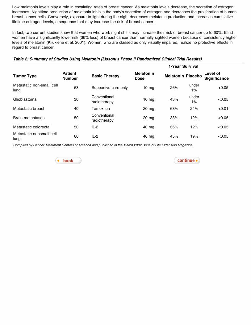
Low melatonin levels play a role in escalating rates of breast cancer. As melatonin levels decrease, the secretion of estrogen increases. Nighttime production of melatonin inhibits the body's secretion of estrogen and decreases the proliferation of human breast cancer cells. Conversely, exposure to light during the night decreases melatonin production and increases cumulative lifetime estrogen levels, a sequence that may increase the risk of breast cancer.
In fact, two current studies show that women who work night shifts may increase their risk of breast cancer up to 60%. Blind women have a significantly lower risk (36% less) of breast cancer than normally sighted women because of consistently higher levels of melatonin (Kliukiene et al. 2001). Women, who are classed as only visually impaired, realize no protective effects in regard to breast cancer.
Table 2: Summary of Studies Using Melatonin (Lissoni's Phase II Randomized Clinical Trial Results)
1-Year Survival
Tumor Type Patient Number Basic Therapy Melatonin
Dose Melatonin Placebo Level of Significance
Metastatic non-small cell lung 63 Supportive care only 10 mg 26% under
1% <0.05
Glioblastoma 30 Conventional radiotherapy 10 mg 43% under
1% <0.05
Metastatic breast 40 Tamoxifen 20 mg 63% 24% <0.01
Brain metastases 50 Conventional radiotherapy 20 mg 38% 12% <0.05
Metastatic colorectal 50 IL-2 40 mg 36% 12% <0.05Metastatic nonsmall cell lung 60 IL-2 40 mg 45% 19% <0.05
Compiled by Cancer Treatment Centers of America and published in the March 2002 issue of Life Extension Magazine.
Cancer Adjuvant Therapy
It appears that melatonin may also reduce the number of estrogen receptors on breast cancer cells. Since estrogen effectively feeds the growth of hormone-responsive breast tumors, reducing the receptors might slow tumor growth. Science News reported that the amount of melatonin required to inhibit breast cell proliferation appears no greater than the amount commonly present in human blood at night (Science News 93; Moss 1995).
Electromagnetic fields (EMFs) are another inhibitor of melatonin production. There is evidence that ELF (extremely low frequency) magnetic fields can act at the cellular levels to enhance breast cancer cell proliferation by blocking melatonin's natural oncostatic action. The mechanism(s) of action is unknown and may involve modulation of signal transduction events associated with melatonin's regulation of cell growth (Liburdy et al. 1993)
Melatonin delivers another anticancer perk through its antioxidant values. Physicians who once credited glutathione and vitamin E as being antioxidants of choice have now given special honor to melatonin. The neurohormone appears to protect against tumors by shielding molecules (especially DNA) from oxidative stress. Melatonin exerts its antioxidant properties by detoxifying the highly reactive hydroxyl radical, as well as singlet oxygen, hydrogen peroxide, and peroxynitrite anions (Kim et al. 2000).
A typical dose for a healthy individual is 300 mcg-6 mg each night. Cancer patients often take between 3-20 mg each night.
Modified Citrus Pectin (MCP)--retards cancer growth and metastasis Modified citrus pectin (MCP), also known as fractionated pectin, is a complex polysaccharide obtained from the peel and pulp of citrus fruits. Through pH and temperature modifications, the pectin is broken down into shorter, nonbranched, galactose-rich, carbohydrate chains. The shorter chains dissolve more readily in water, making them better absorbed than ordinary, long-chain pectin. The short polysaccharide units afford MCP its ability to access and bind tightly to galactose-binding lectins (galectins) on the surface of certain types of cancers. By binding to lectins, MCP is able to powerfully address the threat of metastasis (Strum et al. 1999).
In order for metastasis to occur, cancerous cells must first bind or clump together; galectin is thought responsible for much of cancer's metastatic potential by providing the binding site (Raz et al. 1987; Guess et al. 2003; Pienta et al. 1995). MCP appears small enough to access and bind tightly with galectins, inhibiting (or blocking) aggregation of tumor cells and adhesion to surrounding tissue (Kidd 1996). Deprived of the capacity to adhere, cancer cells fail to metastasize.
Men with prostate cancer who took 15 grams of MCP a day had a slowdown in the doubling time of their PSA levels. (Lengthening of doubling time represents a decrease in the rate of cancer growth.) Interestingly, rats injected with prostate adenocarcinoma and given MCP (in drinking water) showed a significant reduction in metastasis (compared to control animals), although the primary tumor was unaffected. According to Dr. Kenneth Pienta (leader of the Michigan Cancer Foundation), MCP may be the first oral method of preventing spontaneous prostate cancer metastasis (Pienta et al. 1995; Guess et al. 2003).
As with prostate adenocarcinoma, research shows that metastasis of breast cancer cell lines requires aggregation and adhesion of the cancerous cells to tissue endothelium in order for it to invade neighboring structures (Glinsky et al. 2000). To test the anti-adhesive properties of MCP, researchers evaluated (in an in vitro model) breast carcinoma cell lines MCF-7 and T-47D. The study concluded that MCP countered the adhesion of malignant cells to blood vessel endothelium and subsequently inhibited metastasis (Naik et al. 1995). MCP decreased metastasis of melanoma to the lung by more than 90% in laboratory animals (Platt et al. 1992).
Because MCP is a soluble fiber, no pattern of adverse reaction has been recorded in the scientific literature, apart from a self-limiting loose stool at high doses. MCP dosages are usually expressed in grams, with a typical adult dose ranging from 6-30 grams divided throughout the day. MCP’s apparent safety and proven antimetastatic action, and the lack of other proven therapies against metastasis appear to justify its inclusion in a comprehensive orthomolecular anticancer regimen (Kidd 1996). Pecta-Sol is the brand name of the original modified citrus pectin (MCP. The dosage for Pecta-Sol is about 15 grams a day.
N-acetyl-cysteine (NAC)--is an anticarcinogenic and antimutagenic agent; it inhibits IL-6 as well as invasion and metastasis of malignant cells N-acetyl-cysteine (NAC) is the acetylated precursor of the amino acids L-cysteine and reduced glutathione. Historically, it is used as a mucolytic agent in respiratory illnesses as well as an antidote for acetaminophen hepatotoxicity, but more recently its credits have grown. Animal and human studies have shown it to be a powerful antioxidant and a potential therapeutic agent in the treatment of cancer (Bongers et al. 1995; van Zandwijk 1995).
The biological value of NAC is attributed to its sulfhydryl group, while its acetyl-substituted amino group offers protection against
oxidative and metabolic processes (Bonanomi et al. 1980; Sjodin et al. 1989). In vitro studies showed NAC to be directly antimutagenic and anticarcinogenic; in vivo, NAC inhibited mutagenicity of a number of mutagenic materials (De Flora et al. 1986, 1992).
NAC has both chemopreventive and therapeutic potential in malignancies arising in the lung, skin, breast, liver, head, and neck (van Zandwijk 1995; Izzotti 1998). NAC is effective in inhibiting tumor cell growth in melanoma, prostate cells, and astrocytoma cell lines (the latter is a primary tumor in the brain) (Albini et al. 1995; Arora-Kuruganti et al. 1999; Chiao et al. 2000). Neovascularization (new blood vessel growth) is crucial for tumor mass expansion and metastasis. NAC inhibited invasion and metastasis of malignant cells by up to 80% by preventing angiogenesis (De Flora et al. 1996).
A number of cancers express IL-6 and other potentially dangerous cytokines. NAC inhibited (in a dose-dependent manner) the synthesis of IL-6 by alveolar macrophage (Munoz et al. 1996; Gosset et al. 1999).
Peak plasma levels of NAC occur approximately 1 hour after an oral dose; 12 hours after dosing, it is undetectable. Despite a relatively low bioavailability (4-10%), research has shown NAC to be clinically effective (Borgstrom et al. 1986). A suggested NAC therapeutic dosage is usually in the range of 600 mg per day.
Resveratrol--influences cancer at initiation, promotion, and progression stages Resveratrol is one of a group of compounds (called phytoalexins) that are produced in plants during times of environmental stress, such as adverse weather or insect, animal, or pathogenic attack. Resveratrol has been identified in more than 70 species of plants, including mulberries and peanuts, and the skins of red grapes, which are a particularly rich source (Jang et al. 1999). According to Pezzuto, "Of all the plants we’ve tested for cancer chemopreventive activity, this one [resveratrol] has the greatest promise" (Pezzuto 1997).
Resveratrol was effective against cancer during all three phases of the cancer process: initiation, promotion, and progression. For example, resveratrol displayed antimutagenic and antioxidant activity, providing greater protection against DNA damage than vitamins C, E, or beta-carotene. Resveratrol restored glutathione levels, considered by some as the most essential of antioxidants (Jang et al. 1999). It increased levels of a Phase II detoxifying enzyme (quinone reductase), an enzyme responsible for metabolically disassembling carcinogens.
Resveratrol inhibited the activity of cyclooxygenase-2 (COX-2), reducing the inflammatory response in human epithelial cells (Subbaramaiah et al. 1999). Upregulation of COX-2 is associated with the physical manifestations of various human cancers, as well as other inflammatory disorders. Since inflammation is closely linked to tumor promotion, substances with potent anti-inflammatory activities are thought to exert chemopreventive effects, particularly in the promotion stage of the disease.
Resveratrol prompted differentiation of human promyelocytic leukemia cells. The development of preneoplastic lesions in mouse mammary glands was inhibited by resveratrol (Kang et al. 2003; Asou et al 2002; Tsan et al. 2002).
The following studies illustrate the many pathways resveratrol employs to inhibit cancer:
■ Italian researchers recently determined that resveratrol exhibited a protective role against colon carcinogenesis, with the defense attributed to changes occurring in Bax protein, which encourages cell death (apoptosis), and p21 expression (Tessitore et al. 2000). Reduced Bax activity is associated with resistance to cytotoxic therapy (Bosanquet et al. 2002). p21 is able to arrest the cell cycle at the G1 phase by inhibiting DNA replication (Aaltomaa et al. 1999). Suppressing the growth cycle allows for a critical phase in cellular development referred to as differentiation, that is, an abnormal cell becomes more normal.
■ Resveratrol appears a promising anticancer agent for both hormone-dependent and hormone-independent breast cancers. At high concentrations, resveratrol caused suppression of cell growth in three breast cancer cell lines: estrogen-receptor (ER)-positive KPL-1 and MCF-7 and ER-negative MKL-F. Growth inhibition was credited in part to up-regulation of Bax protein and activation of caspase-3 (a key mediator of apoptosis in mammalian cells). Resveratrol was also able to lessen the growth stimulatory effects of linoleic acid, a fatty acid frequently over-consumed in Western diets (Nakagawa et al. 2001).
■ Resveratrol significantly reduced tumor volume (42%), tumor weight (44%), and metastasis (56%) in mice with highly metastatic Lewis lung carcinoma. Resveratrol was able to inhibit angiogenesis and reduce oxidative stress (Kimura et al. 2001; Kozuki et al. 2001).
■ Different wine polyphenols (catechin, epicatechin, quercetin) including resveratrol may be effective against prostate cancer. Prostate cancer cell lines (LNCaP and DU145) produce high concentrations of nitric oxide; PC3 produces low concentrations. Researchers propose that the anti-proliferative effects of polyphenols are due to their ability to adjust nitric oxide production (Kampa et al. 2000). Grape extract, a rich source of resveratrol, inhibited prostate cancer growth up to 98% in a dose- and time-dependent manner (Agarwal et al. 2000b).
■ Resveratrol appears to be promising in the control of acute monocytic leukemia (Tsan et al. 2000). Resveratrol induced
apoptotic cell death in human leukemia cells (HL60) (Clement et al. 1998) and stopped the growth of lymphocytic leukemia cells during the S-phase of the growth cycle (the time of DNA replication) (Bernhard et al. 2000).
■ Resveratrol inhibits NF-kB, thus inhibiting cell proliferation and cytokine production (Gao et al. 2001). The inhibition of cytokine production by resveratrol was found to be irreversible.
If using pure resveratrol, the suggested dosage is 7-50 mg a day. Beware of diluted supplements that provide very little active resveratrol. At the time of this writing, there were only a few sources of pure high-potency resveratrol available as dietary supplements.
Selenium--is protective against many types of cancers, promotes apoptosis, is a powerful antioxidant, and improves quality of life during aggressive cancer therapies Many animal studies have been conducted to evaluate the effects of super nutritional levels of selenium on experimental carcinogenesis using chemical, viral, and transplantable tumor models. Two thirds of these studies found that high levels of selenium reduced the development of tumors at least moderately (14-35% compared to controls) and, in most cases, significantly (by more than 35%) (Whanger 1998).
The impact of selenium supplementation on basal cell carcinoma was studied on 1312 subjects (18-80 years of age, 75% of whom were men) (Clark et al. 1996). Within 6-9 months, the group receiving 200 mcg a day of selenium realized about a 67% increase in plasma selenium levels. The non-supplemented group, although judged "normal" in regard to plasma selenium levels, experienced twice the rate of cancer as those receiving selenium. Researchers concluded that higher amounts of dietary selenium than the amount recommended by the FDA are needed to prevent cancer.
Although the study failed to show the effectiveness of selenium in altering the course of either basal or squamous cell carcinoma, selenium impacted the incidence of other types of malignancies with amazing success. The overall reduction in cancer incidence was 37% in the selenium-supplemented group; a 50% reduction in cancer mortality was observed over a 10-year period (Clark et al. 1996).
The following are the site-specific reductions in cancer incidence observed in the study: colorectal cancers (58%), lung cancer (46%), and prostate cancer (63%). A selenium deficiency appears to increase the risk of prostate cancer four- to five-fold. It was determined that as the male population ages selenium levels decrease, paralleling an increase in prostate cancer (Brooks et al. 2001).
Data is compelling regarding the usefulness of selenium’s protective effects against cancer:
■ Selenium-enriched broccoli is protective against chemically induced mammary and colon cancer in rats (Davis et al. 2002). Note: While selenium is contributing to the lower incidence of malignancy, the anticancer affects of broccoli should also be factored into the defense. Please read the section What Should the Cancer Patient Eat (appearing in this protocol) for valuable information regarding dietary factors affecting patient outcome.
■ The relationship between serum levels of selenium and the development of upper digestive tract cancer was examined (Mark et al. 2000). The relative risk of esophageal cancer was 0.56 in individuals in the highest quartile of selenium level compared with those in the lowest quartile. The corresponding relative risk of gastric cardia cancer was 0.47. Based on the data, it was concluded that 26.4% of esophageal and gastric cardia cancers are attributable to low selenium levels.
■ Adding selenium to salt resulted in a significant reduction in the incidence of cancer (Whanger 1998). ■ A significant increase in apoptosis and a decrease in DNA synthesis in breast cancers cells (MCF-7 and SKBR-3)
occurred with selenium supplementation. The selenium benefit was just as impressive in cancers of the lung (RH2), small intestine (HCF8), colon (Caco-2), and liver (HepG2). Prostate cancers (PC-3 and LNCaP) as well as colon cancer (T-84), although initially less affected by supplementation, became responsive when selenium was coadministered with Adriamycin or Taxol (Vadgama et al. 2000). This study suggests that selenium potentiates the anti-cancer effects of chemotherapy. Selenium supplementation in patients undergoing radiation therapy for rectal cancer improved quality of life and reduced the appearance of secondary cancers (Hehr et al. 1997).
■ It appears that selenium acts as an immunologic response modifier, normalizing every component of the immune system (Ferencik et al. 2003; Arthur et al. 2003)
An important form of selenium is Se-methylselenocysteine. This is the form of selenium found naturally in plants such as broccoli and garlic. A suggested selenium dosage is 200 mcg a day. The optimal dose for cancer patients is unknown at this time, but suggestions have ranged from 200-400 mcg a day depending upon the selenium content of the soil. Foods considered good sources of selenium include Brazil nuts, grains, onions, tomatoes, broccoli, chicken, eggs, garlic, liver, seafood, and wheat germ. Americans typically consume 60-100 mcg of selenium a day from dietary sources.
Silibinin (from milk thistle)--has antioxidant activity, increases sensitivity to chemotherapy while reducing its side effects, assists in arresting the growth of cancer, promotes differentiation, inhibits COX-2 enzyme, and suppresses NF-kB Fourteen years ago, the Life Extension Foundation introduced silymarin, a hepato-protective herb, to members. The major active constituent of silymarin is silibinin; a long-recognized antioxidant with more recently ascribed anticarcinogenic traits. Silibinin inhibits the growth of various cancer cell lines. The silibinin acts synergistically with cisplatin and doxorubicin, common chemotherapeutic drugs, improving their efficacy. By arresting tumor cell division at a strategic stage, silibinin appears to make tumor cells more sensitive to chemotherapy. Also, the harsh side effects associated with cytotoxic chemicals are less damaging when silibinin is utilized (Bokemeyer et al. 1996).
Milk thistle is described as an adaptogenic herb. For example, it encourages new cell growth where repair is needed but arrests cell division in tumor tissue; it increases the activity of certain enzymes but inhibits others. Milk thistle inhibits COX-2 (Zhao et al. 1999). Note: Go to Cyclooxygenase (COX-2) Inhibitors (Naturally Occurring) appearing in this protocol for other nutraceuticals capable of inhibiting the COX-2 enzyme. Also, consult Cyclooxygenase Inhibitors in the protocol entitled Cancer Treatment: The Critical Factors to learn more about the COX-2-cancer connection.
Silibinin arrests cell growth in the early phase of the cycle known as G1, a period of growth before DNA replication. Silibinin discourages cell growth by inhibiting various kinase enzymes (those playing a pivotal role in regulatory mechanisms), enabling a critical stage in cellular development referred to as differentiation. Differentiated cells abandon their primitive façade and assume the physical likeness and behavioral patterns of healthy cells. In fact, silibinin caused differentiation of a significant number of malignant prostate cells to more normal cells, while simultaneously decreasing PSA levels (Zi et al. 1999).
Silibinin inhibits growth of drug-resistant breast and ovarian cancer lines. It binds to type II estrogen binding sites, an action that turns off the proliferative effects of the cell (Scambia et al. 1996). In addition, silymarin inhibited the secretion of VEGF (an angiogenic factor) by malignant cells, thwarting the formation of cancer's vascular network (Jiang et al. 2000).
Cancer Adjuvant Therapy
Silymarin potently suppressed NF-kB, but did not affect TNF-alpha-induced NF-kB, demonstrating a pathway-dependent inhibition by silymarin. It appears the inhibitory effect of silymarin on NF-kB activation is associated with its liver-protecting properties. Suppression of NF-kB, a key regulator in inflammatory and immune reactions, significantly improves the anticarcinogenic status of silymarin (Saliou et al. 1998).
Silymarin/silibinin is remarkable medicine for the liver. Numerous studies show that milk thistle is effective in treating virtually every type of liver disease, including cirrhosis and alcohol or chemical-induced liver damage (Jacobs et al. 2002; Flora et al. 1998). So worthy is the herb in protecting against life-threatening toxins that individuals poisoned by the Amanita mushroom survived when silibinin was utilized (Carducci et al. 1996). A healthy liver is essential to detoxification, a process key to restoring health to cancer patients.
Standardized milk thistle extract usually consists of 35% silibinin, whereas the silymarin concentrate used in Europe contains a minimum of 80% silibinin. The Life Extension Foundation recommends the highly beneficial 80% silibinin extract. A suggested therapeutic dosage of Silibinin Plus is up to 6 capsules daily (1950 mg a day). For protection, use about 1-2 capsules (325-650 mg a day).
Soy--is protective against certain malignancies, appears to be an alternative to signal transduction-inhibiting drugs, and inhibits angiogenesis, cell proliferation, and metastasis
■ Isoflavones ■ For Prostate Cancer ■ For Breast Cancer ■ Soy and Other Types of Cancer ■ Free-Radical Scavenging Effects ■ Soy Precautions and Dosage
Legumes, including the soybean, contain bioactive compounds classified broadly as phytoestrogens as opposed to estrogens. Phytoestrogens are nonsteroidal and can actually inhibit steroids such as aromatase. Most have little or no estrogenic activity. When others have such activity, it is usually beneficial and specific to a certain tissue. For example, some soy isoflavones (a type of phytoestrogen) benefit bone but do not affect the kidney. In pharmacology terms, this is called a selective estrogen receptor modulator (SERM). A compound in soy, genistein, is a natural SERM. Tamoxifen and Raloxifen are chemical SERMs (Setchell et al. 1999).
The most recent studies suggest that the reason that different estrogens have different effects on different tissues is because there is more than one type of estrogen receptor. So far, three variations of the estrogen receptor have been found: one alpha and two betas. They share similar estrogen structure. The estrogen receptor-receptor (ERb) may suppress the action of the estrogen receptor-alpha (ERa) - at least in cancer cells (Maruyama et al. 2001; Saji et al. 2002; Speirs et al. 2002). And, growth-promoting estrogens such as estradiol activate ERa. Phytoestrogens preferentially activate theERb, which is repressive (Barkhem et al. 1998). For this reason, phytoestrogens have been characterized as good estrogens, and whatever estrogenic effect they have (which is estimated to be 1000-10,000 times weaker than estradiol, where it exists) may be nullified by their inhibition of estrogen synthesis and repression of the receptor that allows estradiol into the cell (Shao et al. 2000).
In normal tissue, the two estrogen receptors apparently work together to control both the amount and the use of estrogen in the body. It has been demonstrated that some types of cancer cells lose one type of estrogen receptor, leaving the control mechanism inoperable (Iwao et al. 2000; Sampath et al. 2001). This has been demonstrated in prostate cancer. Some types of prostate cancers do not express their ERaand some lose beta. This is why some will respond to estrogen and stop growing and others will stop growing when an anti-estrogen, such as genistein or Tamoxifen, is added.
The loss or gain of estrogen receptors occurs because of methylation abnormalities that occur in DNA (Lau et al. 2000). DNA methylation abnormalities are caused by three known factors: poor diet (i.e., a diet lacking in methylation factors including folate, vitamins B6 and B12), chemicals, and age.
Phytoestrogens include many diverse plant compounds, including resveratrol from grapes (Kopp 1998), curcumin from roots (Jaga 2001), and polyphenols from tea leaves (Mazur 1998). It is a very broad category that is further broken down into dozens of classifications such as flavonoids and flavones. The anticancer effects of phytoestrogens are the subject of dozens of scientific studies (Adlercreutz 1995).
Soy Isoflavones Soy contains phytoestrogens known as isoflavones, including daidzein, coumestrol, and genistein. Isoflavone supplements contain a mixture of many different types of these compounds. Interest in their anticancer potential stems from the lower occurrence of hormone-related cancers in Asians who eat a lot of soy. It is doubtful that the low rates of breast, prostate, and other hormonally related cancers are due solely to soy, but studies show that compounds isolated from soy have significant anticancer effects (Suthar et al. 2001).
Soy for Prostate Cancer The most dangerous aspect of prostate cancer is metastasis (spreading to other areas). Prostate cancer can be controlled if it can be limited to the prostate gland. Unfortunately, many men with prostate cancer have undetected metastases.
Genistein has powerful and specific effects against the spread of prostate cancer. Genistein significantly activated 832 genes in prostate cancer cells, 13 of which are related to metastasis (Li et al 2002a,b; Sarkar et al. 2002).
Genistein down-regulated multiple genes that dissolve surrounding tissue to enable metastasis and invasion of surrounding tissue, and down-regulated genes that create new tumor blood vessels. Genistein also affected genes important in stopping the cell cycle, differentiation, apoptosis, and cell signaling communication (Li et al. 2002a).
Genistein has “potent anti-proliferative effects” against human prostate cells (Shen et al. 2000), and inhibits metastasis (Schleicher et al. 1999). Genistein is one component of soy. Soy has powerful effects in the prevention and eradication of prostate cancer. Different components of soy have different effects against prostate cancer cells. Genistein blocks an enzyme that destroys an anticancer vitamin D metabolite in cancer cells (Farhan et al. 2002).
Prostate cancer is a hormone-related cancer. In a study mice were fed three different soy products: soy protein without isoflavones, soy phytochemical concentrate (a combination of genistein, daidzein, glycitein, and other compounds), and genistein. All three feeds had a positive effect on hormones as they relate to prostate cancer growth. The androgen receptor, which correlates with tumor weight, was reduced 42% by soy protein. Genistein reduced serum dihydrotestosterone, a form of testosterone associated with hyperplasia and cancer, and caused a 57% reduction in tumor growth. Soy phytochemical concentrate inhibited the overall growth of prostate cancer by 70%. Soy phytochemical concentrate also stopped metastases to lymph nodes and lung. Cell death was induced, and angiogenesis was significantly inhibited (Zhou et al. 2002).
Healthy, normal rodents fed genistein for 2 weeks at a dietary level had significant reductions in androgen and the two estrogen receptors (Fritz et al. 2002). Minimizing the number of hormone receptors reduces levels of cell growth-promoting hormones in the prostate gland. The levels of phytoestrogens in 25 men with and without benign prostatic hyperplasia (BPH), a noncancerous overgrowth of prostate cells, were examined. Genistein levels in men with BPH were significantly lower than in those without BPH (Hong et al. 2002). Adding genistein to prostate tissue taken from men with BPH stops the prostate cancer growth (Geller et al. 1998).
Various soy diets have significant effects against prostate cancer compared to a casein (milk protein) diet. Soy significantly reduced insulin-like growth factor (IGF-1), a protein that helps tumors create blood vessels. Blood vessel density and tumor cell proliferation were decreased. Cell death was increased. Dietary soy works through “a combination of direct effects on tumor cells and indirect effects on tumor neovasculature” (blood vessels) (Zhou et al. 1999). The cell-killing effects of soy components are important not only for men who have been diagnosed with prostate cancer, but for healthy men as well.
Prostate-specific antigen (PSA) is elevated in men with prostate enlargement. PSA is regulated by androgens. Genistein and its precursor, biochanin A, markedly decrease PSA in prostate cancer cells by inactivating testosterone (Sun et al. 1998). A study on rats showed a 38% decline in PSA, along with a significant reduction in metastases when genistein was given subcutaneously (Schleicher et al. 1999; Zand et al. 2002).
The ability of genistein to reduce cellular proliferation in men with elevated PSA is currently under investigation. In addition, the ability of supplemental soy to lower PSA and kill cancer cells in men with localized prostate cancer is being studied. The ability of soy isoflavones to modulate hormones and cancer-related proteins in men with prostate cancer is also being studied.
Population-based studies have shown that men with high levels of soy and other isoflavones in their blood have the lowest risk of prostate cancer. In a study on men from Japan, China, and the United States, it was shown that legumes, including soy, reduce the incidence of prostate cancer by 38%. Eating yellow-orange vegetables reduces it 33%, and cruciferous vegetables reduce it 39%. These findings are consistent across ethnicities, indicating that isoflavones, not genes, are responsible for the reductions in risk (Kolonel et al. 2000). An analysis of data collected from 12,395 Seventh-Day Adventist men indicates that more than one serving per day of soymilk can reduce the risk of prostate cancer 70% (Jacobsen et al. 1998). Note: Seventh-Day Adventists are vegetarians; meat is a known risk factor for prostate cancer. Maintaining a vegetarian diet may have contributed to the low rates of
prostate cancer.
Genistein down-regulates proteins that enhance prostate cancer growth, including HER2 neu. Genistein has no adverse toxicity, and the amount needed to reduce the proteins by half is achieved with supplemental genistein or a diet high in soy products. Genistein inhibits EGF signaling pathway suggesting that this phytoestrogen may be useful in both protecting against and treating prostate cancer (Dalu et al. 1998).
Soy isoflavones clearly work against prostate cancer through several mechanisms, including modulating hormones, blocking metastasis, interfering with cell signaling, stopping cell growth, inducing cell death, and possibly activating and deactivating cancer-related genes.
Soy for Breast Cancer Soy phytoestrogens help to prevent and control hormone-related breast cancer (Zhou et al. 2004; Adlercreutz 2002). It is especially beneficial for Western women, who are exposed to a comparatively high level of environmental estrogens. Soy is anti-estrogenic. It prevents the conversion of estrone to 17-beta-estradiol. Estradiol fuels the growth of breast cancer, whereas estrone is a weaker estrogen. Genistein causes cancer cells to metabolize estradiol to estrogenically weaker or inactive metabolites (Brueggemeier et al. 2001).
Soy phytoestrogens naturally activate the receptor, known as ERb, which in turn suppresses the activation of Era and allows growth-promoting estradiol into cancer cells (Pettersson et al. 2000). ERa is the receptor referred to as “estrogen receptor positive;” “estrogen receptor negative” breast cancer cells have estrogen ERb. Estrogen receptor positive cells have lost their beta-receptors duringthe events leading to breast cancer. Normal cells have both types of estrogen receptors.
Genistein naturally activates ERb, inhibiting cell proliferation. Activating the beta-receptor down-regulates the alpha-receptor, or estradiol-activated, receptor. This negates estradiol's cancer-promoting effects.
The consumption of soy reduced the risk of having ERa positive breast cancer by 56%, whereas the effect on both types of breast cancer was 30% (Dai et al. 2001).
Genistein interferes with cancer's ability to grow blood vessels. A direct link between alpha-receptors and angiogenesis has been discovered in estrogen receptor positive cancer cells (MCF-7). These cells have too many alpha-receptors and not enough beta-receptors. When estradiol attaches to the alpha-receptors, it activates a protein that promotes the formation of new blood vessels (Sampath et al. 2001). Genistein blocks the formation of new blood vessels (Zhou et al. 1998; Wietrzyk et al. 2001). Furthermore, genistein prevents vitamin D from being degraded by cancer cells (Farhan et al. 2002).
In a study on estrogen receptor positive breast cancer cells (MCF-7), genistein competed successfully with estradiol for access to the cells, and once inside, blocked estradiol from inducing cell growth. In a study on Japanese women who drank soymilk containing 100 mg of isoflavones a day, estrone and estradiol levels fell by almost 30% (Nagata et al. 1998).
Breast cancer cells have elevated levels of enzymes that produce estradiol. One of the enzymes, known as 17-beta-hydroxysteroid dehydrogenase type 1 (17HSD1), causes the conversion of "weak estrogen" (estrone) to "strong estrogen" (estradiol) and helps cancer cells grow. A variant known as 17HSD2 does the opposite. Breast cancer cells have elevated amounts of 17HSD1, and insufficient 17HSD2 (Miyoshi et al. 2001). Studies show that if cancer cells are treated with genistein, 17HSD2 will be made, and "strong estrogen" (estradiol) will be converted to "weak" (estrone) (Hughes et al. 1997). A woman with breast cancer may have the same level of estrogen in her blood as a woman without breast cancer. The elevated estradiol levels occur inside cancer cells where abnormalities create imbalances in enzymes. Such 17HSDvariances favor the accumulation of estrogen for cell growth.
Genistein also inhibits an enzyme that is elevated in breast cancer cells known as "aromatase" (Kao et al. 1998; Breuggemeier et al. 2001). Aromatase helps convert testosterone to estrogen. Elevated male hormones, enlarged prostate, and abnormal cell growth do not promote prostate cancer in mice that lack aromatase (McPherson et al. 2001).
Asian women get early protection by eating soy their entire lives (Lamartiniere et al. 1998). The genistein in soy promotes more differentiated tissue in the breast, which leaves less tissue that can become cancerous. Soy isoflavones decrease density in the breast enabling easier detection of cancer by mammogram (Maskarenic et al. 2001). A serving of tofu every week decreases the risk of breast cancer by 15% (Wu et al. 1996). It is well-established that when Asian women abandon their traditional diet, their risk of breast cancer escalates. It is important to realize, however, that while it has been proven that soy components have direct and powerful effects against cancer cells, it cannot be assumed that soy alone is responsible for the reduced risk of hormone-related cancers in Asians. There are many aspects of the Asian diet that undoubtedly play a role, including the low consumption of animal fat. Green tea is another component of the Asian diet that has proven anticancer effects. A polyphenol from black tea has no effect on prostate cancer cells. However, when combined with genistein, it stops proliferation (Sakamoto 2000).
HER2/neu and EGFR are both related to breast cancers resistant to treatment with tamoxifen and other therapies (Ross et al. 1998). Genistein blocks an enzyme that promotes the proliferation of cancer cells. Because protein tyrosine kinases activate other cancer-promoting factors, genistein is a very attractive candidate for the prevention and treatment of various types of cancer. A dietary amount of the soy compound genistein significantly delayed the appearance of the HER2/neu-type cancer. It did not, however, reduce tumor size or number in this study (Jin et al. 2002).
It is important to note that DDT and other chlorine-related chemicals activate tyrosine kinases (TK), including HER2/neu-related ones in human cancer cells. Although DDT was banned decades ago, Americans are still being exposed to it. Genistein and other isoflavones block the activation of TK by DDT and related estrogen-mimicking chemicals, but tamoxifen does not (Enan et al. 1998; Verma et al. 1998).
A mouse study shows that increasing amounts of genistein retard cancer growth, in accordance with the cell studies (Shao et al. 1998). The animals must be implanted with estradiol to make the cancer cells grow (Santell et al. 2000; Allred et al. 2001; Ju et al. 2001). When mice are fed the equivalent of what Asians usually consume in their diets, the appearance of a genetic type breast cancer (as opposed to a chemically induced one) is significantly delayed by genistein, soy isoflavones, and daidzein, another soy compound (Jin et al. 2002).
Studies in monkeys, the closest animal model to humans, show that soy phytoestrogens impede the proliferation of cells responsive to estrogen. "Soybean phytoestrogens are not estrogenic at dietary doses" (Cline et al. 2001). Statistics on the rate of hormone-related cancers in Asians prove that soy is extremely beneficial against hormone-related cancers in humans. They show that people who eat large amounts of soy products have the lowest levels of strong estrogen in their bodies and the lowest rates of breast and prostate cancers.
Cancer Adjuvant Therapy
Soy and Other Types of Cancer
■ Glioma ■ Bladder Cancer ■ Stomach Cancer ■ Melanoma ■ Lung Cancer ■ Colon Cancer ■ Thyroid Cancer ■ Leukemia
Soy has powerful anticancer effects that do not involve hormones. Genistein inhibits a chemical reaction used by many different types of cancer cells to multiply and spread. Compounds that can do this are called tyrosine kinase (TK) inhibitors. Dozens of studies in different types of cancer cells show that genistein is a powerful and effective TK inhibitor.
Glioma. Glioma cancer cells have very high TK activity, which correlates with cancer growth. Several in vitro studies show that genistein inhibits the growth of glioma (Baltuch et al. 1996; Tu et al. 2000; Khoshyomn et al. 2002). Genistein also enhances the effectiveness of the chemotherapeutic drugs carmustine and camptothecin with a 40% decrease in growth and a 50% increased killing effect in some cells (Ciesielski et al. 1999; Khoshyomn et al. 2002). The amount of genistein needed to enhance the effectiveness of carmustine is not high. The appropriate amount of genistein can be obtained by following the supplement program recommended in the Soy Dosing and Precautions section.
Bladder Cancer. Genistein's ability to inhibit TK may be of great benefit in keeping bladder cancer localized. In Asia, the incidence of invasive bladder cancer is much lower than in the United States, leading some researchers to investigate the effects of soy. Invasive bladder cancers have high levels of a protein known as epidermal growth factor receptor (EGFR), which enables the cancer to invade muscle. EGFR is activated by TK and can be reversed by genistein (Theodorescu et al. 1998).
The effects of genistein, soy protein isolate, and soy phytochemical concentrate on human bladder cancer cells and bladder cancer were studied in mice. The three soy products reduced tumor volume 40%, 37%, and 48%, respectively. They blocked tumor blood vessel formation and induced tumor cell death, stopping the cells from growing at the G2-M part of the cell cycle (Zhou et al. 1998).
A mixture of isoflavones work better than a single soy compound for bladder cancer. In a study on seven different cell lines, genistein plus isoflavones inhibited tumor growth and induced cell death at levels obtainable through the diet or soy supplements. Both genistein and combined isoflavones exhibited a significant tumor suppressor effect in vivo. These results justify the potential use of soybean isolateas a practical chemoprevention approach for patients with urinary tract cancer (Su et al. 2000).
Stomach Cancer. The effects of soy products on 10 different types of human gastrointestinal cancer cells found that genistein and biochanin A (a genistein precursor) strongly inhibited proliferation of stomach, colon, and esophageal cancers (Yanagihara et al. 1993). Data from a study involving over 30,000 people was analyzed and it was found that people who eat the most soy products reduced their risk of stomach cancer by half compared to those who eat the least (Nagata et al. 1998).
Melanoma. Studies on the effects of genistein on human melanoma cancer cells showed that genistein is a powerful inhibitor of the growth of this cancer and that it stops the cell cycle as effectively as the chemotherapeutic drugs adriamycin and etoposide (Darbon et al. 2000).
Studying melanoma in mice revealed that genistein reduces the blood supply to lung tumors and has an additive effect with the drug cyclophosphamide. In laboratory rodents, genistein can reduce the growth of tumors by half through supplements and/or diet (Record et al. 1997).
Lung Cancer. Genistein has several actions against small cell and non-small cell lung tumors. In a study in which Lewis lung cancer was transplanted into mice, genistein reduced the tumor colonies by half, and genistein plus cyclophosphamide reduced
them by 90% (Wietrzyk et al. 2001). Several studies show that genistein stops lung cancer cells from growing and induces cell death (Tallett et al. 1996; Fujimoto et al. 2002; Wietrzyk et al. 2000). Genistein inhibits enzymes that help lung cancer cells to proliferate and spread (Leyton et al. 2001). Genistein up-regulates tumor suppressor genes p53 and p21 (Lian et al. 1999). Genistein reverses the multidrug resistance-associated protein, a protein that makes lung cancer cells resistant to daunorubicin, doxorubicin, etoposide, and vinblastine (Versantvoort et al. 1994; Berger et al. 1997).
Researchers in Japan analyzed information from 333 people with lung cancer. They found that eating tofu every day reduced the risk of lung cancer 45% in men and 86% in women (Wakai et al. 1999).
Colon Cancer. Soy has anticancer effects against cells that line the digestive tract. For this reason, it may have beneficial effects against different types of digestive tract cancers. Researchers looking at how three different types of human colon cancer cells react to soy confirmed that colon cancer is susceptible to soy's anticancer effects (Zhu et al. 2002). Some colon cancers may be estrogen dependent. Estradiol activates four kinase enzymes in colon cancer cells, two of which are tyrosine dependent and therefore potentially susceptible to genistein. Genistein blocks at least one of these enzymes and retards cell growth (Di Domenico et al. 1996). Genistein also suppresses the growth of nonestrogen-dependent colon cancer cells, which also respond to treatment with tamoxifen (Arai et al. 2000).
In a study that investigated how tamoxifen, genistein, and estradiol affect intestinal cells, genistein and tamoxifen emerged as the strongest inhibitors of cell proliferation, inhibiting TK and inducing the death of cancer cells (Booth et al. 1999). Genistein reverses resistance to doxorubicin and other chemotherapeutic drugs in at least one type of colon cancer by a "novel drug resistance pathway" (Rabindran et al. 1995). However, a study in mice showed that soy isoflavones may not counteract a bad diet. Mice fed a Western high fat, low fiber, and low calcium diet developed colon cancer despite isoflavones in their food (Sorensen et al. 1998). Soy could not reverse colon cancer (whereas rye lignans could) in mice on high fat diets (Davies et al. 1999).
Thyroid Cancer. Soy may have beneficial effects against thyroid cancer. Six hundred and eight cases of thyroid cancer, found that people who consume soy compounds, genistein and daidzein, in their diet reduced their risk of this cancer by one-third. However, adding soy flour or protein to a Western diet was not effective (Horn-Ross et al. 2002).
Leukemia. A few studies have been done on human leukemia cells treated with genistein. Of nine compounds tested, genistein showed the strongest inhibitory effects against human promyelocytic leukemia (HL-60) cells. All nine compounds are found in miso (Hirota et al. 2000). In human leukemia cells resistant to chemotherapy, genistein was able to reverse the drug resistance almost completely (Nagasawa et al. 1996). The anti-proliferative effect of genistein against human leukemia was significantly augmented by vitamin D analogs (Siwinska et al. 2001).
Free-Radical Scavenging Effects The antioxidant effects of soy were the focus of much of the early research on how soy prevents cancer. The powerful free-radical scavenging effects of soy compounds and how they impact cancer continue to emerge.
Soy has an additive effect with vitamin E; it lowers rather than elevates estrogen levels in women and androgen levels in men (Jenkins et al. 2000). Damage to DNA caused by certain types of free radicals is strongly inhibited by genistein and other soy compounds (Breinholt et al. 1999; Davis et al. 2001). This helps prevent cancer. Dietary amounts significantly lower free-radical damage (Davis et al. 2001; Exner et al. 2001).
In addition to blocking free-radical damage, soy phytoestrogens also block inflammation, a contributor to cancer growth, notably in the colon (Davis et al. 2001; Zheng et al. 2002).
The effects of genistein against the activation of EGFR by free radicals were demonstrated. In this study, genistein reversed the free-radical activation of EGFR in normal cells (Chen et al. 2001). The benefits of genistein against oxidative stress are evident from a study on brain cells exposed to hydrogen peroxide. Free radicals generated by this oxidant degrade phospholipids and activate enzymes, which are crucial for memory and other brain functions. Genistein, through its ability to inhibit a tyrosinekinase enzyme that sets off the reaction, rescues cells from damage (Servitja et al. 2000).
Soy Precautions and Dosage While the data are persuasive regarding the chemoprotective effects of soy, many questions remain. Some nutritionally based oncologists do not permit soy in their patients' regime. Others believe that soy should be avoided by everyone and have launched massive public relations campaigns to discredit soy and discourage even moderate consumption by healthy people.
Breast cancer patients should avoid soy until their estrogen receptor status has been determined. Estrogen receptor alpha-positive breast cancer patients may benefit from genistein, while beta-receptor positive breast cancer patients’ tumors cells may proliferate faster in response to genistein. It has been suggested that patients avoid soy supplements 1 week prior to, during, and 1 week after radiation therapy, although new studies appearing in the Cancer Radiation Therapy protocol indicate a potential benefit to using soy isoflavones during radiation therapy.
Some people believe that soy is toxic to the thyroid gland, yet this may be a concern only in cases of iodine deficiency (Doerge et al. 2002). Some of the more credible arguments deal with soy-based infant formulas (Tuohy 2003).
There are a number of human clinical studies being conducted on the use of soy to both prevent and treat cancer (http://clinicaltrials.gov/ct/search?term=soy). When the findings of these studies are published, perhaps more definitive recommendations can be made about soy supplements. Based on the information available to us as of this writing, those concerned about cancer may consider these guidelines: a suggested dosage is five 700-mg capsules 4 times a day of a soy extract providing a minimum of 40% isoflavones. For prevention purposes, as little as 135 mg of a 40% soy isoflavone extract once a day may be adequate.
Theanine--increases efficacy of chemotherapeutic drugs Researchers speculate that drinking 1 cup of green tea favors a positive mental attitude and increases the efficacy of chemotherapy. However, components of green tea have been identified (caffeine, epigallocatechin gallate (EGCG), flavonoids, and theanine) that better explain the chemotherapeutic advantage beyond its soul-soothing effects (Sadzuka et al. 2000a).
Japanese researchers focused specifically on theanine and its influence on the anti-tumor activity of Adriamycin (doxorubicin). In vitro, theanine inhibited the outflow of Adriamycin (ADR) from cancerous cells, increasing concentrations within the cell by almost three-fold. An increase in ADR concentrations was not observed in normal tissues, suggesting theanine protects healthy organs, such as the heart and liver. (Sadzuka et al. 1996). Illustrative of the enhancing qualities of theanine, injecting ADR into ovarian sarcoma-bearing (M5076) mice did not inhibit tumor growth, whereas a combination of theanine and ADR reduced tumor weight 62% (Sugiyama et al. 1998).
When theanine was added to pirarubicin, intracellular concentrations of pirarubicin increased 1.3-fold and the overall therapeutic efficacy of the drug increased 1.7-fold (Sugiyama et al. 1999). Satisfying results were also found when theanine was used with Idarubicin (IDA), which is highly toxic to bone marrow and an anti-leukemia agent similar to doxorubicin. Risk factors permitted only about one-fourth of the standard IDA dose to be used in combination with theanine. However, theanine reduced toxicities and increased IDA anti-tumor activity, rendering the chemotherapeutic agent a possibility for the treatment of leukemia (Sadzuka et al. 2000b).
Part of theanine's anticancer effects can be attributed to mimicking glutamate, an amino acid that potentiates glutathione. Glutathione detoxifies chemotherapeutic agents, barricading chemicals from cells, and inhibiting tumor cell kill. Theanine is structurally similar to glutathione and crowds out glutamate transport into tumor cells. Cancer cells (in confusion) erringly take in theanine and theanine induces glutathione production. Glutathione (derived from theanine) does not detoxify like natural glutathione, and instead blocks the ability of cancer cells to neutralize cancer-killing agents. Deprived of glutathione, cancer cells cannot remove chemotherapeutic agents, and the tumor cell dies as a result of chemical poisoning (Sadzuka et al. 2001).
Administered with doxorubicin, the suggested dose of theanine is 500-1000 mg a day, although no human studies have been conducted with chemotherapy and theanine.
Thymus Extract--improves T-cell response and regulates the activity of cytokines The thymus gland was at one time removed as an unnecessary appendage. It is an essential organ of the immune system, increasing stamina, energy, well-being, and the ability to ward off infections and cancer. Since 1965, when Burnet was awarded the Nobel Prize for demonstrating the endocrine function of the thymus gland, medical interest has focused on the thymus. It is now largely accepted that the thymus gland plays a central role in the mammalian immune system.
The immune system is made up of B-cells that protect against bacterial and viral infections and T-cells that guard against viral and fungal infections, as well as cancer. This powerful body of cells normally treats a developing cancer as foreign tissue, destroying aberrant cells before rapid multiplication occurs.
The effectiveness of T-cell mediated immunity depends upon the activity of T-lymphocytes (T-cells), which are programmed by proteins from the thymus gland. Immature (naïve) T-4 cells do not function properly until programmed by thymic proteins. As new T-lymphocytes migrate from the bone marrow to the thymus, they are programmed to distinguish between self-tissue (the host) and nonself tissue (an invading pathogen).
The thymus gland, a lymphoid organ situated in the anterior superior mediastinum, reaches its maximum weight near puberty and then undergoes involution, or degenerative change, shrinking to about one-sixth of its original size. By the age of 40, the thymus gland is scarcely functional in many individuals; therefore, the essential thymus-provided protein is no longer available to program T-4 cells. More than 20 years ago, thymic protein A was isolated and purified from bovine thymus cells (by Dr. Terry Beardsley, an immunologist). Dr. Beardsley patented a technology to grow thymus cells in the laboratory and then purify a specific thymus protein (Thymic Protein A) that helps T-cells to mature with immune competency. The active ingredient in Thymic Protein A is the precise thymus protein that programs the T-4 lymphocytes to locate abnormal cells and then directs T-8 killer cells to destroy them.
Three types of cells emerge from the thymus: T-4 helper cells (master regulators), T-8 cytotoxic killer cells (guided by T-4 helper cells to attack and destroy invading cells), and T-8 suppressor cells. T-4 helper cells regulate many key functions, including the activity of IL-2 and interferon.
High dose thymosin, a humoral factor secreted by the thymus, in conjunction with intensive chemotherapy was administered to 21 patients with advanced lung cancer. Ordinarily, patients with late stage lung cancer live about 240 days; the median survival rate more than doubled (500 days) among patients receiving thymosin. Some of the thymosin-treated group were alive and disease-free 2 years after treatment (Chretien et al. 1979).
Blood tests to measure the immune response are extremely valuable when detailing either a preventive or a therapeutic program to fight cancer. While determining T-lymphocyte numbers is important, assessing their activity is even more crucial. It is possible for a person with a total count of 1000 T-4 cells to have only 50% of these cells activated by the thymus. It is important that the patient know the degree of immune impairment in order to structure a corrective program. Tests to evaluate the activity of the immune system are performed at the Immuno-Science Laboratory (Los Angeles), (310) 657-1077.
A suggested dosage for healthy individuals is 1 packet of BioPro Thymic Protein A daily or every other day. Cancer patients may wish to increase this amount. For example, HIV patients use 3 doses a day until blood tests remain normal for 3-6 months. For maintenance, reduce to 1 dose a day. Use the thymic protein under the tongue, retaining for 3 minutes to allow for maximum absorption. Typically, patients undergoing chemotherapy maintain acceptable white blood cell counts if Thymic Protein A accompanies treatment.
Cancer Adjuvant Therapy
Vitamin A--offers protection against radiation induced tissue damage, down-regulates telomerase activity, and is involved at almost every juncture of cancer control Retinoids induce cell differentiation, control cancer growth and angiogenesis, repair precancerous lesions, prevent secondary carcinogenesis and metastasis, and act as an immunostimulant. After FAR therapy (5-fluorouracil-retinol palmitate with radiation and surgery), the disease-specific, 5-year survival was nearly 50% in various head and neck cancers (Yamamoto 2001). Retinoids, at pharmacological levels, assist in preventing the appearance of secondary tumors following curative therapy for epithelial malignancies.
It is well-established that a vitamin A deficiency (in laboratory animals) correlates with a higher incidence of cancer and an increased susceptibility to chemical carcinogens. This is in agreement with epidemiological studies, which indicate that individuals with a lower dietary vitamin A intake are at a higher risk of developing cancer (Sun et al. 2002). The chemotherapeutic possibilities surrounding vitamin A areplentiful.
Two vitamin A analogs currently in large chemoprevention, intervention trials, or epidemiological studies are all-trans-retinoic acid (ATRA) and 13-cis-retinoic acid (13-cis-RA).
Note: Retinoic acid is biologically active in two forms: all- trans- retinoic acid and 9-cis-retinoic acid. Vitamin A and 13-cis-RA are converted to these biologically active forms.
Thirty-two women with previously untreated cervical carcinoma (ages 14-60) were treated for at least 2 months using oral 13-cis-RA (1 mg per kg body weight a day) and alpha-interferon subcutaneously (6 million units daily): 16 of the women (50%) had major reactions, including four complete clinical responses. Remission occurred in 15 of the patients within 2 months and in one patient within 1 month; toxicity to treatment was described as manageable (Espinoza et al. 1994). The positive results were replicated in other studies using a similar model (Hansgen et al. 1998, 1999).
The role of 13-cis-RA on a human prostate cancer cell line (LNCaP) was studied. It was found that 13-cis-RA significantly inhibited PSA secretion and the ability to form new tumors. It was also noted that tumors that appeared (having escaped 13-cis-RA inhibition) were smaller compared to tumors in nontreated animals (Dahiya et al. 1994). During the course of 13-cis-RA therapy, prostate cancer cells became more differentiated, that is, they resembled (microscopically) normal prostate cells.
A combination of phenylbutyrate and 13-cis-RA as a differentiation and anti-angiogenesis strategy against prostate cancer was evaluated. Phenylbutyrate, considered nontoxic, is used to arrest tumor growth and induce differentiation of premalignant and malignant cells. Tissue examination of tumors showed decreased cell proliferation and increased apoptosis, as well as reduced microvessel density in animals treated with 13-cis-RA and phenylbutyrate; tumor growth was inhibited by 82-92%. In contrast, researchers reported 13-cis-RA and phenylbutyrate, when used singularly, were suboptimal in terms of clinical benefit (Pili et al. 2001).
A pilot study conducted at M.D. Anderson Cancer Center found ATRA alone ineffective as a long-term treatment for chronic myelogenous leukemia (CML). Only four of 13 subjects showed a transient, nonsustaining indication of an anti-leukemic effect (Cortes et al. 1997). However, combinations of therapeutic agents that included ATRA were promising in the treatment of CML. The combination included alpha-interferon plus ATRA, which reduced proliferation 50-60% (Marley et al. 2002).
Cisplatin (a popular chemotherapeutic agent) shares a similar chemotherapeutic profile with ATRA (the ability to induce cytotoxicity through apoptosis). A combination of ATRA and cisplatin induced apoptosis in significantly more cancer cells, particularly in ovarian and head and neck carcinomas, than either drug alone (Aebi et al. 1997). A combination of ATRA and IL-2 showed therapeutic value in treating resistant metastatic osteosarcoma, a malignant tumor of the bone (Todesco et al. 2000).
For decades, researchers have searched for ways to minimize the damage to the heart during Adriamycin therapy. Adriamycin, though relatively effective, damages the heart muscle. Several animal studies indicated that supplemental vitamin A reduced Adriamycin-induced inflammation and preserved heart tissue. Vitamin A appears not only to counter Adriamycin damage, but also to increase survival in animals (Tesoriere et al. 1994). Vitamin A extends similar protection to patients using cisplatin, a drug often used for bladder and ovarian cancer, as well as small cell carcinoma.
Radiation-induced lung injury frequently limits the total dose of thoracic radiotherapy that can be delivered to a patient undergoing treatment, restricting its effectiveness. Animal studies suggest that supplemental vitamin A may reduce lung inflammation after thoracic radiation and modify radiotherapy damage to the lungs (Redlich et al. 1998).
Vitamin A (in dosages of 25,000 IU a day) offers significant protection against radiation-induced tissue damage. Various cancer patients use more than 100,000 IU of a water-soluble vitamin A liquid a day, a dosage that must be supervised by a physician. Do not supplement with vitamin A if the cancer involves the thyroid gland or if the liver is damaged. Both professionals and patients should consult Appendix A to read about avoiding vitamin A toxicity. Good food sources of vitamin A include liver and fish liver oils, green and yellow fruits and vegetables such as apricots, asparagus, broccoli, cantaloupe, carrots, collards, papayas, peaches, pumpkins, spinach, and sweet potatoes. High-potency water-soluble vitamin A is available as a dietary supplement.
Vitamin C (ascorbic acid)--has a chemotherapeutic effect on many cancers, promotes collagen production, sequestering the tumor, and reduces the toxicity of conventional therapies Linus Pauling, winner of the Nobel Prize for chemistry in 1954 and the Nobel Prize for Peace in 1963, believed strongly that vitamin C could play an important role in cancer treatment. Dr. Pauling suggested 10 grams of vitamin C a day for patients with advanced cancer for whom conventional treatments had ceased to be of benefit (Cameron et al. 1993). Over an 8-year period, 500 patients with varying stages and types of cancer were treated with vitamin C therapy. Those receiving 10 grams of vitamin C a day improved their state of well-being, as measured by increased appetite and mental alertness, as well as a decreased need for pain-killing drugs. A retrospective analysis showed that those using vitamin C lived considerably longer than those not supplemented.
Various clinics are using intravenous vitamin C and with positive results. Dr. Hugh Riordan, recognized as a world authority on this procedure, practices from Wichita, KS, at the Center for the Improvement of Human Functioning International. Dr. Riordan's vitamin C story began in 1984 when he treated his first cancer patient; a 70-year-old renal cell carcinoma patient with metastasis to the lung and liver, using injectable vitamin C. Renal cell carcinoma has only a 5% response rate.
The initial treatment began with 15 grams of vitamin C administered intravenously 2 times a week; showing excellent tolerance, the vitamin C dosage was increased to 30 grams twice weekly. Within 6 weeks, the patient showed a favorable response to treatment and at the 12-week interval was pronounced tumor-free. The patient lived 14 additional years and died of congestive heart failure with no evidence of tumors.
In light of the favorable initial response to intravenous (IV) vitamin C, ascorbic acid was investigated. Vitamin C is preferentially toxic to tumor cells, that is, it kills tumor cells but not normal cells.
In low doses, vitamin C assumes the nature of an antioxidant; in high dosages, vitamin C changes roles and becomes a prooxidant, inducing peroxide production. Tumor cells have a relative catalase deficiency, an enzyme necessary to detoxify hydrogen peroxide to water and oxygen. A 10- to 100-fold difference in catalase concentrations exists between tumor cells and normal cells. Without the protection of catalase, peroxide accumulates in cancerous cells, along with aldehydes (toxic byproducts of the reaction), causing death to malignant cells. On the other hand, normal, healthy tissues have the protection of the detoxification enzyme and are spared destruction by peroxide and aldehyde. Vitamin C, a virtually nontoxic nutrient (Bowie et al. 2000), could cause a transient diarrhea if not absorbed properly.
Vitamin C is safe compared to standard chemotherapeutics and has an ability to preserve immune function. Many patients succumb, not because of cancer, but rather from a post-chemotherapeutic toxicity, resulting from a damaged immune system. Vitamin C protects the immune system. Vitamin C is preferentially toxic to many types of cancer cells, including 20 different melanoma cell lines. Ovarian cell lines are more susceptible to vitamin C-induced toxicity than pancreatic cells. Breast cancer appears to be one of the most responsive cancers to IV vitamin C.
Much higher concentrations of vitamin C are required to kill cancer cells than originally thought, about 600 mg/dL. Also, as the density of the cells increases, the efficacy of vitamin C decreases. It is extremely difficult to reach vitamin C concentrations greater than 200 mg/dL even when administered intravenously (Riordan et al. 2000). To increase the sensitivity of tumor cells to vitamin C, other approaches need to be employed.
Alpha-lipoic acid, a water- and lipid-soluble antioxidant that recycles vitamin, enhances the toxic effect of ascorbic acid. Lipoic acid decreases the dose of vitamin C required to kill tumor cells from 700 to 120 mg/dL (Riordan et al. 2000). Vitamin C toxicity is further enhanced by 1000 mcg of vitamin B12, which forms cobalt ascorbate, a benign but cancer-cell-toxic agent. Vitamin K, selenium, quercetin, niacinamide, biotin, and grape seed extract are also regarded as potentiation factors.
The goal is to achieve and maintain 400 mg/dL of vitamin C in the plasma. At this concentration, every cancer cell line so far tested has been found to be sensitive to vitamin C. After reaching an ascorbic acid peak, as occurs during infusion, the level returns to near baseline levels 24 hours after the IV infusion.
Vitamin C has an ability to increase collagen production. Vitamin C is required for the hydroxylation of proline, which in turn is required for collagen production. Vitamin C has the ability to inhibit enzymes that degrade or break down the extracellular matrix. Vitamin C dramatically increased the collagen within tumor cells, an act that tended to immobilize the cells
Vitamin C (supported by lipoic acid) has been used as a cancer therapy. It is strongly advised that patients contact a physician trained in administering infusions and monitoring progress. By giving vitamin C intravenously, doctors can achieve a blood saturation that far exceeds that attained by administering vitamin C orally (200% versus 2%). A high dose of vitamin C is critical to achieve tumor cell kill.
A Hickman line allows large doses of vitamin C to be self-administered at home on a daily to weekly basis over a period of months, modulating down or up in frequency according to response. Otherwise the treatment can be administered as an outpatient. Contraindications to vitamin C therapy are few but include individuals with kidney failure and on dialysis, as well as those with hemochromatosis. Also, physicians should screen patients for a red blood cell glucose-6 phosphate dehydrogenase deficiency, a rare condition whose presence can lead to a hemolytic crisis involving red blood cell breakdown.
Large doses of vitamin C should be reached gradually to establish tolerance. For example, 15 grams for one or two sessions and then 50 grams to 100 grams if necessary. The exact dose is determined by the individual's plasma saturation immediately after an infusion. The therapy should not be stopped abruptly because a rebound effect could result in scurvy. Patients should allow weeks or even months to wean off the treatment, with oral vitamin C therapy used on the days between infusions.
A 10-year research project using high dose IV vitamin C has been completed. While a number of orthomolecular physicians are using IV vitamin C therapy, it is recommended that Dr. Riordan's protocol become the backbone of the therapy. Instructions are available to physicians upon request from the center (Riordan et al. 2003).
Center for the Improvement of Human Functioning 3100 North Hillside Avenue Wichita, KS 67219 (316) 682-3100
Other chemotherapeutic credits awarded to vitamin C:
■ Vitamin C prolongs the lives of animals undergoing conventional cancer treatment by protecting normal cells against chemotherapy-induced toxicity; in tandem, vitamin C increases the cytotoxicity targeted at the cancer (Antunes et al. 1998; Giri et al. 1998). When 5-FU was administered together with vitamin C, the tumor cell kill rate was boosted from 38 to 95.5%. X-ray therapy decreased cancer growth 72%, but adding vitamin C to the regime decreased cancer growth by 98.2%. Full spectrum antioxidants rather than isolated nutrients are suggested (Prasad et al. 1999; Moss 2000).
■ Infection: Heliobacter pylori increases the risk of developing stomach cancer (Uemura et al. 2001), as well as pancreatic cancer (Stolzenberg-Solomon et al. 2001). High doses of vitamin C inhibit the growth of H. pylori, both in vitro and in vivo (Zhang et al. 1997). A study showed vitamin C levels to be consistently low in individuals with the H. pylori infection (The Analyst 2002).
■ Frequent intake of vitamin C from food and supplement sources was associated with a protective effect against multiple myeloma, particularly among Caucasians. African Americans benefited less from ascorbic acid intake (Brown et al. 2001).
■ NF-kB is a central mediator of altered gene expression during inflammation and is implicated in cancer. Vitamin C inhibited the activation of NF-kB by multiple stimuli, including IL-1 and TNF-alpha (Bowie et al. 2000).
It should be re-emphasized that oral vitamin C does not bestow equal benefits compared to intravenous vitamin C. If a patient with a solid tumor elects to use oral vitamin C, ascorbic acid buffered with sodium may produce better results. If the cancer is blood-borne (leukemia, lymphoma, or myeloma), ascorbic acid crystals buffered with calcium appears to offer greater efficacy. The majority of the patients use 6-12 grams a day. Food sources of vitamin C are berries, citrus fruits, papayas, and pineapple, as well as tomatoes, broccoli, Brussels sprouts, dandelion and mustard greens, peas, red peppers, and spinach.
Vitamin D--promotes differentiation, inhibits angiogenesis, regulates cell division Current recommendations to avoid natural sunrays to thwart the possibility of deadly melanoma may be allowing other endangerments. For more than 50 years, medical literature has affirmed that regular sun exposure is associated with a substantial decrease in death rates from certain types of cancers. It is estimated that moderate sun exposure without sunscreen - enough to stimulate vitamin D production but not enough to damage the skin - could prevent 30,000 cancer deaths in the United States each year (Ainsleigh 1993). The most damaging of the sun's rays occur between the hours of 10 a.m. and 3 p.m. and are thus the hours demanding the greatest watchfulness.
Evidence points to a prostate, breast, and colon cancer belt in the United States, which lies in northern latitudes under more cloud cover than other regions (Studzinski et al. 1995). Certain regions in the United States, such as the San Joaquin Valley cities and Tucson, AZ; Phoenix, AZ; Albuquerque, NM; El Paso, TX; Miami, FL; Jacksonville, FL; Tampa, FL; and Orlando, FL; have a lower incidence of breast and bowel cancers. Conversely, New York; Chicago; Boston; Philadelphia; New Haven, CT; Pittsburgh; and Cleveland, OH; have the highest rates of breast and intestinal cancer of the 29 major cites in the United States. The greater hours of year-round sunlight correlate to a lower rate of breast and intestinal cancer in the U.S.A.
Vitamin D is formed in the skin of animals and humans by the action of shortwave UV light, the so-called fast-tanning sunrays. Precursors of vitamin D in the skin are converted into cholecalciferol, a weak form of vitamin D3, which is then transported to the liver and kidneys where enzymes convert it to 1,25-dihydroxycholecalciferol, the more potent form of vitamin D3 (Sardi 2000). Although vitamin D exists in two molecular forms, vitamin D3 (cholecalciferol) found in animal skin and vitamin D2 (ergocalciferol) found in yeast, vitamin D3 is believed to exhibit more potent cancer-inhibiting properties and is therefore the preferred form.
Dark-skinned people require more sun exposure to produce vitamin D because the thickness of the skin layer (the stratum corneum) affects the absorption of UV radiation. Black human skin is thicker than white skin and thus transmits only about 40% of the UV rays needed for vitamin D production. Darkly pigmented individuals who live in sunny equatorial climates experience a higher mortality rate from breast and prostate cancer when they move to geographic areas that are deprived of sunlight exposure in winter months (Angwafo 1998; Sardi 2000).
Cancer Adjuvant Therapy
Women with polymorphisms (genetic variations) of the vitamin D receptor gene may be less able to benefit from the nutrient. There is some evidence that vitamin D receptor gene polymorphisms play a role in the breast cancer (Bretherton-Watt et al. 2001); however, recent studies do not support this evidence (Buyru et al. 2003). Identifying the at-risk groups, through the assessment of genetic variations in the vitamin D receptor, appears to be a forthcoming tool for planning intervention strategies.
Human leukemia cells cultured in the presence of vitamin D exhibited a reduced rate of tumor growth when injected into mice. Cells grown in vitamin D3 failed to form detectable tumors in 11 of 12 inoculated mice (Wang et al. 1997). The anticarcinogenic properties of vitamin D, confronts multiple stages of cancer development, including apoptosis, differentiation, angiogenesis, and metastasis, as well as regulating the cell growth cycle (van den Bemd et al. 2002).
Since vitamin D can cause calcium to be released from bones (a condition referred to as hypercalcemia), large doses of vitamin D cannot be used in patients whose medical history or genetics puts them at increased risk. Using a combination of Vitamin D3 and vanadium (a metallic element) enables vitamin D to retain its anticancer activity and vanadium addresses the problem of hypercalcemia (Basak et al. 2000).
Rats were supplemented with vanadium or vitamin D3 or both vanadium and D3 four weeks prior to induced liver cancer and continued thereafter until the 20th week. After 20 weeks of supplementation, the vitamin D3-vanadium combination had significantly reduced the number and size of abnormal hepatic nodules. The combination also showed an additive effect, reducing the number and size of hyperplastic nodes from 83.3% to 37.5%. In addition, vanadium effectively blocked the entry of calcium into cells.
A modified form of vitamin D (referred to as a deltanoid) delays the onset and reduces the number of skin cancers in laboratory mice. The microscopically altered structure of vitamin D produced a potentially effective cancer therapeutic. The vitamin D analog retains its anticancer profile but diminishes the threat of hypercalcemia. The most effective of four analogs tested was a doubly modified hybrid compound containing fluorine (Posner 2000).
During one study, mice painted with a chemical substance, inducing cancerous tumors were concurrently the animals were given the deltanoid. After 20 weeks, the fluorine-containing analog had reduced the incidence of tumors more than 28%, while the actual number fell 63% (Kensler et al. 2000). Deltanoids are in the early stages of development and, unfortunately, it may take 10 years before they become available (Guyton et al 2003). It is possible that deltanoids could lessen the need for hormone treatments or aggressive chemotherapy. Patients could theoretically stay on the treatment for the remainder of their life to keep the cancer from advancing.
Studies indicate that moderate or severe hypovitaminosis D was present in 66% of patients taking daily vitamin D in amounts less than the recommended dosage for their age. Adults may need a minimum of 5 times the 200-IU RDA, (or 1000 IU daily), to protect against cancer (Vieth 1999). Therapeutic dosages of vitamin D typically range from 800-4000 IU a day. Monthly kidney function blood tests (creatine, BUN, etc.) should be performed if daily vitamin D intake exceeds 1400 IU. These tests are included in most standard blood chemistry tests that cancer patients regularly perform to guard against anemia and overt immunosuppression.
Food sources of vitamin D include egg yolks, organ meats, fortified dairy products, butter, cod liver oil, and cold-water fish, such as salmon, herring, and mackerel. Vitamin D enhancers are vitamins A and C, calcium, magnesium, phosphorus, and choline. Antagonists are mineral oil, phenobarbital, and laxatives.
Vitamin E--is an antioxidant that can protect smokers, reduces radiation damage, potentiates chemotherapy, and inhibits many types of cancers The inhibitory role of vitamin E in the growth of a number of human tumor cells, as well as its defensive functions in overcoming treatment-induced toxicity have been examined. The impact of vitamin E (perhaps acting through its antioxidant strengths) is significant, as evidenced by the following studies:
■ After examining 29,000 male smokers in Finland, researchers found that high blood levels of alpha-tocopherol reduced the incidence of lung cancer by approximately 19%. The relationship appears stronger among younger persons and among those with less cumulative smoke exposure. These findings suggest that high levels of alpha-tocopherol, if present during the early critical stages of tumorigenesis, may inhibit lung cancer development (Woodson et al. 1999).
■ A combination of vitamin E and pentoxifylline (PTX), a drug that inhibits abnormal platelet aggregation, allowing more blood to reach irradiated areas, resulted in a 50% regression of superficial radiation-induced fibrosis (the proliferation of fibrous connective tissue) in half of the patients studied (Gottlober et al. 1996; Delanian 1998). A suggested dosage is 800 mg a day of PTX and 1000 IU per day of vitamin E.
■ An anti-melanoma effect obtained from vitamin E succinate in vivo has been reported (Malafa et al. 2002). ■ Gamma-tocopherol inhibits COX-2 activity, demonstrating anti-inflammatory properties (Jiang et al. 2001; Life Extension
Magazine 2002). ■ The use of vitamin E, in combination with vitamins A and C, led to a four-fold reduction in p53 mutations (Brotzman et al.
1999). This is an extremely important finding because p53 mutations indicate a more malignant, aggressive form of cancer. ■ Men with a high intake of vitamin E are 65% less likely to develop colorectal adenomas (precursors to colon cancer)
compared to men with low vitamin E intake (Tseng et al. 1996). ■ Lower morbidity and mortality from prostate cancer in men taking 50 mg of synthetic alpha-tocopherol daily. Subsequent
testing determined gamma-tocopherol to be superior, however, to alpha-tocopherol in terms of tumor cell inhibition (Moyad et al. 1999). Men in the highest fifth of the distribution for gamma-tocopherol had a five-fold reduction in the risk of developing prostate cancer compared to those in the lowest fifth. In addition, statistically significant protection from high levels of selenium and alpha-tocopherol occurred only when gamma-tocopherol concentrations were also high (Helzlsourer et al. 2000).
■ Vitamin E's mode of efficacy in regard to prostate protection: Vitamin E interferes with two proteins (the receptor for testosterone and prostate-specific antigen [PSA]). The fewer androgen receptors there are on a prostate cancer cell, the less capable the remaining receptors are of turning on genes that stimulate prostate cancer growth and progression. PSA serves as a good marker molecule for androgen receptor activity (Mercola 2002b).
■ Tocotrienols, quite similar to a tocopherol (but for the addition of an unsaturated tail in its chemical structure), accumulate in adipose tissues, including mammary glands. If a cell becomes diseased, the tocotrienol is prepared for action, ready to inhibit growth and regulate aberrant cellular activity at onset. Curiously, the more cancerous the cell, the more susceptible it is to tocotrienols. Scientists apparently have been focusing upon the wrong form of vitamin E (the tocopherols), which show little protection against breast cancer. Tocotrienols appear to inhibit proliferation of human breast cancer cells by as much as 50% (Nesaretnam et al. 1998). Results suggest that tocotrienols are effective inhibitors of both estrogen receptor-negative and estrogen receptor-positive cells and that combination with tamoxifen should be considered as a possible improvement in breast cancer therapy. This strategy could significantly reduce the amount of tamoxifen required to affect the cancer (Guthrie et al. 1997).
■ Cortisol (associated with poorer survival) and IL-6 (a negative marker for various cancers) were significantly lower in laboratory animals that received alpha-tocopherol before a cortisol-IL-6 challenge (Webel et al. 1998).
A suggested vitamin E dosage is from 400-1200 IU a day of alpha-tocopherol together with gamma E tocopherol. For optimal results, use 80% alpha-tocopherol and 20% gamma-tocopherol. A tocotrienol dosage is 240 mg each day. Good food sources of vitamin E are cold-pressed vegetable oils, wheat germ, eggs, dark green vegetables, nuts, brown rice, and butter.
Vitamin K--is a growth regulator, promotes apoptosis, and decreases pro-inflammatory cytokines A novel form of vitamin K that appears extremely promising in the treatment of primary liver cancer, a type notoriously resistant to chemotherapy has been discovered by scientists at the University of Pittsburgh Cancer Institute (UPCI). The research published in the Journal of Biological Chemistry described an innovative approach to treat, and possibly prevent, cancer by triggering apoptosis (Ni et al.1998).
The UPCI team found that a vitamin K analog, Compound 5 (CPD5), causes an imbalance in the normal activity of enzymes that controls the addition or removal of small molecules (phosphate groups) from proteins inside cells. Specifically, CPD5 blocks the activity of enzymes (protein-tyrosine phosphatases) that normally remove phosphate groups from selected proteins inside liver cancer cells. CPD5, however, does not interfere with another group of enzymes called protein tyrosine-kinases, which add phosphate groups to the same proteins. The result is an excess of tyrosine-phosphorylated proteins, which triggers a variety of activities within cells, including the shutting down and subsequent death of the cell.
It may be possible to remove some individuals from liver transplant waiting lists if CPD5 is as effective in humans as it is experimentally. However, the vitamin K compound is not limited to killing liver cancer; in tissue culture the compound was also effective against melanoma and breast cancers. Although the new vitamin K is not in clinical testing at this time, clients and physicians may contact the UPCI's Cancer Information and Referral Service at 412-647-2811 for periodic updates regarding the treatment. Inquirers can also visit the University's website at http://www.upci.upmc.edu .
Vitamin K compounds inhibited IL-6 production by lipopolysaccharide-stimulated fibroblasts, which are recognized as rich sources
Vitamin E Chemotherapeutic Agent Combination with Vitamin E47% growth inhibition Bleomycin, 46% tumor reduction 71% reduction 5-FU, 37% tumor reduction 85% reduction Adriamycin, 58% tumor reduction 88% reduction Cisplatin, 57% tumor reduction 82% reduction
of cytokines (Reddi et al. 1995). This finding has significant anticancer implications because over-expression of IL-6 is intricately involved in the inflammatory process, bone resorption, the activation of telomerase, and cancer proliferation. A suggested vitamin K dosage is 10 mg a day. Interesting research relating to the use of vitamin K concurrent with anticoagulant therapy (not usually a recommended practice) appears in the protocol Cardiovascular Disease: Comprehensive Analysis in the section dedicated to vitamin K.
OTHER FACTORS AFFECTING PATIENT OUTCOME
■ What Should Cancer Patients Eat? ■ Cholesterol
What Should Cancer Patients Eat? For a cancer patient who appreciates the importance of a properly planned diet, the task is daunting. The diversity of the population minimizes the likelihood of a universal diet; nonetheless, most diets are hyped as being nutritionally correct for everyone. This section explores dietary variables, conceding that many generalities exist, that is, eat organic when available and eat on schedule to avoid blood glucose swings. Select foods characterized by color and texture. Avoid synthetic and refined foods: white flour products and sugar as well as trans fats (those fats altered by overheating, hydrogenation, and refining). Avoiding well-done meats and exposure to heterocyclic amines (formed during high temperature cooking) eliminates another significant cancer source (Zheng et al. 1998).
Tumors are primarily obligate glucose metabolizers, meaning they require sugar for survival. Even though the brain normally uses high amounts of glucose, hepatomas (a tumor of the liver) and fibrosarcomas (a sarcoma that contains fibrous connective tissue) consume roughly as much glucose as the brain. Some Americans continuously satisfy cancer's appetite, ingesting as much as 295 pounds of sugar a year.
Nobel laureate Otto Warburg, Ph.D., discovered in 1955 that cancer cells use glucose for fuel. But glucose accomplishes another strategic maneuver that strongly favors the cancer: it immobilizes internal defenses, the actions of the immune system. A study involving 10 healthy human volunteers assessed fasting blood glucose levels and the phagocytic index of neutrophils, a type of white blood cell. Glucose, fructose, sucrose, honey, and orange juice all significantly decreased the capacity of neutrophils to engulf bacteria. A diet structured away from sugars deprives cancer of its energy and increases the reliability of the immune response.
Dr. Jeff Bland advises selecting foodstuffs low on the glycemic index to avoid gratifying the tumor's appetite. The glycemic index lists the relative speed at which different foods are digested and raise blood sugar levels. Each food is compared to the effect of the same amount of pure glucose on the body's blood sugar curve. Glucose itself has a glycemic index rating of 100. Foods that are broken down and raise blood glucose levels quickly have higher ratings. The closer to 100, the more the food resembles glucose. The lower the rating, the more gradually that food affects blood sugar levels.
Common foods have the following glycemic ratings: baked potatoes, 95; white bread, 95; mashed potatoes, 90; chocolate candy bar, 70; corn, 70; boiled potatoes, 70; bananas, 60; white pasta, 55; peas, 50; unsweetened fruit juice, 40; rye bread, 40; lentils, 30; soy, 15; green vegetables; and tomatoes, < 15.
Note: The glycemic index should not be relied upon without factoring in the glycemic load, which is the glycemic index of a food times its carbohydrate content in grams, a concept developed at Harvard School of Public Health in 1997. Carrots, for instance, have a high glycemic index, but a very low glycemic load. This means that carrots consumed in moderation usually do not present a problem. Refer to the Obesity protocol for complete information about the glycemic index load.
An admonition, based more on folk medicine than scientific certainty, to avoid the white foods (all sugar-containing foods, as well as rice, and white flour and flour-based products) appears to have validity when applied to the glycemic index. A diet structured principally around carbohydrates that promotes hyperglycemia (high blood sugar level) and hyperinsulinemia (high blood insulin level) provides an environment that feeds the fire of cancer. High blood insulin levels drive protein tyrosine kinase (leading to cell division) and high blood glucose metabolically feeds cancer cells. On the other hand, a diet centered on fiber-, vitamin-, and mineral-rich foods that cause no blood glucose rise or insulin rush is an excellent target for healthy eating.
The diseases such as obesity and diabetes mellitus (often characterized by hyperinsulinemia) are associated with an increased risk of endometrial, colorectal, and breast cancers. The mechanisms underlying insulin-mediated neoplasias appear to include enhanced DNA synthesis (with the resultant tumor cell growth), inhibited apoptosis, and an altered sex hormone milieu. The reduced insulin levels seen with physical activity, weight loss, and a high fiber diet may in fact account for the decreased cancer incidence observed in individuals who maintain normal glucose and insulin levels (Gupta et al. 2002). Comment: Reducing blood insulin levels may result in remarkable improvements in men with prostate disease, with a concurrent drop in PSA levels (Hsing et
al 2001).
Unfortunately, glucose modulation is an under-utilized component of cancer treatment. Some aspects of traditional treatments actually contribute to higher blood levels of glucose. For example, consider hospital meals, often favoring sugar-based foodstuffs. In addition, if the patient is on an IV solution, the infusion is largely dextrose based, feeding the cancer and perpetuating its growth.
The American Cancer Society believes that 30% of all cancer is due to inadequate consumption of vegetables and fruits. About 91% of Americans fail to achieve target recommendations, that is, 5 vegetable servings a day or 2-3 pounds a week. Asians who consume from 15-20 servings of fruits and vegetables a day have a much lower incidence of some cancers.
Vegetables of the cruciferous family isolate the anticarcinogenic constituents of Brassica plants. Glucosinolates (appearing in cruciferous vegetables) can inhibit, retard, or even reverse experimental multistage carcinogenesis (Fimognari et al. 2002). As enzymatic processes hydrolyze glucosinolates, isothiocyanates are released, including sulphoraphane. Sulphoraphane wields a strong arm against cancer, promoting apoptosis, inducing Phase II detoxification enzymes, increasing p53 and participating in the regulatory mechanisms of the cell's growth cycle. Necrosis (localized death of diseased tissues) is typically observed after prolonged exposure to elevated doses of sulphoraphane.
Cancer Adjuvant Therapy
For the past several years, researchers at Johns Hopkins University have urged the inclusion of broccoli sprouts in the diet. According to Dr. Paul Talalay, broccoli sprouts have 20-50 times more anticancer sulphoraphanes than grown vegetables (Fahey et al. 1997). Eating a few tablespoons of sprouts daily can supply the same amount of chemoprotection as 1-2 pounds of broccoli eaten weekly (Talalay 1997).
Broccoli sprouts contain a chemical that kills H. pylori, even in antibiotic-resistant conditions. The release of anticarcinogenic chemicals from Brassica vegetables is a sequential process that occurs as the plant tissue is broken down. Indole-3-carbinol (I3C), a product of cruciferous metabolism, is referred to as a secondary metabolite, meaning it is not found in a preformed state in the vegetables. Rather, I3C is formed after myrosinase (an enzyme inherent to the plant) is exposed to a phytochemical in the vegetable (glucobrassicin), a glucosinolate that subsequently delivers indole-3-carbinol. This occurs only when vegetable cells are crushed or eaten, a process known as enzymatic hydrolysis. I3C, thus formed, is then broken down in the presence of stomach acid to various byproducts including diindolylmethane (DIM), another powerful defense against cancer (Lukaczer 2001). It appears highly possible that the breakdown products of I3C may be delivering as much protection as I3C itself (Katchamart et al. 2001; Lukaczer 2001; Lord et al. 2002).
An undesirable effect is the conversion of estrone to a carcinogenic material called 16-alpha hydroxyestrone that damages DNA and inhibits apoptosis. The ratio of 2-hydroxyestrone to 16-hydroxyestrone indicates a woman's risk for developing breast and ovarian cancer. Levels of 2-hydroxyestrone are typically higher in women who do not get cancer; 16-hydroxyestrone is higher in women with cancer. When breast cancer cells are treated with I3C (in vitro) 90% of cells undergo growth inhibition, whether the cells are estrogen positive or negative (Galland 2000).
Broccoli (500 grams for 12 days) increased the average 2-alpha-hydroxyestrone:16- alpha-hydroxyestrone ratio (Kall et al. 1997). Hence, consuming vegetables rich in indole-3-carbinol gives hope that as 2-hydroxyestrone increases, cancers will be decreased in both men and women. The ability of I3C to neutralize estrogen metabolites as well as to block aflatoxin (a mycotoxin that promotes prostate cancer) makes cruciferous vegetables equally important to men.
By inhibiting protein kinases and other growth factors, restoring p21 activity, and encouraging apoptosis, I3C appears an effective chemopreventive/therapeutic agent against many types of malignancies (Chinni et al. 2001; Roman-Gomez et al. 2002). Evidencing its benefits, I3C reduced the incidence of cervical cancer from 76 to 8% in laboratory mice (Jin et al. 1999), and administered together with tamoxifen, I3C inhibited the growth of estrogen-dependent human MCF-7 breast cancer more effectively than either agent used alone (Cover et al. 1999).
If vegetables providing I3C are in short supply in the diet, indole-3-carbinol capsules are available. For those under 120 pounds, one 200-mg capsule taken 2 times a day is suggested; those between 120-180 pounds could take 200 mg 3 times a day, while those over 180 pounds could take four 200 mg a day. If the diet generally lacks adequate amounts of vegetables, powdered vegetable extracts are available, an example is PhytoFood; a suggested dosage for cancer patients is 1-2 tbsp daily (with food).
Cholesterol (Can It Be Too Low?) Hypocholesterolemia (abnormally low levels of cholesterol) has been shown in several epidemiological studies to be related to increased mortality from human cancer. Cholesterol and triglyceride levels in 135 patients with squamous cell and small cell lung carcinoma were evaluated. All lung cancer patients had higher rates of hypocholesterolemia as well as lower triglyceride levels compared to a healthy control group. Total cholesterol concentrations were lower in both histological types, but triglyceride levels were lower only in patients with squamous cell lung cancer (Siemianowicz et al. 2000).
An article in Hematology and Oncology reported that 90% of 83 patients with acute myeloid leukemia were hypocholesterolemic (Zyada et al. 1990). Additionally, another article in the European Journal of Haemtology reported that remission in acute myelogenous leukemia was associated with a significant increase in cholesterol levels in those patients with low cholesterol concentrations or high leukocyte counts at diagnosis (Reverter et al. 1988).
Various reports have emerged showing that low cholesterol levels are associated with higher death rates (particularly among elderly people), from cancer and infection (Weverling-Rijnsburger et al. 1997; Schatz et al. 2001). These findings raise concerns regarding hypocholesterolemic drug therapy and diet manipulation to drastically lower cholesterol levels in a subset of the population.
STRESS AND CANCER
Few events are as stressful as a diagnosis of cancer. As the stress level increases, the outpouring of the adrenal cortex hormone
(cortisol) also increases. Women with breast cancer who had abnormal cortisol rhythms survived an average of 3.2 years, while those with normal rhythms survived an average of 4.5 years (more than a year longer). The difference in survival times began to emerge about 1 year after the cortisol testing and continued for at least 6 additional years (Richter 2000).
Animal studies, mostly involving rats, demonstrated stress as a causal factor in cancer. The onset of cancer appears similarly allied in humans, with the immune system highly responsive to emotional pitfalls. It is well established that when the individual is emotionally challenged, cancer has a significant advantage (Levy et al. 1987).
Psychobiologist Shamgar Ben-Eliyahu, Ph.D., has been working for the past decade on stress, tumor development, and the activity of NK cells (Ben-Eliyahu et al. 2000). Considering all immune system cells, NK cells show the strongest activity in preventing metastasis and the strongest response to stress. Even short-term stress decreases NK cell activity in laboratory animals, significantly increasing the risk of certain types of cancer and metastasis. Gender plays a significant role in the NK cell response to stress, with men more adversely affected than women (Irwin 2000). The stress of abdominal surgery promotes the growth of cancerous tumors in rats, a sequence thought orchestrated by NK cell suppression (Ben-Eliyahu et al. 1999).
High levels of neuropeptide-gamma are observed in the bloodstream of depressed individuals, an elevation synonymous with immune suppression (Ader et al. 1981; Scanlan et al. 2001). Macrophages (pathogen scavengers) have receptor sites that attract endorphins (mood enhancers with analgesic traits). With the right emotional programming, white blood cells swim through the bloodstream with determination; conversely, under stress, immune competence falters, and the immune attack becomes lethargic.
Breast cancer patients with the most anxiety had a weaker immune response and were less equipped to fight the disease. The following stress-associated situations and personality types are associated with breast cancer: (1) the use of denial or repression as a coping strategy, (2) an experience of separation or loss, (3) a history of stressful life experiences, (4) a tendency toward melancholy and hopelessness (this trait has, since antiquity, been associated with uterine and breast cancers), and (5) a personality type characterized by conflict avoidance. It is theorized that the genes that cause one to avoid conflict are the same genes that increase susceptibility to cancer (Goodkin et al. 1986; Darmon 1993).
Also, psychological stress induces the production of pro-inflammatory cytokines, such as TNF-alpha, IL-6, and IL-10 (Maes et al. 2000). Please refer to the protocol Cancer: Gene Therapies, Stem Cells, Telomeres, and Cytokines for a full discussion regarding pro-inflammatory cytokine's role in malignancies.
The effect of chronic stress on the immune system of 116 recently treated breast cancer patients found (reproducibly) that stress levels significantly predicted (1) lower NK cell activity, (2) diminished response of NK cells to interferon-gamma, and (3) decreased proliferation of lymphocytes, white blood cells considered the army of the immune system (Andersen et al. 1998). Oncologists often suggest stress management, such as meditation, yoga and breathing exercises, guided imagery, or spirituality, to help bring about calm.
Because the cells responsible for cancer surveillance work best in an environment favoring confidence and calm, it is important that the message springing from our thoughts and transmitted to cells is commensurate with healing. Fright, pessimism, and melancholy send uncertain instructions and the cells respond with a feeble effort. The enduring message (fear or assurance, despair or hopefulness, laughter or tears) reflects our hour-to-hour psyche and sets the tone for health victories or failures. Expect little more from your body than the quality of your thoughts at this very moment: "As a man thinks in his heart, so is he" (Proverbs 23:7).
SUMMARY
The drugs, hormones, and nutrients discussed in this protocol have documented mechanisms of action that may benefit the cancer patient. The objective of implementing an adjuvant regimen consisting of multiple agents is to increase the odds of achieving a long remission. Once a remission is achieved, preventing recurrence and secondary cancers becomes a lifetime commitment.
Few oncologists aggressively seek to prevent recurrence once the primary disease appears to have been eradicated. However, the regrettable facts are that colonies of cancer cells can remain dormant in the body for years or decades before reappearing as full-blown disease that is highly resistant to treatment. This has been documented in autopsy studies of people who died of diseases other than cancer but nonetheless showed significant residual metastatic tumors in their bodies.
Nutrient Preventive Dose Cancer Adjuvant DoseR-dyhydro-lipoic acid 150-600 mg/day 600-1200 mg/dayCoenzyme Q10 30-300 mg/day Up to 400 mg/day
In too many cases, a breast, melanoma, or other cancer reemerges that was supposed to have been cured. Scientists speculate that the body has natural anticancer control mechanisms that may diminish with age and exposure to physical and emotional stress factors. It is thus important for cancer patients to be vigilant in maintaining an inhospitable environment for cancer cells to propagate and protecting against age-associated immune dysfunction.
We have prepared the chart above to summarize recommendations on the basic dietary supplements and suggested doses for cancer prevention and adjuvant treatment. In addition to the agents listed here, a number of other potential adjuvant approaches are discussed in this protocol. For long-term control of cancer, some cancer patients attempt to incorporate as many of these adjuvant approaches as are tolerable and affordable. Others pick and choose which drugs, hormones, and supplements they want to consume over the long term.
Patients should read the other cancer protocols in this book, with special attention given to Cancer: Should Patients Take Dietary Supplements? and Cancer Treatment: The Critical Factors. If surgery, radiation, or chemotherapy is being considered, please refer to these specific protocols: Cancer Surgery, Cancer Radiation, and Cancer Chemotherapy.
Note: While it would be wholly inappropriate for the Life Extension Foundation to steer individuals in decisions of omission or commission regarding therapies, it would be equally improper to shun responsibility. Because we are challenged by a professional and moral commitment to assist in overcoming appalling statistics, we have discussed some controversial issues in this protocol. We look forward to new findings to better substantiate optimal therapeutic approaches.
PRODUCT AVAILABILITY
R-dyhydro-lipoic acid, alpha-tocopherol succinate, L-arginine, buffered ascorbic acid, Bio Pro Thymic Protein A, biotin, calcium, Cell Forte with IP-6, Chloroplex, coenzyme Q10, conjugated linoleic acid (CLA), flaxseed oil, Gamma-E-Tocopherol/w Sesame Lignans, glutathione, goldenseal, grape seed-skin extract, green tea bags (organic), Kyolic Reserve Garlic, indole-3-carbinol w/Broccoli Extract, lactoferrin, L-glutamine, Life Extension Mix (caps, powder, or tablets), Super EPA/DHA w/Sesame Lignans, Mega GLA w/Sesame Lignans, melatonin, N-acetyl-cysteine, Pecta-Sol, perilla oil, Phyto-Food, Super Curcumin, selenium, Silibinin Plus, Ultra Soy Extract, Mega Green Tea Extract, Indole 3 Carbinol w/Resveratrol, theanine, tocotrienol w/Sesame Lignans, vitamin A, vitamin B12, vitamin D, and vitamin E are available by calling (800) 544-4440 or by ordering online.
EPA-DHA fatty acids 1400 mg/day 2800-4200 mg/dayKyolic Reserve Garlic 600 mg/day Up to 5400 mg/day Indole 3 Carbinol w/Resveratrol 1-2 caps/day 1-2 caps/dayGreen Tea (725 mg) 1-2 caps/day 5 capsules 3 times/day
Life Extension Mix* 1 tbsp of powder, 9 tabs, or 14 capsules daily
1 tbsp of powder, 9 tabs, or 14 capsules daily
Liquid Emulsified Vitamin A Up to 35,000 IU/day** Up to 100,000 IU/day**Vitamin C (included in LE Mix) 6-12 grams/day Vitamin D3 Up to 1400 IU/day 800-4000 IU/day**Gamma Tocopherol w/Seseame Lignans 1 capsule/day 2-4 capsules/dayGrape Seed Extract 100 mg/day 300 mg/dayPhyto-Food (cruciferous vegetable concentrate) 1 tbsp/day 1-4 tbsp/day
Melatonin 300 mcg-6 mg/day 3-50 mg/daySelenium (included in LE Mix) 200-400 mcg/day 200-400 mcg/daySilibinin 260 mg/day Up to 2000 mg/dayCurcumin 900 mg/day 2700 mg 3 times/dayGLA (gamma-linolenic acid) 1 softge per day 900 mg/day*Those individuals using the Life Extension Mix (powder, tablets, or capsules) are receiving a storehouse of nutrients targeted at maintaining good health. Very few of the cornerstone nutrients are not contained in the Life Extension Mix Formula, but exceptions are alpha-lipoic acid, coenzyme Q10, essential fatty acids, garlic, and melatonin. If indicated, the reader may wish to emphasize these nutrients for maximum support. Some people bolster their nutritional program by incorporating the Life Extension Booster (complete with gamma E tocopherol) together with the Life Extension Mix. These formulas are popular from both financial and convenience perspectives. While individuals with cancer will benefit from these suggestions, a more comprehensive program is recommended, such as supplements with precise anticancer mechanisms, targeted at specific cancer cell lines or established weaknesses. **Refer to safety precautions that appear in this protocol when taking high doses of vitamins A and D.
STAYING INFORMED
The information published in this protocol is only as current as the day the book was sent to the printer. This cancer protocol raises many issues that are subject to change as new data emerge. Furthermore, cancer is still a disease with unacceptably high mortality rates, and none of our suggested treatment regimens can guarantee a cure.
The Life Extension Foundation is constantly uncovering information to provide the cancer patient with more ammunition to battle their disease. A special website has been established for the purpose of updating patients on new findings that directly pertain to the cancer protocols published in this book. Whenever Life Extension discovers information that points to a better way of treating cancer, it will be posted on the website www.lefcancer.org.
Before utilizing the cancer protocols in this book, we suggest that you log on to www.lefcancer.org to see if any substantive changes have been made to the recommendations described in this protocol. Based on the sheer number of newly published findings, there could be significant alterations to the information you have just read. Alternatively, call 1-800-226-2370 and ask a Health Advisor if your topic of interest has been updated on the website - www.lefcancer.org
All Contents Copyright © 1995-2011 Life Extension Foundation All rights reserved.
These statements have not been evaluated by the Food and Drug Administration. These products are not intended to diagnose, treat, cure or prevent any disease. The information provided on this site is for informational purposes only and is not intended as a substitute for advice from your physician or other health care professional or any information contained on or in any product label or packaging. You should not use the information on this site for diagnosis or treatment of any health problem or for prescription of any medication or other treatment. You should consult with a healthcare professional before starting any diet, exercise or supplementation program, before taking any medication, or if you have or suspect you might have a health problem. You should not stop taking any medication without first consulting your physician.
Related Documents