CLINICAL CASE STUDY ARTICLE published: 23 December 2013 doi: 10.3389/fnint.2013.00099 Caloric vestibular stimulation in aphasic syndrome David Wilkinson 1 *, Rachael Morris 1 , William Milberg 2 and Mohamed Sakel 3 1 School of Psychology, University of Kent, Canterbury, Kent, UK 2 Department of Psychiatry, Harvard Medical School, Boston, MA, USA 3 East Kent Neuro-Rehabilitation Service, East Kent Hospitals University NHS FoundationTrust, Kent, UK Edited by: Paul Smith, University of Otago Medical School, New Zealand Reviewed by: Claudio V. Mello, Oregon Health and Science University, USA Paul Smith, University of Otago Medical School, New Zealand Stephane Besnard, INSERM U1075, France *Correspondence: David Wilkinson, School of Psychology, University of Kent, Canterbury, Kent CT2 7NP, UK e-mail: [email protected] Caloric vestibular stimulation (CVS) is commonly used to diagnose brainstem disorder but its therapeutic application is much less established. Based on the finding that CVS increases blood flow to brain structures associated with language and communication, we assessed whether the procedure has potential to relieve symptoms of post-stroke aphasia. Three participants, each presenting with chronic, unilateral lesions to the left hemisphere, were administered daily CVS for four consecutive weeks. Relative to their pre-treatment baseline scores, two of the three participants showed significant improvement on both picture and responsive naming at immediate and 1-week follow-up. One of these participants also showed improved sentence repetition, and another showed improved auditory word discrimination. No adverse reactions were reported. These data provide the first, albeit tentative, evidence that CVS may relieve expressive and receptive symptoms of aphasia. A larger, sham-controlled study is now needed to further assess efficacy. Keywords: neuro-stimulation, stroke, language, communication, rehabilitation INTRODUCTION Aphasia is a language disorder most commonly caused by stroke to the left cerebral hemisphere in right-handed adults (Hamilton et al., 2011). The condition often disrupts compre- hension, speech, reading, and writing, and impacts general rehabilitative outcome (Code and Herrmann, 2003; Paolucci et al., 2005; Hilari, 2011). Aphasia affects approximately 38% of patients who suffer a left hemisphere stroke, persisting in approximately 40–60% of cases (Meinzer et al., 2005). Unfortunately, the con- ventional treatment of speech and language therapy is often not effective, especially in chronic cases (Robey, 1994; Berthier, 2005; Kelly et al., 2010; Brady et al., 2012). Preliminary success in reliev- ing certain symptoms of aphasia has, however, been achieved using transcranial direct current stimulation and transcranial magnetic stimulation (e.g., Naeser et al., 2005b; You etal., 2011). Here we investigated the potential efficacy of another form of non-invasive neuro-modulation, caloric vestibular stimulation (CVS). Caloric vestibular stimulation involves the transmission of either warm or cool temperature, usually via water or air, from the external ear canal to the vestibular organs located in the adjacent labyrinth. These temperatures alter the density of endolymphatic fluid within the semi-circular canals and otolith organs, which in turn modulates the firing rates of vestibular hair cells. The resulting change in vestibular nerve activity is interpreted by the brain as a natural head movement, and increases blood flow across many cortical regions including language areas 44/45 (Broca’s) and 22 (Wernicke’s) (Fasold et al., 2002; Dieterich et al., 2003). These flow changes may be important because there is evidence from brain-injured patients that they correlate with spontaneous improvements in repetition and comprehension (Muira et al., 1999; Heiss and Thiel, 2006; Hamilton et al., 2011). Vestibu- lar stimulation also modulates the release of glutamate (Horii et al., 1994; Holstein et al., 2012), nor-adrenaline (Nishiike et al., 2001), serotonin (Ma et al., 2007), and acetylcholine (Horii et al., 1994), all of which have been implicated in cognitive function and recovery (Klein and Albert, 2004). A small number of studies have monitored language ability during CVS. Magrun et al. (1981) reported spontaneous improve- ment in the speech of developmentally delayed children following 5, 10 min CVS sessions. Schiff and Pulver (1999) reported a case of acute aphasia in whom brief language improvement occurred shortly after a single session of CVS of unspecified duration. This patient presented with severe expressive difficulties, but following CVS was briefly able to produce full sentences using appropri- ate emotion and intonation. By contrast, Vallar et al. (1995) described another case in which CVS did not improve language. However, this observation was based on 1 min sessions of CVS which are much shorter than the durations used in neurostim- ulation studies that have reported favorable language outcomes (Naeser et al., 2005b; Baker et al., 2010). Recently, for example, Barwood et al. (2012) applied low frequency repetitive transcra- nial magnetic stimulation (rTMS) to the right homolog of the pars triangularis of Broca’s area (BA 45) for 20 min per day and found a subsequent improvement in picture naming, repetition, and auditory comprehension (for similar outcomes using rTMS, see Naeser et al., 2005a, b; Hamilton et al., 2010). In a similar vein, You etal. (2011) showed improved auditory comprehension and spontaneous speech following repeated 30 min administrations of cathodal transcranial direct current stimulation (tDCS) to the right homolog of Wernicke’s area (for a similar outcome using tDCS, see Kang et al., 2011). Compared to CVS, however, TMS and tDCS have several shortcomings that limit their rehabilitative potential. Firstly, both techniques involve the stimulation of a specific brain area which requires a priori knowledge of where to position the magnetic coil/electrodes. This can be particularly difficult when working Frontiers in Integrative Neuroscience www.frontiersin.org December 2013 | Volume 7 | Article 99 | 1

Welcome message from author
This document is posted to help you gain knowledge. Please leave a comment to let me know what you think about it! Share it to your friends and learn new things together.
Transcript
“fnint-07-00099” — 2013/12/19 — 20:48 — page 1 — #1
CLINICAL CASE STUDY ARTICLEpublished: 23 December 2013doi: 10.3389/fnint.2013.00099
Caloric vestibular stimulation in aphasic syndromeDavid Wilkinson1*, Rachael Morris1,William Milberg 2 and Mohamed Sakel 3
1 School of Psychology, University of Kent, Canterbury, Kent, UK2 Department of Psychiatry, Harvard Medical School, Boston, MA, USA3 East Kent Neuro-Rehabilitation Service, East Kent Hospitals University NHS Foundation Trust, Kent, UK
Edited by:
Paul Smith, University of OtagoMedical School, New Zealand
Reviewed by:
Claudio V. Mello, Oregon Health andScience University, USAPaul Smith, University of OtagoMedical School, New ZealandStephane Besnard, INSERM U1075,France
*Correspondence:
David Wilkinson, School ofPsychology, University of Kent,Canterbury, Kent CT2 7NP, UKe-mail: [email protected]
Caloric vestibular stimulation (CVS) is commonly used to diagnose brainstem disorder butits therapeutic application is much less established. Based on the finding that CVS increasesblood flow to brain structures associated with language and communication, we assessedwhether the procedure has potential to relieve symptoms of post-stroke aphasia. Threeparticipants, each presenting with chronic, unilateral lesions to the left hemisphere, wereadministered daily CVS for four consecutive weeks. Relative to their pre-treatment baselinescores, two of the three participants showed significant improvement on both pictureand responsive naming at immediate and 1-week follow-up. One of these participantsalso showed improved sentence repetition, and another showed improved auditory worddiscrimination. No adverse reactions were reported. These data provide the first, albeittentative, evidence that CVS may relieve expressive and receptive symptoms of aphasia.A larger, sham-controlled study is now needed to further assess efficacy.
Keywords: neuro-stimulation, stroke, language, communication, rehabilitation
INTRODUCTIONAphasia is a language disorder most commonly caused bystroke to the left cerebral hemisphere in right-handed adults(Hamilton et al., 2011). The condition often disrupts compre-hension, speech, reading, and writing, and impacts generalrehabilitative outcome (Code and Herrmann, 2003; Paolucci et al.,2005; Hilari, 2011). Aphasia affects approximately 38% of patientswho suffer a left hemisphere stroke, persisting in approximately40–60% of cases (Meinzer et al., 2005). Unfortunately, the con-ventional treatment of speech and language therapy is often noteffective, especially in chronic cases (Robey, 1994; Berthier, 2005;Kelly et al., 2010; Brady et al., 2012). Preliminary success in reliev-ing certain symptoms of aphasia has, however, been achieved usingtranscranial direct current stimulation and transcranial magneticstimulation (e.g., Naeser et al., 2005b; You et al., 2011). Here weinvestigated the potential efficacy of another form of non-invasiveneuro-modulation, caloric vestibular stimulation (CVS).
Caloric vestibular stimulation involves the transmission ofeither warm or cool temperature, usually via water or air, from theexternal ear canal to the vestibular organs located in the adjacentlabyrinth. These temperatures alter the density of endolymphaticfluid within the semi-circular canals and otolith organs, whichin turn modulates the firing rates of vestibular hair cells. Theresulting change in vestibular nerve activity is interpreted by thebrain as a natural head movement, and increases blood flow acrossmany cortical regions including language areas 44/45 (Broca’s)and 22 (Wernicke’s) (Fasold et al., 2002; Dieterich et al., 2003).These flow changes may be important because there is evidencefrom brain-injured patients that they correlate with spontaneousimprovements in repetition and comprehension (Muira et al.,1999; Heiss and Thiel, 2006; Hamilton et al., 2011). Vestibu-lar stimulation also modulates the release of glutamate (Horiiet al., 1994; Holstein et al., 2012), nor-adrenaline (Nishiike et al.,
2001), serotonin (Ma et al., 2007), and acetylcholine (Horii et al.,1994), all of which have been implicated in cognitive function andrecovery (Klein and Albert, 2004).
A small number of studies have monitored language abilityduring CVS. Magrun et al. (1981) reported spontaneous improve-ment in the speech of developmentally delayed children following5, 10 min CVS sessions. Schiff and Pulver (1999) reported a caseof acute aphasia in whom brief language improvement occurredshortly after a single session of CVS of unspecified duration. Thispatient presented with severe expressive difficulties, but followingCVS was briefly able to produce full sentences using appropri-ate emotion and intonation. By contrast, Vallar et al. (1995)described another case in which CVS did not improve language.However, this observation was based on 1 min sessions of CVSwhich are much shorter than the durations used in neurostim-ulation studies that have reported favorable language outcomes(Naeser et al., 2005b; Baker et al., 2010). Recently, for example,Barwood et al. (2012) applied low frequency repetitive transcra-nial magnetic stimulation (rTMS) to the right homolog of thepars triangularis of Broca’s area (BA 45) for 20 min per day andfound a subsequent improvement in picture naming, repetition,and auditory comprehension (for similar outcomes using rTMS,see Naeser et al., 2005a,b; Hamilton et al., 2010). In a similar vein,You et al. (2011) showed improved auditory comprehension andspontaneous speech following repeated 30 min administrationsof cathodal transcranial direct current stimulation (tDCS) to theright homolog of Wernicke’s area (for a similar outcome usingtDCS, see Kang et al., 2011).
Compared to CVS, however, TMS and tDCS have severalshortcomings that limit their rehabilitative potential. Firstly, bothtechniques involve the stimulation of a specific brain area whichrequires a priori knowledge of where to position the magneticcoil/electrodes. This can be particularly difficult when working
Frontiers in Integrative Neuroscience www.frontiersin.org December 2013 | Volume 7 | Article 99 | 1
“fnint-07-00099” — 2013/12/19 — 20:48 — page 2 — #2
Wilkinson et al. CVS in aphasia
with stroke patients whose functional anatomy is often alteredby the presence of a lesion (Bolognini et al., 2009). TMS is alsoassociated with an increased risk of seizure and is difficult tominiaturize, while tDCS is contraindicated for individuals withelectronic implants or certain types of metal plates (Rossi et al.,2009).
The traditional method of CVS involves irrigating the exter-nal ear canal with ice-cold water. It is difficult to control thetemperature and rate of flow during irrigation, and perhaps,more important, the presence of cold water in the external earcanal induces vertigo, a strong horizontal nystagmus and nau-sea. Advances in biomedical engineering have, however, led to thedevelopment of a solid state device that can warm or cool the exter-nal ear canal via a small thermal-electric probe. The temperaturecan be easily maintained a few degrees above 15◦C which is theapproximate point at which the vestibular nerves reach asymptote(Reker, 1977). Importantly, this temperature range is not associ-ated with nausea, marked nystagmus or vertigo. An added benefitof solid-state devices is that they can be made highly portable andeasy to use, so are suitable for home-based, self-administration.
The aim of the current study was to seek preliminary evidencefor the hypothesis that the administration of CVS can result inmeasurable improvements in language function in patients withaphasia. We recruited three individuals in the sub-acute/chronicphase (i.e., >6 months post-onset) of stroke who were sufferingfrom receptive and expressive aphasia. All had shown either lit-tle or no improvement in language ability within the preceding3 months. Each individual received 20 days of CVS, with eachdaily session lasting 20 mins. Language and communication wasassessed using subtests of the Boston Diagnostic Aphasia Exam-ination (BDAE; Goodglass et al., 2001) which were administeredacross two baseline sessions, on the final day of stimulation, andthen at 1 and 4 weeks post-stimulation. We did not include a shamcondition because we felt it unreasonable at such an early stage ofstudy to ask brain-injured patients to travel to the clinic every dayfor 4 weeks with no realistic prospect of gain. Given the failure ofmany speech and language rehabilitation programs, we felt it moreappropriate to first establish whether any of our participants actu-ally showed improvement, and for how long. Once the wash-outperiod had been estimated, a subsequent study could implementa cross-over design that exposed all participants to appropriatelyspaced active and sham treatments.
CASE HISTORIESThree right-handed aphasic individuals were recruited via physi-cian referral from the Kent and Canterbury Hospital, East KentHospitals University NHS Foundation Trust. All participants hadsuffered an ischemic stroke at least 6 months prior to study enrol-ment, and had received speech and language therapy during theacute phase. Given the exploratory nature of the study, partici-pants were only excluded if they had not suffered a left, unilateralstroke, had a significant history of neurological or medical illness,or presented with inner ear pathology or hearing difficulties.
Participant 001, a 62-year-old female, was admitted to hospitalfollowing a left middle cerebral artery (MCA) infarct, 11 monthsprior to study enrolment. CT investigation at admission indicateda large area of ischemic damage within the territory of the left
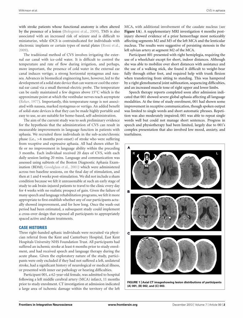
MCA, with additional involvement of the caudate nucleus (seeFigure 1A). A supplementary MRI investigation 4 months post-injury showed evidence of a prior hemorrhage most noticeablyaffecting segments M2 and M5 of the left MCA and the lenticularnucleus. The results were suggestive of persisting stenosis in theleft sylvian artery at segment M2 of the MCA.
Participant 001 presented with right hemiplegia, requiring theuse of a wheelchair except for short, indoor distances. Althoughshe was able to mobilize over short distances with assistance andthe use of a walking stick, she found it difficult to weight-bearfully through either foot, and required help with trunk flexionwhen transferring from sitting to standing. This was hamperedby a right glenohumeral joint subluxation, sequencing difficulties,and an increased muscle tone of right upper and lower limbs.
Speech therapy reports completed soon after admission indi-cated that 001 showed severe global aphasia affecting all languagemodalities. At the time of study enrolment, 001 had shown someimprovement in receptive communication, though spoken outputwas limited to single words and short automatic phrases. Repeti-tion was also moderately impaired; 001 was able to repeat singlewords well but could not manage short sentences. Progress inspeech and physiotherapy had been limited, largely due to 001’scomplex presentation that also involved low mood, anxiety, andtearfulness.
FIGURE 1 | Axial CT imageshowing lesion distributions of participants
(A) 001, (B) 002, and (C) 003.
Frontiers in Integrative Neuroscience www.frontiersin.org December 2013 | Volume 7 | Article 99 | 2
“fnint-07-00099” — 2013/12/19 — 20:48 — page 3 — #3
Wilkinson et al. CVS in aphasia
At the time of study enrolment, 001 was living semi-independently in her own home with the support of her husbandand carers. Her rehabilitation physician advised that her speechand language ability had been stable for several months. She wasstill unable to transfer independently and scored 3/5 on the Mod-ified Ashworth Scale (MAS; Bohannon and Smith, 1987) for theright flexor digitorum profundus and flexor digitorum superfi-cialis. Perceptual assessment revealed a mild, right-sided visualhemi-spatial neglect, as assessed by subtests of the Behavioral Inat-tention Test (Wilson et al., 1987). She also showed a constructionalapraxia, scoring within the moderate-severely impaired range forboth the copying accuracy and planning elements of the Rey–Osterrieth Complex Figure task (Stern et al., 1999), and failing toprogress past the initial trial of the WAIS-R Block Design subtest(Wechsler, 1981).
Participant 002, a 70-year-old male, was admitted to hospital8 months prior to study enrolment, having suffered a left MCAinfarct. A CT investigation revealed low attenuation in the leftbasal ganglia, left parietal, temporal and frontal lobes, plus sul-cal effacement and compression of the anterior horn of the leftlateral ventricle (see Figure 1B). A carotid Doppler test carriedout a month later indicated complete occlusion of the left internalcarotid artery.
Participant 002 presented with right hemiplegia, reduced coor-dination of the right upper limb and reduced balance. At the timeof study enrolment, 002 was able to walk short distances withsupervision and the use of a walking stick, though required theuse of a wheelchair when outdoors and had difficulty mobilizingdue to distractions. He showed difficulty with motor planningand sequencing and over-activity into flexion in his right upperlimb during functional tasks, though this could be overcome byprompting. Poor scapular control and awareness limited the activerange of his right glenohumeral joint.
Speech therapy prior to study enrolment indicated severeexpressive aphasia and apraxia of speech. Spontaneous spokenoutput was largely unintelligible. Repetition was also significantlyimpaired, even at single word level. Apraxic difficulties were evi-dent in attempts to copy lip and tongue movements and repeatsounds. In addition, 002 presented with moderate receptive dif-ficulties. Participant 002 had regularly expressed frustration andlow mood at difficulties during speech therapy sessions. At thetime of study enrolment, 002 was living semi-independently athome with the support of his wife and carers. His rehabilita-tion physician advised that his speech and language ability hadbeen stable for several months. On the Rey–Osterrieth ComplexFigure task, participant 002 scored within the low-average rangefor copying accuracy and below average/mildly impaired for theplanning element (Stern et al., 1999). He showed moderate tosevere impairment on the WAIS-R Block Design subtest (Wechsler,1981).
Participant 003, a-51-year old male, was admitted to hos-pital upon sudden onset of aphasia and right-sided weakness,22 months prior to study enrolment. A CT scan taken uponadmission revealed a low density area in the deep white matterof the left posterior frontal lobe, lateral to the anterior horn ofthe left ventricle (see Figure 1C). An additional MRI scan a weeklater indicated a hyperdense signal in the left temporal lobe, left
basal ganglia, left caudate nucleus, and internal capsule extendinginto the left semiovale. Magnetic resonance angiography (MRA)indicated complete occlusion of the left MCA and left internalcarotid.
Speech assessment prior to discharge from hospital high-lighted severe expressive and receptive aphasia. Follow up with thecommunity speech therapist suggested that his comprehensiondifficulties had mostly resolved following discharge, but spokenoutput remained limited to occasional words and short phrases.Participant 003 also presented with a mild apraxia of speech. Par-ticipant 003’s speech presentation had remained stable up until6 weeks prior to study enrolment, when a period of modest, spon-taneous recovery occurred. During this time participant 003 beganto say more words and sentences, though overall verbal outputstill remained limited. 003 further presented with a mild right-sided weakness resulting in restricted upper limb movement andreduced fine motor skills. There was muscle weakness of the rightlower limb, although he was completely mobile. He scored 5/5on the Functional Ambulation Categories (Holden et al., 1984),4/5 on the MRC muscle power testing (Medical Research Council,1986), and 1/5 on the MAS (Bohannon and Smith, 1987) for hisupper right limb, and 40/40 on the Modified Rivermead MobilityIndex (Lennon and Johnson, 2000). Tests of visual constructionand lateralised attention were not performed because, unlike par-ticipants 001 and 002, there was no mention of these capacitiesat hospital discharge. At the time of study enrolment, Participant003 was living largely independently at home with his wife.
LANGUAGE OUTCOME MEASURESParticipants were assessed on various sub-tests of the BDAE thirdEdition (Goodglass et al., 2001) – see Table 1.
TEST SCHEDULEFavorable ethical approval was gained from the University of KentPsychology Research Ethics Committee, and all participants gavewritten, informed consent prior to study.
Baseline assessments were administered 10 and 3 days prior tostimulation to gage pre-treatment language ability. CVS was per-formed from Monday to Friday for the next 4 weeks, and post-CVSassessments were conducted immediately after the last session andthen 1 and 4 weeks later. The language assessments were adminis-tered in the following order; auditory comprehension, repetition,naming, connected speech.
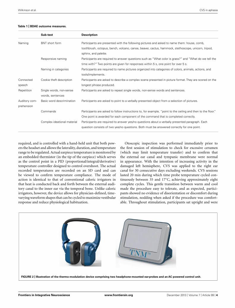
STIMULATIONCaloric vestibular stimulation was administered via a custom-built, experimental device that modulates the temperature ofsmall, thermo-electric, solid-state probes inserted into the rightand left external ear canals (see Figure 2). The probes are mountedon a headset and are too large to enter the bony portion of thecanal, resting instead on the outer fleshy portion. The probes areheld in place by first ensuring that the headphones are properlyseated on the head (which ensures that the probes are seated withinthe ear canals) and then fastening a velcro head-strap to preventfurther movement. Each probe can be warmed/cooled indepen-dently depending on whether unilateral or bilateral stimulation is
Frontiers in Integrative Neuroscience www.frontiersin.org December 2013 | Volume 7 | Article 99 | 3
“fnint-07-00099” — 2013/12/19 — 20:48 — page 4 — #4
Wilkinson et al. CVS in aphasia
Table 1 | BDAE outcome measures.
Sub-test Description
Naming BNT short form Participants are presented with the following pictures and asked to name them: house, comb,
toothbrush, octopus, bench, volcano, canoe, beaver, cactus, hammock, stethoscope, unicorn, tripod,
sphinx, and palette.
Responsive naming Participants are required to answer questions such as “What color is grass?” and “What do we tell the
time with?” Two points are given for responses within 5 s, one point for over 5 s.
Naming in categories Participants are required to name pictures organized into categories of colors, animals, actions, and
tools/implements.
Connected
speech
Cookie theft description Participants are asked to describe a complex scene presented in picture format. They are scored on the
longest phrase produced.
Repetition Single words, non-sense
words, sentences
Participants are asked to repeat single words, non-sense words and sentences.
Auditory com-
prehension
Basic word descrimination Participants are asked to point to a verbally presented object from a selection of pictures.
Commands Participants are asked to follow instructions to, for example, “point to the ceiling and then to the floor.”
One point is awarded for each component of the command that is completed correctly.
Complex ideational material Participants are required to answer yes/no questions about a verbally presented paragraph. Each
question consists of two yes/no questions. Both must be answered correctly for one point.
required, and is controlled with a hand-held unit that both pow-ers the headset and allows the laterality, duration, and temperaturerange to be regulated. Actual earpiece temperature is monitored byan embedded thermistor (in the tip of the earpiece) which servesas the control point in a PID (proportional/integral/derivative)temperature controller designed to control overshoot. The actualrecorded temperatures are recorded on an SD card and canbe viewed to confirm temperature compliance. The mode ofaction is identical to that of conventional caloric irrigators inthat heat is conducted back and forth between the external audi-tory canal to the inner ear via the temporal bone. Unlike caloricirrigators, however, the device allows for physician-defined, time-varying waveform shapes that can be cycled to maximize vestibularresponse and reduce physiological habituation.
Otoscopic inspection was performed immediately prior tothe first session of stimulation to check for excessive cerumen(which may limit temperature transfer) and to confirm thatthe external ear canal and tympanic membrane were normalin appearance. With the intention of increasing activity in thedamaged left hemisphere, CVS was applied to the right earcanal for 30 consecutive days excluding weekends. CVS sessionslasted 20 min during which time probe temperature cycled con-tinuously between 35 and 17◦C, achieving approximately eightcomplete cycles. This gentle transition between warm and coolmade the procedure easy to tolerate, and as expected, partici-pants showed no evidence of disorientation or discomfort duringstimulation, nodding when asked if the procedure was comfort-able. Throughout stimulation, participants sat upright and were
FIGURE 2 | Illustration of the thermo-modulation device comprising two headphone-mounted ear-probes and an AC powered control unit.
Frontiers in Integrative Neuroscience www.frontiersin.org December 2013 | Volume 7 | Article 99 | 4
“fnint-07-00099” — 2013/12/19 — 20:48 — page 5 — #5
Wilkinson et al. CVS in aphasia
not engaged in prolonged conversation or speech or languagetasks.
STATISTICAL APPROACHNinety five percent confidence intervals were constructed to deter-mine whether changes from baseline were greater than that whichcould be attributed to natural variation. For each subtest, upperand lower confidence limits were calculated as two standard errors(SE) above and below the baseline scores, respectively, using theequation SE = SD
√1 − ρ where ρ refers to Cronbach’s alpha
and where the SD was derived from the responses of an alliedsample containing 85 aphasic and 15 elderly normal participants(Jacobson et al., 1999; Goodglass et al., 2001; Reise and Haviland,2005). Given that each participant showed little change acrossthe two baseline administrations of each subtest, the two base-line scores were combined to produce a more reliable estimate oflanguage ability (Weller, 2007). Adjusted reliability estimates (ρ)were therefore calculated using the Spearman–Brown ProphecyFormula (see Nunnally, 1967). To reduce the likelihood of mistak-ing experimentally induced recovery with that which was alreadyoccuring, 95% confidence intervals were only calculated for thosesubtests in which the change between the baseline and post-CVSscores was numerically greater than the change between the twobaseline scores.
There were two instances in which an average baseline wasnot calculated; participant 001’s Basic Word Discrimination andparticipant 003’s Responsive Naming. In both instances, there wasa significant difference between the two baseline scores (thoughthis was a much smaller difference than was seen between preand post-CVS). In these cases it was deemed inappropriate touse an average pre-treatment score and therefore 95% confidenceintervals were calculated around the higher of the two baselinemeasures. In these cases, reliability of the original scale, as opposedto the combined scale, was used.
To provide broader insight into wellbeing and functional recov-ery, written testimonials from participants’ relative/carers arepresented below alongside the inferential statistics. These testi-monials are, of course, anecdotal and subjective, but neverthelesshold corroborative value.
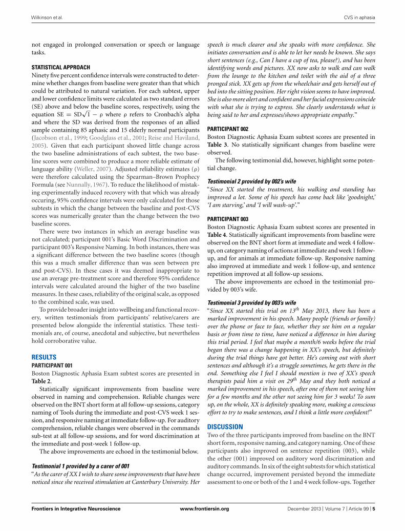
RESULTSPARTICIPANT 001Boston Diagnostic Aphasia Exam subtest scores are presented inTable 2.
Statistically significant improvements from baseline wereobserved in naming and comprehension. Reliable changes wereobserved on the BNT short form at all follow-up sessions, categorynaming of Tools during the immediate and post-CVS week 1 ses-sion, and responsive naming at immediate follow-up. For auditorycomprehension, reliable changes were observed in the commandssub-test at all follow-up sessions, and for word discrimination atthe immediate and post-week 1 follow-up.
The above improvements are echoed in the testimonial below.
Testimonial 1 provided by a carer of 001“As the carer of XX I wish to share some improvements that have beennoticed since she received stimulation at Canterbury University. Her
speech is much clearer and she speaks with more confidence. Sheinitiates conversation and is able to let her needs be known. She saysshort sentences (e.g., Can I have a cup of tea, please?), and has beenidentifying words and pictures. XX now asks to walk and can walkfrom the lounge to the kitchen and toilet with the aid of a threepronged stick. XX gets up from the wheelchair and gets herself out ofbed into the sitting position. Her right vision seems to have improved.She is also more alert and confident and her facial expressions coincidewith what she is trying to express. She clearly understands what isbeing said to her and expresses/shows appropriate empathy.”
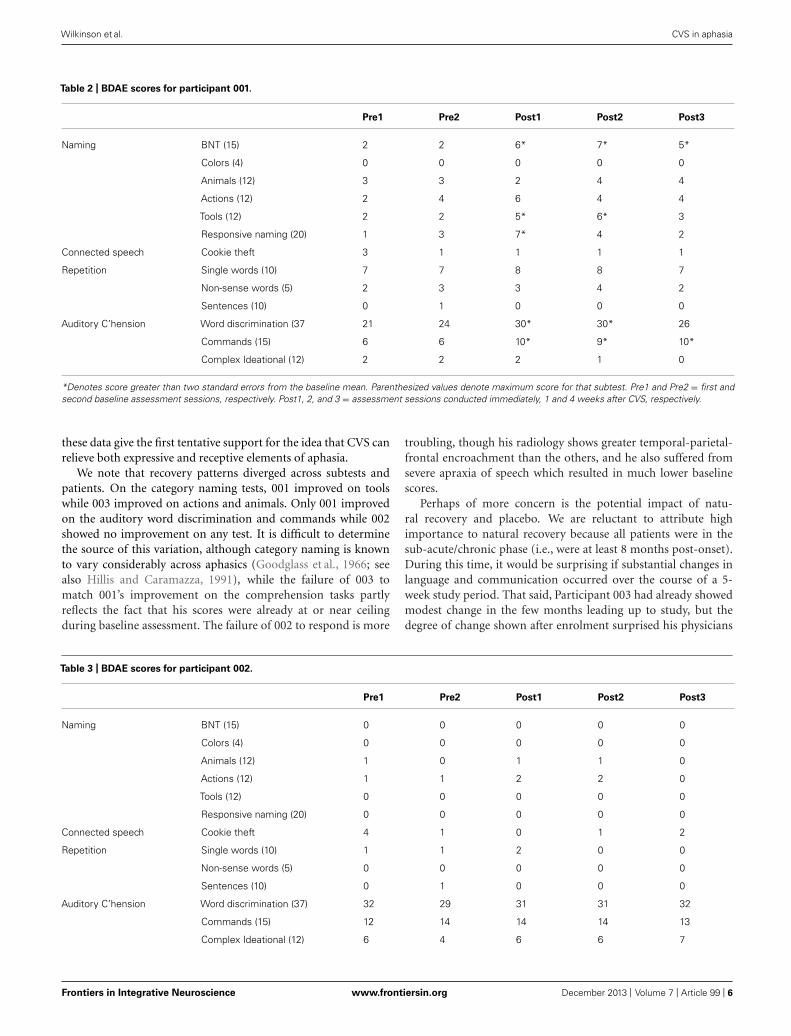
PARTICIPANT 002Boston Diagnostic Aphasia Exam subtest scores are presented inTable 3. No statistically significant changes from baseline wereobserved.
The following testimonial did, however, highlight some poten-tial change.
Testimonial 2 provided by 002’s wife“Since XX started the treatment, his walking and standing hasimproved a lot. Some of his speech has come back like ‘goodnight,’‘I am starving,’ and ‘I will wash-up’.”
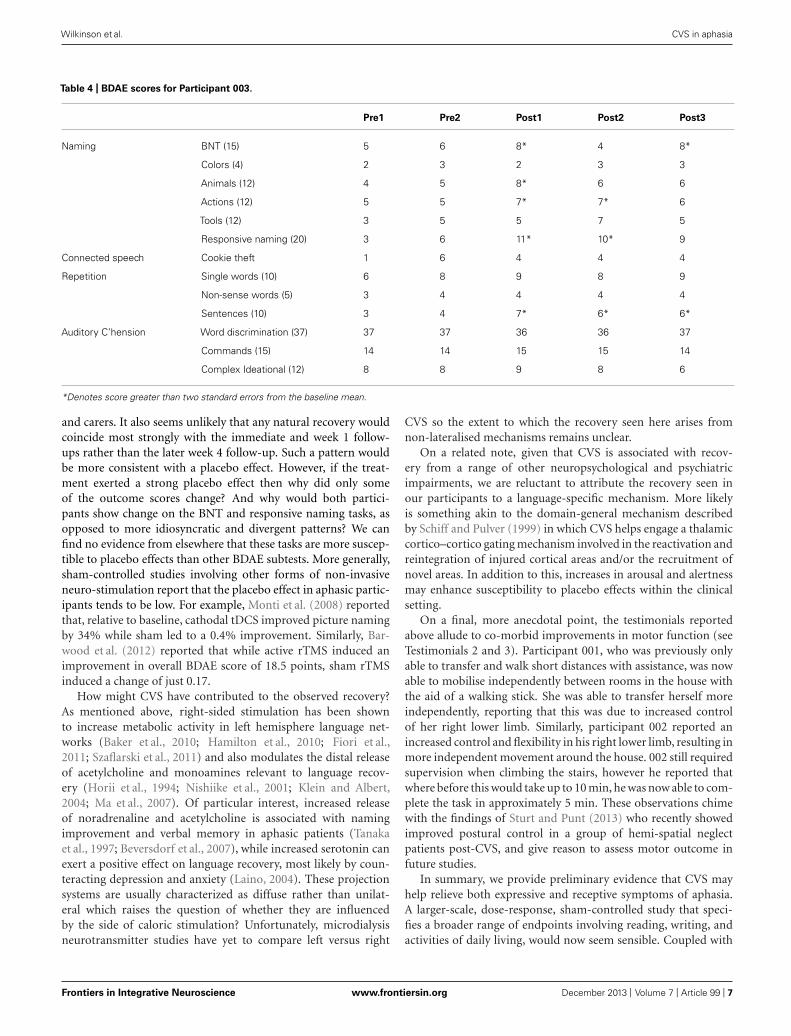
PARTICIPANT 003Boston Diagnostic Aphasia Exam subtest scores are presented inTable 4. Statistically significant improvements from baseline wereobserved on the BNT short form at immediate and week 4 follow-up, on category naming of actions at immediate and week 1 follow-up, and for animals at immediate follow-up. Responsive namingalso improved at immediate and week 1 follow-up, and sentencerepetition improved at all follow-up sessions.
The above improvements are echoed in the testimonial pro-vided by 003’s wife.
Testimonial 3 provided by 003’s wife“Since XX started this trial on 13th May 2013, there has been amarked improvement in his speech. Many people (friends or family)over the phone or face to face, whether they see him on a regularbasis or from time to time, have noticed a difference in him duringthis trial period. I feel that maybe a month/6 weeks before the trialbegan there was a change happening in XX’s speech, but definitelyduring the trial things have got better. He’s coming out with shortsentences and although it’s a struggle sometimes, he gets there in theend. Something else I feel I should mention is two of XX’s speechtherapists paid him a visit on 29th May and they both noticed amarked improvement in his speech, after one of them not seeing himfor a few months and the other not seeing him for 3 weeks! To sumup, on the whole, XX is definitely speaking more, making a consciouseffort to try to make sentences, and I think a little more confident!”
DISCUSSIONTwo of the three participants improved from baseline on the BNTshort form, responsive naming, and category naming. One of theseparticipants also improved on sentence repetition (003), whilethe other (001) improved on auditory word discrimination andauditory commands. In six of the eight subtests for which statisticalchange occurred, improvement persisted beyond the immediateassessment to one or both of the 1 and 4 week follow-ups. Together
Frontiers in Integrative Neuroscience www.frontiersin.org December 2013 | Volume 7 | Article 99 | 5
“fnint-07-00099” — 2013/12/19 — 20:48 — page 6 — #6
Wilkinson et al. CVS in aphasia
Table 2 | BDAE scores for participant 001.
Pre1 Pre2 Post1 Post2 Post3
Naming BNT (15) 2 2 6* 7* 5*
Colors (4) 0 0 0 0 0
Animals (12) 3 3 2 4 4
Actions (12) 2 4 6 4 4
Tools (12) 2 2 5* 6* 3
Responsive naming (20) 1 3 7* 4 2
Connected speech Cookie theft 3 1 1 1 1
Repetition Single words (10) 7 7 8 8 7
Non-sense words (5) 2 3 3 4 2
Sentences (10) 0 1 0 0 0
Auditory C’hension Word discrimination (37 21 24 30* 30* 26
Commands (15) 6 6 10* 9* 10*
Complex Ideational (12) 2 2 2 1 0
*Denotes score greater than two standard errors from the baseline mean. Parenthesized values denote maximum score for that subtest. Pre1 and Pre2 = first andsecond baseline assessment sessions, respectively. Post1, 2, and 3 = assessment sessions conducted immediately, 1 and 4 weeks after CVS, respectively.
these data give the first tentative support for the idea that CVS canrelieve both expressive and receptive elements of aphasia.
We note that recovery patterns diverged across subtests andpatients. On the category naming tests, 001 improved on toolswhile 003 improved on actions and animals. Only 001 improvedon the auditory word discrimination and commands while 002showed no improvement on any test. It is difficult to determinethe source of this variation, although category naming is knownto vary considerably across aphasics (Goodglass et al., 1966; seealso Hillis and Caramazza, 1991), while the failure of 003 tomatch 001’s improvement on the comprehension tasks partlyreflects the fact that his scores were already at or near ceilingduring baseline assessment. The failure of 002 to respond is more
troubling, though his radiology shows greater temporal-parietal-frontal encroachment than the others, and he also suffered fromsevere apraxia of speech which resulted in much lower baselinescores.
Perhaps of more concern is the potential impact of natu-ral recovery and placebo. We are reluctant to attribute highimportance to natural recovery because all patients were in thesub-acute/chronic phase (i.e., were at least 8 months post-onset).During this time, it would be surprising if substantial changes inlanguage and communication occurred over the course of a 5-week study period. That said, Participant 003 had already showedmodest change in the few months leading up to study, but thedegree of change shown after enrolment surprised his physicians
Table 3 | BDAE scores for participant 002.
Pre1 Pre2 Post1 Post2 Post3
Naming BNT (15) 0 0 0 0 0
Colors (4) 0 0 0 0 0
Animals (12) 1 0 1 1 0
Actions (12) 1 1 2 2 0
Tools (12) 0 0 0 0 0
Responsive naming (20) 0 0 0 0 0
Connected speech Cookie theft 4 1 0 1 2
Repetition Single words (10) 1 1 2 0 0
Non-sense words (5) 0 0 0 0 0
Sentences (10) 0 1 0 0 0
Auditory C’hension Word discrimination (37) 32 29 31 31 32
Commands (15) 12 14 14 14 13
Complex Ideational (12) 6 4 6 6 7
Frontiers in Integrative Neuroscience www.frontiersin.org December 2013 | Volume 7 | Article 99 | 6
“fnint-07-00099” — 2013/12/19 — 20:48 — page 7 — #7
Wilkinson et al. CVS in aphasia
Table 4 | BDAE scores for Participant 003.
Pre1 Pre2 Post1 Post2 Post3
Naming BNT (15) 5 6 8* 4 8*
Colors (4) 2 3 2 3 3
Animals (12) 4 5 8* 6 6
Actions (12) 5 5 7* 7* 6
Tools (12) 3 5 5 7 5
Responsive naming (20) 3 6 11* 10* 9
Connected speech Cookie theft 1 6 4 4 4
Repetition Single words (10) 6 8 9 8 9
Non-sense words (5) 3 4 4 4 4
Sentences (10) 3 4 7* 6* 6*
Auditory C’hension Word discrimination (37) 37 37 36 36 37
Commands (15) 14 14 15 15 14
Complex Ideational (12) 8 8 9 8 6
*Denotes score greater than two standard errors from the baseline mean.
and carers. It also seems unlikely that any natural recovery wouldcoincide most strongly with the immediate and week 1 follow-ups rather than the later week 4 follow-up. Such a pattern wouldbe more consistent with a placebo effect. However, if the treat-ment exerted a strong placebo effect then why did only someof the outcome scores change? And why would both partici-pants show change on the BNT and responsive naming tasks, asopposed to more idiosyncratic and divergent patterns? We canfind no evidence from elsewhere that these tasks are more suscep-tible to placebo effects than other BDAE subtests. More generally,sham-controlled studies involving other forms of non-invasiveneuro-stimulation report that the placebo effect in aphasic partic-ipants tends to be low. For example, Monti et al. (2008) reportedthat, relative to baseline, cathodal tDCS improved picture namingby 34% while sham led to a 0.4% improvement. Similarly, Bar-wood et al. (2012) reported that while active rTMS induced animprovement in overall BDAE score of 18.5 points, sham rTMSinduced a change of just 0.17.
How might CVS have contributed to the observed recovery?As mentioned above, right-sided stimulation has been shownto increase metabolic activity in left hemisphere language net-works (Baker et al., 2010; Hamilton et al., 2010; Fiori et al.,2011; Szaflarski et al., 2011) and also modulates the distal releaseof acetylcholine and monoamines relevant to language recov-ery (Horii et al., 1994; Nishiike et al., 2001; Klein and Albert,2004; Ma et al., 2007). Of particular interest, increased releaseof noradrenaline and acetylcholine is associated with namingimprovement and verbal memory in aphasic patients (Tanakaet al., 1997; Beversdorf et al., 2007), while increased serotonin canexert a positive effect on language recovery, most likely by coun-teracting depression and anxiety (Laino, 2004). These projectionsystems are usually characterized as diffuse rather than unilat-eral which raises the question of whether they are influencedby the side of caloric stimulation? Unfortunately, microdialysisneurotransmitter studies have yet to compare left versus right
CVS so the extent to which the recovery seen here arises fromnon-lateralised mechanisms remains unclear.
On a related note, given that CVS is associated with recov-ery from a range of other neuropsychological and psychiatricimpairments, we are reluctant to attribute the recovery seen inour participants to a language-specific mechanism. More likelyis something akin to the domain-general mechanism describedby Schiff and Pulver (1999) in which CVS helps engage a thalamiccortico–cortico gating mechanism involved in the reactivation andreintegration of injured cortical areas and/or the recruitment ofnovel areas. In addition to this, increases in arousal and alertnessmay enhance susceptibility to placebo effects within the clinicalsetting.
On a final, more anecdotal point, the testimonials reportedabove allude to co-morbid improvements in motor function (seeTestimonials 2 and 3). Participant 001, who was previously onlyable to transfer and walk short distances with assistance, was nowable to mobilise independently between rooms in the house withthe aid of a walking stick. She was able to transfer herself moreindependently, reporting that this was due to increased controlof her right lower limb. Similarly, participant 002 reported anincreased control and flexibility in his right lower limb, resulting inmore independent movement around the house. 002 still requiredsupervision when climbing the stairs, however he reported thatwhere before this would take up to 10 min, he was now able to com-plete the task in approximately 5 min. These observations chimewith the findings of Sturt and Punt (2013) who recently showedimproved postural control in a group of hemi-spatial neglectpatients post-CVS, and give reason to assess motor outcome infuture studies.
In summary, we provide preliminary evidence that CVS mayhelp relieve both expressive and receptive symptoms of aphasia.A larger-scale, dose-response, sham-controlled study that speci-fies a broader range of endpoints involving reading, writing, andactivities of daily living, would now seem sensible. Coupled with
Frontiers in Integrative Neuroscience www.frontiersin.org December 2013 | Volume 7 | Article 99 | 7
“fnint-07-00099” — 2013/12/19 — 20:48 — page 8 — #8
Wilkinson et al. CVS in aphasia
the beneficial effects of CVS on other stroke conditions such ashemi-spatial neglect (Cappa et al., 1987), pain (McGeoch et al.,2008), and hemi-anesthesia (Bottini et al., 2005), the current dataalso strengthen the growing idea that CVS triggers a generic com-pensatory response to brain trauma which may be of relevanceto a wide variety of neurological conditions. Given the need formany different brain systems to know if the head is upright, mov-ing and if so, in what direction and at what speed, we believethat the therapeutic reach of vestibular stimulation will proveconsiderable.
ACKNOWLEDGMENTSWe are grateful for the cooperation of the three participants andtheir spouses. We also thank Scion Neurostim LLC for providingthe vestibular stimulation equipment and for methodological andtechnical assistance.
REFERENCESBaker, J. M., Rorden, C., and Fridriksson, J. (2010). Using transcranial direct-
current stimulation to treat stroke patients with aphasia. Stroke 41, 1229–1236.doi: 10.1161/STROKEAHA.109.576785
Barwood, C. H., Murdoch, B. E., Whelan, B. M., Lloyd, D., Riek, S., O’Sullivan, J. D.,et al. (2012). Improved receptive and expressive language abilities in non-fluentaphasic stroke patients after application of rTMS: an open protocol study. BrainStimul. 5, 274–286. doi: 10.1016/j.brs.2011.03.005
Berthier, M. L. (2005). Poststroke aphasia epidemiology, pathophysiology andtreatment. Drugs Aging 22, 163–182. doi: 10.2165/00002512-200522020-00006
Beversdorf, D. Q., Sharma, U. K., Phillips, N. N., Notestine, M. A., Slivka, A. P.,Friedman, N. M., et al. (2007). Effect of propranololon naming in chronic Broca’saphasia with anomia. Neurocase 13, 256–259. doi: 10.1080/13554790701595471
Bohannon, P., and Smith, M. (1987). Interrater reliability of a modified Ashworthscale of muscle spasticity. Phys. Ther. 67, 206.
Bolognini, N., Pascual-Leone, A., and Fregni, F. (2009). Using non-invasive brainstimulation to augment motor training-induced plasticity. J. Neuroeng. Rehabil.6, 1–13. doi: 10.1186/1743-0003-6-8
Bottini, G., Paulesu, E., Gandola, M., Loffredo, S., Scarpa, P., Sterzi, R., et al. (2005).Left caloric vestibular stimulation ameliorates right hemianesthesia. Neurology65, 1278–1283. doi: 10.1212/01.wnl.0000182398.14088.e8
Brady, M. C., Kelly, H., Goodwin, J., and Enderby, P. (2012). Speech and lan-guage therapy for aphasia following stroke. Cochrane Database Syst. Rev. 5. doi:10.1002/14651858.CD000425.pub3
Cappa, S., Sterzi, R., Vallar, G., and Bisiach, E. (1987). Remission of hemineglectand anosognosia during vestibular stimulation. Neurospsychologia 25, 775–782.doi: 10.1016/0028-3932(87)90115-1
Code, C., and Herrmann, M. (2003). The relevance of emotional and psychoso-cial factors in aphasia to rehabilitation and neuropsychologial rehabilitation.Neuropsychol. Rehabil. 13, 109–132. doi: 10.1080/09602010244000291
Dieterich, M., Bense, S., Lutz, S., Drzezga, A., Stephan, T., Bartenstein, P.,et al. (2003). Dominance for vestibular cortical function in the non-dominanthemisphere. Cereb. Cortex 13, 994–1007. doi: 10.1093/cercor/13.9.994
Fasold, O., von Brevern, M., Kuhberg, M., Ploner, C. J., Villringer, A., Lempert,T., et al. (2002). Human vestibular cortex as identified wuth caloric vestibularstimulation in functional magnetic resonance imaging. Neuroimage 17, 1384–1393. doi: 10.1006/nimg.2002.1241
Fiori, V., Coccia, M., Marinelli, C. V., Vecchi, V., Bonifazi, S., Ceravelo, M. G.,et al. (2011). Transcranial direct current stimulation improves word retrieval inhealthy and nonfluent aphasic subjects. J. Cogn. Neurosci. 23, 2309–2323. doi:10.1162/jocn.2010.21579
Goodglass, H., Kaplan, E., and Barresi, B. (2001). The Assessment of Aphasia andRelated Disorders, 3rd Edn. Philadelphia, PA: Lippincott, Williams and Wilkins.
Goodglass, H., Klein, B., and Jones, K. (1966). Specific semantic word categories inaphasia. Cortex 2, 74–89. doi: 10.1016/S0010-9452(66)80029-1
Hamilton, R. H., Chrysikou, E. G., and Coslett, B. (2011). Mechanisms of aphasiarecovery after stroke and the role of noninvasive brain stimulation. Brain Lang.118, 40–50. doi: 10.1016/j.bandl.2011.02.005
Hamilton, R. H., Sanders, L., Benson, Y., Faseyitan, O., Norise, C., Naeser, M., et al.(2010). Stimulating conversation: enhancement of elicited propositional speechin a patient with chronic non-fluent aphasia following transcranial magneticstimulation. Brain Lang. 113, 45–50. doi: 10.1016/j.bandl.2010.01.001
Heiss, W. D., and Thiel, A. (2006). A proposed regional hierarchy in recovery ofpost-stroke aphasia. Brain Lang. 98, 118–123. doi: 10.1016/j.bandl.2006.02.002
Hilari, K. (2011). The impact of stroke: are people with aphasia different to thosewithout? Disabil. Rehabil. 33, 211–218. doi: 10.3109/09638288.2010.508829
Hillis, A. E., and Caramazza, A. (1991). Category-specific naming and com-prehension impairment: a double dissociation. Brain 141, 2081–2094. doi:10.1093/brain/114.5.2081
Holden, M. L., Gill, K. M., Magliozzi, M. R., Nathan, J., and Piehl-Baker, L.(1984). Clinical gait assessment in the neurologically impaired. Reliability andmeaningfulness. Phys. Ther. 64, 35–40.
Holstein, G., Friedrich, V., Martinelli, G., Ogorodnikov, D., Yakushin, S., and Cohen,B. (2012). Fos expression in neurons of the rat vestibulo-autonomic pathwayactivated by sinusoidal galvanic vestibular stimulation. Front. Neurol. 3:4. doi:10.3389/fneur.2012.00004
Horii, A., Takeda, N., Mochizuki, T., Okakura-Mochizuki, K., Yamamoto, A., andYamatodani, A. (1994). Effects of vestibular stimulation on acetylcholine releasefrom rat hippocampus: an in vivo microdislysis study. J. Neurophysiol. 72, 605–611.
Jacobson, N. S., Roberts, L. J., Berns, S. B., and McGlinchey, J. B. (1999). Meth-ods for defining and determining the clinical significance of treatment effects:description, application, an alternatives. J. Consult. Clin. Psychol. 67, 300–307.doi: 10.1037/0022-006X.67.3.300
Kang, E., Kim, Y., Sohn, H., Cohen, L. G., and Paik, N. (2011). Improved picturenaming in aphasia patients treated with cathodal tDCS to inhibit the right Broca’shomologue area. Restor. Neurol. Neurosci. 29, 141–152. doi: 10.3233/RNN-2011-0587
Kelly, H., Brady, M. C., and Enderby, P. (2010). Speech and languagetherapy for aphasia following stroke. Cochrane Database Syst. Rev. 5.doi:10.1002/14651858.CD000425.pub2
Klein, R. B., and Albert, M. L. (2004). Can drug therapies improve language func-tions of individuals with aphasia? A review of the evidence. Semin. Speech Lang.25, 193–204. doi: 10.1055/s-2004-825655
Laino, C. (2004). Serotonergic therapy shows promise for aphasia. Neurol. Today 4,54. doi: 10.1097/00132985-200406000-00017
Lennon, S., and Johnson, L. (2000). The modified rivermead mobility index: validityand reliability. Disabil. Rehabil. 22, 833–839. doi: 10.1080/09638280050207884
Ma, F. R., Liu, J. X., Li, X. P., Mao, J. J., Zhang, Q. D., Jia, H. B., et al. (2007). Effects ofcaloric vestibular stimulation on serotononinergic system in the media vestibulatnuclei of guinea pigs. Chin. Med. J. 120, 120–124.
Magrun, W. M., Ottenbacher, K., McCue, S., and Keefe, R. (1981). Effects of vestibu-lar stimulation on spontanouse use of verbal language in developmentally delayedchildren. Am. J. Occup. Ther. 35, 101–104. doi: 10.5014/ajot.35.2.101
McGeoch, P. D., Williams, L. E., Lee, R. R., and Ramachandran, V. S. (2008).Behavioural evidence for vestibular stimulation as treatment as treatment forcentral post-stroke pain. J. Neurol. Neurosurg. Psychiatry 79, 1298–1301. doi:10.1136/jnnp.2008.146738
Medical Research Council. (1986). Aids to the Examination of the Peripheral NervousSystem. London: Baillière Tindall.
Meinzer, M., Djundja, D., Barthel, G., Elbert, T., and Rockstroh, B.(2005). Long term stability of improved language functions in chronic apha-sia after constraint-induced aphasia therapy. Stroke 36, 1462–1466. doi:10.1161/01.STR.0000169941.29831.2a
Monti, A., Cogiamanian, F., Marceglia, S., Ferrucci, R., Mameli, F., Mrakic-Sposta, S., et al. (2008). Improved naming after transcranial direct currentstimulation in aphasia. J. Neurol. Neurosurg. Psychiatry 79, 451–453. doi:10.1136/jnnp.2007.135277
Muira, K., Nakamura, Y., Muira, F., Yamada, I., Takahashi, M., Yoshikawa, A., et al.(1999). Functional magnetic resonance imaging to word generation task in apatient with Broca’s aphasia. J. Neurol. 246, 939–942. doi: 10.1007/s004150050486
Naeser, M. A., Martin, P. I., Nicholas, M., Baker, E. H., Seekins, H., Helm-Estabrooks,N., et al. (2005a). Improved naming after TMS treatments in a chronic, globalaphasia patient. Neurocase 11, 182–193. doi: 10.1080/13554790590944663
Naeser, M. A., Martin, P. I., Nicholas, M., Baker, E. H., Seekins, H., Kobayashi,M., et al. (2005b). Improved picture naming in chronic aphasia after rTMS
Frontiers in Integrative Neuroscience www.frontiersin.org December 2013 | Volume 7 | Article 99 | 8
“fnint-07-00099” — 2013/12/19 — 20:48 — page 9 — #9
Wilkinson et al. CVS in aphasia
to part of Broca’s area: an open protocol study. Brain Lang. 93, 95–105. doi:10.1016/j.bandl.2004.08.004
Nishiike, S., Takeda, N., Kubo, T., and Nakamura, S. (2001). Noradrenergic pathwaysinvolved in the development of vertigo and dizziness – a review. Acta Otolaryngol.Suppl. 545, 61–64. doi: 10.1080/000164801750388135
Nunnally, J. C. (1967). Psychometric Theory. New York: McGraw-Hill.Paolucci, S., Matano, A., Bragoni, M., Coiro, P., De Angelis, D., Fusco, F. R., et al.
(2005). Rehabilitation of left brain-damaged ischemic stroke patients: the role ofcomprehension language deficits. A matched comparison. Cerebrovasc. Dis. 20,400–406. doi: 10.1159/000088671
Reise, S. P., and Haviland, M. G. (2005). Item response theory and the measure-ment of clinical change. J. Pers. Assess. 84, 228–238. doi: 10.1207/s15327752jpa8403_02
Reker, U. (1977). Caloric diagnosis. Arch. Otolaryngol. 214, 247–256.Robey, R. R. (1994). The efficacy of treatment for aphasic persons: a meta analysis.
Brain Lang. 47, 582–608. doi: 10.1006/brln.1994.1060Rossi, S., Hallett, M., Rossini, P. M., Pascual-Leone, A., and Safety of TMS Consensus
Group. (2009). Safety, ethical considerations, and application guidelines for theuse of transcranial magnetic stimulation in clinical practise and research. Clin.Neurophysiol. 120, 2008–2039. doi: 10.1016/j.clinph.2009.08.016
Schiff, N. D., and Pulver, M. (1999). Does vestibular stimulation activate thalam-ocortical mechanisms that reintegrate impaires cortical regions? Proc. R. Soc. BBiol. Sci. 266, 421–423. doi: 10.1098/rspb.1999.0654
Stern, R., Javorsky, D., Singer, E., Singer-Harris, N., Somerville, J., Duke, L., et al.(1999). The Boston Qualitative Scoring System. Florida: Psychological AssessmentResources.
Sturt, R., and Punt, T. D. (2013). Caloric vestibular stimulation and postural controlin patients with spatial neglect following stroke. Neuropsychol. Rehabil. 23, 299–316. doi: 10.1080/09602011.2012.755831
Szaflarski, J. P., Vannest, J., Wu, S. W., DiFrancesco, M. W., Banks, C., and Gilbert,D. L. (2011). Excitatory repetitive transcranial magnetic stimulation inducesimprovements in chronic post-stroke aphasia. Med. Sci. Monit. 17, 132–139.doi: 10.12659/MSM.881446
Tanaka, Y., Miyazaki, M., and Albert, M. (1997). Effects of increased cholinergicactivity on naming in aphasia. Lancet 350, 116–117. doi: 10.1016/S0140-6736(05)61820-X
Vallar, G., Papagno, C., Rusconi, M. L., and Bisiach, E. (1995). Vestibular stimulation,spatial hemineglect and dysphasia. Selective effects? Cortex 31, 589–593. doi:10.1016/S0010-9452(13)80070-6
Wechsler, D. (1981). Manual for the Wechsler Adult Intelligence Scale – Revised. NewYork: Psychological Corporation N.
Weller, S. C. (2007). Cultural consensus theory: applications and frequently askedquestions. Field Methods 19, 339–368. doi: 10.1177/1525822X07303502
Wilson, B. A., Cockburn, J., and Halligan, P. W. (1987). Behavioural Inattention Test.Titchfield: Thames Valley Test Company Ltd.
You, D. S., Kim, D. Y., Chun, M. H., Jung, S. E., and Parks, S. J. (2011).Cathodal transcranial direct current stimulation of the right Wernicke’s areaimproves comprehension in subacute stroke patients. Brain Lang. 119, 1–5. doi:10.1016/j.bandl.2011.05.002
Conflict of Interest Statement: The authors declare that the research was conductedin the absence of any commercial or financial relationships that could be construedas a potential conflict of interest.
Received: 20 September 2013; paper pending published: 08 November 2013; accepted:08 December 2013; published online: 23 December 2013.Citation: Wilkinson D, Morris R, Milberg W and Sakel M (2013) Caloricvestibular stimulation in aphasic syndrome. Front. Integr. Neurosci. 7:99. doi:10.3389/fnint.2013.00099This article was submitted to the journal Frontiers in Integrative Neuroscience.Copyright © 2013 Wilkinson, Morris, Milberg and Sakel. This is an open-access articledistributed under the terms of the Creative Commons Attribution License (CC BY). Theuse, distribution or reproduction in other forums is permitted, provided the originalauthor(s) or licensor are credited and that the original publication in this journal is cited,in accordance with accepted academic practice. No use, distribution or reproduction ispermitted which does not comply with these terms.
Frontiers in Integrative Neuroscience www.frontiersin.org December 2013 | Volume 7 | Article 99 | 9
Related Documents