Journal of Neurology, Neurosurgery, and Psychiatry, 1980, 43, 127-135 Brain damage and neurological outcome after open-heart surgery K A SOTANIEMI From the Department of Neurology, University of Oulu, Oulu, Finland S U M M A RY Cerebral damage remains a major hazard of open-heart surgery. A one-year follow-up investigation of 100 consecutive patients who underwent open-heart operation for valve replace- ment revealed an incidence of postoperative cerebral disorders of 37%. The occurrence of brain damage was clearly related to the presence of a history of previous neurological diseases, to operative hypoxia, and to unexpected events during operation, but long perfusion time proved to be the most significant risk factor. In contrast to previous findings, age and moderate operative hypotension proved unimportant. The abnormalities tended to resolve rapidly but, one year after operation seven patients still displayed residual signs. An interesting interhemispheric difference in susceptibility to damage was evident, the clinical signs indicating lesions of the right hemisphere in 71 % of the damaged cases. The nature of both preoperative and postoperative signs and symptoms, the determinants of brain damage and the significance of the observed disparity between the hemispheres are discussed. The continuous occurrence of brain damage obliges us to develop preventive measures more efficient than those now available. Cerebral disorders related to cardiac surgery still occur despite recent advances in surgical tech- nique and extracorporeal circulation devices, these methods being far from perfect.`' With the ever-increasing number of applications of open- heart procedures, there is a continuing need to recognise the potential threat of brain damage and to consider the factors which determine the outcome. The reported incidence of cerebral damag.e after open-heart surgery varies widely. In pro- spective investigations incidences of 5% to 53% have been reported.2 47 In retrospective studies the incidence has usually been smaller: 19-2% in a study of 417 patients8 and 7-4% in a more recent study in 538 patients.1 By contrast, in an earlier investigation of 245 patients no obvious neurological disorders were found.9 The results are, however, not exactly compara.ble with each other because of differences in patients' age groups and cardiological diagnoses, in performed Address for reprint requests: Dr Kyosti Sotaniemi, MD, Depart- ment of Neurology, University of Oulu, 90220 Oulu 22, Finland. Accepted 7 August 1979 op.erative procedures, and, particularly, in the criteria of neurological evaluation: furthermore, the evaluation has not always been carried out by a neurologist. The complications detected have been found to affect all levels of the nervous system. Hemi- spheral damage, resulting in various forms of motor and sensory defects or cognitive and in- tellectual impairment or psychiatric disorders, cranial nerve disturbances, and cerebellar or brain stem lesions are among the abnormalities most often reported.5 8 10 Peripheral neuropathy has also been reported1'" as well as involvement of the brachial plexus.'3 The causes and mechanisms of brain damage have been thoroughly discussed in several re- ports.' 3 6 " Microembolisation and inadequate cerebral perfusion seem to be regarded as the most probabl.e mechanisms. Microembolisation may originate from the heart or from the extra- corporeal circuit.6 1..8 .. The composition of emboli has been recognised to be varied: air, fat, antifoam particles, fragments of valvular debris, aggregates of platelets or leucocytes or denatured blood proteins.7 18 20 21 Gaseous microembolism 127 c Protected by copyright. on 5 June 2018 by guest. http://jnnp.bmj.com/ J Neurol Neurosurg Psychiatry: first published as 10.1136/jnnp.43.2.127 on 1 February 1980. Downloaded from

Welcome message from author
This document is posted to help you gain knowledge. Please leave a comment to let me know what you think about it! Share it to your friends and learn new things together.
Transcript
Journal of Neurology, Neurosurgery, and Psychiatry, 1980, 43, 127-135
Brain damage and neurological outcome afteropen-heart surgery
K A SOTANIEMI
From the Department of Neurology, University of Oulu, Oulu, Finland
S U M M A R Y Cerebral damage remains a major hazard of open-heart surgery. A one-year follow-upinvestigation of 100 consecutive patients who underwent open-heart operation for valve replace-ment revealed an incidence of postoperative cerebral disorders of 37%. The occurrence of braindamage was clearly related to the presence of a history of previous neurological diseases, to operativehypoxia, and to unexpected events during operation, but long perfusion time proved to be the mostsignificant risk factor. In contrast to previous findings, age and moderate operative hypotensionproved unimportant. The abnormalities tended to resolve rapidly but, one year after operation sevenpatients still displayed residual signs. An interesting interhemispheric difference in susceptibility todamage was evident, the clinical signs indicating lesions of the right hemisphere in 71 % of thedamaged cases.The nature of both preoperative and postoperative signs and symptoms, the determinants of brain
damage and the significance of the observed disparity between the hemispheres are discussed. Thecontinuous occurrence of brain damage obliges us to develop preventive measures more efficient thanthose now available.
Cerebral disorders related to cardiac surgery stilloccur despite recent advances in surgical tech-nique and extracorporeal circulation devices,these methods being far from perfect.`' With theever-increasing number of applications of open-heart procedures, there is a continuing need torecognise the potential threat of brain damageand to consider the factors which determine theoutcome.The reported incidence of cerebral damag.e
after open-heart surgery varies widely. In pro-spective investigations incidences of 5% to 53%have been reported.2 47 In retrospective studiesthe incidence has usually been smaller: 19-2% ina study of 417 patients8 and 7-4% in a morerecent study in 538 patients.1 By contrast, in anearlier investigation of 245 patients no obviousneurological disorders were found.9 The resultsare, however, not exactly compara.ble with eachother because of differences in patients' agegroups and cardiological diagnoses, in performed
Address for reprint requests: Dr Kyosti Sotaniemi, MD, Depart-ment of Neurology, University of Oulu, 90220 Oulu 22,Finland.
Accepted 7 August 1979
op.erative procedures, and, particularly, in thecriteria of neurological evaluation: furthermore,the evaluation has not always been carried outby a neurologist.The complications detected have been found
to affect all levels of the nervous system. Hemi-spheral damage, resulting in various forms ofmotor and sensory defects or cognitive and in-tellectual impairment or psychiatric disorders,cranial nerve disturbances, and cerebellar orbrain stem lesions are among the abnormalitiesmost often reported.5 810 Peripheral neuropathyhas also been reported1'" as well as involvementof the brachial plexus.'3The causes and mechanisms of brain damage
have been thoroughly discussed in several re-ports.'3 6 " Microembolisation and inadequatecerebral perfusion seem to be regarded as themost probabl.e mechanisms. Microembolisationmay originate from the heart or from the extra-corporeal circuit.6 1..8.. The composition ofemboli has been recognised to be varied: air, fat,antifoam particles, fragments of valvular debris,aggregates of platelets or leucocytes or denaturedblood proteins.7 18 20 21 Gaseous microembolism
127
c
Protected by copyright.
on 5 June 2018 by guest.http://jnnp.bm
j.com/
J Neurol N
eurosurg Psychiatry: first published as 10.1136/jnnp.43.2.127 on 1 F
ebruary 1980. Dow
nloaded from

128
originating from the pump has been verified byultrasonic methods22 and it has been shown tobe reduced by microfiltration.23 Negative pressureconditions in the circuit supposedly assist ingenerating gaseous microemboli."7 Differencesbetween the types of oxygenators seem to havebeen observed by some,2 19 but findings have notalways been consistent wit-h one another.
Cerebral perfusion inadequacy may be causedby hypothermia,24 hypoxia and hypotension6" oreven by obstruction of the circulation due tomalposition of the intravascular cannulae.2"
Brain metabolic rate has been shown to fallduring cardiopulmonary bypass both in clinical5 26and in animal1827 studies. There have also beenreports of significant and not immediately re-versible depression of cerebral blood flow andmetabolism when associated with even shortperiods of bypass and supposedly caused by micro-em,bolisation."8 On the other hand, fall of meta-bolic rate thought to be due to decreased oxygenavailabillity caused by haemodilution or loss ofpulsatile pressure has been described.26This study, part of a larger research programme
investigating the effects of cardiac surgery on thecentral nervous system, was initiated to study thedeterminants and clinical characteristics of neuro-logical complications related both to severevalvular disease and to open-heart surgery.
Patients and methods
One hundred consecutive patients admitted forvalvular replacement surgery were examinedduring a period of over two years from May1975. The sample comprised 37 women and 63men whose ages ranged from 15 to 64 years atoperation, mean age 44 years. The aortic valvewas replaced (AVR) in 72 cases, the mitral valve(MVR) in 21, and both the valves (AVR+MVR)in seven. The cardiological diagnoses (table 1)were determined clinically and confirmed by apreo,perative cardiac catheterisation in all of the
Table 1 Main cardiological diagnoses and operativeprocedures
Diagnosis and procedures No ofpatients
Aortic valve insufficiency (Al) 26Aortic valve stenosis (AS) 22Al +AS 24Mitral valve insufficiency (MI) 8Mitral valve stenosis (MS) 13Combined aortic and mitral disease 7
Aortic valve reolacement (AVR) 72Mitral valve replacement (MVR) 21AVR + MVR 7
K A Sotaniemi
patients.In most cases (91) a Bjork-Shiley prosthesis
was used and a Hall-Kaster model in the re-mainder. Cardiopulmonary bypass was carriedthrough in moderate hypothermia, lowering theoesophageal temperature to 30-32 'C. During by-pass mean arterial pressure was recorded at five-minute intervals and blood gases were analysedat given periods. Preoperative bleeding was esti-mated to the accuracy of 50 ml. The duration ofperfusion was registered in minutes. The degreeof valvular calcification was classified as absent,moderate, or heavy. The methods of premedi-cation, anaesthesia, postoperative care and con-trols, and any eventual dissimilarities in thesemeasures were registered. Practice was, however,virtually uniform with no essential alterationsduring the period under survey. Cardiopulmonarybypass was carried out with the aid of the Rygg-Kyvsgard bubble oxygenator in 90 cases and theTravenol TMO membrane oxygenator in 10patients. Moderate haemodilution technique wasused in all cases and the oxygenator was primedwith a mixture of blood, gelatine, and Ringer'ssolution. Non-pulsatile flow was used in allpatients.
Neurological evaluation Preoperative neuro-logical evaluation consisted of two examinationswhich were always performed on the fifth andthe second day before surgery. Postoperativelythe first scheduled examination was carried outon the 10th day, up to which time the patienthad already spent a few days on a regular surgicalward. The next contact took place at the out-patient clinic two months after operation andthe following examinations were performed everythree months uip to one year. The follow-upperiod was therefore one year and included fiveneurological evaluations at fixed intervals. Everyexamination consisted of a complete clinicalneurological investigation, a standardised inter-view to ascertain neurological symptoms, and anevaluation of mental status, awareness, orien-tation, speech, praxis, and memory. Owing tothe fact that the patients were investigated uni-formly according to the established protocol andthat the evaluation was always made by the sameperson, all the observable abnormalities and notonly severe or obvious disorders could be regis-tered. Signs present before operation were notrecorded as new findings later; abnormalitieswhich could be interpreted as a part of a largerentity were not listed separately.
In addition to the clinical evaluation describedabove, all of the patients underwent an electro-encephalographic follow-up, and a detailed neuro-
Protected by copyright.
on 5 June 2018 by guest.http://jnnp.bm
j.com/
J Neurol N
eurosurg Psychiatry: first published as 10.1136/jnnp.43.2.127 on 1 F
ebruary 1980. Dow
nloaded from
Brain damage and neurological outcome after open-heart surgery
Table 2 Preoperative and postoperative history andsymptoms. Results are numbers ofpatients
History Before After operationoperation Early Late
Major CVA:subarachnoid haemorrhage I 0 0cerebral thrombosisor embolism:
spontaneous 4 0 2after a previouscardiac operation 3 - -
TIA 5 0 1Migraine 12 4 8Other forms of headache If 5 12Vertigo, dizziness 11 29 14Syncope 9 0 1Epilepsy 3 2 2Sleep disturbances 4 23 7Neurovegetative disorders 2 7 3
psychological and psychiatric assessment was
performed in most cases according to a largerresearch programme. Student's T-test was used.
Results
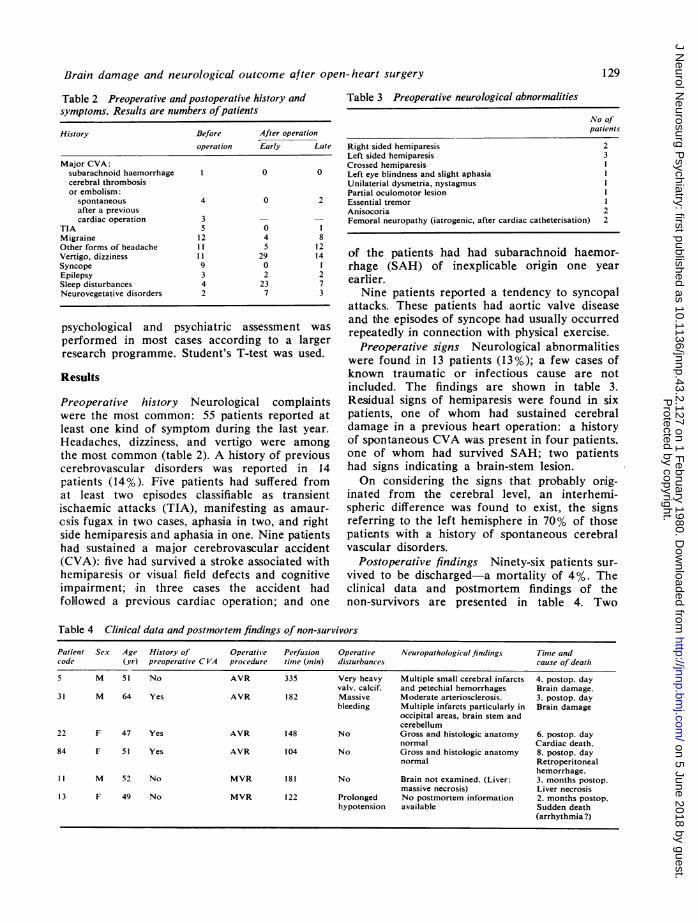
Preoperative history Neurological complaintswere the most common: 55 patients reported atleast one kind of symptom during the last year.Headaches, dizziness, and vertigo were amongthe most common (table 2). A history of previouscerebrovascular disorders was reported in 14patiients (14%). Five patients had suffered fromat least two episodes classifiable as transientischaemic attacks (TIA), manifesting as amaur-esis fugax in two cases, aphasia in two, and rightside hemiparesis and aphasia in one. Nine patientshad sustained a major cerebrovascular accident(CVA): five had survived a stroke associated withhemiparesis or visual field defects and cognitiveimpairment; in three cases the accident hadfolilowed a previous cardiac operation; and one
Table 3 Preoperative neurological abnormalities
No ofpatients
Right sided hemiparesisLeft sided hemiparesisCrossed hemiparesisLeft eye blindness and slight aphasiaUnilaterial dysmetria, nystagmusPartial oculomotor lesionEssential tremorAnisocoriaFemoral neuropathy (iatrogenic, after cardiac catheterisation)
23
1
22
of the patients had had subarachnoid haemor-rhage (SAH) of inexplicable origin one yearearlier.Nine patients reported a tendency to syncopal
attacks. These patients had aortic valve diseaseand the episodes of syncope had usually occurredrepeatedly in connection with physical exercise.
Preoperative signs Neurological abnormalitieswere found in 13 patients (13%); a few cases ofknown traumatic or infectious cause are notincluded. The findings are shown in table 3.Residual signs of hemiparesis were found in sixpatients, one of whom had sustained cerebraldamage in a previous heart operation: a historyof spontaneous CVA was present in four patients.one of whom had survived SAH; two patientshad signs indicating a brain-stem lesion.On considering the signs that probably orig-
inated from the cerebral level, an interhemi-spheric difference was found to exist, the signsreferring to the left hemisphere in 70% of thosepatients with a history of spontaneous cerebralvascular disorders.
Postoperative findings Ninety-six patients sur-
vived to be discharged-a mortality of 4%. Theclinical data and postmortem findings of thenon-survivors are presented in table 4. Two
Table 4 Clinical data and postmortem findings of non-survivors
Patient Sex Age History of Operative Perfusion Operative Neuropathological findings Tinme andcode (yr) preoperative C VA procedure time (min) disturbances cause of death
5 M 51 No AVR 335 Very heavy Multiple small cerebral infarcts 4. postop. dayvalv. calcif. and petechial hemorrhages Brain damage.
31 M 64 Yes AVR 182 Massive Moderate arteriosclerosis. 3. postop. daybleeding Multiple infarcts particularly in Brain damage
occipital areas, brain stem andcerebellum
22 F 47 Yes AVR 148 No Gross and histologic anatomy 6. postop. daynormal Cardiac death.
84 F 51 Yes AVR 104 No Gross and histologic anatomy 8. postop. daynormal Retroperitoneal
hemorrhage.11 M 52 No MVR 181 No Brain not examined. (Liver: 3. months postop.
massive necrosis) Liver necrosis13 F 49 No MVR 122 Prolonged No postmortem information 2. months postop.
hypotension available Sudden death(arrhythmia?)
129
Protected by copyright.
on 5 June 2018 by guest.http://jnnp.bm
j.com/
J Neurol N
eurosurg Psychiatry: first published as 10.1136/jnnp.43.2.127 on 1 F
ebruary 1980. Dow
nloaded from
130
Table 5 Major neurological complications after surgery
Hemiparesis probably originating fromcerebral hemispheral level
right sided 6left sided 16
Heiniparesis of uncertain originright sidedleft sided
Confusion 4Aphasia only 2Intellectual impairment 2Brain stem lesion 6Diverse signs of uncertain etiology
Anisocoria 6Nystagmus 5Trigeminal sensory impairmentUnilateral peripheral lesion
(probably brachial plexus) 12
patients died within four days of surgery withoutgaining consciousness; both showed massive focalsigns of brain damage. Two more patients diedof evidently non-cerebral causes at the end oftihe first week, having got through the operationwithout manifest neurological complications. Twolater deaths occurred within three months: inone of the cases cardiac aetiology was suspected,the other patient dying of massive liver necrosis.Focal cerebral signs had been present in thesetwo patients after surgery but they had recoveredcompletely before the second postoperativeexamination.The brains of the patients who died in the
hospital were available for necropsy. In the casesof operative death marked brain damage was
found to be the principal cause: multiple smallhaemorrhages and acute ichaemic neuronaldamage was seen both in the cerebral hemispheresand it the brain sterm and cerebellum. By con-trast, no pathological findings could be seen inthe brains of those two patients who died ofevidently non-cerebral causes and who had notshown any neurological disorders.
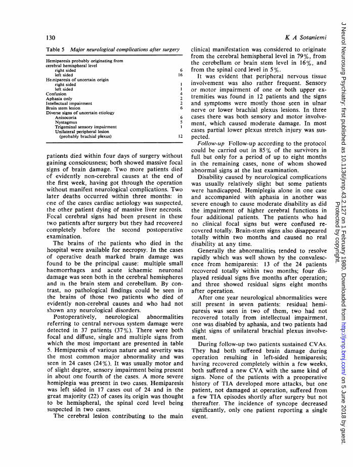
Postoperatively, neurological abnormalitiesreferring to central nervous system damage weredetected in 37 patients (37%). There were bothfocal and diffuse, single and multiple signs fromwhich the most important are presented in table5. Hemiparesis of various nature and severity wasthe most common major abnormality and wasseen in 24 cases (24%). It was usually motor andof slight degree, sensory impairment being presentin about one fourth of the cases. A more severehemiplegia was present in two cases. Hemiparesiswas left sided in 17 cases out of 24 and in thegreat majority (22) of cases its origin was thoughtto be hemispheral, the spinal cord level beingsuspected in two cases.The cerebral lesion contributing to the main
K A Sotaniemi
clinical manifestation was considered to originatefrom the cerebral hemispheral level in 79%, fromthe cerebellum or brain stem level in 16%, andfrom the spinal cord level in 5%.
It was evident that peripheral nervous tissueinvolvement was also rather frequent. Sensoryor motor impairment of one or both upper ex-tremities was found in 12 patients and the signsand symptoms were mostly those seen in ulnarnerve or lower brachial plexus lesions. In threecases there was both sensory and motor involve-ment, which caused moderate damage. In mostcases partial lower plexus stretch injury was sus-pected.
Follow-up Follow-up according to the protocolcould be carried out in 85% of the survivors infull but only for a period of up to eight monthsin the remaining cases, none of whom showedabnormal signs at the last examination.
Disability caused by neurological complicationswas usually relatively slight but some patientswere handicapped. Hemiplegia alone in one caseand accompanied with aphasia in another wassevere enough to cause moderate disability as didthe impairment of higher cerebral functions infour additional patients. The patients who hadno clinical focal signs but were confused re-covered totally. Brain-stem signs also disappearedtotally within two months and caused no reald-isability at any time.
Generally the abnormalities tended to resolverapidly which was well shown by the convalesc-ence from hemiparesis: 13 of the 24 patientsrecovered totally within two months; four dis-played residual signs five months after operation;and three showed residual signs eight monthsafter operation.
After one year neurological abnormalities werestill present in seven patients: residual hemi-paresis was seen in two of them, two had notrecovered totally from intellectual impairment,one was disabled by aphasia, and two patients hadslight signs of unilateral brachial plexus involve-ment.During follow-up two patients sustained CVAs.
They had both suffered brain damage duringoperation resulting in left-sided hemiparesis;having recovered completely within a few weeks,both suffered a new CVA with the same kind ofsigns. None of the patients with a preoperativehistory of TIA developed more attacks, but onepatient, not damaged at operation, suffered froma few TIA episodes shortly after surgery but notthereafter. The incidence of syncope decreasedsignificantly, only one patient reporting a singleevent.
Protected by copyright.
on 5 June 2018 by guest.http://jnnp.bm
j.com/
J Neurol N
eurosurg Psychiatry: first published as 10.1136/jnnp.43.2.127 on 1 F
ebruary 1980. Dow
nloaded from
Brain damage and neurological outcome after open-heart surgery
Table 6 Prognostic values ofpreoperative neurologichistory and signs
History of sign Number of Postoperative braincases damage observed in
N %
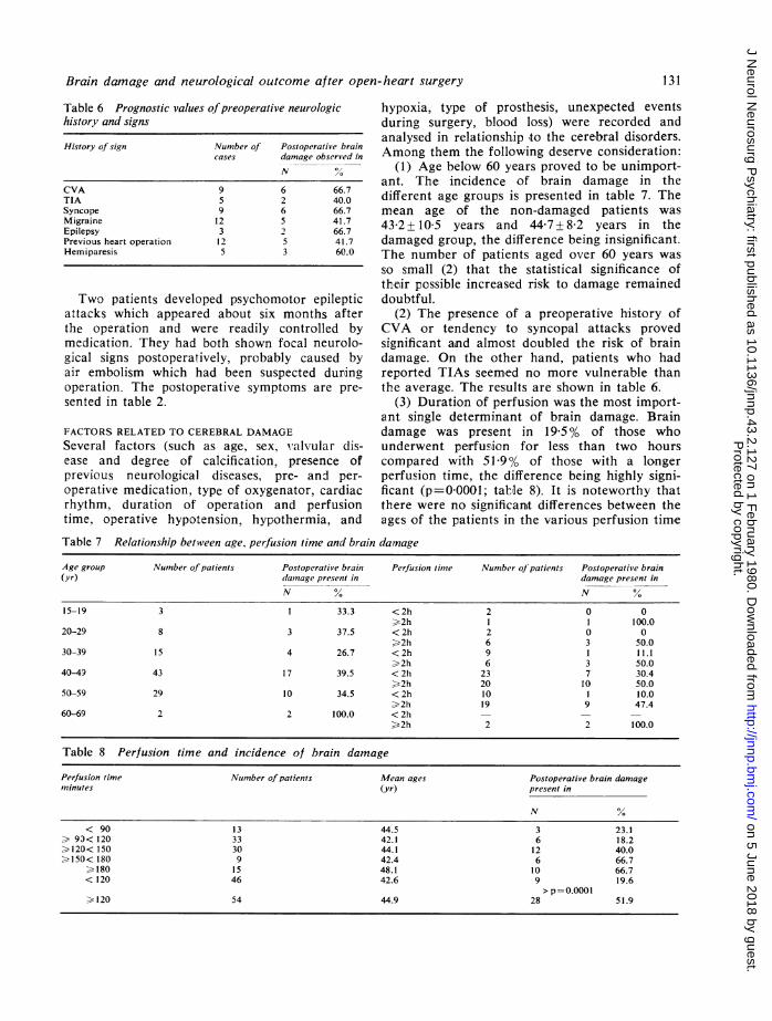
CVA 9 6 66.7TIA 5 2 40.0Syncope 9 6 66.7Migraine 12 5 41.7Epilepsy 3 2 66.7Previous heart operation 12 5 41.7Hemiparesis 5 3 60.0
Two patients developed psychomotor epilepticattacks which appeared about six months afterthe operation and were readily controlled bymedication. They had both shown focal neurolo-gical signs postoperatively, probably caused byair embolism which had been suspected duringoperation. The postoperative symptoms are pre-sented in table 2.
FACTORS RELATED TO CEREBRAL DAMAGE
Several factors (such as age, sex, valvular dis-ease and degree of calcification, presence ofprevious neurological diseases, pre- and per-
operative medication, type of oxygenator, cardiacrhythm, duration of operation and perfusiontime, operative hypotension, hypothermia, and
hypoxia, type of prosthesis, unexpected eventsduring surgery, blood loss) were recorded andanalysed in relationship to the cerebral disorders.Among them the following deserve consideration:
(1) Age below 60 years proved to be unimport-ant. The incidence of brain damage in thedifferent age groups is presented in table 7. Themean age of the non-damaged patients was43-2 + 10-5 years and 44-7 + 8-2 years in thedamaged group, the difference being insignificant.The number of patients aged over 60 years was
so small (2) that the statistical significance oftheir possible increased risk to damage remaineddoubtful.
(2) The presence of a preoperative history ofCVA or tendency to syncopal attacks provedsignificant and almost doubled the risk of braindamage. On the other hand, patients who hadreported TIAs seemed no more vulnerable thanthe average. The results are shown in table 6.
(3) Duration of perfusion was the most import-ant single determinant of brain damage. Braindamage was present in 19-5% of those whounderwent perfusiion for less than two hourscompared with 51-9% of those with a longerperfusion time, the difference being highly signi-ficant (p=00001; tabile 8). It is noteworthy thatthere were no significanit differences between theages of the patients in the various perfusion time
Table 7 Relationship between age, perfusion time and brain damage
Age group Number of patients Postoperative brain Perfusion tinme Number of patients Postoperative brain(yr) damnage present in damage present in
N % N %15-19 3 1 33.3 < 2h 2 0 0
>2h 1 1 100.020-29 8 3 37.5 < 2h 2 0 0
>2h 6 3 50.030-39 15 4 26.7 < 2h 9 1 11.1
>2h 6 3 50.040-49 43 17 39.5 < 2h 23 7 30.4
. 2h 20 10 50.050-59 29 10 34.5 < 2h 10 1 10.0
> 2h 19 9 47.460-69 2 2 100.0 < 2h - - -
>2h 2 2 100.0
Table 8 Perfusion time and incidence of brain damage
Perfusion time Nunmber of patients Mean ages Postoperative brain damagemninutes (yr) present in
N %
< 90 13 44.5 3 23.1. 93< 120 33 42.1 6 18.2.120<150 30 44.1 12 40.0.150< 180 9 42.4 6 66.7
.180 15 48.1 10 66.7< 120 46 42.6 9 19.6
> p =0.000I.120 54 44.9 28 51.9
131
Protected by copyright.
on 5 June 2018 by guest.http://jnnp.bm
j.com/
J Neurol N
eurosurg Psychiatry: first published as 10.1136/jnnp.43.2.127 on 1 F
ebruary 1980. Dow
nloaded from
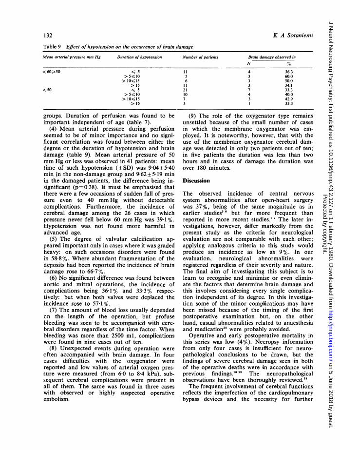
Table 9 Effect of hypotension on the occurrence of brain damage
Mean arterial pressure mm Hg Duration of hypotension Number of patients Brain damage observed in
N /
<60>50 S 5 11 4 36.3> 5610 5 3 60.0>10.15 6 3 50.0
> 15 1 1 3 34.1<50 6 5 21 7 33.3
> 5610 10 4 40.0> 10615 7 3 42.9
> 15 3 1 33.3
group's. Duration of perfusion was found to beimportant independent of age (table 7).
(4) Mean arterial pressure during perfusionseemed to be of minor im,portance and no signi-ficant correlation was found between either thedegree or the duration of hypotension and braindamage (table 9). Mean arterial pressure of 50mm Hg or less was observed in 41 patients: meantime of such hypotension (± SD) was 9 04 + 540min in the non-damage group and 9-62±5-19 minin the damaged patients, the difference being in-significant (p=0 38). It must be emphasised thatthere were a few occasions of sudden fall of pres-sure even to 40 mm Hg without detectablecomplications. Furthermore, the incidence ofcerebral damage among the 26 cases in whichpres.sure never fell below 60 mm Hg was 39 1 %.Hypotension was not found more harmful inadvanced age.
(5) The degree of valvular calcification ap-peared important only in cases where it was gradedheavy: on such occasions disorders were foundin 58-8 %. Where abundant fragmentation of thedeposits had been reported the incidence of braindamage rose to 66-7 %.
(6) No significant difference was found betweenaortic and mitral operations, the incidence ofcomplications being 36- 1 % and 33-3 % respec-tively: but when both valves were deplaced theincidence rose to 571 %.
(7) The amount of blood loss usually dependedon the len.gth of the operation, but profusebleeding was seen to be accompanied with cere-bral disorders regardless of the time factor. Whenbleeding was more than 2500 ml, complicationswere found in nine cases out of ten.
(8) Unexpected events during operation wereoften accompanied with brain damage. In fourcases difficulties with the oxygenator werereported and low value.s of arterial oxyge.n pres-sure were measured (from 6-0 to 8-4 kPa), sub-sequent cerebral complications we-re present inall of them. The same was found in three caseswith observed or highly suspected operativeembolism.
(9) The role of the oxygenator type remainsunsettled because of the small number of casesin which the membrane oxygenator was em-ployed. It is noteworthy, however, that with theuse of the membrane oxygenator cerebral dam-age was d.etected in only two patients out of ten;in five patients the duration was less than twohours and in cases of damage the duration wasover 180 minutes.
Discussion
The observed incidence of central nervoussystem abnormalities after open-heart surgerywas 37 %, being of the same magnitude as inearlier studies5 6 but far more frequent thanreported in more recent studies."2 The later in-vestigations, however, differ markedly from thepresent study as the criteria for neurologicalevaluation are not comparable with each other;applying analogous criteria to this study wouldproduce an incidence as low as 9 %. In ourevaluation, neurological abnormalities wereregistered regardless of their severity and nature.The final aim of investigating this subject is tolearn to recognise and minimise or even elimin-ate the factors that determine brain damage andthis involves considering every single complica-tion independent of its degree. In this investiga-ticn some of the minor complications may havebeen missed because of the timing of the firstpostoperative examination but, on the otherha,nd, casual abnormal,ities related to anaesthesiaand medication28 were probably avoided.
Operative and early postoperative mortality inthis series was low (4%). Necropsy informationfrom only four cases is insufficient for neuro-pathological conclusions to be drawn, but thefindings of severe cerebral damage seen in bothof the operative deaths were in accordance withprevious findings.6 29 The neuropathologicalobservations have been thoroughly reviewed."The frequent involvement of cerebral functions
reflects the imperfection of the cardiopulmonarybypass devices and the necessity for further
132 K A Sotaniemi
Protected by copyright.
on 5 June 2018 by guest.http://jnnp.bm
j.com/
J Neurol N
eurosurg Psychiatry: first published as 10.1136/jnnp.43.2.127 on 1 F
ebruary 1980. Dow
nloaded from
Brain damage and neurological outcome after open-heart surgery
efforts to develop and improve both the equip-ment and the operative procedures. Severalcperating centres have carefully studied andpractised preventive measures and reduced com-plications considerably,'2 but complete elimina-tion has not yet been achieved.The factors most often put forward as res-
ponsible for brain damage are embolisation andinadequate cerebral perfusion.8 Among the vari-ables most likely to increase the degree of riskadvanced age, long duration of perfusion, lowperfusion pressure, and the presence of a historyof previous neurological diseases have proved tobe the most important."6 30 However no singlefactor has proved absolutely decisive alone"4 andthe roles of the determinants are somewhat con-troversial. In a retrospective study8 the possiblecause of brain damage could be named in 40% ofoccurrences: in this study it can be stated in 12cases out of 37 (32-4%): hypoxia in five cases,massive bleeding, and technical difficulties in twocases and embolisation in three survivors and intwo non-survivors.The presence of a history of preoperative CVA
seemed to correlate positively with the incidenceof brain damage, which ocurred in 66-7% cofcases, regardless of whether or not clinical signscould still be seen preoperatively. This supportsthe findings of Branthwaite8 but there are papers
reporting prognostic values only if the clinicals,igns are still present.' 6 On the other hand,previous cardiac operations, independent ofneurological outcome, did not increase the risk,and the same was found to be true in cases witha history of transient ischaemic attacks. Thisleads to the conclusion that cerebrovascular dis-eases cannot be considered as a homogeneousentity for prognostic purposes in evaluating theneurological risks of extracorporeal circulation.A history of syncopal attacks appeared dis-
advantageous, being associated with cerebraldisorders in 66-7% of cases. The importance ofbilackout spells has not been discussed in previousreports and the prognostic significance must bereassessed with more subjects but it is noteworthythat there existed another known factor possiblycontributing to damage in only one of the cases
(table 6).The importance of age has been emphasised
in several communications' 68 but could not beconfirmed in this study as presented in table 7.Some proof could be seen however in the un-
favourable outcome of the two oldest patients(both aged 64) but both of them also had a longperfusion time (154 and 182 min). In additionthere was a trend increasing with advancing age
to higher susceptibility to damage after only ashort perfusion time (table 8). But if age wereof major importance there should have been aparticularly clear-out correlation between ageand frequency of complications after long dura-tions of perfusion and this could not be verified.There is no evidence of the increased harmful-ness of hypotension with advancing age whichprovided further su,pport for the minor value ofage. The present results cannot be explained byany variables specially favourable to the olderpatient groups and disadvantageous to theyounger groups (as for age and perfusion time,see table 8). It may be concluded that age itselfis not a matter of importance but rather the stateof the reguilatory mechanisms of circulation.
Duration of perfusion proved the most signi-ficant of the operative variables directly verifyingthe cumulatively damaging character of cardio-pulmonary bypass conditions. Perfusion timeshorter than two hours was accompanied by cere-bral disorders in 19-6% of cases in contrast to51-9% after longer duration of bypass, the differ-ence being highly significant (p=00001). Thesame time limit has been found earlier5 and thedefinite benefits of short perfusion time policyhave been well documented.2 '" In the presentstudy, the significance is further accentuated bythe relatively slight weight of other factors, suchas hypotension and age. The final harmful causesrelated to cardiopulmonary bypass have not beendefinitely ascertained and cannot be solved byclinical investigations-for example, studies onmicroembolisation" have shown that it does notnecessarily cause any detectable damage. Untilwe have more detailed solutions the practicalccnclusion is to use as short a perfusion time aspossible.
Information on the role of hypotension duringbypass is controversial. Hypotension has beenregarded as a major determinant of cerebraldamage by some authors5830 3' and less importantby others.2 4 Our results show indisputably thatmoderate hypotension alone is not a decisivedeterminant contributing to brain dysfunction.During perfusion severe hypotension (below
40 mm Hg) could be effectively avoided but meanarterial pressure below 50 mm Hg was recordedin more than one third of the patients and therewere no significant differences between thedamaged and non-damaged patient groups in theduration or degree of hypotension. There was nopo.sitive correlation between age and harmfulnessof moderate hypotension and there were oc-casions when pressure fell suddenly to 40 mmHg without detectable disorders. Cerebral cir-
133
Protected by copyright.
on 5 June 2018 by guest.http://jnnp.bm
j.com/
J Neurol N
eurosurg Psychiatry: first published as 10.1136/jnnp.43.2.127 on 1 F
ebruary 1980. Dow
nloaded from
134
culation is known to be able to adopt to hypoten-sion with a mean arterial pressure of 50 mmHg32 33 but our findings suggest that the criticallimit is idiosyncratic and may be influenced byother coincidental factors. Our results lead to theconclusion that the factors of actual importanceare first the causes of hypotension and second,the individual's abilities to tolerate the conditionand to respond to it.The observed interhemispheric difference in
susceptibility to damage deserves consideration,particularly because it has not been reportedbefore. Some references to similar results can beseen in previous studies5 8 but the matter has notbeen discus.sed. In this study, the preoperativesigns and case histories referred to the left hemi-s.phere in 70% of cases but, by contrast, the post-operative signs originated from the right hemi-sphere in 71 %. Hemiparesis, which was the mostusual major complication, was left-sided in 16of the 22 cases in which the origin could belocalised to the cerebral level. These cases wereanalysed with special care in relation to pre-operative, intraoperative, and postoperative vari-ables but no explanation could be stated onclinical grounds to the disparity between rightand left hemispheral involvement. All thepatients were right-handed, no cervical vasculardisease could be verified clinically, and no posi-tional or technical cause favouring right sideinvolvement could be determined. It is unlikelythat emboli of any kind would prefer the rightcarotid artery to the left side or that a largegroup of patients would have similarvascular anomalies. The present knowledge ofinterhemispheral diffe.rences includes mainlyneuropsychological information343 and electro-encephalographic findings3 39, but knowledge ofthe conditions of microcirculation and metabol-ism as well as of their regulators is practicallynon-existent. Because the hemispheres aredifferent from each other in dominance, neuro-psychological functions, and electrical charac-teristics there may also be differences in main-taining normal functions under exceptionalconditions and in tolerating damage. The mech-anism of dissimiliarities remains unsettled on aclinical basis, but either the dominant and morespecialised le.ft hemisphere may be more resistantto harmful factors such as are present duringcardiopulmonary bypass, or conversely theremay be a direct cause damaging the right sidealone, or perhaps the hemispheres are uniformlyinvolved but the threshold in producing clinicalmanifestations is different for each hemisphere.An indirect involvement of this kind seems un-
K A Sotaniemi
likely to be strong enough to lead to clinicalsigns in the undamaged hemisphere if there areno signs from the primarily affected hemisphere.However,. there are reports of bilateral abnor-malities in unilateral brain damage cause bymicroem,bolisation" 38 and by irradiation." Theinterhemispheric difference observed by clinicalneurological methods remains to be confirmedand its significance reassessed after evaluation ofthe results of electroencephalographic and psy-chometric tests, the analysis of which is alreadyunder way.The final brain damaging effects of cardio-
pulmonary bypass are at least partly uncertainand because clinical methods are of limited valuein expIaining the basic events, investigations mustbe focused on the nerve cell level and on theregulators of its metabolism. Open-heart opera-tions with cardiopulmonary bypass have provedto be a potent multifactorial hazard for all thelevels of the nervous system and the advancesthus far still seem inadequate.
References
1 Branthwaite MA. Prevention of Neurologicaldamage during open-heart surgery. Thorax 1975;30:258-61.
2 Aberg T, Kihlgren M. Cerebral protectionduring open-heart surgery. Thorax 1977; 32:525-33.
3 Ross Russell RW, Bharucha N. The recognitionand prevention of border zone cerebral ischaemiaduring cardiac surgery. Q J Med, New Series1978; 47:303-23.
4 Gilman S. Cerebral disorders after open-heartoperations. N Engl J Med 1965; 272:489-98.
5 Javid H, Tufo HM, Najafi H, Dye WS, HunterJA, Julian OC. Neurologic abnormalities follow-ing open-heart surgery. J Thorac Cardiov Surg1969; 58:502-9.
6 Tufo HM, Ostfeld AM, Shekelle R. Centralnervous system dysfunction following open-heartsurgery. J Am Med Ass 1970; 212:1333-40.
7 Lee WH, Krumhaar D., Fonkalsrud EW,Schjeide OE, Maloney IV. Denaturation ofplasma proteins as a cause of morbidity anddeath after intracardiac operations. Surg 1961;50:29-39.
8 Branthwaite MA. Neurological damage relatedto open-heart surgery. Thorax 1972; 27:748-53.
9 Patrick RT, Kirklin JW, Theye RA. The effectsof extracorporeal circulation on the brain. In:Allen JG, ed. Extracorporeal circulation.Springfield, Ill. Charles G Thomas 1958; 272.
10 Aberg T, Kihlgren M. Effect of open-heartsurgery on intellectual function. Scand JCardiov Surg 1974; Suppl 15.
11 Walsh JC. Mononeuritis multiplex complicating
Protected by copyright.
on 5 June 2018 by guest.http://jnnp.bm
j.com/
J Neurol N
eurosurg Psychiatry: first published as 10.1136/jnnp.43.2.127 on 1 F
ebruary 1980. Dow
nloaded from
Brain damage and neurological outcome after open-heart surgery
the post-perfusion syndrome. A ust NZ J Med1968; 17:327-30.
12 Keates JRW, Innocenti DM, Ross DN. Mono-neuritis multiplex, a complication of open-heartsurgery. J Thorac Cardiov Surg 1975; 69:816-9.
13 Honet JC, Raikes JA, Kantrowitz A, Pursel SE,Rubenfire M. Neuropathy in upper extremityafter open-heart surgery. Arch Phys MedRehabil 1976; 57:264-7.
14 Aguilar MJ, Gerbode F, Hill J. Neuropathologiccomplications of cardiac surgery. J ThoracCardiov Surg 1971; 61:676-85.
15 Branthwaite MA. Cerebral blood flow andmetabolism during open-heart surgery. Thorax1974; 29:633-38.
16 Brierley JB. Neuropathological findings inpatients dying after open-heart surgery. Thorax1963; 18:291-304.
17 Bass RM, Longmore DB. Cerebral damageduring open-heart suirgery. Nature 1969; 222:30-33.
18 Brennan RW, Patterscn RH, Kessler J. Cerebralblood flow and metabolism during cardio-pulmonary bypass. Neurology (Minneap) 1971;21:655-72.
19 Carlson RG, Lande AJ, La1idis B, Rogoz B,Baxter J, Patterson RH, Stenzel K, Lillehei CW.The Lande-Edwards membrane oxygenatorduring heart surgery. J Thorac Carciov Surg1973; 66:894-905.
20 Helmsworth JA, Gall EA, Perrin EV, BraleySA, Flege JB, Kaplan S, Keirle AM. Occurrenceof emboli during perfusion with an oxygenatorpump. Surg 1963; 53:177-85.
21 Brunon AM, Setiey A, Courjon J. Cerebralarterial embolism during extracorporeal circula-tion. Ann Anesthiol Fr 1977; 18:111-7.
22 Gallagher EG, Pearson DT. Ultrasonic identifi-cation of sources of gaseous microemboli duringopen-heart surgery. Thorax 1973; 28:295-305.
23 Hill JD, Osborn JJ, Swank RL, Aguilar MJ,de Lanerolle P, Gerbode F. Experience using anew Dacron wool filter during extracorporealcirculation. Arch Surg 1970; 101:649-52.
24 Bjork VO, Hultquist G. Brain damage inchildren after deep hypothermia for open-heartsurgery. Thorax 1960; 15:284-91.
25 Salerno TA, Lince DP, White DN, Lynn RB,Clharette EJP. Monitoring of electroencephalo-
gram during open-heart surgery. J ThoracCardiov Surg 1978; 76:97-101.
26 Wright G, Sanderson JM. Brain damage andmortality in dogs following pulsatile and non-pulsatile blood flows in extracorporeal circula-tion. Thorax 1972; 27:738-49.
27 Vise WM, Schuier F, Hossman K-A, Takagi S,Zulch KJ. Cerebral microembolization. ArchNeurol (Chicago) 1977; 34:660-5.
28 Bedford PD. Adverse effects of anaesthesia onold people. Lancet 1955; 2:259-61.
29 Witoszka MM, Tamura H, Indeglia R, HopkinsRW, Simeone FA. Electroencephalographicchanges and cerebral complications in open-heart surgery. J Thorac Cardiov Surg 1973; 66:855-64.
30 Stockard JJ, Bickford RG, Myers RR, AungMH, Dilley RB, Schauble JF. Hypotension in-duced changes in cerebral function during open-heart surgery. Stroke 1974; 5:730-46.
31 Stockard JJ, Bickford RG, Schauble JF. Pres-sure-dependent cerebral ischaemia during cardio-pulmonary bypass. Neurology (Minneap) 1973;23:521-9.
32 Harper AM, Glass HI. Effects of alterations inthe arterial carbon dioxide tension on the bloodflow through the cerebral cortex at normal andlow arterial pressures. J Neurol, NeurosurgPsychiatry 1965; 28:449-52.
33 Meyer JS, Gilroy J. Regulation and adjustmentof the cerebral circulation. Diseases of the chest1968; 53:30-6.
34 Milner B. Interhemispheric differences andpsychological processes. Br Med Bull 1971;27:272-7.
35 Kimura D. The asymmetry of the human brain.Sci Am 1973; 228:70-8.
36 Busse EW, Obrist WD. Significance of focalelectroencephalographic changes in the elderly.Postgrad Med 1963; 34:179-82.
37 Butler SR, Glass A. Asymmetries in the electrc-encephalogram associated with cerebral domin-ance. Electroencephalogr Clin Neurophysiol1974; 36:481-91.
38 Meyer JS, Gotoh F, Tazaki Y. Circulation andmetabolism following experimental cerebralembolism. J Neuropathol Exp Neurol 1962; 21:4-24.
39 Hfakansson CH, Lindgren M, Sulg IA EEGeffects of postoperative irradiation treatment ofbrain tumours. Acta Radiol Ther 1969; 8:301-10.
135
Protected by copyright.
on 5 June 2018 by guest.http://jnnp.bm
j.com/
J Neurol N
eurosurg Psychiatry: first published as 10.1136/jnnp.43.2.127 on 1 F
ebruary 1980. Dow
nloaded from
Related Documents


![Brain-to-brain loop 2020: è ancora utile il ciclo di Lundberg?...128 Riv Ital Med Lab (2017) 13:127–133 processo che la origina, attiva e utilizza [1]. Il punto chiave è l’interfaccia](https://static.cupdf.com/doc/110x72/60c5e0569a5fe824ae305572/brain-to-brain-loop-2020-ancora-utile-il-ciclo-di-lundberg-128-riv-ital.jpg)