Abstract book BioMediTech Research Day 2014

Welcome message from author
This document is posted to help you gain knowledge. Please leave a comment to let me know what you think about it! Share it to your friends and learn new things together.
Transcript

Abstract book
BioMediTech Research Day 2014
Table of contents
Contents
Table of contents Pages 2‐3 Defne Us 35 Kivinummi Kati 74Programme Pages 4‐5 Vainio Iina 36 Penkki Sanna 75Exhibition Pages 6‐7 Luoto Suvi 37 Rahikainen Rolle 76Keynote speakers Pages 8‐9 Tervonen Aapo 38 Salpavaara Timo 77Abstracts Pages 10‐110 Septimia Sarbu 39 Vornanen Inkeri 78
Juuti‐Uusitalo Kati 1 Moradi Elaheh 40 Haflidadottir Benedikta S. 79Viiri Keijo 2 Iftikhar Haider 41 Annala Matti 80Lloyd‐Price Jason 3 Räisänen Eero A. 42 Cordova Zuzet Martinez 81Peltola Marja 4 Kytölä Ville 43 Virjula Sanni 82Tanja Paavilainen 5 Ampuja Minna 44 Dhandapani Praveen Kumar 83Ryynänen Tomi 6 Luukinen Bruno 45 Esteves Luís 84Pajula Juha 7 Sorkio Anni 46 Mäkinen Meeri 85Santos Florentino 8 Saari Sina 47 Häyrynen Sergei 86Haaparanta Anne‐Marie 9 Scaravilli Mauro 48 Bremer Katharina 87Laaksonen Maria 10 Eerola Sini 49 Palanivel Suresh 88Ojala Marisa 11 Lehtonen Soili 50 Rantapero Tommi 89Penttinen Kirsi 12 Ojansivu Miina 51 Romagnoli Dario 90Lin Jake 13 Kartasalo Kimmo 52 Granberg Kirsi 91Saralahti Anni 14 Ortutay Zsuzsanna 53 Luukinen Hanna 92Ojanen Markus 15 Kaukoniemi Kirsi M. 54 Abdollahzadeh Ali 93Turunen Sanna 16 Seppä Ville‐Pekka 55 González de Cozar Jose M. 94Latonen Leena 17 Lehto Kalle 56 Tamminen Ilmari 95Auer Sanna 18 Liuksiala Thomas 57 Kukkurainen Sampo 96Kallio Heini ML 19 Riihimäki Tiina 58 Andjelkovic Ana 97Cannino Giuseppe 20 Vartiainen Suvi 59 Pitkänen Sanna 98Soto de la Cruz Ana María 21 Valanne Susanna 60 Kemppainen Esko 99Harjula Sanna‐Kaisa 22 Nättinen Janika 61 Narra Nathaniel 100Määttä Juha 23 Kuusisto Kirsi M. 62 Shcherban Anastasia 101Sjöblom Liisa 24 Salonen Johanna 63 Halonen Heidi 102Gnanavel Mutharasu 25 Laurila Eeva 64 Joers Priit 103Vuorinen Elisa 26 Joki Tiina 65 Lehti‐Polojärvi Mari 104Koivisto Janne 27 Ahola Antti 66 Vuornos Kaisa 105Kohvakka Annika 28 Laine Kaisa 67 Kapucu Fikret E. 106Kiamehr Mostafa 29 Annala Matti 68 Kekonen Atte 107Aittomäki Saara 30 Gracia Javier 69 Hannula Markus 108Afyounian Ebrahim 31 Ruusuvuori Pekka 70 Lillsunde Päivi 109Neeli‐Venkata Ramakanth 32 Patrikoski Mimmi 71 Abstracts in alphabetical order Pages 112‐113Cannnino Giuseppe 33 Järvelä‐Stölting Mirva 72Kuusela Tanja 34 Johansson Laura 73 Speakers: X

Programme:
08.30–18.30 Poster Exhibition, Tietotalo 2nd Floor
09.30–16.00 Commercial/corporateexhibition,Tietotalo2ndfloor
08:30–08.35 Opening words, Prof. Olli Yli-Harja, Vice Director, BioMediTech
08:35–09.15 Keynote 1: Prof. Roger W. Beuerman, Singapore Eye Research Institute and School of Medicine, UTA: Proteomics: Leading to Personalized Medicine
09:15–10.15 Session 1: Cancer, Chair: Prof. Matti Nykter
• Ville Kytölä/ Nykter: Enhancer RNAs reveal widespread chromatin reorganization in prostate cancer cell lines
• Annika Kohvakka/ Visakorpi: Transcriptome sequencing reveals PCAT5 - new ERG-regulated non-coding transcript in prostate cancer
• Mutharasu Gnanavel/ Meenakshisundaram Kandhavelu: Decoding the Glioblastoma Multiforme mechanism by deciphering the GPR17 networks
• Leena Latonen/ Visakorpi: In vivo role of miR-32 in prostate cancer
10.15–10.45 Coffee and Commercial/corporate exhibition
10.45–12.00 Session 2: Modelling Health and Disease, Chair: Docent Meenakshisundaram Kandhavelu
• Keijo Viiri/ Mäki: Polycomb regulate the intestinal stem cell niche – aberrancies implicated in celiac disease
• Ana Andjelkovic/Jacobs: AOX and the JNK signalling pathway in Drosophila
• Anni Saralahti/ Rämet: A forward genetic screen for zebrafish genes involved in pneumococcal infection
• Jason Lloyd-Price/ Ribeiro: Predictability and randomness of paw usage in mice
• Aapo Tervonen/ Hyttinen: Dynamics of Epithelial Tight Junction as Molecular and Electric Barrier – A Computational Approach
12.00–13.00 Lunch and Commercial/corporate exhibition
13.00–13.20 Keynote 2: Frank Emmert-Streib
13.20–14.20 Session 3: Computational Biology, Chair: Project Researcher Kerstin Lenk
• Sergei Häyrynen/ Nykter: Uncovering unannotated splicing sites in large RNA-sequenced sample sets
• Kirsi Penttinen/ Aalto-Setälä: A novel analysis software to detect Ca2+ signaling abnormalities in cardiomyocytes
• Elaheh Moradi/ Tohka: Machine learning framework for early MRI-based Alzheimer’s conversion prediction in MCI subjects
• Sampo Kukkurainen/ Hytönen: Flexible loop in talin head domain contributes to integrin activation
14.20–15.00 Coffee and Commercial/corporate exhibition
15.00–16.15 Session 4: Biomaterials and Imaging, Chair: Docent Heli Skottman
• Kalle Lehto/ Hyttinen: Contrast Enhanced X-ray Microtomography in Virtual 3D Histology of Eye
• Anni Sorkio/ Skottman: Biomimetic Collagen I and IV double layer Langmuir-Schaeffer films as microenvironment for human pluripotent stem cell derived retinal pigment epithelial cells
• Miina Ojansivu/ Miettinen: Bioactive Glass Ions as Strong Enhancers of Osteogenic Differentiation in Human Adipose Stem Cells
• Laura Johansson/ Kellomäki: Analysis of the novel blended polymeric materials for tissue engineering applications
• Timo Salpavaara/ Kellomäki, Lekkala: Fully biodegradable resonance circuit
16.15–16.40 Best PhD theses of BMT/UTA
16.40–16.50 Closing words: Hannu Hanhijärvi, Director, BioMediTech
16.50–18.30 Researchers available at their posters
Programme

Exhibition

Keynote Speakers
Keynote Speakers
Roger W. Beuerman, PhDSeniorScientificDirector
Singapore Eye Research Institute Duke-NUS, Professor Ophthalmology and Emerging Infectious Diseases
FiDiPro Professor Ophthalmology, University of Tampere School of Medicine
Proteomics: Leading to Personalized MedicineA focus my work has been to develop proteomic biomarkers of disease processes to understand individual differences in disease and to accommodate individual differences in response to therapy. Thegoalinpersonalizedmedicineistodefinethediseasephenotypeandmassspectrometryisanexcellent tool of this purpose as it allows an unbiased evaluation of disease at the highest levels of geneexpression-theproteome.MylabispartoftheHUPOefforttodefinethenormalproteomeandwe are currently working on white papers in the area of biomarkers and dry eye.
Frank Emmert-Streib

ABSTRACTS
Kati Juuti-Uusitalo
kati.juuti‐[email protected]
Biomaterials and Regenerative Medicine ‐ Heli Skottman
Käpylä, Sorkio, Teymouri, Lahtonen, Vuori, Valden, Skottman, Kellomäki
BioMediTech of UTA and TUT; Surface Science Laboratory TUT
Ormocomp® -modified glass as culture substratum for human embryonic stem cell-de
hESC‐RPE, Ormocomp®, APTES, MAPTES, silane, coating, In in vitro live‐cell imaging, it would be beneficial to grow and assess human embryonic stem cell‐derived retinal pigment epithelial (hESC‐RPE) cells on thin, transparent and rigid surfaces such as cover glasses.
In this study, we assessed how silanization of glass with 3‐aminopropyltriethoxysilane (APTES), 3‐(trimethoxysilyl)propyl methacrylate (MAPTMS) or the polymer‐ceramic material Ormocomp® affects the surface properties, protein binding and maturation of hESC‐RPE cells.
The surface properties were studied by contact angle measurements, Xray photoelectron spectroscopy (XPS), atomic force microscopy (AFM) and a protein binding assay. The cell adherence and proliferation were evaluated by culturing hESC‐RPE cells on collagen IV coated untreated or silanized surfaces for 7 or 42 days.
The Ormocomp® treatment significantly increased the hydrophobicity and roughness of glass surfaces compared to the APTES and MAPTMS treatments. The XPS results indicated that the Ormocomp® treatment changes the chemical composition of the glass surface by increasing carbon content and the number of C‐O/=O bonds. The protein binding test confirmed that the Ormocomp® treated surfaces bound more collagen IV than APTES or MAPTMS treated surfaces. All the silane treatments increased the number of attached cells compared to untreated glass but the highest cell numbers were detected on Ormocomp® treated surfaces. There were no differences in cell numbers compared to smoother to rougher Ormocomp® surfaces suggesting that the surface chemistry, and more specifically the collagen binding in combination with Ormocomp® is beneficial for hESC‐RPE cell culture.
This study clearly demonstrates that Ormocomp® treatment combined with collagen coating significantly increases hESC‐RPE cell attachment compared to the commonly used silanizing agents APTES and MAPTMS. Ormocomp® silanization could thus enable the use of microscopic live cell imaging methods for hESC‐RPE cells.
Kati Juuti-UusitaloAbstracts 1-109

Keijo Viiri
Other ‐ Disease Epigenomics PI Keijo Viiri
Mikko Oittinen, Alina Popp, Kalle Kurppa, Katri Lindfors, Markku Mäki
University of Tampere, School of Medicine, Tampere, Finland
Polycomb regulate the intestinal stem cell niche – aberrancies implicated in celiac disease
Keyword(s): Intestinal stem cells, Gastroenterology Epigenomics, Polycomb, H3K27me3, Intestinal stem cells, Celiac disease
Polycomb proteins regulate embryogenesis and maintain stem cell pluripotency and differentiated cell state by placing the repressive H3K27me3 mark on developmentally important genes. Polycomb function is also necessary for regulating the cell differentiation from adult stem cells to fully differentiated tissues throughout the organism’s life‐span. The crypt‐villus axis constitutes the functional unit of the small intestine where mature absorptive epithelial cells are confined to the villi. Intestinal stem cells (ISC) and transit amplifying and differentiating epithelial cells, on the other hand, are restricted to the crypts. Epithelial cells undergo rapid turnover (3 – 5 days) and are thus tightly regulated to maintain homeostasis between proliferation, differentiation and apoptosis.
We have found that inhibition of polycomb activity triggers enterocyte differentiation in mouse intestinal organoid cultures suggesting that polycomb proteins maintain the tissue homeostasis in the gut. ChIP‐seq with H3K27me3 antibody in ISC and fully mature enterocytes revealed that polycomb regulates the stem cell niche in the small intestine by governing the expression of master regulators of differentiation and stemness. In addition, immunohistochemistry analyses indicate that polycomb protein SUZ12 is expressed in a significantly wider region in a crypt‐villous axis in celiac compared to healthy suggesting that out‐of‐bounds polycomb activity might contribute to the crypt hyperplasia manifested in celiac disease. Finally, due to the almost sole preference of polycomb to regulate genes involved in development and signalling, our data provide a vetted list of candidate genes directly involved in the regulation of crypt stem cells.
Jason Lloyd-Price
jason.lloyd‐[email protected]
Other ‐ Animal Behaviour ‐ Andre Ribeiro
Fred G. Biddle, Brenda A. Eales, Andre S. Ribeiro
Tampere University of Technology; University of Calgary
Predictability and randomness of paw usage in mice
Keyword(s): paw preference; mouse; adaptability; information entropy; learning and memory
In a food‐reaching experiment, mice will generally prefer to use one paw over the other, even in an unbiased test setting. The acquisition of this preference is based on a gradual reinforcement of weak, randomly‐occurring asymmetries in paw choice early in training. This reinforcement has been shown to rely on strain‐dependent memory abilities, resulting in strain‐specific patterns of paw choices. From two training sessions of 50 reaches each, separated by a one week interval, and using an information‐theoretic measure of the predictability of paw usage from past choices, we characterized how information of previous choices is incorporated into future choices, for six mouse strains, differing in their learning abilities. We found that each choice is based on a limited number of previous choices, which differs between strains. The maximum predictability of paw reaches is also limited and strain‐dependent, implying that there is a strain‐dependent degree of randomness in paw choice that is not lost with training. For most strains, the number of previous choices used decreases in the second training session, while the maximum predictability increases. Two strains, 9XCA and BTBR, with brain defects resulting in memory deficiencies, do not show any differences between sessions. We conclude that paw choices are regulated by at least three strain‐dependent components: short‐ and long‐term memory, and a random component that is not lost with training. This random component may be a critical source of behavioural plasticity in paw preferences, allowing the mouse to adapt its paw usage in fluctuating environments.
Keijo Viiri Jason Lloyd-Price
Session 2: Modelling Health and Disease Session 2: Modelling Health and Disease

Marja Peltola
Regenerative Medicine ‐ Susanna Narkilahti
Paavilainen Tanja, Fayuk Dmitriy, Ylä‐Outinen Laura, Narkilahti Susanna
University of Tampere, BioMediTech, Tampere, Finland
Characterizing the Human Embryonic Stem Cell –Derived Neural Cultures: the Effect of Astrocytes on Network Properties
Keyword(s): Astrocytes, MEA, embryonic stem cells
Background
During the last ten years the prevailing neuron centric research of CNS has started to focus to the role of astrocytes, and new more dynamic roles have been suggested to them. Thus, in addition to their traditional role as structural and nutritional supporters, astrocytes are now considered to be involved in the regulation of synaptic function and information processing. This research aims to gain understanding how astrocytes affect to the development and functionality of human neuronal networks in vitro. In this study we used human embryonic stem cell (hESC)‐derived neuronal cells and MEA (microelectrode array) platform.
Methods
The cells for the experiment were derived from hESCs. Briefly, embryonic stem cell colonies were cut and allowed to form spheres in neural differentiation medium. The formed neurospheres were cut to small pieces, plated onto MEAs and cell culture well plates. Before cell plating, the MEAs were coated with PEI and laminin and the well plates with laminin. In addition to the functional characterization with MEAs, the cultures were characterized using viability analysis, gene and immunocytochemical staining. The functional development of the networks was followed by MEA measurements twice a week for 5 weeks.
Results/Conclusions
According to the preliminary data, there seems to be a difference in the development of electrical activity in networks with different proportions of astrocytes. The data also suggests that the network maturation pattern differs according to the proportion of astrocytes in the culture. The protein expression data confirmed that the two cultures had different proportions of astrocytes.
Paavilainen Tanja
Regenerative Medicine ‐ Susanna Narkilahti
Fayuk Dmitriy, Peltola Marja, Heikkilä Juha, Narkilahti Susanna
BioMediTech, University of Tampere, Tampere, Finland
Simultaneous Microelectrode Array Recording and Intracellular Ca2+ Imaging of Human Pluripotent Stem Cell-Derived Neural Cultures
Keyword(s): Stem cells, neural cells, MEA, Ca2+ imaging
Background
Human pluripotent stem cells (hPSCs) can be differentiated into neural cell types and they are able to form functional networks in vitro. Experimental investigation of network dynamics can gain us a better insight into basic mechanisms of neural network functioning in the brain. Here, we present our experiments where we combined micro electrode array (MEA) and calcium imaging measurements for functional characterization of neural networks.
Methods
HPSCs were differentiated into neural cell types and maturated by plating on MEA dishes. The functional development of the networks was followed with MEA recordings every week after plating. Combined MEA and calcium imaging recordings were made 3‐4 weeks after plating when network electrical activity demonstrated mature patterns. For fluorescent imaging of intracellular Ca2+ signals the neural cultures were loaded with fluo‐4 AM calcium fluorophore and were imaged using fast fluorescent imaging system.
Results
When studied with MEA the hPSC‐derived networks demonstrated spontaneous electrical activity in the form of spike trains starting from week two after plating. In addition, spontaneous intracellular Ca2+ signaling was revealed in Ca2+ imaging experiments. Despite the electrical and Ca2+ signals were detected in the same network regions we did not find strong temporal correlation between electrical activity recorded by particular electrode and Ca2+ signals in the nearby neurons.
Conclusions
Simultaneous use of MEA measurements and Ca2+ imaging is feasible approach and it promises to provide us more detailed information about particular neurons involved in generation of electrical activity in the network.
Marja Peltola Paavilainen Tanja

Tomi Ryynänen
Tomi Ryynänen
Measurement and stimulation systems and methods ‐ Jukka Lekkala
Manoj Sivasubramaniapandian (1), Marja Peltola (2), Susanna Narkilahti (2), Jukka Lekkala (1)
(1) Department of Automation Science and Engineering and BioMediTech, Tampere University of Technology, (2)
NeuroGroup, BioMediTech, University of Tampere
Impedance considerations on MEA – the effect of electrode materials and coatings
Keyword(s): MEA, impedance, noise, coating
We have evaluated how different electrode materials and coatings used to improve cell attachment affect on impedance and noise levels of microelectrode arrays (MEAs). Titanium nitride, titanium, and ALD iridium oxide (IrOx) were experimentally studied as electrode materials and, in addition, several other materials were reviewed from the literature. From MEA coatings polyethylenimine (PEI) and laminin, gelatin, and MatrigelTM were studied. The results support titanium nitride’s and platinum black’s position as the most commonly used low impedance electrode materials on MEAs, whereas IrOx and carbon nano tube based materials are promising alternatives as the microelectrode materials. PEI and laminin, and gelatin coatings did not cause any significant change in the overall performance of the MEAs, but a substantial increase in impedance was observed with MatrigelTM especially when applied as thick layer.
Juha Pajula
Images, signals and models ‐ Ulla Ruotsalainen / Jussi Tohka
jukka‐Pekka Kauppi, Jussi Tohka
Tampere University of Technology, University of Helsinki
A Versatile Software Package for Inter-subject Correlation Based Analyses of fMRI
Keyword(s): Complex naturalistic stimuli, Matlab, higher brain function, analysis toolbox
In the inter‐subject correlation (ISC) based analysis of the functional magnetic resonance imaging (fMRI) data, the extent of shared processing across subjects during the experiment is determined by calculating correlation coefficients between the fMRI time series of the subjects in the corresponding brain locations. This implies that ISC can be used to analyze fMRI data without explicitly modelling the stimulus and thus ISC is a potential method to analyze fMRI data acquired under complex naturalistic stimuli such as movies, music, or aesthetic performances. Despite of the suitability of ISC based approach to analyze complex fMRI data, no generic software tools have been made available for this purpose, limiting a widespread use of ISC based analysis techniques among neuroimaging community. Here a graphical user interface (GUI) based software package, ISC Toolbox is presented.
The ISC toolbox is designed for generic ISC based analysis of fMRI data. No information about the stimulus is required to carry out the analysis, making the toolbox suitable to analyze nearly any kind of fMRI data including at least two subjects. The construction of the whole‐brain ISC maps as well as more advanced analyses including the comparison of ISCs between different stimuli, time‐window ISC analysis, frequency‐speci_c ISC analysis and intersubject phase synchronization analysis are supported. The analyses are coupled with re‐sampling based statistical inference. The ISC Toolbox is available in https://code.google.com/p/isc‐toolbox/ for Matlab under the MIT open source licence.
Juha Pajula
Florentino Santos Anne-Marie Haapa-ranta
Florentino Santos
Images, signals and models ‐ Hannu Eskola
Atte Joutsen, Hannu Eskola, Juha Salenius
Tampere University of Technology, BioMediTech, Tampere University Hospital and Medical School
Fusion of edge enhancing algorithms for atherosclerotic carotid wall contour detection in computed tomography angiography
Keyword(s)Atherosclerosis, Medical image processing, CTA,
The aim of this study is to assess the feasibility and performance of the fusion of edge enhancers in in vivo computed tomography angiography (CTA) images for automatic segmentation of outer and inner vessel walls, in the presence of atherosclerotic plaques. From four patients’ CTA exams (stenosis degrees 70% – 95%) 223 slices representing plaques were extracted. A trained operator segmented the contours of the inner and outer vessel wall. The analysed slices depict the common and internal carotid arteries and the carotid bifurcation. The automatic protocol exploits two different categories of image edge enhancers: Five edge detectors (Sobel, Prewitt, Roberts, Laplacian of Gaussian (LOG) and Canny) and five filters/mapping functions (Laplacian filter, gradient map (GM), Otsu thresholding (OT), local range map (LRM) and standard deviation (STD) map). The mean correlation coefficient between the manual and the automatic masks is 48% [17%, 64%]. By selecting the GM, LRM and STD algorithms only, the mean performance is improved up to 58%. The proposed approach result was an accurate representation of the manual outline for the carotid wall. The correct selection of the edge enhancers is critical for the performance optimization: GM, LRM and STD proved to be the most suitable for our purpose.
Anne-Marie Haaparanta
anne‐[email protected]
Biomaterials ‐ Minna Kellomäki
Virpi Muhonen2, Ville Ellä1, Elina Järvinen2, Ilkka Kiviranta2, and Minna Kellomäki2
1 Department of Electronics and Communications Engineering, Tampere University of Technology, BioMediTech,
Tampere, Finland 2Department of Orthopaedics and Traumatology, University of Helsinki, Helsinki, Finland
Highly porous collagen / polylactide hybrid scaffolds for cartilage tissue engineering
Keyword(s): Hybrid scaffold, collagen, polylactide, cartilage tissue engineering
Native cartilage possesses very low self healing capacity when injured and therefore the repair of cartilage lesion is challenging. Tissue engineering has emerged as a promising method to repair damaged cartilage. For successful tissue engineering highly porous scaffolds with interconnected pore network is required. Collagen is the most abundant component in native cartilage and therefore an optimal scaffold material for cartilage tissue engineering. However, natural polymers often lacks the ability to work properly in load bearing applications, and therefore the hybridization with synthetic polymer component is used to enhance the mechanical properties of the scaffolds. In this study, highly porous freeze‐dried collagen was used together with polylactide (PLA 96/4) fibrous mesh to manufacture highly porous hybrids for cartilage tissue engineering applications. We studied the effect of the structure of fibrous PLA96/4 component on the hybrids as well as the structure of the hybrid together with freezedried collagen. The method, freeze‐drying of collagen into porous PLA96/4 fibrous mesh, resulted to highly porous scaffold structure with interconnected pores. The hybridization was found to improve the compression strength of the hybrids and optimal hybrid structure with needle punched PLA96/4 fibrous mesh was found to be the most favourable structure for the hybrids. The hybrids were also tested in large animal model with good results to verify the successful tissue engineering of articular cartilage with these kinds of hybrids.
Maria Laaksonen Marisa Ojala
Maria Laaksonen (1)
Cancer ‐ Matti Nykter
Birgitta Lehtinen(1), Janne Seppälä(1), Tommi Rantapero(1), Kirsi Granberg(1,2), Matti Nykter(1)
(1) BioMediTech, University of Tampere, (2) Department of Signal Processing, Tampere University of Technology
RNA-seq Data Reveals Novel lncRNAs in Glioblastoma
Keyword(s): glioblastoma, lncRNA, glioma, The Cancer Genome Atlas, RNA‐seq
Long non‐coding RNAs (lncRNAs) have been recognized as critical components of cancer biology. They have the potential to unveil new perspectives on tumor biology, for example they could be used in the stratification of cancer subtypes. In this study we uncovered 53 unannotated transcripts from RNA‐seq data of 169 primary glioblastoma patient samples acquired from The Cancer Genome Atlas (TCGA) by using a in‐house designed computational tool called Novellette. Based on the length of these transcripts they can be assumed to be lncRNAs. The gene structures of these lncRNAs were manually checked and association studies revealed correlation to IDH1 mutation for 13 lncRNAs, which marks glioblastoma tumors that have developed from low‐grade glioma. Based on these findings we chose 22 lncRNAs for further studies. Their expression in cell lines, SNB19, LN229, U118 and T98G, were analyzed by PCR and qPCR, 20 out of 22 were expressed in at least one cell line.
Expression and survival analyses were done to the RNA‐seq dataset of these 22 lncRNAs. We found that four of these lncRNAs are differentially expressed in grade 2, 3 and 4 gliomas. One lncRNA has an effect on survival, when the grade of the tumor is taken into account. The next step is to do RNA interference (RNAi) assays to determine the role of these lncRNAs in glioblastoma. Hitherto, in this study, we have detected novel lncRNAs, confirmed that most of them are really expressed in glioma cell lines and that one has an effect on patient survival.
Marisa Ojala
Biotechnology ‐ Katriina Aalto‐Setälä
Kristiina Rajala, Risto‐Pekka Pölönen, Chandra Prajapati, Kim Larsson and Katriina Aalto‐Setälä
BioMediTech, University of Tampere, Tampere, Finland. Heart Center, Tampere University Hospital, Tampere,
Finland.
hiPSC Model for Hypertrophic Cardiomyopathy
Keyword(s): iPS cells, hypertrophic cardiomyopathy, disease modelling
Hypertrophic cardiomyopathy (HCM) is a complex autosomal‐dominant disease associated with significant
genotypic and phenotypic heterogeneity. HCM is the most common inherited cardiovascular disorder and the leading cause of sudden cardiac death in young adults. Typically hypertrophy affects the left ventricle and interventricular septum and may eventually lead to left ventricular outflow tract obstruction, arrhythmias, diastolic dysfunction, and sudden death. Other hallmark features are myocyte disarray and fibrosis. No specific therapy is available to prevent the onset or regression of hypertrophy.
The two most predominant founder mutations for HCM in Finland are in cMYBPC (Q1061X) accounting for 11,4% and in α‐tropomyosin (TPM1, D175N) accounting for 6,5% of the HCM cases. Mutation of each sarcomeric protein is likely to result in a distinct set of clinical characteristics. The functional consequences and the mechanisms by which mutations cause diverse phenotypes in HCM are still only partly understood. Another important question is why some members of the family carrying the same gene mutation develop different severity of clinical symptoms while some remain completely asymptomatic.
We have reprogrammed human induced pluripotent stem cells (hiPSCs) from patients carrying Finnish founder mutations and differentiated hiPSCs into cardiomyocytes. We have studied the morphology and functionality of HCM hiPSCs by immunocytochemistry, Ca2+ imaging and patch clamp. With our HCM hiPSC model we can demonstrate common and new pathophysiological mechanisms of the HCM disease in humans regarding the two founder mutations in TPM1 and cMYBPC.
Kirsi Penttinen Jake Lin
Kirsi Penttinen
Biotechnology ‐ Katriina Aalto‐Setälä
Jorge Avalos Salguero, Harri Siirtola, Martti Juhola and Katriina Aalto‐Setälä
BioMediTech, University of Tampere, Tampere, Finland. Tampere Unit for Computer‐Human Interaction, University of Tampere, Tampere, Finland. Heart Center, Tampere University Hospital, Tampere, Finland.
A novel analysis software to detect Ca2+ signaling abnormalities in cardiomyocytes
Keyword(s): induced pluripotent stem cells, cardiomyocytes, calcium cycling analysis
Calcium (Ca2+) signaling plays major role in cardiac contractility. Alterations in Ca2+ signaling can be seen in arrhythmogenesis associated with cardiac disorders and heart failures. By analyzing the Ca2+ cycling of cardiomyocytes with the help of Ca2+ imaging, basic cardiac functionality, cardiac disorders and drug responses can be studied more thoroughly.
We have generated spontaneously beating cardiomyocytes from induced pluripotent stem cell lines derived from patients with different cardiac disorders. Ca2+ cycling studies have revealed substantial defects and abnormalities in Ca2+ signaling of these cardiomyocytes, presumably reflecting the cardiac phenotype observed in the patients. Ca2+ signaling abnormalities can be seen as variable frequency and amplitude and they can be categorized by their form. The analysis of these abnormalities is extremely important to study and understand different cardiac diseases and drug responses. So far this analysis has been done manually by researcher without any generally accepted analysis criteria’s. However this way of analysis is subjective and slow and repeatability of analysis may be poor. To overcome these issues, we have developed Ca2+ data analysis software based on interactive visualization.
The Ca2+ data analysis software allows to explore and distinguish different Ca2+ signal patterns from recordings and categorize the abnormalities objectively. It will speed up the analysis of cardiomyocyte Ca2+ signals and provide a specific analysis criteria’s to analyze Ca2+ abnormalities in cardiomyocytes. This analysis software can be exploited to study basic disease pathology, to screen drugs, and to optimize drug therapy in a patient‐specific manner.
Jake Lin
Health informatics ‐ Matti Nykter
Patrick May, Olli Yli‐Harja, Merja Heinäniemi
BioMediTech UTA, Signal Processing, TUT, LCSB University of Luxembourg, 5A. I. Virtanen Institute for Molecular
Sciences, University of Eastern Finland
HEMAP: A platform for visualizing cancer maps and E-Staining
Keyword(s): Interactive Visualization, Exploration and Intergration
Methods and tools for categorizing and visualizing biological data are in demand as the adoption and decreased cost of high throughout technologies have resulted in a big data reality. In parallel, systems medicine there is great incentive within systems medicine in applying novel machine learning methods to public released data in gleaming for potential new and salient insights. Over the years and particularly with microscopy, genomic and protein browsers, there have been great advances with tools in combining advanced web technologies. In addressing the need for a visualization tool to support labelled and rank samples, we have developed HEMAP using HTML5 standards. The open sourced web project has been deployed for visual exploration of more than 10,000 blood cancer samples classified with t‐Distributed Stochastic Neighbor Embedding method. HEMAP is method independent and the results can be explored at different spatial scales and importantly with interactive annotation filtering and dynamic E‐Staining functions. E‐Staining is defined as selection of a gene or pathway or annotations and then overlaying the custom classifications against the original map. The platform is customizable via web configurations. We anticipate that the HEMAP platform will be able to help a diverse range of biomedicine domains as we are working with a comparative metagenomics lab with visual exploration of microbial markers and diversity.
The application is available at: http://compbio.uta.fi/hemap Please contact authors* for access.
Session 3: Computational Biology

Anni Saralahti Markus Ojanen
Anni Saralahti(1)
Immunology ‐ Mika Rämet
Jenni Jouppila(1), Sanna‐Kaisa Harjula(1), Mataleena Parikka(1), Samuli Rounioja(2), Mika Rämet(1,3,4,5)
(1)University of Tampere, BioMediTech(2)Fimlab Laboratories, Tampere(3)Department of Pediatrics, Tampere
University Hospital(4)Department of Children and Adolescents, Oulu University Hospital(5)Department of
A forward genetic screen for zebrafish genes involved in pneumococcal infection
Keyword(s): Streptococcus pneumoniae, zebrafish, infection model
Streptococcus pneumoniae (pneumococcus) is a major human pathogen and one of the leading causes of pneumonia, septicemia, and meningitis. The complex interactions occurring between the pneumococcus and the immune system are only partly understood and, therefore, the optimal treatment and prevention methods are lacking. Previously, we showed that zebrafish (Danio rerio) are valuable hosts in the study of innate immune response against pneumococcus. In the present study we have employed the zebrafish embryo model for the forward genetic screen to identify novel innate immunity components that play a role in the defense against pneumococcus. Low cost, high fecundity, and relatively short generation time make the zebrafish a practical model for large‐scale genetic screens. In this study, the gene‐breaking transposon mutagenesis is used to introduce random mutations into the zebrafish genome. These mutants are crossed further to generate the F3 mutant families which are tested for the altered susceptibility for pneumococcal infection. The effect of the mutation is primarily tested by survival assays. So far, we have screened about 120 mutant families and the preliminary results have revealed six families with an interesting phenotype. These mutant families will be further tested and genotyped by inverse‐PCR. Eventually, the screen is likely to expand our understanding of the innate immune response against pneumococcal infection, providing new insights into the treatment of pneumococcal diseases.
Markus Ojanen (1)
Immunology ‐ Mika Rämet
Hannu Turpeinen (1), Milka Hammaren (1), Sanna‐Kaisa Harjula (1), Mataleena Parikka (1), Mika Rämet (1,2,3,4),
Marko Pesu (1,5)
(1) BioMediTech, University of Tampere (2) Dept. of Pediatrics, Tampere University Hospital (3) Dept. of Children
and Adolescents, Oulu University (4) Dept. of Pediatrics, MRC Oulu (5) Fimlab Laboratories, Pirkanmaa Hospital
The Proprotein Convertase Subtilisin/Kexin FurinA Regulates Zebrafish Host Response against Mycobacterium marinum
Keyword(s): Tuberculosis, Proprotein convertase, Furin, Zebrafish, Mycobacterium marinum
Human tuberculosis is an epidemic disease caused by Mycobacterium tuberculosis. Immunity against the bacterium is an interplay of both innate and adaptive immunity, including T helper (Th) type 1 cell response. FURIN is a proprotein convertase subtilisin/kexin (PCSK) enzyme which is involved in a large variety of functions by cleaving proproteins into their biologically active form. FURIN is highly expressed in Th1 type cells and it is crucial for maintaining T cell mediated peripheral immune tolerance. The role of FURIN in innate immunity and infections is still however unclear. The Mycobacterium marinum infection model of zebrafish is nowadays well established in tuberculosis studies in both embryos and adults. In our study, we used morpholino gene knockdown technique as well as heterozygous furinAtd204e mutant (furinAtd204e/+) zebrafish to study Furin in a mycobacterial infection. In steady state furinAtd204e/+ mutants decreased furinA mRNA levels associated with lowered granulocyte counts and up‐regulation of Th cell transcription factor expressions. furin gene silencing accompanied with M. marinum infection reduced the survival of zebrafish embryos. Adult zebrafish infected with mycobacterium showed upregulation of furinA, and furinAtd204e/+ mutants displayed a pro‐inflammatory phenotype with elevated tnfa, lta and il17a/f3 expression. This boosted innate immune response in the furinAtd204e/+ zebrafish correlated with a decreased bacterial burden in a chronic M. marinum infection. Our results indicate that furin up‐regulation could be used to diagnose mycobacterial infection. Moreover, our results encourage FURIN inhibitors to be studied as possible novel mycobacterial drugs, since FURIN impedes early host responses against mycobacteria and promotes bacterial outgrowth.
Session 2: Modelling Health and Disease
Sanna Turunen Leena Latonen
Sanna Turunen (1, 2)
Biomaterials ‐ Minna Kellomäki
Tiina Joki (2, 3, 4), Susanna Narkilahti (2, 3, 4), Minna Kellomäki (1, 2)
(1) Biomaterials and Tissue Engineering Group, Department of Electronics and Communications Engineering, TUT, (2) BioMediTech, (3) NeuroGroup, Institute of Biomedical Technology, UTA, (4) The Science Center of Pirkanmaa
Direct laser writing of bioactive protein surface patterns for neuronal cell growth guidance
Keyword(s): Direct laser writing, two‐photon polymerization, proteins, surface patterns, cell growth guidance, neuronal cells
As the complex architecture of nervous tissue is lost during the dissociation procedures used to create primary cell cultures, it is obvious that neuronal cells were among the first cells to be plated onto patterned substrates to study cell attachment and outgrowth. Indeed, 2D in vitro models mimicking the organization of in vivo neural networks would be a convenient way to study their properties in a controlled environment. It is a challenging task to organize individual cells so that one can control the polarity of neurons at predefined locations. However, direct laser writing (DLW) by two‐photon absorption (2PA) induced crosslinking of proteins into microscale bioactive patterns provides an intriguing means to affect to the organization and polarity of cells in culture. To test the ability of protein patterns to guide the growth of human pluripotent stem cell (hPSC) derived neurons, avidin and biotinylated BSA surface patterns having a round node area for the cell soma, continuous lines radiating from the node to promote the differentiation of neurites into axons, and interrupted lines to encourage the formation of dendrites, were polymerized.
The protein patterns were functionalized with ECM peptides via avidin‐biotin interaction. The functionality of the patterns was tested in a proof‐of‐concept cell culture study with hPSC derived neurons. The first tests highlighted some issues, such as the need for the passivation of the glass background, and improvement of the adhesion of the patterns to the glass, which need to be solved before further cell experiments are worth of conducting.
Leena Latonen
Cancer ‐ Tapio Visakorpi
Sanni Jalava1, Mauro Scaravilli1, Teuvo LJ Tammela2, Fuping Zhang3, Matti Poutanen3 and Tapio Visakorpi1
1Prostate Cancer Research Center and Institute of Biomedical Technology, University of Tampere, 2Department of Urology, University of Tampere and Tampere University Hospital, 3Department of Physiology and Turku Center for
In vivo role of miR-32 in prostate cancer
The androgen receptor (AR) signaling pathway is central to the emergence of castration‐resistant prostate cancer
(CRPC). We set out to identify androgen‐regulated microRNAs (miRNAs) that may contribute to the development of CRPC. By microarray approach, we found miR‐32 to be an androgen‐regulated miRNA differentially expressed in CRPC compared to benign prostatic hyperplasia (BPH). In vitro, miR‐32 is able to provide a significant growth advantage to LNCaP cells by reducing apoptosis. To study how increased miR‐32 expression contributes to prostate cancer formation and/or progression in vivo, and to search for in vivo targets of miR‐32 in the prostate tissue, we have established transgenic mice expressing miR‐32 specifically in the prostate. FVB/N mouse strain was used to create transgenic mice with probasin promoter (ARR2PB) driving expression of miR‐32 androgen‐responsively in prostate epithelium post‐puberty. The mice develop and breed normally, and express the transgene specifically in the ventral and dorsolateral lobes of the prostate. To provoke lesions in prostate epithelium, the miR‐32 mice were crossbred with mice heterozygous for tumor suppressor Pten. Histological analysis of the prostates of ARR2PB‐miR‐32xPten+/‐ mice shows increased number of prostatic intraepithelial neoplasia (PIN) lesions in the dorsal prostate compared to Pten+/‐ mice. In addition, transgenic miR‐32 can induce intestinal metaplasia and stromal responses in the prostate in the Pten+/‐ background.
We find that miR‐32 is potentially an important gene in the progression of prostate cancer and a putative drug target. We are currently analyzing histology of the miR‐32 transgenic mouse prostates further, and assessing tissue targets of miR‐32. With the transgenic mouse model, we aim to determine whether miR‐32 is an oncomiR for prostate cancer in vivo, and will assess the potency of miR‐32 as a therapeutic target.
Session 1: Cancer

Sanna Auer Heini ML Kallio
Sanna Auer
Other – Diagnostics ‐ Vesa Hytönen
Tiia Koho, Hanni Uusi‐Kerttula, Timo Vesikari, Vesna Blazevic and Vesa P. Hytönen
BioMediTech and School of Medicine, University of Tampere; Fimlab Laboratories Ltd., Tampere
Rapid and sensitive detection of antibodies against norovirus using VLP-functionalized biolayer interferometry biosensor
Keyword(s): biolayer interferometry, norovirus, virus‐like particles (VLPs), p‐particles, antibody, non‐labelled detection, serum
We describe the use of a biolayer interferometry biosensor (ForteBio Octet Red) for fast and sensitive detection of virus antibodies from human serum samples. Norovirus‐like particles and norovirus p‐particles were used to functionalize biosensor surface to detect norovirus antibodies from human serum. Detection of antibody binding directly from serum samples was difficult, but use of metal chelator (DAB) enhancement in combination with antihuman HRP‐tagged antibody enabled detection nearly as sensitively as with enzyme‐linked immunoassay (ELISA) using serum dilution up to 1:100 000. With Octet system the analysis is though much faster compared to the conventional ELISA: the analysis could be performed in 10‐20 minutes using prefunctionalized sensors. Therefore, BLI offers attractive method for situations where quick and sensitive quantification from complicated sample matrix is required.
Heini ML Kallio (1)*
Cancer ‐ Prof. Tapio Visakorpi
Matti Annala (1)*, Kati Kivinummi (1), Gunilla Högnäs (1), Matti Nykter (1), Tapio Visakorpi (1), G. Steven Bova (1)
(1) Prostate Cancer Research Center and Institute of Biosciences and Medical Technology–BioMediTech, University of Tampere, Tampere, Finland, *Equal contribution
Clonal Evolution of a Lethal Prostate Cancer: A Case Study
Keyword(s): lethal metastatic prostate cancer, personalized medicine, whole genome sequencing, evolution of cancer
Prostate cancer (PC) is the most common male malignancy and the second most common cause of cancer related death in many Western countries. In EU, annual cost of PC care has been estimated to be 8.43 billion euros. Prostate cancer is highly prevalent in both indolent and lethal forms. Although many prostate cancer patients diagnosed today have organ‐confined disease curable by prostatectomy or radiation therapy, 20‐30% of PCs will relapse to lethal disease within 5‐years of treatment. Relatively little scientific attention has been paid to distinguishing the molecular characteristics of proven lethal metastatic prostate cancer from non‐lethal cancers. A better understanding of the origins and evolution of lethal cancers should allow screening and treatment to be better tailored to the needs of each patient. To this end, we performed an integrative molecular profiling of a lethal prostate cancer from one patient, A21. We were able to show that whole genome sequence from metastatic and primary tumor foci from the same patient can be used to define the origin and evolution of cancer in individual patients. Surprisingly, androgen receptor sequence revealed convergent evolution, presumably in response to androgen deprivation, and transcriptome sequencing showed inception of p.L702H mutation in the androgen receptor in a small liver metastatic subclone, with parallel increases in AR regulated transcripts. These findings suggest the untapped power of integrated clinical‐genomic studies of cancer in general, and support the use of metastatic tracing as a critical foundation for establishing clinical trials of personalized medicine for prostate cancer patients.
Giuseppe Cannino Ana María Soto de la Cruz
Giuseppe Cannino
Mitochondria ‐ Howy Jacobs
Atsushi Fukuoh, Mike Gerards, Eric Dufour and Howard T Jacobs
Institute of Biosciences and Medical Technology, University of Tampere, Finland
A new role in mitochondrial DNA maintenance for ATP synthase
Mitochondrial DNA (mtDNA) is important for energy production as it encodes some of the key genes of the electron transfer chain, where the majority of cellular energy is generated through oxidative phosphorylation. MtDNA replication and expression is mediated by nuclear DNA‐encoded proteins, which translocate to the mitochondria. This machinery, strictly regulated throughout development, constitutes a separate apparatus for genome maintenance and gene expression. The number of mitochondria and mtDNA in a cell varies depending on the energetic requirements but in some pathological states, collectively described as ‘mtDNA depletion syndrome’, mtDNA copy number is drastically decreased. The genetic defect underlying mtDNA depletion syndrome remains unknown in many cases, and the associated pathological mechanisms are not well understood. Therefore identifying the full set of gene products involved in faithful mtDNA maintenance is of broad interest and importance. To this end, we implemented a genome‐wide screen of Drosophila S2 cells, using dsRNA‐based RNA interference. The screening strategy has identified otherwise non‐essential genes whose products are involved in the faithful maintenance of mtDNA copy number. Almost all of the nuclear‐coded subunits of ATP syntase (cV) were revealed as positives, by this screen. The effect on mitochondrial DNA maintenance is associated with decreased growth, cellular respiration, mitochondrial mass and mitochondrial membrane potential, plus increased production of mitochondrial superoxide. Finally analyzing mitochondrial turnover, we discovered that cV knockdown causes increased mitochondrial fragmentation and increased lysosomal content that colocalizes with mitocondria. These findings suggest that cV knockdown produces copy number depletion by inducing an increased rate of mitochondrial turnover via pathways linked to lysosomes, leading to decreased mitochondrial content in response to oxidative stress.
Ana María Soto de la Cruz
Images, signals and models ‐ Jari Hyttinen
A. M. Soto, J. Koivisto, J. E. Parraga, J. Silva‐Correia, J. M. Oliveira, R. L. Reis, M. Kellomäki, J. Hyttinen, E. Figueiras
Tampere University of Technology, University of Tampere, University of Minho, ICVS/3B’s ‐ PT Government
Associate Laboratory
Optical Projection Tomography as a Tool for Visualizing Hydrogels Microstructures
Keyword(s): hydrogels, images, optical projection tomography, entropy, kurtosis
Optical Projection Tomography (OPT), is a non‐destructive 3D imaging technique, where a suspended specimen, immersed in an index‐matching liquid, is rotated and an image is taken at each orientation. The 3D volume of the samples can be reconstructed using back projection algorithm. We present preliminary results of OPT imaging of different Gellan Gum (GG) hydrogels. Four different hydrogels with physical crosslinking [e.g. monovalent cations or spermine (SPM)], different crosslinker quantities and chemical modifications with methacrylate (MA), and one hydrogel with combination of chemical and physical crosslinking, were imaged. Statistical information, as kurtosis (a measure of the shape of the probability distribution of the image histogram) and entropy (a measure of the randomness of the pixels intensities), were calculated from the projections and reconstructed slices in four samples of each hydrogel type. The hydrogels that present higher visual density of microstructures have lower values of kurtosis and higher values of entropy while the opposite happens for the transparent hydrogels. High kurtosis values are related with a more homogeneous pixels intensities distribution in transparent hydrogels while high entropy is related with a more random intensity distribution in high density hydrogels. We show OPT is a suitable tool for imaging hydrogel microstructure and statistical data, such as kurtosis and entropy, could be used to characterize hydrogels according to their textural properties.

Sanna-Kaisa Harjula
Sanna-Kaisa Harjula
Immunology ‐ Mika Rämet
Heather Mathie, Nicholas Halfpenny, Anni Saralahti, Markus Ojanen, Olli Lohi, Mataleena Parikka, Mika Rämet
SKH, HM, NH, AS, MO, MP, MR: UTA, BioMediTech; OL: TACC, UTA Medical School and TAUH; MR: Department of Children and Adolescents, OUH; MR: Department of Pediatrics, Medical Research Center Oulu, UO; MR:
Genes affecting mycobacterial infection in zebrafish
Keyword(s): zebrafish, Mycobacterium marinum, tuberculosis
Tuberculosis, caused by Mycobacterium tuberculosis, is a worldwide health issue. In 2013, 1.5 million people died of tuberculosis and 9 million developed the disease. Mycobacterium marinum is a natural zebrafish pathogen genetically similar to M. tuberculosis. M. marinum causes a systemic disease in zebrafish. This disease is partly similar to human tuberculosis. Thus M. marinum infection in zebrafish is a potentially good model for tuberculosis. The aim of this study is to identify genes underlying defence mechanisms against M. marinum infection in zebrafish by carrying out a gene‐breaking transposon (GBT)‐based forward genetic screen.
For GBT‐based mutagenesis, we inject GBT RP2 construct (a generous gift from Professor Stephen C. Ekker’s laboratory (Mayo Clinic, Rochester, USA)) with synthetic mRNA of Tol2 transposase into fertilized wild‐type zebrafish eggs to induce random mutations into the zebrafish genome. Successfully injected embryos are raised and crossed further. In F3 and further generations heterozygous and homozygous adult zebrafish will be screened for resistance against M. marinum.
We have infected approximately 90 adult mutant zebrafish families with a low bacterial dose and followed their survival for 14 weeks. The fish are euthanized at the humane endpoint. Some of the families have repeatedly showed either impaired or improved resistance against M. marinum infection compared to the wild‐type. Interesting mutants will be characterized further with the methods we have developed to study the progression of M. marinum infection in zebrafish. We expect this study to provide novel information about host mechanisms that are involved in resistance against mycobacterial infection.
Juha Määttä
Biotechnology ‐ Vesa Hytönen
Niklas Kähkönen
BioMediTech, University of Tampere, Biokatu 6, 33520, Tampere, Finland
Protein Technologies
Keyword(s): protein expression, purification, affinity, interaction
Protein Technologies offers recombinant protein expression, protein purification and biophysical characterization for life science research. The proteins may be used in structural biology, protein‐ligand and protein‐protein interaction studies, and in development of diagnostics.
Protein Technologies is situated in BioMediTech (Tampere) and the mammalian expression system is operated by Haartman Institute (Helsinki).
The facility offers hands‐on counseling concerning protein expression methods and expression vectors, but customer is typically responsible for the preparation of the DNA plasmid. Typical workflow includes pilot scale protein production and purification, scaleup to liter scale, protein isolation and purification and finally, protein characterization by various biochemical and biophysical methods including interaction assays.
Juha Määttä
Liisa Sjöblom Mutharasu Gnanavel
Liisa Sjöblom (1)
Cancer ‐ Tapio Visakorpi
Outi Saramäki (1), G. Steven Bova (1), Hans Lilja (1), Tapio Visakorpi (1)
1) Prostate Cancer Research Center, Institute of Biosciences and Medical Technology‐BioMediTech, University
of Tampere
Validation of beta-microseminoprotein as a prostate cancer biomarker
Keyword(s): beta‐microseminoprotein, prostate cancer, biomarker
BACKGROUND.
Prostate‐specific antigen (PSA) is widely used prostate cancer (PC) biomarker but it distinguishes poorly aggressive cases from less aggressive ones. Therefore, more specific markers are needed. MSP has been suggested to be a PC biomarker due to observations of its reduced expression in prostate cancer tissue compared with benign prostate tissue. Our aim was to assess the expression of MSP at protein and mRNA levels in different stages of prostate cancer.
METHODS.
Immunohistochemistry with two antibodies was used to study MSP expression in specimens of prostatectomies, diagnostic needle biopsies from patients treated with androgen deprivation therapy (ADT), as well as locally recurrent and metastatic castration‐resistant prostate cancers (CRPCs). Transcript levels of MSMB, NCOA4 and MSMB‐NCOA4 fusion were examined with Q‐RT‐PCR from prostatectomy samples and based on RNA‐sequencing data of benign prostatic hyperplasia (BPH), prostatectomy and CRPC samples. In addition, serum MSP level was measured and SNP rs10993994 was genotyped from the blood of 369 prostate cancer patients and 903 controls.
RESULTS.
Positive MSP protein expression was found in 29% of prostatectomies, 21% of needle biopsies, 9% of locally recurrent CRPC and 9% of CRPC metastases (p<0.0001). MSP protein, but not mRNA, expression was inversely correlated with the Gleason score (p=0.024). MSP expression was not associated with progression‐free survival in either prostatectomy or ADT treated patients. Read‐trough fusion gene MSMB‐NCOA4 was expressed at low level in prostate cancer specimens. Genotype of rs10993994 was associated with the serum levels of MSP in prostate cancer patients and controls.
Mutharasu Gnanavel 1
Computational biophysics and systems biology ‐ Meenakshisundaram Kandhavelu 1
Olli Yli‐Harja 1.2
1Molecular Signaling Lab, Computational Systems Biology, Signal Processing Department,Tampere University of
Technology, 33101 Tampere, Finland. 2Institute for Systems Biology, 1441N 34th St, Seattle, WA 98103‐8904, USA.
Decoding the Glioblastoma Multiforme mechanism by deciphering the GPR17 networks
Keyword(s): Glioblastoma Multiforme, GPCR, GPR17, network analysis, signaling
Cancer states are mainly arising due to the inappropriate signaling pathways triggered from cell surface G‐Protein Coupled Receptors (GPRs). As a result of recent reports from many neuronal cancer cells, GPR17 captured emerging interest of having potential impact in altering the tumor conditions. Signal transductions mediating through this membrane protein and its consequent biochemical processes are attained through complex networks, i.e. protein‐protein, protein‐nucleotide and protein‐small molecular interactions. For the better clarification in this, we present the complexity of signaling networks mediated by or through GPR17, and possible effects in metabolic pathways of Glioblastoma Multiforme (GBM) as a case study. We used sequence and network analysis methods that reveal several molecular connections of GPR17 with diverse protein partners. Also understanding the dynamic nature of this receptor considering the conservation of interactions in GBM is giving new insights to decode the cancer mechanism. We also identified many other genes products having possible connections with this receptor captured from expression patterns. For this study, we developed a regulatory network of GBM focusing GPR17 as a critical node generated by integrating differential gene expression analysis from RNA‐Seq data, protein‐protein interaction from computational docking and cell‐like simulation environment. Molecular level map of GBM revealed from this study expands the possibilities of therapeutic targets in multiple dimensions including membrane proteins, secondary intracellular signaling proteins, signaling RNAs and protein‐protein interfaces.
Session 1: Cancer
Elisa Vuorinen Janne Koivisto
Elisa Vuorinen
Cancer ‐ Anne Kallioniemi
Nina Rajala, Hanna Rauhala, Anne Kallioniemi
BioMediTech, University of Tampere, Finland
Karyopherin alpha 7 (KPNA7) mediated nuclear transport in pancreatic cancer
Keyword(s): KPNA7, nuclear import, pnacreatic cancer
The correct subcellular localization of proteins is essential for their proper function. Nuclear import of classical nuclear localization signal containing proteins from the cytosol into the nucleus takes place by binding to an adaptor protein, karyopherin alpha (KPNA) and to a transport protein, karyopherin beta 1 (KPNB1). Changes either in the transport machinery or in the cargo proteins themselves can lead to the mislocalization of the cargos abolishing their function. Such changes are relatively commonly observed in numerous diseases, including cancer. Karyopherin alpha 7 (KPNA7) is the newest member in the KPNA family with no known human cargos. KPNA7 is expressed in ovarian and cervical tissues as well as during embryogenesis. It is also expressed in a subset of pancreatic cell lines and tumors that harbor KPNA7 amplification. We have earlier shown that silencing KPNA7 in these pancreatic cells causes G1 cell cycle arrest and reduces their anchorage‐independent growth. We hypothesize that this results from the mislocalization of critical KPNA7‐transported proteins. Here, we aim to identify KPNA7 interaction partners in human cells. To this end, we generated stable, inducible pancreatic cancer
cell lines that overexpress KPNA7 with a Twin‐Strep®‐tag at its C‐terminal end. The functionality of the recombinant KPNA7 was confirmed with co‐immunoprecipitation experiment with anti‐KPNB1 antibody. Strep‐Tactin pull down was carried out in search for KPNA7 interaction partners and mass spectrometry was used for protein identification. In addition, the possible effect of KPNA7 on pancreatic cell line phenotypes was assessed.
Janne Koivisto(1,2)
Biomaterials ‐ Minna Kellomäki
Shokoufeh Teymouri(1), Jenny E. Parraga(1), Teemu O. Ihalainen(3), Katriina Aalto‐Setälä(2), Minna Kellomäki(1)
(1)Biomaterials and Tissue Engineering Group, BMT, ELT, Tampere University of Technology, (2) Heart Group, BMT, University of Tampere, (3) Neuro Group, BMT, University of Tampere
Development of Bioamine Cross-linked Gellan Gum Hydrogels as Soft Scaffolds for Tissue Engineering
Keyword(s): Hydrogel, Tissue Engineering, Material Characterization, Neural Cell Culture
Gellan gum (GG) is a potential hydrogel for many tissue engineering applications due to biocompatibility, transparency, tuneable mechanical characteristics, and easy processing. In addition to biomedical applications, it is also FDA approved for use as food additive. However, conventionally the crosslinking is done with Ca2+ and K+‐ions, but a high ion content will interfere with cell electrophysiology in case of neurons or cardiomyocytes. Photocrosslinking can be done with GG‐methacrylate modification, but UV‐light can cause cytotoxic effects.
We have crosslinked GG with positively charged bioamine, spermine, to avoid the aforementioned problems. Spermine is a small molecule found in most eukaryotic cells and has a role in DNA conservation. We have tested several composition suitable for forming a hydrogel. The mechanical properties of these hydrogels were determined by compression testing and the compressive modulus is close to that of real brain tissue. Diffusion profile was established by fluorescence recovery after photobleaching (FRAP), a confocal microscopy method, using different sized FITC‐labelled dextran molecules as model diffusing agent. Biocompatibility was demonstrated with neurosphere cell culture on top of the hydrogel. Incorporation of small amount of laminin‐1protein inside the hydrogel seems to enhance the cell attachment. The spermine crosslinked gellan gum can be used for cell culture, but chemical modification is needed for enhanced cell response.
Annika Kohvakka Mostafa Kiamehr
Mostafa Kiamehr
Regenerative Medicine ‐ Katriina Aalto‐Setälä
Katriina Aalto‐Setälä, Leena Viiri
Heart Group, BioMediTech, University of Tampere
Predicting acute coronary events by novel lipid biomarkers gained from in vitro hepatocyte model
Keyword(s): Atherosclerosis, induced pluripotent stem cells, in vitro hepatocyte model, novel biomarkers
Coronary artery disease (CAD) remains asymptomatic for decades. At least 10% of events occur in apparently healthy individuals in the absence of traditional risk factors. Conventional laboratory tests such as LDL‐C are known as poor predictors of chronic diseases and it has been suggested that molecular lipid species such as distinct phospholipids and sphingolipids could be considerably better predictors of clinical outcome. In this study we aim to develop a patient‐specific in vitro hepatocyte model to study the lipids involved in the development of atherosclerosis. We are investigating new biomarkers to diagnose patients with high risk CAD. To achieve our aim, induced pluripotent stem cell (iPSC) lines have been developed from the skin biopsies of three patient groups: acute, stable CAD and control. To find the best method for hepatic differentiation, iPSCs have then been differentiated to functional hepatocyte‐like cells using 3 different methods.
Produced hepatocyte‐like cells were all able to uptake LDL, store lipids, and secrete albumin. However, we did observe that various methods produced hepatocytes with different morphology and functionality. In addition, analyzing their lipidomics by mass spectrometry showed that the lipid profile varies between hepatocytes produced by different methods. In conclusion, we have successfully set up a functional in vitro hepatocyte model to study novel lipid biomarkers. We already have some candidates for novel reliable predictors to identify the patients with high risk CAD.
Annika Kohvakka
Cancer ‐ Tapio Visakorpi
Antti Ylipää, Kati Kivinummi, Matti Annala, Leena Latonen, Mauro Scaravilli, Kimmo Kartasalo, Simo‐Pekka
Leppänen, Serdar Karakurt, Janne Seppälä, Olli Yli‐Harja, Teuvo L.J. Tammela, Wei Zhang, Matti Nykter
BioMediTech, UTA; Department of Signal Processing, TUT; Department of Urology, Tampere University Hospital
and Medical School, UTA; Department of Pathology, University of Texas M.D. Anderson Cancer Center
Transcriptome sequencing reveals PCAT5 - new ERG-regulated non-coding transcript in prostate cancer
Keyword(s): prostate cancer, long non‐coding RNA
Background:
Prostate cancer (PC) is the second most common cancer among men. Most PC‐related deaths are due to invasive tumors that are treated with therapies inhibiting androgen production or androgen receptor (AR) activity. After an initial response, tumors invariably progress to castration resistant prostate cancers (CRPCs) for which no effective cure exists.
Results:
We report a transcriptome sequencing study characterizing both untreated prostate cancers (PCs) and castration‐resistant prostate cancers (CRPCs). Expression analysis of protein‐coding transcripts and small RNAs revealed that the expression pattern of PCs and CRPCs differ in key pathways such as cell cycle regulation and AR pathway. Additionally, we identified 145 previously unannotated intergenic PC and CRPC associated long non‐coding transcripts (lncRNAs) or isoforms. One third of the transcripts were CRPC‐specific. We showed that one of the novel transcripts, Prostate Cancer Associated Transcript 5 (PCAT5), expressed in half of the tumors, was likely regulated by ERG, the key transcription factor in ~50% of prostate cancers. Genome‐wide expression analysis of a PCAT5‐positive prostate cancer cell line after PCAT5 knockdown suggested significant alterations in proliferation pathways. In vitro validation of the pathway alterations revealed concordantly dramatic effects in phenotype: stalling of cell growth, migration, invasion, and colony forming potential, and increase in the rate of apoptosis.
Conclusions:
We identified the key differences between PC and CRPC in transcriptome level, and validated the oncogenic potential of a novel lncRNA in ERG‐positive prostate cancers, PCAT5. Our study presents a number of putative lncRNA biomarkers for CRPC, and opportunities for therapeutic intervention.
Session 1: Cancer

Saara Aittomäki Ebrahim Afyounian
Saara Aittomäki
Immunology ‐ Marko Pesu
Saara Aittomäki 1), Tapio Lehtinen 1), Susanna Valanne 2), Mika Rämet 2,3,4,5), Marko Pesu 1,3)
1) Immunoregulation, BioMediTech, UTA 2) Experimental Immunology, BioMediTech, UTA 3) Pirkanmaa Hospital
District, Tampere 4) Dept of Children & Adolescents, Oulu University Hospital 5) Dept of Pediatrics, Medical
The role of Furin1 in Drosophila immunity
Keyword(s): Innate immunity, Drosophila
Many important biological processes involve proteolysis of larger protein precursors by proprotein convertase enzymes. We have investigated the role of the proprotein convertase family member Furin1 of Drosophila melanogaster in innate immunity. Furin1 expression was knocked down in the fat body by crossing transgenic UASFurin1‐RNAi flies with C564‐GAL4 or Fb‐GAL4 flies. Adult flies from these crosses and control crosses were infected with Gram negative Enterobacter cloacae or Gram positive Micrococcus luteus plus Enterococcus faecalis and their survival was monitored. Our preliminary results indicate that flies with decreased expression of Furin1 in the fat body are more sensitive to bacterial infection than control flies. This is possibly due to the lack of induction of antimicrobial peptide expression via the Toll and Imd signalling pathways, as analysed by quantitative RT‐PCR. The fat body structure of Furin1‐RNAi x Fb‐GAL4;UAS‐GFP larvae appeared normal when compared to the w1118 x Fb‐GAL4;UAS‐GFP control cross larvae. Both RNAi‐mediated knockdown of Cactus and overexpression of IMD rescued the antimicrobial peptide expression from flies with decreased levels of Furin1 in the fat body. This suggests that Furin1 may function upstream of the Toll pathway member Cactus and the IMD pathway member IMD. Further studies will be carried out to elucidate the mechanism for Furin1 mediated regulation of the immune signaling pathways.
Ebrahim Afyounian
Computational biophysics and systems biology ‐ Matti Nykter
Matti Annala, Matti Nykter
Institute of Biosciences and Medical Technology, University of Tampere
Segmentum: an easy-to-use and fast bioinformatics software for segmenting the aberrant cancer genome
Keyword(s): Cancer; allele‐specific copy number analysis; whole‐genome sequencing
Genomic copy number alteration (CNA) and loss of heterozygosity (LOH) are two essential features of cancer genomes, and accurate detection of these abnormalities is a crucial step in identifying novel oncogenes and tumor suppressor genes. Recent advances in sequencing technology have opened up new, cost‐effective opportunities in the detection of such aberrations. Segmentum is an easy‐to‐use bioinformatics software developed to identify such abnormalities in tumor samples using whole genome sequencing data.
Segmentum, written in the Python programming language, segments the genome by analyzing the read depth and genome‐wide B‐allele frequency profiles using a sliding window method. It requires a matched normal sample to correct for biases such as GCcontent and mapability and to discriminate somatic from germline events. The software is fast and performs the segmentation of the whole genome in less than two minutes. Segmentum’s output can be loaded in Integrative Genomics Viewer (IGV) for visual inspection of the results.
Ramakanth Neeli-Venkata Giuseppe Cannnino
Ramakanth Neeli-Venkata
Computational biophysics and systems biology ‐ Andre S.Ribeiro
Nadia Goncalves, Antti Martikainen
Department of Signal Processing, Tampere University of Technology, 33101 Tampere, Finland.
Effects of perturbing the nucleoid structure on the robustness of polar retention of protein aggregates in Escherichia Coli
Keyword(s): Protein aggregation, Nucleoid, Polar Retention, Segregation
Protein aggregates have deleterious effects on cell physiology and have consequences on cellular aging. It has been recently established that Escherichia coli cells accumulate unwanted protein aggregates at the polar regions. However, there is no evidence for a transport mechanism responsible for this behavior. Instead, strong evidence suggests that this process is due to the presence of the nucleoid at midcell. Here, we describe our results from ongoing studies aiming to understand the intracellular spatial organization of protein aggregates and the role of the nucleoid in their retention at the cellular poles. In particular, we describe ongoing measurements wherein we alter the nucleoid size by subjecting cells to inhibitors of protein synthesis that cause nucleoid condensation/elongation. We also test growing bacteria in media of varying richness. We use E. coli MGAY strain expressing the fluorescent chaperone IbpA‐YFP, which tag the protein aggregates, and hupA‐mCherry under the control of a plasmid, which tags the nucleoid. Our preliminary results support the hypothesis that nucleoid occlusion, coupled with diffusion and protein aggregation, are the mechanisms responsible for the long‐term spatial distribution of the aggregates. Overall, we conclude that the mechanism of segregation and retention of unwanted aggregates at the cell poles in E. coli is not immune to environmental stress that affects the relative size of the nucleoids.
Giuseppe Cannnino
Mitochondria – Eric Dufour
Giuseppe Cannnino1, Eric Dufour1, A. Sriram3, T. Annila2, M. Pirinen1, A. Ribeiro2, H.T. Jacobs1, A. Sanz3
1Institute of Biosciences and Medical Technology, University of Tampere, Finland. 2Tampere University of
Technology, Finland. 3 Institute for ageing and Health, Newcastle University, UK.
Mitochondrial maintenance during the cell cycle
Keyword(s): metabolic regulation, oxydative stress, autophagy, mitochondrial diseases
In aerobic conditions, oxidative phosphorylation is the most efficient mode of ATP production. This efficiency comes at a price: when energy demand increases, saturation of the mitochondrial respiratory chain (RC) can lead to increased production of reactive oxygen species (ROS) and permeabilization of the mitochondrial membrane; respectively causing accumulation of damage and induction of apoptosis. We recently showed that this overload is sensed by the cell which protects itself by preemptively down‐regulating RC complex I activity (Cannino 2012), shutting‐down mitochondrial respiration. Expressing an alternative NADH dehydrogenase (NDI1; the yeast’s equivalent of complex I) we supressed this shut‐down. NDI1 expression increased mitophagy (mitochondrial autophagy) during the S/G2 stage of the cell‐cycle, unambiguously associating mitochondrial quality‐control to the cell‐cycle. During this S/G2‐dependent mitophagy, mitochondria aggregate in large vesicles through a process relying on endo/exocytosis as well as autophagosome formation. We demonstrate that these vesicles can be exchanged between cells. This novel mitophagy pathway is inhibited by Bcl2 overexpression but not by Z‐VAD treatment (a pan‐caspase inhibitor), distinguishing it from apoptotic bodies formation which is caspase‐3 dependent. NDI1‐expression did not alter O2 consumption or mitochondrial membrane potential but it increased superoxide production in G0/G1. Following the S/G2 induction of mitophagy, this ROS excess was eliminated, suggesting that the intermediate messenger could be a ROS signal.
Our findings reveal a new pathway for adaptation to mitochondrial dysfunction and provide insight on the regulation of mitochondrial turnover. These findings have important implications for the treatment of mitochondrial pathologies.
Tanja Kuusela Defne Us
Defne Us
Other ‐ Medical Instrumentation ‐ Defne Us
Amalia Moreno Galera, Sanaz Nazari Farsani, Karri Palovuori, Ulla Ruotsalainen
Department of Signal Processing
AvanTomography: A Compact, Multipurpose PET Demonstrator
Keyword(s): Axial PET, positron emission tomography
Aim of this project is to achieve an axial PET scanner with compact and modular structure, with a similar performance in energy resolution and sensitivity compared to the commercial PET scanners. For this purpose, research findings from Axial PET project, which was conducted in CERN, was taken as a basis. AvanTomography design eliminates parallax error by enabling precise localization of photon annihilation inside the crystals and promises a versatile arrangement for scanning distance and sensitivity according to desired measurement configuration. This study demonstrates the first results obtained from the front end electronics of AvanTomography modules. Each module consists of scintillating crystals and wavelength shifter strips placed orthogonally to the crystals. Every scintillating crystal and wavelength shifter strip are one‐to‐one coupled with photodetectors, after which signal is converted into a digital format. Energy and time stamp are recorded for each channel in list‐mode format and then processed in MATLAB in order to determine the coincidence information within a timing window. Temperature of the system is regulated by changing the bias voltage of the photodetectors. Intrinsic radioactivity of crystals was measured and the performance of the system with 2 modules was evaluated in terms of coincidence events. Results from intrinsic radioactivity measurements and energy calibration and energy resolution of crystals will be presented.
Tanja Kuusela
Cancer ‐ Matti Nykter
Francesco Tabaro, Robert L. Vessella, Olli Kallioniemi, Tapio Visakorpi, Matti Nykter
Tampere University, BioMediTech
Characterization of prostate cancer model series LuCaP xenografts
Keyword(s): prostate cancer, xenograft model, gene enrichment analysis
Prostate cancer (PC) is one of the most frequent cancers in men. While most PC cases grow slow, it is yet one of the leading causes of cancer death in men in developed countries. The genomic, molecular and phenotypic heterogeneity of the PC make it an exceptionally difficult target for treatment as well as research. During the past ten years, several prostate cancer xenograft models have been established. Here we focus on LuCaP xenograft series. Some of the LuCaP xenograft lines have been characterized to detail, but no comprehensive study on the molecular features across the lines has been conducted. We have collected data on these lines’ gene expression, copy number alteration and mutation features in hope to learn about the phenotypic features present in these disease models.
We have divided the xenograft models into distinct groups according to the expression and copy number alterations and mutations present in a few key genes. Gene set enrichment analysis, or GSEA, was used to learn the pathways differentially expressed within each group. For comparison, parallel analysis using dataset of clinical samples (Taylor et al. 2010, Cancer Cell) has been performed.
Iina Vainio Suvi Luoto
Iina Vainio 1,3
Other – Electrophysiology ‐ Jari Hyttinen
Kati Juuti‐Uusitalo 2,3, Heli Skottman 2,3, Jari Hyttinen 1,3, Soile Nymark 1,3
1 Department of Electronics and Communications Engineering, Tampere University of Technology, Tampere,
Finland; 2 Institute of Biomedical Technology, University of Tampere, Tampere, Finland; 3 BioMediTech, Tampere,
Characterization of Calcium Currents in Stem Cell Derived Retinal Pigment Epithelium
Keyword(s): retinal pigment epithelium, stem cells, voltage‐dependent calcium channels, patch clamp
Purpose:
Physiology of retinal pigment epithelium (RPE) is determined by the activity of various ion channels, and especially important is the role of calcium for RPE functionality. Calcium signaling is associated to several important RPE functions, such as transepithelial transport of ions and water, dark adaption of photoreceptor activity, phagocytosis, secretion, and differentiation. In this study, we characterized the voltage‐dependent calcium channels (VCa) in human embryonic stem cell (hESC) derived RPE cells by patch clamp technique. More information about the functionality of stem cell derived RPE cells is needed as they provide a potential therapeutic approach for the treatment of several degenerative eye diseases.
Methods:
The pluripotent embryonic cell lines were derived and maintained using existing protocols in the laboratory of Heli Skottman (Tampere, Finland). The differentiation of RPE from pluripotent hESCs was performed in the Skottman laboratory as described before. The barium currents through VCa:s were characterized by whole‐cell patch clamp recordings of mature hESC‐RPE monolayers showing pigmentation and typical cobblestone morphology.
Results:
Whole‐cell voltage‐clamp recordings demonstrated a heterogeneous pattern of voltage‐dependent calcium channels in hESC‐RPE. Some cells showed sustained currents resembling L‐type calcium channels whereas other cells showed transient currents typical to T‐type calcium channels.
Conclusions:
Our results indicate the presence and function of at least two types of voltage‐dependent calcium channels in hESC‐RPE. These measured L‐ and T‐type currents are comparable to the recordings of RPE from multiple species in the literature.
Suvi Luoto
Cancer ‐ Matti Nykter
Suvi Luoto 1, Kirsi Granberg 1,2, Juha Kesseli 1, Matti Nykter 1
1)Institute of Biomedical Technology, University of Tampere. 2) Department of Signal Processing, Tampere
University of Technology
The Role of Glioblastoma Microenvironment
Keyword(s): glioblastoma, RNA‐seq, tumor microenvironment, computational biology
Tumor microenvironment has been recognized as an important factor for cancer progression, angiogenesis, development of drug resistance and cancer metastasis among other things. Interactions between various components in the microenvironment and dysregulated immune responses are thought to play important roles in cancer development. For example immunosuppression is thought to have a major role in the aggressive nature of gliomas. In this study, we model the microenvironment of glioblastoma computationally using RNA‐seq data of glioblastoma patients. We want to get better understanding of the role of the glioblastoma microenvironment especially in terms of the immune system function and the aggressiveness and the phenotype of the disease. We did the cluster analysis by using the Markov Cluster Algorithm and based on the results of the enrichment analysis we found 27 interesting clusters, which had enriched terms that are related to the immune response. We are focusing on these interesting clusters in the follow‐up analyses. In the ongoing work we are using regression analysis to computationally model the composition of the cell types in the glioblastoma samples. We will also examine whether the possible differences in the compositions of the cell types and in the genes they express have an effect on the phenotype or the aggressiveness of the disease
Aapo Tervonen
Aapo Tervonen
Images, signals and models ‐ Jari Hyttinen
Daniel Garcia Leon, Niina Onnela and Jari Hyttinen
ELT and BioMediTech, Tampere University of Technology, Tampere, Finland and Atherothrombosis, Imaging and
Epidemiology Department, Spanish National Cardiovascular Research Center (CNIC), Madrid, Spain
Dynamics of Epithelial Tight Junction as Molecular and Electric Barrier – A Computational Approach
Keyword(s): Tight junctions, epithelium, permeability, impedance
Epithelial tissues separate compartments from each other and regulate the solute transfer between compartments. Tight junctions (TJs) between the epithelial cells are the main structure forming the barrier and consist of networking strands of transmembrane molecules that interact with the neighboring cells’ respective molecules. TJs are a highly dynamic structure: the strands break and remodel constantly. This leads to two diffusion pathways that discriminate by solute size. However, it is not well known how these dynamics govern the TJ molecular permeability or its electrical resistance. Thus, we are constructing a computational model, which aims to describe how the TJ remodeling affects the epithelial permeability and conductivity, based on the TJ strand structure and dynamics.
The model consists of two models with different size scales: TJ‐scale model of the strand structure and dynamics, and the epithelial‐scale model to describe how TJ properties affect the epithelium as a whole. The strand dynamics is modeled by stochastic model with parameters describing physiological TJ properties, such as the molecular stability and dynamics time scale. In the end, the results of the TJ‐scale model will be implemented into an epithelial‐scale model describing the molecular and electric hindrance.
The model shows clear discrimination of permeability based on the solute size. In addition, using our model we hope to describe the relationship between molecular permeability and electrical impedance of the TJs. In the future we hope to combine this model with intracellular regulatory model to connect the regulatory processes with the TJ structure and dynamics.
Sarbu Septimia
Computational biophysics and systems biology ‐ Signal processing ‐ Olli Yli‐Harja
Matti Nykter, Ilya Shmulevich
Tampere University of Technology
Information-theoretic analysis of structure-dynamics relationships in complex biological networks
Keyword(s): signal processing, information theory, systems biology, computational models
The general question of how the structure and the dynamics of complex biological systems relate to each other has not been studied extensively. Moreover, complex systems have been studied with methods specific to each type of system. Such an approach makes it difficult to import methods from one type of system to the analysis of another. Our aim is to provide generic, all‐purpose methods and algorithms that can be used regardless of the type of the system. To this end, we use information‐theory as our analysis method. We prove that our information‐theoretic approach is suitable for characterizing complex biological systems, by analysing an executable model of the human immune system. The executable model describes how the heat shock protein HSP60 affects the development of two populations of T‐cells of the human immune system. Our methodology was able to distinguish between different perturbations of the wild‐type system and to provide predictions of the dynamical states of the system. We are extending this methodology to Boolean networks, which are simple and efficient models used to describe the dynamical behaviour of complex biological networks. Theoretical analyses of such models are cumbersome and difficult to implement. As a simpler computational approach, we use algorithmic information theory to solve this problem. We are performing a joint analysis of the structure and the dynamics of Boolean networks using the normalized compression distance (NCD). Our goal is to illustrate how information propagates in biological networks and to measure the amount of information that is processed and transferred.
Sarbu Septimia
Session 2: Modelling Health and Disease

Elaheh Moradi Haider Iftikhar
Elaheh Moradi
Health informatics ‐ Jussi tohka
Elaheh Moradi, Antonietta Pepe, Christian Gaser, Heikki Huttunen, Jussi Tohka
Tampere University of Technology,Tampere, Finland. Aix Marseille Université, Marseille, France. University of Jena, Jena, Germany
Machine learning framework for early MRI-based Alzheimer’s conversion prediction in MCI subjects.
Keyword(s): Mild cognitive impairment, Feature selection, Magnetic resonance imaging, Classification, Semi‐supervised learning, Alzheimer’s disease, Early diagnosis
Mild Cognitive Impairment (MCI) is a transitional stage between age‐related cognitive decline and Alzheimer’s disease (AD). For the effective treatment of AD, it is important to identify MCI patients at high risk for conversion to AD. This work presents a novel Magnetic Resonance Imaging (MRI)‐based method for predicting MCI‐to‐AD conversion from one to three years before clinical diagnosis. First, we used a semi‐supervised learning method (low density separation) for constructing MRI biomarker. Next, we performed feature selection on MRI data of AD and healthy subjects via regularized logistic regression. Then, we removed the aging effects from MRI data before the classifier training to prevent possible confounding between AD and age related atrophies. Finally the aggregate biomarker is constructed by learning a separate MRI biomarker and combining it with age and cognitive measures about the MCI subjects at the baseline by applying a random forest classifier. The experiments demonstrate the added value of these novel characteristics in predicting the MCI‐to‐AD conversion on Alzheimer’s Disease Neuroimaging Initiative dataset. The MRI biomarker achieved a 10‐fold cross‐validated area under the receiver operating characteristic curve (AUC) of 0.7661 in discriminating progressive MCI from stable MCI patients. Our aggregate biomarker based on MRI data together with age and cognitive measurements achieved an even higher predictive performance, a 10‐fold cross‐validated AUC score of 0.9020, in discriminating progressive MCI from stable MCI. The results demonstrate the potential of the suggested approach for early AD diagnosis and an important role of MRI in the MCI‐to‐AD conversion prediction.
Haider Iftikhar
Measurement and stimulation systems and methods ‐ Jukka Lekkala
Tomi Ryynanen, Jukka Lekkala
TUT, BMT
Transparent ITO Microelectrode Arrays
Keyword(s): ITO, MEAs,Transparent
ITO MEAs are designed to measure electrical activity of viable cells without obscuring their physical appearance as is the case with contemporary electrodes. ITO MEAs are capable of measuring real‐time electrical activity of cells (specifically cardiomyocytes), and simultaneously facilitate a real time video feed of the corresponding physiological changes taking place in the cells.
Session 3: Computational Biology

Eero A.Räisänen Ville Kytölä
Eero A.Räisänen
Computational biophysics and systems biology ‐ Jari Hyttinen
Kerstin Lenk, Jari A.Hyttinen
Tampere University of Technology, Department of Electronics and Communications Engineering, BioMediTech,
Finland
Combining spiking neuronal network model with presynaptic and astrocyte interface models
Keyword(s): Neuroal network, Astrocyte, Modeling, gliotransmission,
Astrocytes have gained an increased interest in neuroscience due to their ability to influence synaptic transmission through gliotransmitters. The effects of gliotransmitters have been modeled before but models of their effects at network level are lacking. Here we introduce a simulation scheme of astrocyte control of single synapses and of the astrocytic effects on neuronal network behavior. A version of Tsodyks‐Markram presynaptic model and astrocytic effects are used as described by De Pittá et al. (2011). These astrocytic effects are applied to the spiking neuronal network model INEX by Lenk (2011). These two simulators are combined by modifying values of synaptic strengths in the INEX model according to neurotransmitters released in presynaptic models attached to each synapse. At an event of spike an amount of neurotransmitters is released. Astrocytic glutamate changes the amount of transmitter released at a spike. Astrocytes release glutamate according to presynaptic releases. As in many other models we assume that gliotransmission reduces the strength of the synapse. This is the first work to combine astrocytes and neurons to a network level model.
We simulated a small network with and without astrocyte effect on synapses. Our results show that network with astrocytes and with high neural activity is modulated by astrocytes so that the activity is reduced according to astrocytic glutamate releases to single synapses. As astrocytic glutamate is taken up, the activity increases. This leads to periodic bursting of network. Our results show that astrocytes can regulate network activity by regulating individual synapses.
Ville Kytölä
Computational biophysics and systems biology ‐ Matti Nykter
Annika Kohvakka, Juha Kesseli, Kati Kivinummi, Minna Kaikkonen, Merja Heinäniemi, Tapio Visakorpi, Matti Nykter
University of Tampere, University of Eastern Finland
Enhancer RNAs reveal widespread chromatin reorganization in prostate cancer cell lines
Keyword(s): Enhancer RNA, Transcriptional Regulation, Prostate Cancer
Chromatin conformation determines the gene regulatory programs and enables the diversity of cell types. The characterization of chromatin state across different cell lines has been central focus of major projects such as ENCODE. These studies have revealed number of insights into cellular programs in cell differentiation and disease related dysregulation. However, the degree of chromatin variation between individuals is less studied and the diversity of chromatin organization in cancer is not known.
In order to gain insight into diversity of chromatin organization in prostate cancer we characterized 11 prostate and prostate cancer cell lines under different culture conditions using Global Run‐On sequencing (GRO‐seq). This assay allows us to identify the active enhancer areas from each cell line through detection of nascent transcription of enhancer RNA (eRNA) molecules. To this end, we developed a new computational algorithm to identify eRNA signals in genome wide manner by utilizing the unique bi‐directional pattern of nascent transcription. Identified eRNA sites show high consistency with areas of open chromatin from DNase I sequencing (DNase‐seq) data as over 80% of the sites are covered by open chromatin signals in LNCaP cells.
We present a comparison of eRNA signals across prostate cancer cell lines. Our analysis reveals extensive variation in enhancer activity between disease models. On average approximately 3,000 active eRNA loci were identified from each cell line with the number of detected sites varying from 1,300 to 8,000 eRNAs. Based on the detection results the cell lines clustered according to androgen receptor (AR) status. When cultured in the presence of androgens the number of identified eRNA sites in LNCaP and VCaP cells doubled in comparison to cells without androgens. Overall, we identified nearly 25,000 distinct loci of which only 33% where shared between more than two cell lines. We find an unexpectedly high number of loci which eRNA activity correlates with the expression of nearby genes. Interestingly, from among these sites we were able to extract a subset of over a hundred extremely highly correlating ( > 0.9) connections, strongly indicating that these eRNAs are contributing to the phenotypic diversity of prostate cancer. Taken together, these analyses highlight several new patterns of active enhancer regions that associate with specific prostate cancer subtypes. We are integrating eRNA activities with DNA methylation and transcriptome data from the same cell lines to uncover detailed regulatory programs in prostate cancer.
Session 1: Cancer

Minna Ampuja Bruno Luukinen
Minna Ampuja
Cancer ‐ Anne Kallioniemi
Tommi Rantapero, Alejandra Rodríguez‐Martínez, Emma‐Leena Alarmo, Matti Nykter, Anne Kallioniemi
BioMediTech, University of Tampere, Tampere, Finland
NGS technologies to decipher BMP4-elicited transcriptional regulation in breast cancer cells
Keyword(s): Breast cancer, BMP4, NGS, ChIP‐seq, DNase‐seq, RNA‐seq
Breast cancer is the most common cancer in women in the world. We have found that bone morphogenetic protein 4 (BMP4) reduces breast cancer cell proliferation and induces migration, in a cell line‐dependent manner. BMPs are a group of growth factors belonging to the TGF‐β superfamily. They are versatile regulators of cell proliferation, differentiation and motility. BMPs bind their receptors on the cell surface and activate inside the cell the SMAD‐mediated signaling pathway, which involves DNA‐binding common mediator SMAD4. In order to further elucidate the effects of BMP4 on both proliferation and migration in breast cancer cells, we used ChIP‐seq, DNaseseq and RNA‐seq to uncover chromatin regulation in response to BMP4 signaling pathway activation. We used two breast cancer cell lines; T‐47D which responds to BMP4 by a dramatic decrease in proliferation, whereas MDA‐231 cells mainly exhibit increased migration and invasion. We treated the cells with BMP4 and vehicle control, and compared mRNA expression levels (RNA‐seq), open chromatin locations (DNase‐seq) and open chromatin sites associated with BMP‐signaling pathway mediator SMAD4 (ChIP‐seq). RNA‐seq revealed a unique set of differentially expressed genes in MDA‐231 compared to T‐47D cell line, although some common genes were also found. Data from DNase‐seq and ChIP‐seq is being currently analyzed. These studies will reveal important new information on BMP4 target genes in breast cancer.
Bruno Luukinen
Other ‐ Infection biology ‐ Mika Rämet
Milka Hammarén, Mika Rämet, Mataleena Parikka
BioMediTech, University of Helsinki
Fish tuberculosis enlightened
Keyword(s): tuberculosis, zebrafish, mycobacterium marinum
Tuberculosis (TB) as a human disease is everything but beaten. Annually, more than a million people die of TB and one third of people worldwide are infected. The pathogen, Mycobacterium tuberculosis, is a challenging bacterium: its diverse lifestyles include metabolically active and dormant behaviour of both intracellular and extracellular bacteria. This has the unfortunate outcome of ineffective clearance of the infection in host and difficulties in disease diagnosis, treatment and prevention in terms of disease control. An established zebrafish–Mycobacterium marinum infection model can be used to study TB safely, ethically and cost‐efficiently. In this study, an M. marinum fluorescence reporter plasmid was developed and used to monitor fish TB dissemination. Dissemination is known to occur ubiquitously in human TB but is poorly characterised due to lack of experimental tools. Due to small size, infection was relatively easy and fast to be comprehensively monitored from zebrafish abdominal organs; only intestine was too intensively auto‐fluorescent to be included in the analysis. Although no consistency was noticed in bacterial dissemination, fluorescent bacilli in blood circulation and in tissues distant to primary infection site strongly suggest that dissemination occurred hematogeneously. Also, differences between male and female zebrafish infections may imply that the i.p. infection method is more errorprone with female zebrafish. Another aim of this study was to develop a double reporter concept plasmid. By adding a conditional reporter unit next to a constitutive one, different mycobacterial lifestyles could be monitored within bacterial populations in vivo. Optimisation of the plasmid is still ongoing.

Anni Sorkio Sina Saari
Anni Sorkio
Regenerative Medicine ‐ Heli Skottman
Elina Vuorimaa‐Laukkanen2, Hanna Hakola2, Huamin Liang3, Tiina Ujula3, Juan José Valle‐Delgado3, Monika
Österberg3, Marjo Yliperttula4 and Heli Skottman1
1.BioMediTech, University of Tampere 2.Department of Chemistry and Bioengineering, Tampere University of
Technology 3.Department of Forest Products Technology, Aalto University 4. Division of Biopharmaceutical
Biomimetic Collagen I and IV double layer Langmuir-Schaeffer films as microenvironment for human pluripotent stem cell derived retinal pigment epithelial cells
Keyword(s): Biomimetic material, Collagen structure, Langmuir Blodgett film, Epithelial cell, Ophthalmology, Retina, Human embryonic stem cell
Human embryonic stem cells (hESC) are invaluable and renewable source of retinal pigment epithelial cells (RPE) that have great potential in the cell therapy for retinal degenerations. However, the classic cell culture substrates do not provide the natural environment for the cultured cells in vitro, potentially effecting on the clinical outcome of these cells in tissue engineering applications. In the eye, the RPE cells rests on a laminar extracellular matrix sheet called Bruch’s membrane. In this study, we applied the Langmuir‐Schaeffer (LS) deposition to manufacture a biomimetic microenvironment mimicking the structure and organization of native Bruch’s membrane for the production of the functional hESC‐RPE cells. Surface pressure‐area isotherms were measured simultaneously with Brewster angle microscopy to investigate the assembly of human collagens type I and IV on air‐subphase interface. The structure of the collagen LS films was characterized with scanning electron microscopy, atomic force microscopy, surface plasmon resonance measurements and immunofluorescence staining. The barrier properties of hESC‐RPE on double layer LS films were investigated by measuring transepithelial resistance and permeability of small molecular weight substance. Maturation and functionality of hESC‐RPE cells on double layer collagen LS films was further assessed by RPE‐specific gene and protein expression, growth factor secretion, and phagocytic activity. Here, we showed that the collagen LS films have layered structure with oriented fibers corresponding to architecture of the two uppermost layers of Bruch’s membrane and result in increased barrier properties as well as functionality of hESC‐RPE cells as compared to the commonly used dip‐coated controls.
Sina Saari
Mitochondria ‐ Howard T. Jacobs
Marcos T. Oliveira, Howard T. Jacobs
Dietary Restriction Affects the Development of Drosophila melanogaster Expressing the Ciona intestinalis Alternative Oxidase
Keyword(s): mitochondria, alternative oxidase
We have expressed the mitochondrial alternative oxidase (AOX), a respiratory chain alternative enzyme from the sea squirt Ciona intestinalis, in the fruit fly Drosophila melanogaster. In this system, AOX is able to counteract deleterious effects of dysfunction in complexes III and/or IV of the oxidative phosphorylation (OXPHOS) system. These features suggest that AOX could potentially be used in gene therapies for human mitochondrial diseases that currently have no treatment. However, the natural lack of alternative enzymes in the mitochondria of arthropods and vertebrates suggests a possible impediment to such an approach. The focus of our work was to investigate possible disadvantages of AOX expression on the metabolism and development of D. melanogaster, under conditions that mimic ecological scenarios in which resources are usually limited. The flies were reared on restricted diets that, unlike the standard laboratory diet (rich on a variety of carbon sources), only included glucose (5‐10 %) and yeast extract (1‐10 %), as a source of amino acids. The results show that on restricted diets AOXexpressing flies had lower pupal viability (10‐40 %) compared to AOX‐nonexpressing flies, including lines expressing other transgenes, such as GFP. This suggests that AOX may decrease maximal mitochondrial ATP production needed for the development of fast‐moving metazoans, such as D. melanogaster, which in turn may account for the loss of the gene coding for the enzyme during evolution. Whether this developmental defect is due exclusively to calorie restriction or lack of specific supplements in the diet is yet to be determined.
Session 4: Biomaterials and Imaging

Mauro Scaravilli Sini Eerola
Mauro Scaravilli
Cancer ‐ Tapio Visakorpi
Olvedy, M.1, Scaravilli, M.2, Hoogstrate, Y.1, Visakorpi, T.2, Martens‐Uzunova, E.S.1, Jenster, G.1
1Department of Urology, Erasmus MC, Rotterdam, The Netherlands 2 Prostate Cancer Research Center, Institute of Biosciences and Medical Technology ‐ BioMediTech, University of Tampere and Tampere University Hospital,
tRNA-derived fragments are deregulated in prostate cancer
Keyword(s): prostate cancer, tRNA‐derived fragment, gene expression
Prostate cancer is the most common cancer among men in developed countries. Although the genetic background of prostate cancer is well studied, there is very little known about the role of small non‐coding RNAs (sncRNA) in this disease. Next to microRNAs, the most abundant non‐coding RNAs in prostate cancer cell lines were found to be fragments derived from tRNAs, therefore named tRNA‐derived RNA fragments or tRFs. The characteristic and abundant expression of these fragments, as well as their precise sequence, indicate that these molecules are not random products of tRNA degradation. The precise role of tRFs is still unclear, although cleavage of normally functional tRNAs into tRNA fragments has been reported to be induced in mammalian cells by oxidative stress conditions. Within the EU‐FP7‐ProspeR (Prostate cancer: profiling and evaluation of non‐coding RNAs) program, the first expression profile of tRFs was generated by high throughput RNA‐sequencing from freshly frozen patient samples of normal adjacent prostate and different stages of prostate cancer. A total of more than 550 unique tRFs were identified and about one quarter of them showed a relatively high expression level. Interestingly, several tRFs were found to be differentially expressed in cancer samples when compared to matching healthy prostate. The most differentially expressed tRFs were selected for further analysis and the expression levels were validated using custom qRT‐PCR LNA primer sets (Exiqon, Denmark) in two cohorts of clinical samples obtained from the Erasmus Medical Center, Rotterdam, The Netherlands and from Tampere University Hospital. A specific fragment named tRNA‐PheGAA was confirmed to be significantly down‐regulated in cancer when compared to normal samples in both cohorts. Our findings form the basis of further analysis of tRFs in prostate cancer as more studies are needed to clarify the function of this new class of non‐coding RNAs in the onset and progression of the disease.
Sini Eerola
Cancer ‐ Tapio Visakorpi
Santio Niina, Tuomela Johanna, Seppänen Jani, Luostarinen Anniina, Moreau Pascale, Härkönen Pirkko & Koskinen Päivi
Department of Biology, University of Turku, FinPharma Doctoral Programme, Pharmatest Services Ltd, Clermont
Université, Université Blaise Pascal and CNRS, France, Institute of Biomedicine and Anatomy, University of Turku
Pim inhibitor DHPCC-9 decreases Pim-dependent tumor formation in an orthotopic mouse model for prostate cancer
Keyword(s): Pim kinase, prostate cancer, an orthotopic mouse model
The oncogenic serine/threonine‐specific Pim kinases have been shown to regulate cell survival and motility. Since Pim kinases promote cancerous behavior of cells, they have become targets for drug development. We have previously identified a novel Pim‐selective kinase inhibitor DHPCC‐9, which decreases Pim‐dependent cell survival in hematopoietic cells, and reduces Pim‐dependent migration and invasion of adherent cancer cells. We also established stable Pim‐overexpressing PC‐3 prostate cancer cell lines, in which tumor formation and metastatic potential in vivo are higher than in control cells. Here we used these cell lines to study the effect of DHPCC‐9 on Pim‐dependent cell survival and invasion in an orthotopic mouse model for prostate cancer.
The potential DHPCC‐9 to decrease Pim kinase activity was tested both in vitro and in cell based assays. The cytotoxicity of the compound was examined in different cell lines, zebrafish embryos and with male mice. Prior to testing the potential of DHPCC‐9 to decrease Pim‐dependent cell survival and motility in vivo, we first established PC‐3 cell lines, stably overexpressing either Pim‐1, Pim‐3 or the vector control Mock, and then inoculated these cells into the prostates of male mice. After 24 h the mice were treated daily by intraperitoneally injecting 50 mg/kg of DHPCC‐9 in 20 μl of DMSO. Mice were treated for three weeks after which tumor size was measured and all the substantial organs were collected for further analysis.
According to the results Pim inhibitor DHPCC‐9 decreases Pim dependent tumor growth and metastizing in the orthotopic mouse model for prostate cancer.
Soili Lehtonen Miina Ojansivu
Soili Lehtonen (1)
Biotechnology ‐ Markku Kulomaa
Antti Tullila(2), Sampo Kukkurainen(1,3), Niklas Kähkönen(1), Masi Koskinen(1), Tarja Nevanen(2), Markku S.
Kulomaa(1), Tiina A. Riihimäki(1) and Vesa P. Hytönen(1,3 )
1) BioMediTech, University of Tampere and Tampere University Hospital, Finland; 2) VTT Research Centre of
Finland, Espoo, Finland; 3) Fimlab Laboratories, Tampere, Finland
Development of artificial avidin-based receptors for small molecules
Keyword(s): alternative scaffolds, directed evolution, protein engineering, targeted random mutagenesis, avidin‐based receptors, biosensors
The number of applications using proteins for the biomolecular recognition is increasing fast and the demands for the proteins used in these applications have diversified (Banta S. et al. 2013). Chicken‐derived avidin has many desirable properties for an alternative scaffold: it is a small (~60 kDa) and rigid protein that tolerates extensive genetic engineering (Laitinen O.H. et al. 2005). The tetrameric beta‐barrel protein can be efficiently produced in E. coli (Hytönen V.P. et al. 2004), and it has high thermal stability (Gonzalez M. et al. 1999). Importantly, different topological modifications exist such as single and dual chain avidins expanding the usability of avidin. Furthermore, a variety of medical and biotechnological applications already utilize avidin (Laitinen O.H. et al. 2007; Dundas C.M. et al. 2013).
We used a large‐scale phage display screening method to select small ligand binders from targeted random mutagenesis libraries. Mutagenesis was targeted to the loop regions of avidin known to be important for the biotin binding. New avidin‐based binders showing micromolar to nanomolar affinities towards the small molecules progesterone, testosterone, cholic acid, and folic acid were captured. Furthermore, the biotin‐binding ability of these avidin forms was significantly reduced.
To further increase the affinity and sensitivity of the avidinbased binders, the synthetic combinatorial libraries were designed. Prepared libraries will be used to select highaffinity binders with phage display method utilizing magnetic particle processor. Captured avidin‐based receptors with tailored ligand‐binding preferences may serve as cost‐effective, stable protein reagents for the development of novel diagnostics and biomedical applications.
Miina Ojansivu (1,2,3)
Regenerative Medicine ‐ Susanna Miettinen
Sari Vanhatupa (1,2,3), Leena Björkvik (4), Heikki Häkkänen (5), Minna Kellomäki (6), Reija Autio (7), Janne
Ihalainen (5), Leena Hupa (4), Susanna Miettinen (1,2,3)
Affiliations 1 Adult Stem Cell Research Group, University of Tampere, Tampere, Finland 2 BioMediTech, University of Tampere, Tampere, Finland 3 Science Center, Tampere University Hospital, Tampere, Finland 4 Process Chemistry Center, Åbo Akademi University, Turku, Finland 5 Nanoscience Center, University of Jyväskylä, Jyväskylä, Finland 6 Department of Electronics and Communications Engineering, Tampere University of Technology, Tampere, Finland 7 School of Health Sciences, University of Tampere, Tampere, Finland
Bioactive Glass Ions as Strong Enhancers of Osteogenic Differentiation in Human Adipose Stem Cells
Keyword(s): adipose stem cell, bioactive glass, bone tissue engineering, metal ion, osteogenic differentiation
Bioactive glasses are known for their ability to induce osteogenesis of stem cells. To elucidate the mechanism of the osteoinductivity in more detail, we assessed whether ions dissolved from a commercial glass S53P4 and from three experimental glass compositions (2‐06, 1‐06 and 3‐06) are alone sufficient to induce osteogenesis of human adipose stem cells. Cells were cultured using basic medium or osteogenic medium as extract basis. Our results indicate that cells stay viable in all the glass extracts for the whole culturing period, 14 days. At 14 days all the four osteogenic medium extracts showed a decrease in cell amount, but induced excessive mineralization compared to the control. Among these studied osteogenic medium extracts, 2‐06 and 3‐06 induced the best responses of osteogenesis. Surprisingly, the extract conditions were not enhancing the alkaline phosphatase activity of adipose stem cells. However, parallel to the increased mineral formation, the osteogenic medium extracts increased osteocalcin production and collagen‐I accumulation in the extracellular matrix, supporting the mature osteoblastic phenotype of the cells. Raman spectroscopy and Laser Induced Breakdown spectroscopy analyses confirmed that the mineral consisted of calcium phosphate. No shift towards osteoblastic phenotype was detected in basic medium extracts implying that osteogenic supplements are required in combination with the bioactive glass ions to induce the boost in differentiation efficacy. Our results indicate that the osteogenic medium extracts could provide a fast and effective way to differentiate human adipose stem cells in vitro prior to their utilization in clinical bone tissue engineering applications.
Session 4: Biomaterials and Imaging
Kimmo Kartasalo
Images, signals and models ‐ Pasi Kallio, Jukka Lekkala
Jyrki Rasku, Risto‐Pekka Pölönen, Marisa Ojala, Katriina Aalto‐Setälä, Martti Juhola, Pasi Kallio, Jukka Lekkala
Department of Automation Science and Engineering, Tampere University of Technology, Korkeakoulunkatu 3,
33720 Tampere, Finland; School of Information Sciences, University of Tampere, 33014 Tampere, Finland;
Spectral analysis of oriented features on cellular and subcellular levels
Keyword(s): Image analysis, orientation analysis, spectral analysis, cardiomyocytes
Oriented features such as various fibrillar structures are widespread in biological systems but their analysis is often limited to subjective assessment of micrographs. Such procedures are not only typically too arduous for highthroughput studies but their reliability and repeatability are also questionable. Computational image analysis tools are scarcely available for the purpose, and many of the proposed methods still require considerable manual operation or careful tuning of parameter values that lack any intuitive physical or biological interpretation. To address this issue, a user‐friendly software tool for analyzing oriented features on both cellular and subcellular levels with minimal user intervention has been developed.
The software is based on spectral analysis, in which images are transformed into a frequency representation by decomposing them into a spectrum of periodic components. By analyzing the spectrum, one can estimate both orientation and spatial frequency distributions of the different features in the image. Features present at different scales and orientations are separately extracted from the spectrum using an approach resembling a Bayesian classifier. An integrated segmentation module allows this analysis and extraction of morphological parameters to be performed on a cell‐by‐cell basis. The analysis can be controlled via a graphical user interface that allows images to be analyzed either interactively or in fully automatic batch mode.
To quantitatively assess the performance of the proposed method, simulations will be performed using synthetic images. Finally, the computational results will be validated using a set of brightfield and epifluorescence images of iPS‐derived cardiomyocytes, visually analyzed by human experts.
Zsuzsanna Ortutay
Immunology ‐ Marko Pesu
Anna Oksanen, Csaba Ortutay, Saara Aittomäki, Marko Pesu
1Institute of Biomedical Technology, Biokatu 8, FI‐33014 University of Tampere, Finland
Proprotein convertase furin regulates T cell mediated immune responses via affecting T cell receptor induced transactivation
Keyword(s): proprotein convertase, T cell signaling, transcription factor
Furin is a ubiquitously expressed member of the proprotein convertase enzyme family. It activates physiological proproteins, like growth factors, hormones, plasma proteins and receptors, and additionally, several pathological proteins, for example bacterial toxins and viral envelope glycoproteins. Furin is the most prominently expressed proprotein convertase in T cells, and T cell activation upregulates its expression. Furin cleaves and thus activates several proteins with important roles in T cell functions (e.g. TNF_ converting enzyme (TACE), Notch1, and TGF_family cytokines). T cell specific furin knockout mice develop age‐related autoimmunity; effector T cells from those animals express high amounts of T cell activation hallmark cytokines, while furin deficient regulatory T cells have reduced suppressor activity.
This study has focused on the effects of furin on TCR mediated signaling. Naïve T cells from furin deficient animals respond faster and with more prominent cytokine production to activation than their wild type counterparts. While the phosphorylation processes seems to be intact, the activation level of T cell receptor associated transcription factors AP‐1, NFAT and NF‐_B are higher in the presence of overabundant furin, the TCR signal following gene expression pattern and transcription factor preference varies in the presence or absence of furin in genome‐wide analyses of different helper T cell subsets.
These results affirm that furin has an indispensable role in T cell physiology. Next to its role in maintenance of immune homeostasis, furin promotes the T cell mediated immune responses.
Kimmo Kartasalo Zsuzsanna Ortutay
Kirsi M. Kaukoniemi Ville-Pekka Seppä
Kirsi M. Kaukoniemi1*,2
Cancer ‐ Tapio Visakorpi
Hanna E. Rauhala1*, Mauro Scaravilli1,2, Leena Latonen1,2, Matti Annala1, Robert L. Vessella3, Matti Nykter1,
Teuvo L.J. Tammela4, and Tapio Visakorpi1,2
1Institute of Biosciences and Medical Technology ‐ BioMediTech, University of Tampere 2Fimlab Laboratories,
Tampere University Hospital, Tampere, Finland 3Department of Urology, University of Washington, Seattle, WA,
USA 4Department of Urology, University of Tampere and Tampere University Hospital, Tampere, Finland
*Equally contributing authors
Epigenetically Altered miR-193b Targets Cyclin D1 in Prostate Cancer
Keyword(s): prostate cancer, micro‐RNA, Cyclin D1
miRNAs are important regulators of gene expression and often differentially expressed in cancer and other diseases. We have previously shown that miR‐193b is hypermethylated in prostate cancer and suppresses the cell growth. It has been suggested that miR‐193b targets cyclin D1 in several malignancies. Here, our aim was to find out whether it targets cyclin D1 also in prostate cancer. Our data show that miR‐193b is commonly methylated in cancer samples compared to benign prostate hyperplasia. In a cohort of prostatectomy specimens we found reduced miR‐193b expression (p<0.05) in the pT3 compared to pT2 ‐stage tumors. In prostate cancer cell lines and xenografts, miR‐193b and cyclin D1 had an opposite expression pattern. In 22Rv1 prostate cancer cells with low endogenous miR‐193b expression, the overexpression of miR‐193b reduced CCND1 mRNA levels and cyclin D1 and phospho‐RB protein levels. Moreover, according to reporter assay, miR‐193b targeted 3’UTR of CCND1 in prostate cancer cells and the CCND1 activity was rescued by expressing CCND1 without its 3’UTR. Immunohistochemical analysis of cyclin D1 showed that castration‐resistant prostate cancers have significantly (p=0.0237) higher expression of cyclin D1 compared to hormone‐naïve ones. Furthermore, when prostate cancer cell lines were treated with CDK4/6 inhibitor, 22Rv1 and VCaP cells with low miR‐193b and high CCND1 expression showed significant growth retardation. In contrast, the inhibitor had no effect on the growth of PC‐3 and DU145 cells with high miR‐193b and low CCND1 expression. Taken together, our data demonstrate that cyclin D1 is one of the miR‐193b targets in prostate cancer.
Ville-Pekka Seppä
ville‐[email protected]
Measurement and stimulation systems and methods ‐ Jari Viik
AS Pelkonen (1), A Kotaniemi‐Syrjänen (1), MJ Mäkelä (1), J Viik (2), LP Malmberg (1)
(1): Helsinki University Central Hospital, Department of Allergology, (2) Biomeditech
Tidal breathing flow measurement in awake young children by using impedance pneumography
Keyword(s): lung function; tidal breathing; wheezing; children; impedance pneumography
(NOTE TO ORGANIZERS: First published October 3, 2013; doi:10.1152/japplphysiol.00657.2013.)
Characteristics of tidal breathing (TB) relate to lung function and may be assessed even in young children. Thus far, the accuracy of impedance pneumography (IP) in recording TB flows in young children with or without bronchial obstruction has not been evaluated. The aim of this study was to evaluate the agreement between IP and direct flow measurement with pneumotachograph (PNT) in assessing TB flow and flow‐derived indices relating to airway obstruction in young children.
Tidal flow was recorded for 1 min simultaneously with IP and PNT during different phases of a bronchial challenge test with methacholine in 21 wheezy children aged 3 to 7 years.
The agreement of IP with PNT was found to be excellent in direct flow signal comparison, the mean deviation from linearity ranging from 2.4 to 3.1% of tidal peak inspiratory flow. Methacholine‐induced bronchoconstriction or consecutive bronchodilation induced only minor changes in the agreement. Between IP and PNT, the obstruction‐related tidal flow indices were equally repeatable, and agreement was found to be high, with intraclass correlation coefficients for TPTEF/TE, VPTEF/VE, and parameter S being 0.94, 0.91, and 0.68, respectively. Methacholine‐induced changes in tidal flow indices showed significant associations with changes in mechanical impedance of the respiratory system assessed by the oscillometric technique, with the highest correlation found in VPTEF/VE (r=0.54; p<0.005 and r=0.55; p<0.005 by using IP or PNT, respectively).
The results indicate that IP can be considered as a valid method for recording tidal airflow profiles in young children with wheezing disorders.
Kalle Lehto Thomas Liuksiala
Kalle Lehto
Images, signals and models ‐ Jari Hyttinen
Tamminen I. (1), Skottman H. (2,3), Uusitalo H. (4,5), Nymark S. (1), Aula A. (1,6), Hyttinen J. (1)
1. Department of Electronics and Communications Engineering 2. Institute of Biomedical Technology 3.
BioMediTech 4. TAUH Eye Center 5. SILK, Department of Ophthalmology 6. Department of Medical Physics, Imaging
Contrast Enhanced X-ray Microtomography in Virtual 3D Histology of Eye
Keyword(s): X‐ray microtomography, 3D histology, retina, dystrophy
Inherited retinal dystrophies such as age‐related macula degeneration, retinis pigmentosa and glaucoma cause irreversible vision loss for millions of people annually. In dystrophy, the light‐sensitive photoreceptor layer degrades as a result of retinal pigment epithelium malfunction. Present methods for pre‐clinical animal retina imaging are restricted. Non‐destructive X‐ray microtomography (μCT) has proven its utility in 3D histology assessment of soft tissue morphology. In this report, contrast enhanced μCT methodology is further developed for ex‐vivo eye imaging and used for assessing anatomical retinal layers.
In the preliminary study, control mouse and rat eyes were used for optimizing staining and imaging parameters. The optimized image sequence was applied to control (n=5) and dystrophic (n=5) rat eyes. The eyes were post‐mortem harvested, fixed in Davidson’s solution and dehydrated in rising ethanol series. Before the imaging, the eyes were immersed in 15 ml of absolute ethanol and augmented with 1% w/v iodine for at least 10 days. The eyes were then scanned with μCT. Reconstructed 3D image stacks were post‐processed and segmented. Seven anatomical layers were recognized and thickness of the retina was calculated. Results indicate that contrast enhanced μCT allows high‐resolution imaging, visualization and morphological analysis of the eye tissues. Dystrophic changes in the retina can be clearly assessed from the images. Overall thickness of the retina was 10% lower in the dystrophic eye. The acquired image sets can be used for 3D histology without physically sectioning the sample. The contrast enhanced μCT is promising imaging modality for preclinical ex‐vivo studies of the eye.
Thomas Liuksiala
Computational biophysics and systems biology ‐ Matti Nykter
Kirsi Granberg, Sergei Häyrynen, Juha Mehtonen, Petri Pölönen, Susanna Teppo, Jake Lin, Patrick May, Olli Lohi,
Matti Nykter, Merja Heinäniemi
Tampere University and Tampere University Hospital, Tampere University of Technology, University of Eastern
Finland, University of Luxembourg
Recurrent TF activation underlies the diversity of hematological and lymphoid malignancies
Keyword(s): hematological malignancies, transcription factors, data‐analysis
The availability of diverse genome‐wide data in cancer motivates exploiting their untapped potential for new knowledge. We present a comprehensive analysis of transcriptomes from hematological and lymphoid malignancies across a curated collection of 10,669 samples. We propose the t‐SNE algorithm generated “cancermaps” as a central tool to organize heterogeneous diseases based on sample molecular states.
These cancer‐maps revealed recurrent events of TF locus activation discriminating disease subtypes, including ERG, IRX1 and CEBPE. We further integrated data from other resources, including multilevel data from the Cancer Genome Atlas, and enable inclusion and interpretation of new samples.
The computational methodology, analysis results and integrated data types provide a rich resource for searching subgroups with concomitant identification of cell surface markers, genetic aberrations and derailed cellular pathways. We anticipate our results to be particularly relevant for extending towards clinical utility and therapeutic innovations.
Session 4: Biomaterials and Imaging
Tiina Riihimäki Suvi Vartiainen
Tiina Riihimäki
Biotechnology ‐ Markku Kulomaa
Adyary Fallarero (2), Petri Ihalainen (3), Mataleena Parikka (1), Kristen Kurtzeborn (1), Meri Uusi‐Mäkelä (1), Jouko Peltonen (3), Pia Vuorela (2), and Markku Kulomaa (1)
1) BioMediTech, University of Tampere; 2) Anti‐Infective Research unit, Faculty of Pharmacy, University of Helsinki; 3) Department of Physical Chemistry, Centre of Excellence in Functional Materials, Åbo Akademi University
Functionalization of artificial bacterial biofilms via bio tools – the ArtFilm-project
Keyword(s): Synthetic biology, bacterial biofilms, artificial protein tools, alternative lifestyles, antimicrobial tolerance, Streptococcus aureus, Mycobacterium marinum, functionalization
Synthetic biology can be understood as the construction of new biological systems not found in nature. The ArtFilm project (FinSynBio programme of the Academy of Finland) aims to fabricate artificial biofilms with sophisticated functionalization to detect changes in the bacterial growth and production of biomass.
Biofilms are matrixenclosed lifestyle of bacteria. Inside this multicellular community, bacteria use complex signaling systems to communicate with one another. This behavior enables bacteria to effectively respond to environmental threats. In the human body bacterial biofilms cause recalcitrant infections (65‐80% of all bacterial infections). Planktonic cultures have been shown to be more sensitive to antibiotics than their counterparts in biofilms.
In the ArtFilmproject we have developed methods for the detection of bacterial attachment and growth (Oja T et al. in press). Furthermore, we have screened effects of different surfaces for the biofilm formation of Streptococcus aureus and Mycobacterium marinum strains (Juvonen H et al. manuscript; Riihimäki T & Parikka P, unpublished results). For the functionalization we have generated new protein tools with multiple specificities (Kurtzeborn K, unpublished results) and we are currently tuning the affinity of previously generated avidin‐based recognition molecules (Uusi‐Mäkelä, unpublished results). Bacterial infections and antimicrobial tolerance are major problems in health care. In many infections, antibiotics are effective only for a fraction of planktonic bacteria, the surviving population has a phenotypic resistance (e.g. biofilm). The project aims to increase urgently needed knowledge on the bacterial biofilms and provide diagnostic platforms to screen effects of different conditions on the biofilm harboring bacteria.
Suvi Vartiainen
Mitochondria ‐ Howard Jacobs
Babu Reddy Janakaloti Narayanareddy, Neema Hariri, Diane O’Dowd, Steven Gross
University of Tampere, BioMediTech, University of California, Irvine, USA
Movement of mitochondria in neurons differs between the soma and the processes
Keyword(s): Neuron, Mitochondria, transport
Mitochondrial positioning in cells is crucial as mitochondria produce ATP needed for multiple cellular activities and may locally buffer calcium transients. Motor proteins transport mitochondria along microtubules. The movement is thought to be triggered by local physiological changes in the cell which can be subdued by mobilising the mitochondria. In our study we used primary neuron cultures derived from pupal stage Drosophila melanogaster. The mitochondria were visualised with green fluorescent protein (GFP) localised to mitochondrial matrix. The parameters of mitochondrial movement were analysed separately for neuronal cell bodies, proximal neuronal processes and distal neuronal processes. We report that mitochondria move faster and have longer run lengths in the soma than in the aforementioned processes. As a possible explanation for this, we propose that the confinement in the process is causing resistance to the mitochondrial transport which results in decrease in both run length and speed of transportation.

Susanna Valanne Janika Nättinen
Susanna Valanne
Other ‐ Drosophila, biotechnology
Mika Rämet (a,d,e) & Seppo Parkkila (a,b,f)
Leo Syrjänen (a,b), Marianne Kuuslahti (a,b), Ashwin Sriram (a,c), Tea Tuomela (a), Alberto Sanz (a,c), Howard T.
Jacobs (a)
a) BioMediTech UTa; b) School of Medicine UTa & TaUH; c) Newcastle University, UK; d) TaUH; e) Oulu University & Oulu Univ. Hospital; f) Fimlab.
β carbonic anhydrase is required for female fertility in Drosophila melanogaster
Keyword(s): Drosophila, carbonic anhydrase, RNA interference
Carbonic anhydrases (CAs, EC 4.2.1.1) are ubiquitous enzymes that catalyze the reversible hydration reaction of carbon dioxide. CAs are present as five structurally divergent enzyme families: α, β, γ, δ and ζ. β‐CAs have wide distribution across different species. Recently, β‐CAs were identified in invertebrate species, including Drosophila melanogaster, where β‐CA was shown to be a highly active, mitochondrial enzyme. In this study, we utilized UAS/GAL4 based RNA interference (RNAi) to study the in vivo significance of Drosophila β‐CA. Crossing β‐CA RNAi lines over flies expressing the ubiquitous Actin‐GAL4 driver did not produce viable progeny indicating that β‐CA expression is required for fly development. However, silencing β‐CA expression by RNAi in adult flies utilizing the inducible GeneSwitch‐GAL4 system did not affect survival or function of mitochondrial electron transport chain. Noteworthy, β‐CA RNAi led to impaired reproduction and eventually sterility in adult females, indicating that Drosophila β‐CA is dispensable for survival of adult flies but essential for female fertility. Our study implies that invertebrate β‐CAs are indeed important enzymes and encourages for further studies regarding anti‐parasitic drug development: since silencing the expression of β‐CA causes sterility of female flies, drugs interfering with the function or expression of invertebrate β‐CAs could be useful in controlling the amount of insects that cause or distribute harmful diseases.
Janika Nättinen (1,2)
Other ‐ Computational Biology ‐ Matti Nykter
Antti Jylhä (1), Ulla Aapola (1), Roger Beuerman (1), Matti Nykter (2), Juha Kesseli (2), Hannu Uusitalo (1,3)
1) SILK, Department of Ophthalmology, University of Tampere, Finland, 2) BioMediTech, Tampere, Finland, 3)
Tays
Eys Center, Tampere University Hospital, Finland
Effect of glaucoma medication to proteomic expression levels in corneal and conjunctival epithelial cells
Keyword(s): proteomics, benzalkonium chloride, glaucoma
Treatment of many eye diseases is based on the targeted topical therapy. In case of glaucoma it can last several years or decades. In chronic treatment both the active compounds and adjunctive agents e.g. preservative of the eye drops could cause adverse reactions in most cases either toxic or allergic. Clinically these adverse reactions present themselves as inflammation of the anterior surface of the eye. Benzalkonium chloride (BAK), is the most common preservative used in eye drops and it is known to not only protect the ophthalmic solution from microbial contamination but also to be cytotoxic to cells of the surface of the eye.
The purpose of this study was to identify proteins which play a role in the cellular reactions caused by preserved and/or preservative‐free glaucoma medication in human corneal and conjunctival cell lines. Both cell lines were exposed to either preservative‐free prostaglandin tafluprost, preserved latanoprost or preservative BAK for 24 hours and the proteomic profiles of treated and untreated cells were analyzed with NanoLC‐TripleMSTOF mass spectrometer using iTRAQ labeling. The normalized proteomic data was analyzed using linear mixed effects model in R software taking into account the non‐independence on technical replicates and nested nature of the data. Specific interest was in identifying if any of the differentially expressed proteins are further on linked to any inflammation pathways which could explain the negative side effects of BAK in eye drops.

Kirsi M. Kuusisto Johanna Salonen
Kirsi.M.Kuusisto (1, 2)
Cancer ‐ Johanna Schleutker
Tommi Rantapero (1, 2), Minna Kankuri‐Tammilehto (3), Satu‐Leena Laasanen (4), Johanna Schleutker (1, 2, 5)
1) BioMediTech, University of Tampere, Tampere, Finland 2) Fimlab Laboratories, Pirkanmaa Hospital District,
Tampere, Finland 3)Department of Clinical Genetics, Turku University Hospital, Turku, Finland 4) Department of
Pediatrics, Genetics Outpatient Clinic, and Department of Dermatology, Tampere University Hospital, Tampere,
Finland 5) Department of Medical Biochemistry and Genetics, Institute of Biomedicine, University of Turku, Turku, Finland
Exome sequencing of Finnish hereditary breast and ovarian cancer families
Keyword(s): breast cancer, ovarian cancer, exome sequencing
Background:
Genetic predisposition to breast and ovarian cancer remains mostly unresolved in high‐risk hereditary breast and ovarian cancer families that are tested negative for BRCA1‐ and BRCA2‐mutations. We aimed to identify novel gene variants contributing to breast and ovarian cancer susceptibility in the Finnish population by using family‐based exome sequencing approach.
Methods:
Germline DNA of 37 individuals from 13 high‐risk hereditary breast and/or ovarian cancer families was analyzed using targeted exome capture and massively parallel sequencing. Identified variants in the protein coding region were further prioritized based on the frequency, type, predicted functionality, and presence in the family members.
Results:
Altogether 41,439 variants were observed in 37 individuals after extensive filtering. Of the identified variants, 1.2% were frameshift deletions/insertions, 0.1% resulted stoploss, 3.1% were of unknown type and 95.6% were non‐synonymous. Variants were further analyzed by focusing on each family separately and giving high‐priority on variants that were shared between diseaseaffected family members but were not present in a healthy relative (if available). Variant analysis is currently ongoing.
Johanna Salonen
Immunology ‐ Matti Nykter
Johanna Salonen1*, Gunilla Rönnholm2, Nisse Kalkkinen2 and Mauno Vihinen3
1Institute of Biosciences and Medical Technology (BioMediTech), University of Tampere, Finland 2Institute of Biotechnology, University of Helsinki, Finland 3Department of Experimental Medical Science, Lund University, Sweden
Proteomic changes during B cell maturation: 2D-DIGE approach
Keyword(s): B cell antigen receptor, B cell differentiation, bioinformatics, B lymphocyte, two‐dimensional differential gel electrophoresis
Introduction
B cells play a pivotal role in adaptive immune system, since they maintain a delicate balance between recognition and clearance of foreign pathogens and tolerance to self. During maturation, B cells progress through a series of developmental stages defined by specific phenotypic surface markers and the rearrangement and expression of immunoglobulin (Ig) genes. Defects in genes and proteins important for B cell maturation may block the maturation on certain stage and cause primary immunodeficiency diseases (PID).
To get insight into B cell proteome during the maturation pathway, we studied differential protein expression in eight human cell lines, which cover four distinctive developmental stages; early pre‐B, pre‐B, plasma cell and immature B cell upon anti‐IgM stimulation in a long time series. Our two‐dimensional differential gel electrophoresis (2D‐DIGE) and matrix‐assisted laser desorption/ionization‐time of flight‐mass spectrometry (MALDITOF‐MS) based approach provides deeper insights into B cell ontogeny and may open new avenues for novel therapeutic strategies to treat PID patients.
Results
Out of the 233 identified proteins, the expression of 188 proteins changed at least 2‐fold with relation to internal standard. The gene ontology term enrichment analysis in the clusters of co‐expressed proteins revealed many cellular processes related to B cell maturation. Briefly, the proteome comprises cytoskeleton and ontogeny related proteins in pre‐B cell stages, mitochondrial and ER proteins in immature B cell stage, and ER chaperones and folding enzymes in plasma cell stage. Representative 2DDIGE maps of different B cell maturation stages are available online at http://structure.bmc.lu.se/BcellProteome/.
Reference:
Salonen J, Rönnholm G, Kalkkinen N and Vihinen M (2013). PloS One 8, e77894.
Eeva Laurila Tiina Joki
Eeva Laurila
Regenerative Medicine ‐ Katriina Aalto‐Setälä
Ahola Antti (1,3), Kiviaho Anna (1,2), Baker Bethany (1,2), Aalto‐Setälä Katriina (1,2,4), Hyttinen Jari (1,3)
BioMediTech, Tampere, Finland (1), University of Tampere, Tampere, Finland (2), Tampere University of Technology, Tampere, Finland (3), Heart Center, Tampere University Hospital, Tampere, Finland (4)
Analysis of the beating behavior of human iPS cell derived cardiomyocytes
Keyword(s): iPS cells, cardiomyocytes, single cell analysis, motion analysis
The functionality of a cardiomyocyte is traditionally measured by analyzing electrophysiological properties of the cell. Analysis of the beating behavior of cardiomyocytes is essential in the testing of novel drugs, since cardiac side effects are the most common reason for withdrawal of drugs from the market.
Current methods used for analysis of single cardiomyocyte beating behavior have all drawbacks, especially in terms of high throughput analysis. Patch clamp is precise but extremely slow and laborious, the use of voltage sensitive dyes is invasive (based on the interaction of the dye and the ion channels), and the microelectrode arrays are not very suitable for studying single cells, and are also somewhat unsensitive. It is obvious that there is an urgent need for a fast and reliable method, which does not require special technical skills.
Here, we describe a novel method for analysis of the beating behavior of single cardiomyocytes. The beating of iPS derived cardiomyocytes is recorded on a video, which is analyzed to identify the beating phases of the cell. We demonstrate that this method is accurate and well in concordance with patch clamp, which can be described as the current “golden standard” of cardiomyocyte functionality assays. In conclusion, we provide a fast, non‐invasive and label‐free method to analyze the functionality of cardiomyocytes without extensive technical skills and instrumentation
Joki Tiina
Regenerative Medicine ‐ Narkilahti Susanna
Salonen Pauliina2, Toimela Tarja2, Äänismaa Riikka1, Ylikomi Timo2, Narkilahti Susanna1
1) NeuroGroup, BioMediTech, UTA, 2) FICAM, UTA
Human Pluripotent Stem Cell Derived Neural Cells Obtained Either Using Adherent or Neurosphere Based Differentiation Have Ability to Form Spontaneously Active Neuronal Networks.
Keyword(s): Neural differentiation, Neural network activity
Background Human pluripotent stem cell derived neural cultures offer great opportunity to study human brain development. In in vivo the main function of neurons is to transmit and process electrical signals. Due to this, it is important to show that produced neural cultures have electrical activity typical for neuronal networks. In vitro the stem cell differentiation into neuronal cells is mainly performed using two different growth factor based methods 1) suspension culturing as floating cell aggregates (embryoid bodies or neurospheres) or 2) adherent culturing as cell monolayer. The aim of this work was to study spontaneous activity of human pluripotent stem cell derived neural networks produced via two different methods.
Methods Types of neural cultures used in this experiment were
1) Regea 08/023 human embryonic stem cell (hESC) derived neurons, differentiated using neurosphere based differentiation, and
2) hNP1 (ArunA Biomedical, Inc. Athens, GA), a commercial neural precursor cells from human embryonic stem cell line WA09, differentiated using adherent method.
Neural maturation was studied using immunocytochemistry and network level spontaneous activity using micro electrode array (MEA, MultiChannel Systems, MCS GmbH, Reutlingen, Germany).
Results Both types of studied neural cultures showed neuronal maturation in specific protein expression and formed spontaneously active neural networks. The activity developed from single spiking level to burst level during the 5 weeks of culturing.
Conclusion The activity development between the two different types of cultures was quite similar and also comparable to the activity development earlier reported for stem cell derived neural cultures.

Antti Ahola Kaisa Laine
Antti Ahola
Measurement and stimulation systems and methods ‐ Jari Hyttinen
Paruthi Pradhapan (1), Eeva Laurila (2), Katriina Aalto‐Setälä (2, 3), Jari Hyttinen (1)
(1) Department of Electronics and Communications Engineering, and BioMediTech, Tampere University of Technology, Finland. (2) BioMediTech, University of Tampere, Finland. (3) Heart Hospital, Tampere University
Motion analysis method for determining cardiomyocyte beating properties based on digital image correlation and templates
Keyword(s): Motion analysis, Cardiomyocytes, iPS cells, Single cell analysis
The development of induced pluripotent stem cells (iPSC) has opened new avenues for in‐vitro modelling for disease conditions and drug screening. Non‐invasive and label‐free video image‐based analysis can be used to analyze the beating characteristics of human iPSC derived cardiomyocytes (CMs). We demonstrate that the use of averaged signal templates improves the determination of these characteristics.
We performed video recordings at 60 frames per second (fps) and 120 fps of human iPSC derived CMs. Displacement vector fields, obtained using our previously published digital image correlation based video analysis method, were used to determine beating patterns from different sectors of the cell. For the velocity signals from each individual sector, a cross‐correlation template based average waveform was computed. We further studied the effect of video sampling frequency and video duration on template formation.
Comparison of the average waveforms from different sectors of the same cell revealed that the beating cycle does not happen simultaneously throughout the cell. The fusiform nature of the iPSC derived CMs can be effectively observed using our video analysis method. Our results on the template formation indicate that measurement time can be reduced when using templates, as the template did not change drastically after 12 beats. However, especially then the sampling frequency should be prioritized, and should be at least 60 fps for high quality single cell dynamics. To conclude, the sector approach is beneficial for analysis of iPSC derived CMs. Also, the presented methods improve the parameterization of the signal.
Kaisa Laine
Biomaterials ‐ Minna Kellomäki
Minna Kellomäki
Biomaterials and Tissue Engineering Group. Department of Electronics and Communications Engineering, Tampere University of Technology, Tampere, Finland. BioMediTech, Tampere, Finland.
Textiles in tissue engineering
Keyword(s): textile, tissue engineering
New textile applications in tissue engineering are currently investigated and developed. Textiles are used due to their softness, strength and adjustable properties which are achieved by different structures. Fibrous materials are also shown to stimulate the cell growth along the fiber direction. All textile structures, woven, knitted, braided and nonwoven, are used in commercial implantable textile products. Both, resorbable and permanent materials, are used in these applications. For example, woven structures are introduced to permanent endovascular stent grafts and resorbable nerve guide prosthesis. Weft knitted structures can be used in permanent cardiovascular or heart valve sewing ring applications when warp knits are used as hernia or prolapse repair meshes. Braided structures are common in tubular stents and sutures, when nonwoven structures are appropriate for wound treatment, hernia repair, patches and filter applications. During the Human Spare Parts‐project new textile machines are purchased. By using new machines along with already existed machines, it is now possible to fabricate small diameter tubular weft knits, single layer warp knits, needle punched nonwovens and braids in The Department of Electronics and Communications Engineering. Also the fabrication of combined structures has shown to be promising. New applications for implantable textiles, as well as enhanced properties of textile structures, are under investigation.

Matti Annala Javier Gracia
Matti Annala [1,2]
Cancer ‐ Prof. Matti Nykter
Kati Kivinummi [1], Katri Leinonen [1], Joonas Tuominen [1], Wei Zhang [3], Tapio Visakorpi [1], Matti Nykter [1, 2]
[1] Institute of Biosciences and Medical Technology, University of Tampere, Tampere, Finland [2]
Institute of Biosciences and Medical Technology, Tampere University of Technology, Tampere, Finland [3]
University of Texas MD Anderson Cancer Center, Houston, TX, USA
Hit character limit, see abstract
DOT1L-HES6 fusion drives androgen independent growth in prostate cancer
Keyword(s): prostate cancer, androgen independence, fusion gene
Therapies targeting androgen signaling are effective against aggressive, non‐localized prostate cancer, but tumors eventually develop resistance, resulting in clinical relapse and death. Reported mechanisms of castration resistance include amplification and mutation of AR, neuroendocrine differentiation, and aberrant activation of the glucocorticoid receptor. Recently, overexpression of the transcription factor HES6 has been implicated as a new mechanism in the induction of castration resistance. We present new evidence for the role of HES6 in castration resistant prostate cancer by describing a case of ARnegative prostate cancer driven by a DOT1L‐HES6 fusion gene. The 70‐year old patient was originally diagnosed with a Gleason 4+5 non‐metastatic tumor with positive AR expression and a PSA of 62 ug/l. The patient was treated with orchiectomy, but the tumor relapsed and metastasized within 12 months. A TURP sample of the relapsed tumor was negative for AR and ERG expression, and transcriptome sequencing identified a DOT1L‐HES6 fusion that was not present in the diagnostic biopsy. The TURP sample did not show elevated expression of CHGA, SYP or ASCL1, known markers of neuroendocrine differentiation. We hypothesized that the fusion results in ASCL1‐independent activation of HES6, and showed that HES6 overexpression is sufficient to induce a dramatic phenotype of androgen independent growth in vitro.
Javier Gracia
Measurement and stimulation systems and methods ‐ Jari Viik
Combining forced oscillation technique and multi-lead impeance pneumography for regional analysis of lung mechanics
Medical respiratory researchers agree that one of today’s main challenges is the diagnosis and treatment of small airways narrowing. Hardly detectable by traditional assessment techniques, narrowing of the small airways is the first indicator of asthma and chronic obstructive pulmonary diseases (COPD). The early diagnosis of these diseases not only would improve patients’ quality of life, but also decrease hospitalisation leading to a reduction of the economic burden.
We propose a new non‐invasive technique that has the potential to detect small airways narrowing. It is based on the simultaneous use of conventional FOT and multi‐lead impedance pneumography (MLIP). In FOT a pressure oscillating signal is generated by a loudspeaker and superimposed to patient’s natural breathing. This pressure signal produces airflow changes in the mouth, which are recorded with a pneumotachograph. Consequently, pressure and airflow signals are used to calculate mechanical properties of the respiratory system. MLIP is a novel method developed by our group at Tampere University of Technology (TUT). It uses skin surface electrodes placed on the patient’s thorax to measure air volume changes in different areas of the lung tissue.
The airflow signal measured with FOT in the patient’s mouth contains a mixture of the mechanical properties of all the elements of the whole respiratory system, i.e. airways, thoracic wall and pleural space. Therefore, information regarding the small airways is usually hidden underneath the contribution of the larger elements. We propose to measure the airflow response to FOT’s pressure oscillations, instead of in the mouth, directly on the lung tissue by using MLIP. This will enable us to extract mechanical information solely related to the small airways.
Pekka Ruusuvuori Mimmi Patrikoski
Pekka Ruusuvuori
Images, signals and models ‐ Matti Nykter
Leena Latonen, Matti Nykter
BioMediTech, University of Tampere
Quantitation of lesion parameters in stacked histological sections
Digital pathology combined with image processing allows accurate, quantitative analysis of tumor morphology from histological images. In addition to estimating the tumor area/volume, in digital pathology it is also possible to extract features revealing other, less obvious changes in the morphology. Features telling about more fine‐grained differences between morphologies can be useful especially when dealing with small lesions and early pathological changes in tissue. In our work, we develop methods for volumetric estimation and reconstruction, as well as for quantitation of morphological properties in sequential histological images. As a model we use murine prostate tissue with lesions of high grade prostatic intraepithelial neoplasia (PIN). The tissue samples were fixed with PAXgene™ molecular fixative, embedded in paraffin and cut into sections. Hematoxylin and eosin stained tissue slides were scanned as virtual pathology slides. Second, the regions of interest (lesion) were extracted from the image by an expert. The lesion masks were used for aligning the lesions across different stack layers for volume estimation and 3D visualization, and further, for determining the hotspot area for quantitative image analysis. In the image analysis step, various statistics of the local tissue properties are extracted. The presented workflow allows quantitation of lesion parameters and visualization of the lesion in the stacked section images, improving the possibilities to analyze lesions and early changes in pathology.
Mimmi Patrikoski 1,3
Regenerative Medicine ‐ Susanna Miettinen
Jyrki Sivula 1,3, Heini Huhtala 2,3, Mika Helminen 2,3, Fanny Salo 1,3, Bettina Mannerström 1,3, Susanna Miettinen1,3
1) Adult Stem Cell Group, Institute of Biosciences and Medical Technology (BioMediTech), University of Tampere, Tampere, Finland 2) School of Health Sciences, University of Tampere 3) Science Center, Tampere University
Different Culture Conditions Modulate Immunological Properties of Adipose Stem Cells
Keyword(s): Adipose stem cells, Cytokines, Immunogenicity, Immunosuppression, Serum‐free
The potential of human adipose stem cells (ASCs) for regenerative medicine has received recognition due to the ease of isolation and multilineage differentiation capacity of the cells. Additionally, low immunogenicity and immunosuppressive properties make them a relevant cell source considering immunosuppressive therapies and allogeneic stem cell treatments.
In the current study, immunogenicity and immunosuppression of ASCs were determined through mixed lymphocyte reactions. The immunogenic response was analyzed after cell isolation and expansion in fetal bovine serum (FBS), human serum (HS) supplemented medium, and in xeno‐free and serum‐free (XF/SF) conditions. Additionally, the immunophenotype as well as the cytokine secretion (CXCL8, CXCL9, CXCL10, CCL2, CCL5, IL‐2, IL‐4, IL‐6, IL‐10, IL‐17A, TNF‐α, and IFN‐γ) of cells was analyzed.
Results showed that ASCs were weakly immunogenic when expanded in any of the three conditions. The significantly strongest suppression was observed with cells expanded in FBS conditions, whereas higher ASC numbers were required to display suppression in HS or XF/SF conditions. Also, statistically significant differences in cytokine secretion were observed between different culture conditions, and between direct versus indirect co‐cultures. The characteristic immunophenotype of ASCs was maintained in all conditions. However, in XF/SF conditions significantly lower expression of CD54 (ICAM‐1) and higher expression of CD45 (LCA) was observed at low passage number.
Although culture conditions have an effect on the immunogenicity, immunosuppression and cytokine secretion of ASCs, our findings demonstrated that ASCs have low immunogenicity and promising immunosuppressive potential whether cultured in FBS, HS or in XF/SF conditions.

Mirva Järvelä-Stölting Laura Johansson
Mirva Järvelä-Stölting
mirva.jarvela‐[email protected]
Immunology ‐ Mika Rämet
Susanna Valanne, Henna Myllymäki, Sanna‐Kaisa Harjula & Mika Rämet
Experimental Immunology Research Group, BioMediTech, University of Tampere
Brahma complex modulates immune signalling in Drosophila melanogaster
Keyword(s): Drosophila melanogaster, innate immunity, NF‐kappaB, Toll pathway
Drosophila is a widely used model in immunology research. Genome‐wide RNAi screens have played a significant
role in the progress of the field. Drosophila defence against yeast and bacteria is mainly mediated by two evolutionarily conserved NF‐kappaB signalling pathways, the Toll and the IMD pathway. Although the core Toll signalling is well‐known, negative regulation of Toll signalling is poorly understood.
In a luciferase reporter‐based genome‐wide RNAi in vitro screen we identified 14 genes for negatively regulating the Toll pathway activity. Then we studied the in vivo significance of these 14 genes using the UAS‐GAL4 method to silence genes in specific tissues in Drosophila. The RNAi flies were crossed to the C564‐GAL4 driver, to silence the expression of these genes mainly in the fat body (equivalent to mammalian liver). The progeny flies were infected first with gram positive bacteria, Micrococcus luteus, to activate the Toll pathway. 24h post infection, flies were further infected with more virulent gram positive bacteria, Enterococcus faecalis.
The infection experiments demonstrate that silencing of components of the chromatin remodelling Brahma complex increases survival of the flies suggesting they have a function in negative regulation of the Toll pathway. In our assays, silencing the expression of at least three members of the Brahma complex (brahma, osa and moira) creates a similar phenotype both in vitro and in vivo. These results indicate that DNA modification may play a role in Drosophila Toll signalling. Further studies will elucidate the more specific function of the Brahma complex in Drosophila immune response.
Laura Johansson
Biomaterials ‐ Minna Kellomäki
Laura Johansson*, Miina Ojansivu**, Sari Vanhatupa**, Susanna Miettinen**, Minna Kellomäki*
*Biomaterials and Tissue Engineering Group, Department of Electronics and Communication Engineering, Tampere University of Technology **Adult Stem Cell Group, University of Tampere BioMediTech, Tampere
Analysis of the novel blended polymeric materials for tissue engineering applications
Keyword(s): polymer, extrusion, bone tissue engineering, melt spinning, blend, adipose stem cell
Blending of polymers is usually done to alter the properties of the material. In most cases blending of two different polymers results in material properties with an average of the original polymers. This gives very interesting possibilities to easily customize a material for certain applications.
In this study the matrix material was polylactide (PLA) which is commonly used in medical products and the other components of the blend were poly(butylene succinate) (PBS), poly(ε‐caprolactone) (PCL) and poly(ethylene glycolide) (PEG). The blending was done in a twin‐screw extruder and thin fibers were spun by using melt spinning procedure. The fibers mechanical, thermal and degradation properties were analyzed. Knitted 3D scaffolds were formed from the fibers, seeded with human adipose stem cells (hASC) and cultured for 14 days.
The viability, proliferation and osteogenesis of hASCs on the polymeric scaffold were analyzed with Live/dead staining, CyQUANT cell proliferation assay and quantitative alkaline phosphatase assay, respectively. Analyses indicated that the hASCs stayed viable in all the materials studied. Furthermore, the cell seeding succeeded well since the cells had spread throughout the scaffolds. Based on the qALP activity at 14d, 25 wt‐% PBS supported the osteogenesis of hASCs best. Cells amount was high on all the other materials except PLA/PEG blends on which there were only few cells left at 14d. Our findings indicate thet blended materials studied have high potential for further hASC studies and for the tissue engineering applications in the future.
Session 4: Biomaterials and Imaging
Kati Kivinummi Sanna Penkki
Kati Kivinummi
Cancer ‐ Matti Nykter
Kati Kivinummi1,2, Matti Annala1,2, Joonas Tuominen2, Leena Latonen2, Outi Saramäki2, Teuvo L.J. Tammela4,
Tapio Visakorpi2,3 and Matti Nykter1,2,3
1Department of Signal Processing, Tampere University of Technology, Tampere, Finland 2Institute of Biomedical
Technology, University of Tampere, Tampere, Finland 3BioMediTech, Tampere, Finland 4Department of Urology,
SKIL fusions define a new ETS/SPINK1 negative prostate cancer subtype
Keyword(s): Prostate cancer, genefusion, SnoN, ski‐like oncogene
We transcriptome sequenced fresh‐frozen tissue specimens from 12 BPHs, 28 PCs, and 13 CRPCs from Tampere University Hospital and identified a novel TMPRSS2‐SKIL fusion gene in one CRPC sample. Known fusion events involving an ETS family transcription factor or SPINK1 overexpression were also identified in 32 of 41 tumors. Novel TMPRSS2‐SKIL fusion was validated using Sanger sequencing and fluorescence in situ hybridization. The fusion merged the first three exons of TMPRSS2 with full length SKIL and led to SKIL overexpression due to the androgen regulated TMPRSS2 promoter. SKIL encodes a SKI‐like oncogenic protein that inhibits TGF‐beta signaling by binding to and disrupting the heteromeric SMAD complex. Analysis of transcriptome sequencing data from the Cancer Genome Atlas prostate adenocarcinoma project revealed MIPOL1‐SKIL and ACPP‐SKIL fusions in 2 of 140 samples. To search for more positive cases, we screened 76 additional tumors and 22 LuCaP xenografts with qRT‐PCR, and identified SKIL overexpression in one xenograft and one clinical sample. Transcriptome sequencing of these samples revealed a SLC45A3‐SKIL fusion in LuCaP77 and a MIPEP‐SKIL fusion in the clinical sample, confirming SKIL as a recurrent 3’ fusion partner in prostate cancer. The five fusion positive samples were all negative for ETS and SPINK1 overexpression, which suggests that SKIL fusions may define a new prostate cancer subtype with 1‐2% prevalence. To determine the biological role of SKIL in prostate cells, we knocked SKIL down in PC‐3 cells using two different siRNA, and observed reduced cell growth, invasiveness, and colony formation relative to scrambled siRNA. The effect on cell growth was replicated in LNCaP cells.
Sanna Penkki
Cancer ‐ Anne Kallioniemi
Emma‐Leena Alarmo, Sergei Häyrynen, Johanna Ketolainen, Minna Ampuja, Matti Nykter, Anne Kallioniemi
BioMediTech
BMP4-induced microRNAs in breast cancer cell lines
Keyword(s): bone morphogenetic protein, breast cancer, microRNA
Transforming growth factor B (TGF‐B) family members, including bone morphogenetic proteins (BMPs) are known to be deregulated in cancer. We have previously shown that BMP4 decreases proliferation but also increases migration in breast cancer cell lines in monolayer and in 3D conditions. In this study we aim to determine the possible effect of microRNAs on BMP4‐induced phenotypes.
Several studies have provided evidence of aberrant microRNA expression patterns in breast cancer cells. We performed a miRNA microarray (Agilent) on seven different breast cancer cell lines along with one immortalized breast epithelial cell line that were treated with BMP4 and compared to control treated cells. Altogether 229 miRNAs were differentially expressed (log2 fold change ≥ 1 or ≤‐1) in at least one cell line and there we notable variation between cell lines. Among the most interesting miRNAs were miR‐16‐5p and miR‐106b‐5p that were selected for further functional studies of BMP4‐induced cell proliferation and migration.
The functional studies showed that inhibition of miR‐106‐5p expression slightly reversed the BMP4 induced growth reduction in MDA‐MB‐231 and BT‐474 breast cancer cells. Similar phenomenon was detected with miR‐16‐5p in MDA‐MB‐231 but not in BT‐474 cells. The analysis of migration data is currently ongoing. This study shows that BMP4 signaling regulates miRNA expression in breast cancer in context dependent manner.
Rolle Rahikainen Timo Salpavaara
Rolle Rahikainen
Other ‐ Molecular biology, Cell biology ‐ Vesa Hytönen
Magdalena Von Essen (1), Teemu Ihalainen (1), Bernhard Wehrle‐Haller (2), Jenita Pärssinen, Vesa Hytönen (1)
(1) BioMediTech and Institute of Biomedical Technology, University of Tampere, Finland (2) Department of Cellular
Physiology and Metabolism, University of Geneva, Switzerland
Talin-vinculin interaction in cellular mechanosensing
Keyword(s): Mechanosensing, Mechanotransduction, Talin, Vinculin
Cellular mechanosensing is known to be essential for many cellular processes, e.g. cell migration, stem cell differentiation and tissue remodeling, but the molecular mechanisms behind it are currently only poorly understood. Focal adhesion protein talin directly participates in the transmission of mechanical forces between the cellular cytoskeleton and the extracellular matrix. It has been hypothesized that a mechanically activated interaction between talin and another focal adhesion protein, vinculin, may act as the primary mechanosensory switch in many animal cells.
To study the importance of the talin rod domain stability for the talin‐vinculin interaction, we have designed six truncated talin mutants, each harboring a defined number of vinculin binding sites (VBS). The functionality of the truncated talin proteins was analyzed by co‐expressing them in talin‐1 ‐/‐ MEF cells together with talin‐2 shRNA. Co‐expression of the truncated talin‐1 mutants rescued the spreading of cells to varying degrees, while the expression of the talin head domain alone did not rescue the spreading of the talin deficient cells.
To mechanically modify the VBSs in the studied talin mutants, we have designed both stabilizing and destabilizing point mutations by using steered molecular dynamics simulations. The effects these mutations have on focal adhesion protein recruitment, cell attachment and cell morphology will be analyzed by using elastic cell culture substrates with tunable mechanical properties. The research is in progress.
Timo Salpavaara
Biomaterials ‐ Jukka Lekkala
Jukka Lekkala, Minna Kellomäki
TTY
Fully biodegradable resonance circuit
Keyword(s): biodegradable, sensor
The fabrication of biodegradable resonance circuits is studied. The circuit is made of non‐conductive PLA sheets and conductive patterned magnesium layers. In addition, evaporated silicon dioxide is used as an insulator material between magnesium layers. Magnesium layers are patterned by using e‐beam evaporation and 3D‐printed masks. The magnesium patterned PLA sheets are assembled together by heat sealing. This assembly will also complete the resonance circuit by forming capacitive contacts between the layers. The assembled resonance circuit can be measured wirelessly by using magnetic field even if it is submerged in saline. The concept is still under development and the long‐term stability of the measurement has to be improved. The modifications of this concept could be utilized as implantable sensors for measuring pressure, strain, temperature or even biopotential signals.
Session 4: Biomaterials and Imaging

Inkeri Vornanen BenediktaS.Haflida-dottir
Inkeri Vornanen
Images, signals and models ‐ Jari Hyttinen
Jari Hyttinen, Kerstin Lenk
Tampere University of Technology, Department of Electronics and Communications Engineering, BioMediTech, Tampere
Neural network dynamics affected by the spatial range of connectivity
Keyword(s): neuronal network, computational model, spatial topology
The spatial structures of in vitro neuronal networks are hard to control and therefore it is difficult to inspect, how different spatial structures affect the network behavior. In this study, we inspected, how longer range connections affect the neuronal network behavior by building computational model networks with different connection lengths.
The model was based on INEX, a simple neuronal network model with excitatory and inhibitory neurons. The neurons were scattered in virtual 2D or 3D space. Then the neurons were connected inside a varied radius, with a specific probability, so that the resulting network was approximately 10%‐connected. We also added linear delays to the connections: the longer the distance between the neurons, the longer it takes for the action potential to reach the target neuron.
In general, the spike and burst rate as were higher in 3D networks than in 2D. The effect of longer range connections was similar in 2D and 3D: The burst duration and number of spikes in burst increased almost linearly as longer range connections were added to the network. So in networks with only short connections, bursts die away fast. However, the spike and burst rates decreased slightly, when the connections became longer. Since the longer bursts are not explained by increased spike rate, there must be less individual spikes, and more of the spikes belong to the bursts. As a conclusion, the longer range connections make the bursts in neuronal networks longer, but do not greatly affect the spike and burst rates.
Benedikta S. Haflidadottir
Cancer ‐ Tapio Visakorpi
Andreas Lenshof1, Cecilia Magnusson1, Per Augustsson1, Benedikta S. Haflidadottir2, Hans Lilja1,2,3,4 and Thomas Laurell1,5
1Lund University, Sweden. 2University of Tampere, Finland.3 Memorial Sloan‐Kettering Cancer Center, USA. 4
University of Oxford, UK. 5 Dongguk University, South Korea.
Acoustophoretic separation of circulating tumor cells (CTCs)
Keyword(s): Acoustophoresis, CTC, prostate cancer
During advanced cancer progression tumor cells detach from the primary tumor, invade into the blood stream and then extravasate at new sites where they establish new metastatic tumors. These circulating tumor cells (CTCs) in the blood are very scarce, in clinical samples there are often <10 cancer cells /ml and ≈10(6) white blood cells (WBCs)/ml. Detection of CTC surface markers predicts recurrence and survival in colorectal, breast and prostate cancer.
The Acoustophoretic system, developed at Lund University in Sweden, makes it possible to separate cancer cells from blood regardless of their molecular profile. A sample, including WBCs and cancer cells is driven through a microfluidic channel using a pressure driven flow. Ultrasonic resonance ministered to the channel generates forces that separate the cells as they move through the channel based on the acoustic properties, i.e. size, density and compressibility.
The system was optimized and evaluated by spiking a large number of tumor cells (250 000 cells/ml) into WBC samples and the acoustophoretic power was set to yield a cancer cell recovery of 88% which resulted in a WBC contamination of only 0.03%. The total cancer cell loss in the system was measured to 2%. The main contaminants are granulocytes, the largest WBCs. To simulate a clinical sample with few cancer cells/ml, 1ml samples of WBCs were spiked with 10‐15 prostate cancer cells. After running the samples through the acoustic separator, in principle all tumor cells were accounted for with only 0.4% WBC contamination. The current acoustophoretic system demonstrates a performance that can meet the requirements to investigate clinical samples for CTC analyses.

Matti Annala Zuzet Martinez Cor-dova
Matti Annala[1,2]*
Cancer ‐ Prof. Matti Nykter
Kirsi Granberg[1,2]*, Joonas Haapasalo[3], Hannu Haapasalo[4], Matti Nykter[1,2]
* = shared first authors
Affiliations: [1] Institute of Biosciences and Medical Technology, University of Tampere, Tampere, Finland [2] Institute of Biosciences and Medical Technology, Tampere University of Technology, Tampere, Finland [3] Unit of Neurosurgery, Tampere University Hospital, Tampere, Finland [4] Fimlab Laboratories and Department of Pathology, University of Tampere, Tampere, Finland
Characterization of genomic changes in secondary glioblastoma progression
secondary GBM, glioma, sequencing
Glioblastoma (GBM) is the most common and lethal form of brain cancer in humans. Median survival is 15 months with best available treatment. Most GBMs arise de novo (primary GBM), but 5‐10% progress from lower grade gliomas (secondary GBM). As progression of low grade glioma into secondary GBM significantly impacts prognosis, a better understanding of this process is paramount for treatment. In this study, we applied whole genome and transcriptome sequencing to primary glioma and relapsed secondary GBM tissue from seven patients with progression. All primary gliomas carried IDH1 mutations, and in all cases the mutation was inherited by the secondary GBM. ATRX alterations in 5 of 5 astrocytomas and TERT promoter mutations in 2 of 2 oligoastrocytomas were also inherited in all cases. One secondary tumor exhibited a hypermutation signature caused by a mutation in the proofreading domain of DNA polymerase epsilon. 2 of 2 oligoastrocytomas acquired focal inactivating deletions of the protein tyrosine phosphatase PTPRD at progression, suggesting a novel driver of GBM progression. One secondary GBM originated from a PDGFRA‐amplified subclone that was already present in the low‐grade astrocytoma. The most common genomic alterations acquired at progression were CDKN2A deletion, TP53 mutation, RB1 deletion, PTEN deletion, and deletion of genes crucial to the double strand break repair pathway.
Zuzet Martinez Cordova
Immunology ‐ Marko Pesu
Anna Oksanen , Sanna Hämäläinen , Valentina Taverniti , Ilkka Junttila , Marko Pesu
Institute of Biomedical Technology, Biomeditech, University of Tampere, Department of Food Science and Microbiology, Universita` degli Studi di
Myeloid cell expressed proprotein convertase Furin attenuates
Keyword(s): Furin, TGFb‐1, inflammation
Proprotein convertase Furin converts proteolytically immature proproteins into functional end products. Previously we have shown that Furin is a key regulator of T cell mediated peripheral immune tolerance and that it is upregulated upon the activation of monocytes. The aim of this study is to assess the role of Furin in innate immunity using a mouse model with a conditional deletion for Furin in myeloid cells (LysMcre‐furf/f).
LysMcrefurf/f mice remain healthy during the first year of life. Flow cytometry analysis of the immune cells populations in WT and KO mice did not reveal marked differences in the lymphocyte, macrophage and neutrophil populations. To address the consequence of Furin deficiency in myeloid cells in vivo, mice were intraperitoneally injected with LPS and survival and serum cytokines were monitored. LPS injection resulted in significantly higher mortality of KO mice, and elevated levels of TNFa and IL‐6 in serum. In contrast, IL10 cytokine levels were markedly lower. Furthermore, the in vitro experiments with peritoneal macrophages demonstrated higher production of proinflammatory cytokines and diminished upregulation of IL‐10 upon TLR activation. We observed that Furin deficient myeloid cells produced significantly lower levels of bioactive TGF‐β1 in unstimulated cells and hyper‐activation of TACE and Caspase‐1 p20 upon LPS stimulus.
We conclude that deletion of Furin in myeloid cells results in a proinflammatory immune phenotype, which is due at least in part to a deficiency in TGF‐β1 production and hyperactivation of TACE and Caspase‐1.
Sanni Virjula Praveen Kumar Dhan-dapani
Sanni Virjula1
Measurement and stimulation systems and methods ‐ Susanna Miettinen
Joni Leivo2, Joose Kreutzer2, Sari Vanhatupa1, Pasi Kallio2, Susanna Miettinen1
1 Adult Stem Cell Group, BioMediTech, University of Tampere; PSHP Science Center. 2 Micro and Nanosystems Group, BioMediTech, Tampere University of Technology
Mechanical and topographical cues for stimulating human adipose stem cells
Keyword(s): human adipose stem cells, cell stretching device, covalent coating
The developement and optimization of the culture conditions of human adipose stem cells (hASCs) in vitro, is a critical point to ensure the controlled, safe and efficient differentiation procedures. Besides commonly used biochemical signals, also environmental factors and mechanical forces have shown to influence hASC behavior. Here, we have studied the effect of stretching and different topographical grooves of the growth platform on the attachment, proliferation and osteogenic differentiation of hASCs.
Tailor‐made, pneumatic cell stretching devices with polydimethylsiloxane (PDMS) membrane were used to expose hASCs up to 10 days to cyclic equiaxial stretching with maximum strain of 5 %. Prior to actual stretching experiments, new covalent coating method was developed to enable and support cell attachment and proliferation. The cell attachment was validated both in stretching and static conditions by fluorescent and light microscopy imaging. The effect of stretching on proliferation and osteogenic differentiation with or without osteogenic culture medium were analyzed quantitatively by using CyQUANT and alkaline phosphatase activity assays. The focal adhesions in stretched and static reference samples were visualized by using vinculin staining in immunocytochemistry. Additionally, the cell morphology and organization of actin cytoskeleton was studied with different groove widths (5 – 200 μm) by falloidin stainings. Our results demonstrate the functionality of the novel coating method and the importance of the topographical cues for the cytoskeletal orientation of hASCs . Our preliminary results also indicate that stretching restrains proliferation, and might promote the osteogenic differentiation of the hASCs.
Praveen Kumar Dhandapani
Mitochondria ‐ Prof. Howard Jacobs
I. Salwig, I. Wittig, Z. Gizatullina, E. Dufour, P. Rustin, F.N. Gellerich, H.T. Jacobs, T. Braun,Marten Szibor
Research Program of Molecular Neurology, University of Helsinki, Biomedicum Helsinki 1, Helsinki, Finland
Expression of Ciona intestinalis alternative oxidase (AOX) in mouse
Keyword(s): Alternative oxidase, mitochondrial complex IV dysfunction
Plants and many lower organisms, but not mammals, express alternative oxidases (AOX) that branch the respiratory chain by accepting electrons directly from the ubiquinol pool to reduce oxygen to water. Transgenic expression of AOX from Ciona intestinalis in Drosophila melanogaster and human cell lines showed effective electron bypass under conditions of cytochrome c oxidase inhibition. Here we investigate the consequences of AOX expression on mitochondrial functions in mice.
AOX mice were generated by site‐directed integration of the Aox coding sequence into the Rosa26 locus (AoxRosa26), where the strong and ubiquitous CAG promoter controls its expression. AOX is widely expressed in most organs with high levels in heart, muscle, liver, pancreas and lung. Despite its substantial expression, AOX has no obvious adverse effect on the general mouse phenotype or the expression pattern of other mitochondrial respiratory chain subunits. AOX forms multimers but does not comigrate with one of the mitochondrial respiratory chain complexes. Where expressed, AOX confers resistance to inhibitors of the complexes III or IV.
AoxRosa26 mice are designed to serve as a resourceful and novel tool to study mitochondrial dysfunctions in vivo. Analysis of AOX mitochondria reveals alternative perspectives on the organization and regulation of the mitochondrial respiratory chain. In combination with other mitochondrial disease models, it will aid to decipher mitochondrial disease mechanisms

Luís Esteves Meeri Mäkinen
Luís Esteves
Mitochondria ‐
Professor Howard Jacobs
Marten Szibor
Effect of cigarette smoke extract on mamalian cell
Keyword(s): AOX, CSE
Toxic smoke and exposure to cigarette smoke (CS), is one of the biggest threats to public health. Around the globe, 6 million deaths per year were attributed to smoking, according to world health organization (WHO) . CS contains cyanide and carbon monoxide, both known inhibitors of complex IV of the respiratory chain , which allows to speculate that a blockade in the cytochrome chain (complex III and IV) happens when cells are exposed to CS. It is theoretically possible that the oxidative damage caused by the CS can be diminished if the blockade is bypassed.
The alternative oxidase (AOX), a non‐proton motive inner mitochondrial membrane protein that transfer electrons directly from ubiquinol (QH2) to O2, preventing the ROS overproduction by bypassing the complexes III and IV of the respiratory chain. Due to the low affinity to the ubiquinol, when compared with complex III, this protein is only active under specific conditions that disables the cytochrome respiratory chain (complex III and IV) activity, thus not interfering with the mitochondria ATP production under normal conditions.
We test cell culture susceptibility to cigarette smoke extract (CSE), produced by bubbling a research cigarette (Kentucky university, 3R4F, 0.7 mg nicotine and 9.5 mg tar) through 10mL of culture medium, by evaluating the physiological parameters between cell lines expressing a functional AOX protein and their respective wild types after CSE exposure.
Meeri Mäkinen
Regenerative Medicine ‐ Susanna Narkilahti
Ylä‐Outinen, Laura; Fayuk, Dmitriy; Narkilahti, Susanna
NeuroGroup, BioMediTech, University of Tampere
Pharmacological Studies of Synaptic Communication in Maturing Human Pluripotent Stem Cell Derived Neural Networks
Keyword(s): Microelectrode array, calcium imaging, neural network, development, human embryonic stem cells
During embryogenesis, the developing brain produces spontaneous synchronous network activity. Similar synchronous network activity develops spontaneously in vitro in the human pluripotent stem cell (hPSC) derived neural networks. Our aim is to understand how neurons communicate to give rise to this activity in vitro. In this study, neural network were derived from human embryonic stem cells. The development of electrical activity of the networks was followed for several weeks with microelectrodes and high speed imaging. The participation and development of gap junctions and GABAergic signaling was studied with drug applications.

Sergei Häyrynen Katharina Bremer
Sergei Häyrynen
Cancer ‐ Matti Nykter
Tampere University of Technology, University of Tampere, BioMediTech
Uncovering unannotated splicing sites in large RNA-sequenced sample sets
Keyword(s): Prostate cancer, splicing, RNA‐sequencing
Aberrant splicing patterns have been recently more and more often linked to producing malfunctioning proteins contributing to cancer tumors. Cryptic exons in androgen receptor have been linked to tumor progression in absence of androgens and worse treatment outcomes. While a growing number of published methods for analysing splicing exist, tools providing fast screening and easily interpretable results are scarce.
In this work, a tool using RNA‐sequenced samples for finding novel splice sites not reported in public databases was developed. It efficiently scans over a set of samples and reports splice sites not found in databases, defining boundaries for unannotated exons and calculating their expression. Results can be used to assess the effect of novel splice sites on: transcript truncation due to premature stop codons, alternative or truncated untranslated regions, alternative transcript start sites. We ran our tool on prostate cancer samples from Tampere and a set of samples from The Cancer Genome Atlas. Tool reported a number of recurrent and previously unannotated patterns with strong signal on the level of RNA‐seq data.
Katharina Bremer
Mitochondria ‐ Howard T. Jacobs
BioMediTech
The function and importance of alternative mitochondrial enzymes in the vase tunicate Ciona intestinalis (Chordata)
Most of the energy of eukaryotic organisms is produced in mitochondria via oxidative phosphorylation. This pathway comprises a network of enzyme complexes, forming the electron transport chain (ETC), and is finely regulated on cellular and biochemical levels, always geared to balance energy demand and supply. Thus, any kind of ETC impairment has severe metabolic consequences, with some forms of mitochondrial diseases being the most detrimental ones in humans. However, some species possess one or two alternative mitochondrial enzymes within the ETC: the alternative NADH dehydrogenase and the alternative oxidase. Those alternative respiratory pathways allow animals to cope with many stressors impairing their energy production. The phylogenetically closest species to mammals possessing both alternative enzymes is the vase tunicate, Ciona intestinalis (Chordata). As a sessile marine invertebrate, it is constantly affected by various abiotic stressors (i.e. extreme temperatures, O2 and CO2 levels, elevated trace metal concentrations) that challenge the functionality of the ETC. In my project I elucidate the role of both alternative enzymes in Ciona on the ecological and physiological/cellular level caused by aforementioned stressors during development and in a tissue‐specific manner in adults. Collectively, my project has diverse applicability: from basic biology over ecology to mitochondrial medical research, it adds to the knowledge of mitochondrial respiration, impact of global change on marine invertebrates and may serve in future treatments of mitochondrial diseases.
Session 3: Computational Biology

Suresh Palanivel Tommi Rantapero
Suresh Palanivel
Health informatics ‐ Meenakshisundaram Kandhavelu
Molecular Signaling Lab
Analysis of Ca2+ and cAMP signaling regulated by GPR17 at single cell level.
G Protein‐Coupled Receptors signal transduction pathways plays crucial role in the many neuronal diseases including Alzheimer’s disease, Parkinson’s disease and Multiple Sclerosis. Recently, GPR17 found to be a unique target for human neurodegenerative disease. This receptor is typically present in neurons and acts as a classical ligand‐activated GPCR, which responds to uracil nucleotides and cysteinyl‐leukotrienes. GPR17 acts as a mediator in neurotic progression as well as involved in cell death mechanism in diseased state. Thus, GPR17 is considered as a predetermined therapeutic target. Existence of divergent functions of GPR17 been reported. It was previously reported that activation of GPR17 leads to the adenylyl cyclase inhibition which also results in the increased intracellular calcium concentration. However, detailed molecular mechanism of GPR17 not well‐studied. The central focus of this study is to analyse Ca2+ and cyclic AMP level regulated by GPR17 at single cell level using biosensors and computational biology approaches. For this we optimized the transfection method and single cell imaging protocol which allows us to detect minimal changes in single cell. In the present report we will discuss our preliminary finding on how GPR17 signalling affects Ca2+ and cAMP at single cell level.
Tommi Rantapero
Cancer ‐ Matti Nykter
Minna Ampuja,Alejandra Rodriguez Martinez,Emma‐Leena Alarmo, Matti Nykter, Anne Kallioniemi
BioMediTech
Studying BMP4 response in breast cancer cell lines using integrative sequencing
Keyword(s): Breast cancer, BMP4, epigenetics,integrative sequencing
Bone morphogenic protein(BMPs) signalling has been shown to have a dual role in cancer development and progression. In some cancer types BMP4 acts as a tumour suppressor while in other cancer types BMP4 promotes metastasis. In vitro studies have also shown that BMP4 stimulation increases migration and invasion of breast cancer cell line MDA‐MB‐231. However, in another breast cancer cell line: T47‐D these two cancer progression related properties have not been observed.The aim of this study is to elucidate the underlying epigenomic background to explain differences in BMP4 response in MDA‐MB‐231 and T47‐D cell lines. The differences observed in these two cell lines will give more insight why BMP4 response varies across different cancer types and stages of cancer progression.In this study MDA‐MB‐231 and T47‐D were stimulated by BMP4 for 3 hours. RNA‐seq, ChiP‐seq and DNase‐seq were conducted for stimulated and unstimulated samples for both cell lines. The read alignment for RNA‐seq data was done using TopHat2 and the downstream analysis steps for finding the differentially expressed genes between unstimulated and stimulated conditions were done using in house tools.ChIP‐seq was utilized for finding the binding sites of SMAD4, which is a transcription factor and common mediator of the canonical BMP pathway. DNase‐seq was used to uncover other transcription factors involved in the transcriptional regulatory machinery and the differential chromatin structural patterns in BMP4 response. The read alignment was done using Bowtie2 and peak calling for ChIP‐seq and DNase‐seq were done using MACS2 and DFilter respectively.
Dario Romagnoli Kirsi Granberg
Dario Romagnoli
Cancer ‐ Matti Nykter
Antti Ylipää, Kirsi Granberg
Institute of Biosciences and Medical Technology, University of Tampere
Genetic and epigenetic characterization of twelve subtypes of glioblastome multiforme
Keyword(s): cancer, glioblastoma
Glioblastoma multiforme is the most common and aggressive form of primary brain tumor. Despite some improvements due to the introduction of temozolomide chemotherapy, the median overall survival is only 14 months. For this reason, identification and characterization of clinically meaningful subtypes with different survival rates and response to therapies are strongly required.
In this on‐going project we studied 12 GBM subgroups identified by Ylipää, Yli‐Harja, Zhang and Nykter (2013) from of a cohort of 582 GBM samples provided by The Cancer Genome Atlas. We looked for differentially expressed genes characterized by at least one statistically significant aberration among DNA methylation, copy number variations and genetic mutations.
This task was accomplished calculating the p‐values of the difference between each subgroup and the remaining samples for every data type and then integrating each p‐value using Fisher’s combined probability test. Genes were considered of interest if their mean expression level were above a given cutoff and if they were characterized by a statistically significant integrated p‐value. Pathways which said genes belonged to, were also highlighted.
At the end of the analysis we would be able to underline differences among subgroups and to provide a genetic and epigenetic background that could give a possible explanation for the observed aberrant gene expression.
This project can improve our understanding of how cancers vary among same types and provide new insights for targeted therapy research.
Kirsi Granberg
Cancer ‐ Matti Nykter
Kirsi Granberg1,2,3 Birgitta Lehtinen1,2, Matti Annala1,2, Juha Kesseli 1,Hannu Haapasalo4, Matti Nykter1, Wei
Zhang3
1) Institute of Biosciences and Medical Technology (BioMediTech), University of Tampere, Tampere, Finland 2) Department of Signal Processing, Tampere University of Technology, Tampere, Finland 3) Department of
Using immunohistochemistry to evaluate FGFR3 gene fusion recurrence and clinical associations in astrocytomas
Keyword(s): gene fusion, oncogene, FGFR3, TACC3
Aberrant activation of fibroblast growth factor receptor 3 (FGFR3) has been reported to be tumorigenic in various cancer types. Among brain cancers, gene fusions involving FGFR3 and transforming acidic coiled coil 3 (TACC3) have been detected in glioblastoma and certain low grade gliomas. Furthermore, FGFR3‐TACC3 fusion positive glioma cells have responded well to targeted therapies. Endogenous expression of FGFR3 is extremely low in brain partly due to miR‐99a‐mediated regulation. Gene fusions lead to strong FGFR3 protein expression and ligandindependent activation. In previous studies, FGFR3 fusion recurrence and fusion partners have been analyzed in small cohorts and in the absence clinical association analyses. In this study, we performed FGFR3 immunohistochemistry on tissue microarrays with over 750 clinical astrocytoma samples (including grade 2‐3 astrocytomas and glioblastomas). FGFR3 positive staining was detected in subpopulations of all included tumor grades. The observed recurrences (1.8%‐4.8%, depending on the grade) were consistent with previous sequencingbased studies. According to our analyses, FGFR3 staining associates with poor survival (log rank p‐value 0.042 and COX regression p‐value 0.051) and is more common in females than males (8.6% vs 2.5%, fisher test p‐value 0.0004, n=676). In addition, FGFR3‐staining negatively associates with aberrant p53 staining characteristic of p53 mutation (FGFR3‐positive cases: 8.7% vs 2.1% in p53 low vs high groups, p‐value 0.0059, n=385). We are now confirming the presence of gene fusion in FGFR3‐positive cases with Western blotting and targeted sequencing. This study characterizes the clinical behavior of FGFR3‐positive gliomas and evaluates the suitability of IHC for fusion gene detection.
Hanna Luukinen Ali Abdollahzadeh
Hanna Luukinen
Immunology ‐ Mika Rämet
Milka Hammarén, Mika Rämet, Mataleena Parikka
BioMediTech
Protective Immunity Against Tuberculosis
Keyword(s): tuberculosis, zebrafish, mycobacterium marinum, protective immunity
Tuberculosis is still one of the top killers worldwide. Disease control suffers from lack of effective vaccination, antibiotics and reliable diagnostic methods. Currently, it is unknown what kind of immune response is needed for the bacterial elimination at any state of the mycobacterial infection. In this project, the protective immune response is studied in the Mycobacterium marinum–zebrafish ‐infection model. We have previously noticed that individuals with low mycobacterial load or bacterial clearance have more type‐2 T helper cell (Th2) ‐orientated immune response whereas zebrafish sensitive to mycobacterial infection have low Th2/Th1 ratio. At late time points, spontaneous reactivation with apparent symptoms was characterized by a low Th2/Th1 ratio and a substantial induction of regulatory T cells. In addition, about 10 % of the adult zebrafish can restrict the bacterial amount under the detection limit of qPCR in 4 weeks when infected with low bacterial dose. Now, our aim is to look the details of protective immunity against mycobacteria. One approach is immunomodulation by injecting different immunoactivators prior the infection and evaluating the bacterial loads and immune response mechanisms at various time points. The idea is to study whether protective immunity can be induced experimentally and what kind of immune activation is needed for bacterial elimination. Another approach is to cross zebrafish resistant to mycobacteria and study if this resistance can be inherited or enriched from one generation to another. This experiment can give valuable information about the host genetics of protective immunity to tuberculosis.
Ali Abdollahzadeh
Images, signals and models ‐ Hyttinen Jari
Narra Girish N, Aula A, Hyttinen J
Department of Electronics and Communications Engineering, Tampere University of Technology, Tampere, Finland
X-Ray Micro-CT Reconstruction Improvement Based on Sinogram Enhancement and Iterative Reconstruction Methods
Keyword(s): micro‐CT, metal artifacts, Discrete Algebraic Reconstruction Technique (DART) , reconstruction
By increasing the use of metal objects such as prosthesis, dental implants, nails and screws need for reduction of metal artifacts in medical images has raised. Knowing that the scanned object consist of metals or alternatively only a few different compositions ‐ as is the case in biomaterial assessment ‐ is a useful prior knowledge, one can apply in reconstruction process. In this set of work, effect of enhancing sinograms by reducing metal artifacts and employing Discrete Algebraic Reconstruction Technique (DART) as a reconstruction of objects with known composites are surveyed. Reduction of metal artifacts is implemented by segmenting out the metal objects accurately by Otsu’s thresholding method in micro‐CT images and compensating created gaps with a gap filling method such as inpainting. To employ DART as a reconstruction method, number of homogenous compositions of the scanned object should be known (<5). To approximate the gray level of each homogenous area for reconstruction with DART, initially, the scanned object is reconstructed by Simultaneous Iterative Reconstruction Technique (SIRT) and then the gray levels are exploited from the resultant image. Evaluating the results qualitatively, metal artifact reduction recovers new information about the scanned object adjacent to the metal particle. Fulfilling requirements of DART method, resultant images converge to the original image more robustly in comparison to other reconstruction methods. In presence of metal, considering metal artifacts, applying DART is not recommended due to high number of misclassified pixels. Results indicate that in case of low number of projections, DART reconstructs more quality images.

Jose M. González de Cozar
Ilmari Tamminen
Jose M. González de Cozar
Mitochondria ‐ Howy Jacobs
Priit Jõers, Howard Jacobs
Why is RNase H1 localized in mitochondria and nucleus in Drosophila?
Keyword(s): RNase H1, MTS, mitochondria
Ribonucleases (RNase) are enzymes widely present in all organism. RNase H family degrades RNA of RNA/DNA hybrids during genome maintenance. If this degradation process does not occur, this could generate genomic instabilities. There are two types of RNase H: type 1 degrades RNA fragments that have at least 4 ribonucleotides and type 2 degrades a single ribonucleotide in DNA duplex. RNase H1 seems to play an important role in mitochondrial genome replication as it degrades RNA fragments that primes DNA amplification. Lethality on higher eukaryotes has been published in cases of lack of RNase H1 expression along mitochondrial genome instability, while nuclear genome does not show any deficiency. Preliminary data suggests that Drosophila RNase H1 is colocalized in mitochondria and nucleus when it is overexpressed in S2 cells. Therefore, it is possible that two AUG codons on the same open reading frame placed at positions 1 and 16 could regulate mitochondrial targeting sequence (MTS) presence to control RNase H1 levels in mitochondria and nucleus.
When RNase H1 was translated at the Met1 AUG, MTS would send it to mitochondria and/or nucleus; whereas if translation was initiated at Met16 AUG, it would originate RNase H1 present exclusively in nucleus. Modified MTS via mutation of methionine codons in these positions could elucidate necessary cellular targeting sequences for RNase H1 co‐localization and its maintenance levels in mitochondria. Also, this approach could provide new tools to understand nuclear RNase H1 function if it was possible to express RNase H1 only in mitochondria.
Ilmari Tamminen (1,2)
Images, signals and models ‐ Jari Hyttinen (1,2)
Skottman H. (2), Rämet M. (2), Mataleena P. (2), Miettinen S. (2), Sivula J. (2), Ihalainen T. (2), Aula A. (1,2,3),
Hyttinen J. (1,2)
1. Department of Electronics and Communications Engineering, Tampere University of Technology, Tampere,
Finland 2. BioMediTech, Tampere, Finland 3. Department of Medical Physics, Imaging Centre, Tampere University
Toolbox for Biomedical μCT Imaging
Keyword(s): X‐ray microtomography, contrasting agents, histochemistry, immunocytochemistry
Biological systems, both native and artificial ones, are made of sophisticated 3D interconnections between living cells, organic material and inorganic substances. At best, 2D visual examination of these systems can reveal only the part of the truth. Instead, microcomputed tomography (μCT), or 3D Xray microscopy, is a versatile tool for imaging 3D objects and objects that are not transparent. μCT‐technology can be used to image the density variations within opaque samples with micrometer accuracy in a nondestructive way. Our aim is to improve the contrast of the μCT for samples with natively low density variation. We have applied and developed specific and nonspecific contrasting methods for reaching this goal. Currently this chemical‐immunological toolbox includes several background, semi‐specific and specific contrasting protocols for making initially invisible details distinguishable. By choosing and optimizing a right tool for a certain sample, for example, one can examine general structural details in different tissues, semispecifically map adipose tissues in organisms, or even distinguish biomolecular details inside of cells. Examples of these tools are introduced from the three types of mentioned approaches. The acquired imaging data shown here advocates the usefulness and versatility of our contrasting toolbox for making the otherwise invisible details in biomedical samples visible.

Sampo Kukkurainen Ana Andjelkovic
Sampo Kukkurainen 1
Computational biophysics and systems biology ‐ Vesa Hytönen
Adam Orłowski 2, Bernhard Wehrle‐Haller 3, Tomasz Róg 2, Ilpo Vattulainen 2, Vesa Hytönen 1
1) University of Tampere, BioMediTech 2) Tampere University of Technology, Department of Physics 3) University of Geneva, Department of Cellular Physiology and Metabolism
Flexible loop in talin head domain contributes to integrin activation
Keyword(s): talin, integrin, molecular dynamics, cell adhesion
The integrin‐talin interaction mediates signals both outside‐in and inside‐out of the cell. The head domain of talin binds the β‐integrin cytoplasmic tail and participates in integrin activation. Acidic phospholipids are important for talin‐mediated integrin activation, but the exact activation mechanism is not known.
Recent studies show that although talin head is a FERM domain by sequence homology, it can adopt an atypical, non‐FERM‐like fold. Moreover, the talin head contains a large, flexible loop that is not present in the canonical FERM proteins, and this loop has been shown to bind membrane lipids. We therefore studied the conformation of the talin head domain and the function of the flexible loop in the context of a cell membrane.
We built a structural model of talin head domain by combining published X‐ray and NMR structures of its subdomains. A complex of the talin head domain with integrin transmembrane and cytoplasmic domains was built and embedded in a lipid bilayer. The stability and interactions of the complex were studied in long, atomistic molecular dynamics simulations.
Ana Andjelkovic
Mitochondria ‐ Howard T. Jacobs
Kia K. Kemppainen
Institute of Biosciences and Medical Technology, University of Tampere, Finland
AOX and the JNK signalling pathway in Drosophila
Keyword(s): AOX, Drosophila, JNK, cleft thorax
Most eukaryotes possess an alternative mitochondrial respiratory chain which can bypass the mitochondrial oxidative phosphorylation system under specific physiological conditions. One of the alternative respiratory enzymes is the alternative oxidase (AOX). AOX is found in plants, fungi and some metazoans, although not in vertebrates or arthropods. We found that the ubiquitously acting, drug‐inducible transgene expression driver, α‐ tub‐GeneSwitch (tubGS), in combination with high doses of the inducing drug ‐ the synthetic steroid mifepristone, but in the absence of any target transgene, produces detrimental phenotypes in Drosophila melanogaster including pupal death, cleft thorax, defective bristle morphology, impaired leg development and notched wing phenotype. However, when the expression of Ciona intestinalis AOX is concomitantly driven by tubGS or by a driverindependent promoter, it alleviates all mentioned phenotypes produced by mifepristone‐activated tubGS. Some specific mutations in the pannier and ultraspiracle genes also give rise to cleft thorax in Drosophila melanogaster, as does downregulation of JNK pathway components in epidermal cells during development. We therefore tested whether AOX expression can rescue cleft thorax also in these cases. We found that AOX can significantly alleviate cleft thorax phenotypes induced by knockdown of JNK pathway components hep and bsk, as well as the semilethality caused by pvr knockdown. These observations are of potential medical relevance, since failure of midline closure is a common developmental abnormality in humans, recognized in congenital disorders such as spina bifida, cleft lip and palate. Our current aim is to elucidate the molecular mechanism by which AOX rescues this developmental defect. My starting hypothesis is that the failure of midline closure is due to excessive production of reactive oxygen species (ROS) in specific cells, leading to the dysregulation of downstream signaling, and that AOX can correct the defect by decreasing ROS production at complex III of the mitochondrial respiratory chain, for which it provides a by‐pass.
Session 3: Computational Biology Session 2: Modelling Health and Disease
Sanna Pitkänen Esko Kemppainen
Sanna Pitkänen 1,2,3,4
Regenerative Medicine ‐ Susanna Miettinen
Kaisa Vuornos1,2,3, Laura Kyllönen1,2,3, Kaarlo Paakinaho1,4, Niina Ahola1,4, Minna Kellomäki1,4, Susanna Miettinen1,2,3, Suvi Haimi1,2,3,5
1 BioMediTech, Finland 2 Adult Stem Cells, University of Tampere, Finland 3 Science Centre, Tampere University Hospital, Finland 4 Dept. of Electronics and Communications Engineering, Tampere University of Technology, Finland 5 Biomaterials Science and Technology Group/MIRA Institute, University of Twente, The Netherlands
Adipose Stem Cells and Porous Composite Scaffolds in the Engineering of Vascularized Bone Tissue
Keyword(s): Adipose stem cells, bone tissue engineering, vascularization
The lack of vascularization in large tissue engineering constructs is a major challenge in bone tissue engineering. Vascularization is essential for the viability of implanted cells especially in large tissue constructs. Adipose stem cells (ASCs) have been shown to have potential as a single cell source in the engineering of vascularized bone. In current study, the aim was to evaluate human ASC (hASC) differentiation towards both osteogenic and endothelial lineages on porous composite scaffolds engineered for bone tissue engineering.
Supercritical carbon dioxide (ScCO2) processing was used to manufacture scaffolds with the average pore size of 570±220 μm from poly(lactideco‐ε‐caprolactone) 70L/30CL and β‐tricalcium phosphate (β‐TCP, 40 wt‐%). hASCs were cultured in maintenance medium (MM), osteogenic medium (OM), endothelial medium (EM) and in 2 different cocktail conditions for three weeks. Cell number was studied by analyzing the total DNA amount. Osteogenic differentiation was evaluated by alkaline phosphatase (ALP) activity, Alizarin Red staining and analysis of gene expression by qRT‐PCR. Endothelial differentiation was evaluated with evaluation of gene expression by qRT‐PCR and by immunocytochemical staining of endothelial proteins.
Proliferation of hASCs was the strongest in OM and cocktail groups in comparison to MM and EM. All experimental groups supported mineralization of hASCs. ALP activity and ALP gene expression were stronger in EM group in comparison to other groups. Moreover, EM was essential in the expression of endothelial proteins and genes. To conclude, EM together with the ScCO2 processed porous composites was the most promising combination in simultaneous osteogenic and endothelial differentiation of hASCs.
Esko Kemppainen
Mitochondria ‐ Howy Jacobs
Jack George
BioMediTech, University of Tampere, Tampere, Finland
Nutritional Modulation of Mitochondrial Dysfunction in a Drosophila Model of Human Mitochondrial Disorders
Keyword(s): mitochondria, nutrition, development, growth signaling
Mutations in genes encoding the components of the mitochondrial translational apparatus give rise to various human mitochondrial disorders with a range of clinical phenotypes. We have used a Drosophila mutant harbouring a missense mutation in the gene technical knockout (tko) which encodes the highly conserved mitoribosomal protein S12, to elucidate the pathology and adaptation to such defects in multicellular organisms. The tko25t mutant flies exhibit a phenotype of developmental delay, sensitivity to paralytic and epileptic seizures and hearing impairment, features analogous common clinical manifestations of mitochondrial disease in humans. Transcriptome‐wide analysis of the mutant flies revealed several compensatory changes in the expression of genes required for the transport and catabolism of fats and proteins, and in anaplerotic pathways. The mutant flies also exhibit transcriptional changes with the hypothetical physiological outcome of reduced intestinal absorption and increased renal clearance of sugars. In agreement with this, the mutant flies respond favorably to low dietary sugar content during development while high sugar diet exacerbates the developmental defects. Based on a metabolomics analysis of wild‐type and mutant larvae reared on high and low sugar diets, we believe that the combination of mitochondrial dysfunction and high sugar diet deprive the tko25t‐mutants of NADPH required to drive anabolic and redox reactions during development. This detrimental metabolic state is at least partially rescued on a low sugar diet through a mechanism likely to involve dietary modulation of oxidative stress and a few key enzymes regulating the cellular NADP/NADPH ratio.
Nathaniel Narra Anastasia Shcherban
Nathaniel Narra
Images, signals and models ‐ Prof. Jari Hyttinen
Sébastien B.G. Blanquer, Suvi P. Haimi, Dirk W. Grijpma, Jari Hyttinen
aDept. of Electronics and Communications Engineering, Tampere University of Technology, BioMediTech Tampere, Finland
μCT based assessment of mechanical deformation of designed PTMC scaffolds
Keyword(s): microCT; scaffold design; in‐situ deformation; 3D‐printing; poly(trimethylene carbonate); PTMC
Advances in rapid‐prototyping and 3D printing technologies have enhanced the possibilities in preparing designed architectures for tissue engineering applications. A major advantage in custom designing is the ability to create structures with desired mechanical properties. While the behaviour of a designed scaffold can be simulated using bulk material properties, it is important to verify the behaviour of a printed scaffold at the microstructure level. In this study we present an effective method in validating the mechanical behaviour of designed scaffolds using a μCT with an in‐situ mechanical deformation device. The scaffolds were prepared from biodegradable poly(trimethylene carbonate) (PTMC) by stereolithography and images obtained using a high‐resolution μCT with 12.25 μm isometric voxels. The data was processed (filtering, segmentation) and analysed (surface generation, registration) to extract relevant deformation features. The computed local deformation fields, calculated at sub‐pore resolutions, displayed expected linear behaviour within the scaffold along the compressions axis. On planes perpendicular to this axis, the deformations varied by 150‐200 μm. μCT based imaging with in‐situ deformation provides a vital tool in validating the design parameters of printed scaffolds. Deformation fields obtained from micro‐tomographic image volumes can serve to corroborate the simulated ideal design with the realized product.
Shcherban A.
Other ‐ Computational Biology ‐ Matti Nykter
Kesseli J., Nykter M.
BioMediTech, Tampere, Finland
Feature detection algorithm incorporating the correlation term between two tracks of the high-throughput sequencing data.
Data integration, feature detection, EM algorithm, ZINBA, negative binomial distribution
A number of algorithms have been developed to perform feature detection on high‐throughput sequencing data. However, most of such computational approaches for finding active regulatory regions process one track of e.g. ChIP‐seq, DNase‐seq or FAIRE‐seq data at a time. Therefore, devising a feature detection algorithm analyzing several data tracks simultaneously is desirable for data integration. In this work, a previously proposed approach called ZINBA (Zero‐inflated negative binomial algorithm) is used as a starting point. The algorithm uses the negative binomial distribution to model the observed overdispersed Poisson counts, taking into account a number of factors influencing the readouts using what are called covariates such as local background, GC‐content and mappability. We extend the ZINBA model by incorporating a correlation term between two tracks, so that the algorithm can be run for two tracks in parallel with improved results. To estimate the parameters of this model, the iterative process (EM‐type algorithm) is extended from the original by including correlation estimation based on results of logistic regression and generalized linear model fitting steps. Currently, we are optimizing our parameter estimation algorithm, developed with simulated data, to work effectively on data publicly available from the ENCODE project. In general, analyzing two tracks simultaneously with simulated data tends to give improved performance over two separate ZINBA runs for the tracks. Since there are many ways of combining e.g. replicates or two different kinds of high‐throughput measurement data for feature detection, we believe our approach can improve performance in a variety of data integration tasks.
Heidi Halonen 1, 2
Measurement and stimulation systems and methods ‐ professor Jari Hyttinen
Jyrki Sivula 3, Teemu O. Ihalainen 4, Susanna Miettinen 3, and Jari Hyttinen 1, 2
1: ELT, Tampere University of Technology; 2: CBIG‐group, BioMediTech, Tampere University of Technology; 3: Adult
Stem Cell Group, BioMediTech, University of Tampere; 4: Stem Cells in Neurological Applications, BioMediTech,
High performance mechanical actuator - a subwoofer speaker
Keyword(s): high frequency, impact loading, mechanical stimulation, subwoofer
Introduction
High frequency impact loading has been shown to intensify osteogenic cell responses during stem cell differentiation in vitro. In previous studies, stimulation has been applied both in vertical and horizontal directions. However, the effect of the directionality of the mechanical stimulation has not yet been studied in detail.
Methods
Two separate systems were constructed to carry out either horizontal or vertical mechanical stimulation of the stem cell cultures. The linear movement of the sample vehicles was achieved by using a commercial subwoofer (Lab12, Eminence). We prepared a LabVIEW‐based program that drove the systems simultaneously according to the user‐selected parameters and cycles. The three‐dimensional real‐time acceleration waveforms could be followed together with their power spectral densities. When needed, the measurement data could also be saved.
Mechanical stimulator systems were designed using SolidWorks. Stimulators consisted of frameworks and moving parts. The frameworks were manufactured from aluminum profiles and plastic, and the moving parts were 3D printed from PLA.
Results
Both stimulators were capable of producing high magnitude and high frequency vibration loading, even when placed inside cell culture incubators. During 21 days, the magnitude of the 100 Hz sinusoidal vibration was preserved adequately at 2.5 Gpeak and all the unwanted vibration to other directions remained less than 0.25 Gpeak.
Discussion
We were able to construct stimulator systems that were capable of producing longterm high magnitude impact loading in either in horizontal or vertical directions for large sample sizes in cell culturing conditions.
Priit Joers
Biomaterials ‐ Howard Jacobs
Sampsa Järvinen, Mike Gerards, Cagri Yalgin, Mikael Parhiala, Howard T. Jacobs
Institute of Biomedical Technology
Diverse tissue-specific responses to multiple types of mitochondrial genome stress induced by Type I endonuclease.
Keyword(s): mtDNA, genomic stress, mitochondrial disease
Modifying the mitochondrial genome by mitochondrially‐targeted endonucleases has been achieved either through use of Type II endonucleases, with existing binding sites in mtDNA, or by using tailored zinc‐finger and TALEN nucleases to target specific sequences within mt genome. We have designed an approach utilising the random digestion capability of bacterial Type I endonuclease (EcoBI). Type I nucleases translocate DNA while staying in contact with its binding site until DNA is cleaved. This unique feature coupled with powerful genetics and fast generation time in Drosophila, allows us to modify the mt genome at random positions with the use of a single enzyme. In addition, we can separate DNA translocation from cleavage by introducing specific point mutations to vital positions within the Type I enzyme, generating endonuclease‐deficient (endo‐) and inactive (endo/trans‐) versions. Whilst ubiquitous early expression of fully functional EcoBI results in mtDNA fragmentation and embryonic/L1 larval lethality, the endo‐ version has intact mtDNA and increased survival to L3 stage, albeit with stunted growth and decreased electron transport chain (ETC) activity. Moreover, endo‐ EcoBI expression is capable of inducing cell‐autonomous reprogramming of mature hematopoietic cell types bypassing the stem‐cell niche in the lymph gland. This effect is associated with increased ROS, most likely caused by inhibition of mt transcription due to the EcoBI translocation activity. Induced ubiquitous expression in adult flies causes lethality in an identical fashion in both fully functional and endo‐ EcoBI strains, without any effects on mtDNA integrity, ETC complex levels or activity. This is suggestive of a mitochondrial stress signal that leads to failure of cellular function in postmitotic tissues before any adverse effects can be observed in mitochondrial function. However, there is a marked difference between adult tissues – muscle expression of fully functional EcoBI causes lethality considerably faster than endo‐ EcoBI and induces significant degradation of mtDNA.
Heidi Halonen Priit Joers

Mari Lehti-Polojärvi (1)
Images, signals and models ‐ Jari Hyttinen (1)
Mari Lehti‐Polojärvi (1), Edite Figueiras (1), Janne Koivisto (2,3),Jyrki Sivula (4), Minna Kellomäki (2), Susanna Miettinen (4), Jari Hyttinen (1)
(1) CBIG, Tampere University of Technology, Department of Electronics and Communications Engineering, BioMediTech, Finland
(2) Biomaterials and Tissue Engineering Group, Tampere University of Technology, Department of Electronics and Communications Engineering, BioMediTech, Finland
(3) Heart Group, University of Tampere, BioMediTech, Tampere, Finland
(4) Adult Stem Cell Group, University of Tampere, BioMediTech, Tampere, Finland
Assessing the viability and cell amount in 3D hydrogels by bioimpedance
Keyword(s): electrical impedance spectroscopy, gellan gum hydrogels, adipose stem cells
There is a great need for new imaging methods for monitoring cell growth in the fields of tissue engineering and regenerative medicine. Electrical impedance spectroscopy (EIS) is a label free, non‐invasive and fast method for cell culture monitoring. In this study we explore the possibility of using EIS as a tool to monitor 3D hydrogel scaffolds.
In order to do that, a four‐terminal measurement setup using an impedance spectroscope HF2IS device (Zurich Instruments AG, Switzerland) is developed. Measurements are done in samples with different amounts of adipose stem cells encapsulated in 1 ml of gellan gum hydrogel: 0.5 million, 1 million and 2 million. The possibility to detect cell viability by EIS is also measured from samples containing 0.5 million adipose stem cells encapsulated in 1 ml of gellan gum hydrogel. All cell samples are incubated in 1 ml of cell culture medium.
The EIS experiments indicate that the measured impedance is dependent on the amount and viability of the cells. Impedance increases as the amount of cells increases. The average impedance in viability measurements drops 13 % after samples are exposed to lethal 43 °C temperature for 2 hours. The results suggest that EIS can be used as a tool for assessing cells encapsulated in 3D hydrogel scaffold. However, further studies are needed to enhance the statistical significance of the results.
Kaisa Vuornos
Regenerative Medicine ‐ Susanna Miettinen
Björninen M, Talvitie E, Kellomaki M, Paakinaho K, Miettinen S, Haimi S
BioMediTech, University of Tampere, Tampere University of Technology
Human adipose stem cells cultured in tenogenic differentiation medium on braided polylactide scaffold is a potential approach for tendon tissue engineering
Keyword(s): adipose stem cells, tenogenic differentiation
There is a growing demand for an efficient tendon tissue engineering construct due to an increasing amount of tendon injuries in relation to sports activities and degenerative conditions. This in vitro study proposes a strategy to differentiate multipotent mesenchymal human adipose stem cells (hASCs) towards tenogenic lineage on braided poly‐L‐D‐ lactide (P(L/D)LA) scaffold stimulated with soluble factors.
A suitable tenogenic differentiation medium (TM) containing tenogenesis promoting growth factor combined with ascorbic acid was chosen based on significantly higher cell proliferation and total collagen content together with positive tenogenic marker gene and protein expression profile in phase 1 of study. The braided 8‐filament PLA scaffold was chosen based on higher cell proliferation and total collagen content alongside positive gene and protein expression profile in phase 2 of study. The TM for hASCs was combined with the braided 8‐filament PLA scaffold for phase 3, where the cell proliferation and total collagen content results were significantly higher compared to control, in addition to which the hASC tenogenic differentiation gene expression profile was gained.
The results demonstrated that growth factor and ascorbic acid supplementation significantly increased hASC proliferation and ECM collagen content. This tissue engineering strategy of pretreating hASCs with TM in braided PLA scaffolds prior to in vivo transplantation might be potential for functional musculoskeletal tissue engineering applications. However, more investigations are needed to confirm the optimal TM composition and scaffold architecture required to support further the hASC tenogenic differentiation process and to study the in vivo functionality of the engineered tendon construct.
Mari Lehti-Polojärvi Kaisa Vuornos
Fikret E. Kapucu
Measurement and stimulation systems and methods ‐ Jari A. K. Hyttinen
Jarno M. A. Tanskanen, Yuting Yuan, and Jari A. K. Hyttinen
Tampere University of Technology, Department of Electronics and Communications Engineering, BioMediTech
A Fast Stimulation Protocol for Stimulatability Screening of Neuronal Cultures on Microelectrode Arrays
Keyword(s): microelectrode array, electrical stimulation, neuronal culture
INTRODUCTION
Microelectrode arrays (MEAs) are used to study the electrical activity in brain slices and neuronal cultures. MEA experiments for the analysis of electrical stimulation responses require the sample to be prone to stimulation. For brain slices, potential stimulation sites may be identified in a microscope, and the determination of stimulatability at those locations is sufficient. In less structured neuronal cultures, potential stimulation sites may not be known a priori, and spatial stimulatability screening should be performed. Considering 59 microelectrode sites, each to be stimulated a few times with, e.g., 1 min between the bipolar stimulus pulses to allow network recovery, the screening time would be unacceptably long without an integrated CO2 incubator.
AIM
Our aim is to screen the entire MEA for the best stimulation sites in the sense of producing consistent responses while affecting the network responses as little as possible.
METHODS
Here, we propose a fast stimulation protocol employing pseudorandom stimulation site switching for fast stimulatability screening. The protocol is based on stimulating every MEA electrode once per run with short stimulus interval (here, 5 s) so that no adjacent electrodes are stimulated one after another. The protocol is run a few times to determine the consistency of responses.
CONCLUSION
Although any stimulation may affect the network and its responses, the proposed protocol has been successfully employed in identification of good stimulation sites.
This research has been supported by the 3DNeuroN project in the European Union’s Seventh Framework Programme, Future and Emerging Technologies, grant agreement n°296590.
Atte Kekonen
Measurement and stimulation systems and methods ‐ Jari Viik, Heimo Ylänen
Jari Viik (TUT), Heimo Ylänen (TUT), Annikki Vaalasti (Tampere University Hospital)
Quantitative method for monitoring status of chronic wound
Keyword(s): Bioimpedance, array, wound, chronic, quantitative, mapping
The assessment and monitoring of chronic wounds is currently based primarily on visual inspection by medical professionals. This method is very subjective and its reliability is dependent entirely on the expertise of the evaluator. Moreover, the wound dressing has to be removed for each assessment. Consequently the wound dressings are often changed without medical necessity. This disturbs the delicate wound healing process and increases costs substantially.
Our research group is developing a quantitative method for evaluation and monitoring of the wound healing process. It’s based on impedance mapping of the wound area. An array of small carbon‐ink electrodes is screen printed on a perforated film‐like polyethylene terephthalate (PET) structure. The impedance values measured with the electrodes illustrate the integrity and tissue type found under each electrode. The measured values can be used to form a map of the wound. This method allows the evaluation of the wound without removing the dressings. The electrode array can also be used for low intensity direct current (LIDC) stimulation of the wound and for assessment of effectiveness of compression therapy. A clinical proof of concept trial concerning the method is currently underway in Tampere University Hospital.
Quantification of the wound status removes the bias related to visual evaluation of the wound and reduces significantly the work effort of medical staff. More emphasis can be put on home care. It also increases the lifecycle of wound dressings and by reducing unnecessary dressing changes it may improve the healing rate of hard to heal wounds. The method enables cost reductions and improved wound care.
Fikret E. Kapucu Atte Kekonen
Markus Hannula
Images, signals and models ‐ Jari Hyttinen
Markus Hannula(1,2), Kaarlo Paakinaho(1,2), Minna Kellomäki(1,2), Antti Aula(1,2,3), Jari Hyttinen(1,2)
(1) Department of Electronics and Communications Engineering, Tampere University of Technology, Tampere,
Finland. (2) BioMediTech, Tampere, Finland. (3) Department of Medical Physics, Imaging Centre, Tampere
The effect of threshold value selection for reliable structure analysis of porous biomaterials
Keyword(s): X‐ray microtomography, biomaterials
X‐ray microtomography (μCT) imaging enables three‐dimensional non‐destructive analysis of biomaterial structures. The present μCT imaging devices and methods allow effective analyses with resolutions down to one micrometer. Image segmentation is needed before the analysis. Commonly used segmentation method for material separation is thresholding. Selection of threshold value is critical step in the image analysis and will have an effect on the results.
In this study, porous tissue engineering scaffold structures were imaged with Zeiss Xradia MicroXCT‐400 device. The porous structures were manufactured by a supercritical carbon dioxide foaming process from melt‐extruded poly(lactice‐co‐ε caprolactone) ‐ β‐tricalcium phosphate composites. Pixel size varied from 2 to 30μm and the effect of threshold value on morphological parameters of the material was assessed. Morphological parameters, such as porosity and thickness, were calculated at different threshold values. It is possible to detect material and resolution dependent sensitivity for threshold value selection. This will help in finding the optimal imaging resolution and threshold value.
Päivi Lillsunde
Mitochondria ‐ Laurie S. Kaguni
Marcos T. Oliveira (1,2), Jack D. Griffith (3), Howard T. Jacobs (1) and Laurie S. Kaguni (1,2)
1) Institute of Biomedical Technology, University of Tampere, Finland; 2) Michigan State University, USA; 3) University of North Carolina, USA
Modes of mitochondrial DNA replication and its regulation in Drosophila melanogaster
Keyword(s): Mitochondria, mtDNA, replication, Drosophila melanogaster
Although replication of the mitochondrial DNA genome has been studied extensively in mammalian and yeast models, the mechanisms of mitochondrial DNA (mtDNA) replication in the widely‐used model organism Drosophila melanogaster remain largely unknown. We employ a comparative approach to analyze mtDNA replication intermediates using two‐dimensional neutral agarose gel electrophoresis (2DNAGE), and to visualize their architecture by transmission electron microscopy (TEM). Our studies target the nuclearencoded replisome proteins (DNA polymerase γ, mtDNA helicase and single‐stranded DNA‐binding protein), as well as transcription factors (mTTF and mTerf5), and their roles in the regulation of mtDNA replication through overexpression of both wild type proteins and biochemical variants in cultured insect cells and fly strains. Whereas 2DNAGE allows the analysis of minimally manipulated DNA preparations, TEM provides molecular details regarding the length of Okazaki fragments and on the presence of double‐stranded DNA and/ or RNA loops at the replication fork. In addition to the evaluation of the modes of replication in normal and perturbed cells, biotin labeling of the replisome proteins enables the determination of their positions on the replicating mtDNA molecule. Together, these methods can be used to identify novel structures and proteins involved in the progression of the replication process.
Markus Hannula Päivi Lillsunde
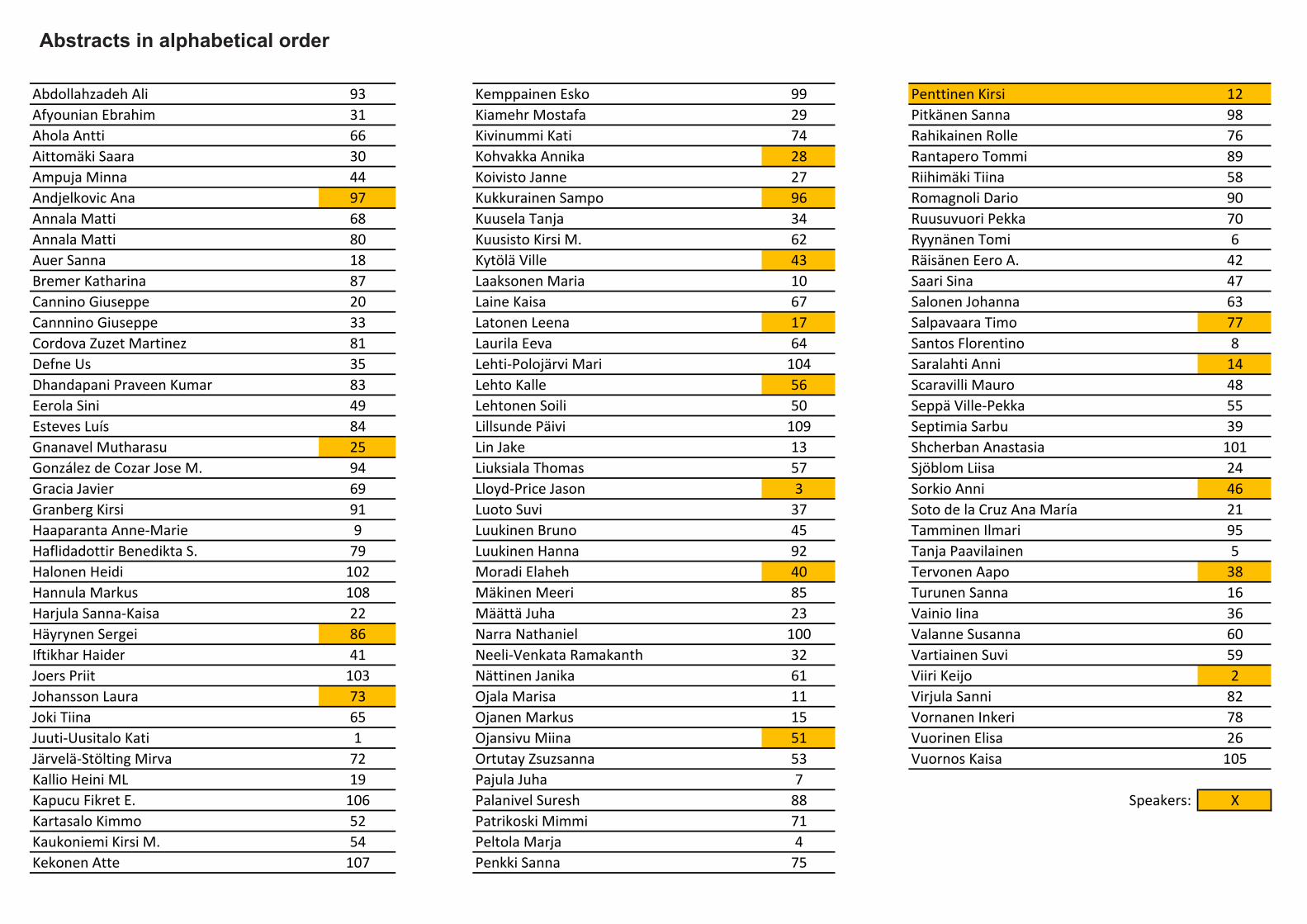
Abstracts in alphabeti-cal order
Abstracts in alphabetical order
Abdollahzadeh Ali 93 Kemppainen Esko 99 Penttinen Kirsi 12Afyounian Ebrahim 31 Kiamehr Mostafa 29 Pitkänen Sanna 98Ahola Antti 66 Kivinummi Kati 74 Rahikainen Rolle 76Aittomäki Saara 30 Kohvakka Annika 28 Rantapero Tommi 89Ampuja Minna 44 Koivisto Janne 27 Riihimäki Tiina 58Andjelkovic Ana 97 Kukkurainen Sampo 96 Romagnoli Dario 90Annala Matti 68 Kuusela Tanja 34 Ruusuvuori Pekka 70Annala Matti 80 Kuusisto Kirsi M. 62 Ryynänen Tomi 6Auer Sanna 18 Kytölä Ville 43 Räisänen Eero A. 42Bremer Katharina 87 Laaksonen Maria 10 Saari Sina 47Cannino Giuseppe 20 Laine Kaisa 67 Salonen Johanna 63Cannnino Giuseppe 33 Latonen Leena 17 Salpavaara Timo 77Cordova Zuzet Martinez 81 Laurila Eeva 64 Santos Florentino 8Defne Us 35 Lehti‐Polojärvi Mari 104 Saralahti Anni 14Dhandapani Praveen Kumar 83 Lehto Kalle 56 Scaravilli Mauro 48Eerola Sini 49 Lehtonen Soili 50 Seppä Ville‐Pekka 55Esteves Luís 84 Lillsunde Päivi 109 Septimia Sarbu 39Gnanavel Mutharasu 25 Lin Jake 13 Shcherban Anastasia 101González de Cozar Jose M. 94 Liuksiala Thomas 57 Sjöblom Liisa 24Gracia Javier 69 Lloyd‐Price Jason 3 Sorkio Anni 46Granberg Kirsi 91 Luoto Suvi 37 Soto de la Cruz Ana María 21Haaparanta Anne‐Marie 9 Luukinen Bruno 45 Tamminen Ilmari 95Haflidadottir Benedikta S. 79 Luukinen Hanna 92 Tanja Paavilainen 5Halonen Heidi 102 Moradi Elaheh 40 Tervonen Aapo 38Hannula Markus 108 Mäkinen Meeri 85 Turunen Sanna 16Harjula Sanna‐Kaisa 22 Määttä Juha 23 Vainio Iina 36Häyrynen Sergei 86 Narra Nathaniel 100 Valanne Susanna 60Iftikhar Haider 41 Neeli‐Venkata Ramakanth 32 Vartiainen Suvi 59Joers Priit 103 Nättinen Janika 61 Viiri Keijo 2Johansson Laura 73 Ojala Marisa 11 Virjula Sanni 82Joki Tiina 65 Ojanen Markus 15 Vornanen Inkeri 78Juuti‐Uusitalo Kati 1 Ojansivu Miina 51 Vuorinen Elisa 26Järvelä‐Stölting Mirva 72 Ortutay Zsuzsanna 53 Vuornos Kaisa 105Kallio Heini ML 19 Pajula Juha 7Kapucu Fikret E. 106 Palanivel Suresh 88 Speakers: XKartasalo Kimmo 52 Patrikoski Mimmi 71Kaukoniemi Kirsi M. 54 Peltola Marja 4Kekonen Atte 107 Penkki Sanna 75


Related Documents