Biomaterials Science PAPER Cite this: Biomater. Sci., 2017, 5, 1558 Received 22nd December 2016, Accepted 2nd February 2017 DOI: 10.1039/c6bm00935b rsc.li/biomaterials-science Phosphatase-triggered cell-selective release of a Pt(IV)-backboned prodrug-like polymer for an improved therapeutic index† Shao-Lu Li,‡ a Yingqin Hou,‡ a Yali Hu, a Jin Yu, a Wei Wei* b and Hua Lu* a We describe here the synthesis and cell-selective delivery of a cationic Pt(IV)-backboned prodrug-like polymer P(DSP-DAEP). P(DSP-DAEP) features excellent aqueous solubility, unusually high (44.5%) drug loading, can be rapidly reduced to release the active cisplatin, and is more potent than its small molecular Pt(IV) precursor DSP. P(DSP-DAEP) can be formulated with an oppositely charged methoxyl poly(ethylene glycol)-block-poly(L-phosphotyrosine) (mPEG-b-PpY) to afford a polyion micelle (Pt-PIC) by taking advantage of polyelectrolyte coacervation. Preliminary in vitro cellular uptake and cytotoxicity assays indi- cate that Pt-PIC exhibits receptor (surface alkaline phosphatase)-dependent uptake and cytotoxicity. Overall, our results suggest a new approach to the improved therapeutic index of platinum-based anti- cancer drugs via cell-selective delivery. Introduction Cisplatin, or cis-diamminedichloridoplatinum(II) (CDDP), is a more-than-forty-years-old chemotherapy drug for lung, gastro- intestinal and genitourinary cancers. 1 Despite its broad clinical application, CDDP often leads to severe side-effects such as nephrotoxicity and neurotoxicity, which necessitates drug delivery systems for an improved therapeutic index. 2–11 Moreover, because of the intrinsically labile square-planar structure of Pt(II), CDDP can undergo rapid ligand exchange with water and sulfur-containing small molecular ligands (e.g. GSH) before it takes effect. To improve the therapeutic window of CDDP, the six-coordinated Pt(IV) compounds have emerged as promising prodrugs because they are less toxic and more inert in a biological environment than Pt(II) species. In the intracellular reductive environment, these Pt(IV) compounds can be activated by regenerating the anticancer Pt(II) species. 12–23 Importantly, the octahedral Pt(IV) prodrugs have two additional “axial” ligand sites compared to Pt(II) drugs, which offer convenient chemical handles for further functionalization. 24,25 In the past decade, many Pt(IV) pro- drugs 19,23,26,27 and delivery systems have been developed including single walled carbon nanotubes, 12 gold nano- particles, 14 polymeric nanoparticles, 18,28 and Pt(IV) nanoscale coordination polymers (NCPs) coated with a silica or liposome surface. 13,29–31 For polymeric delivery systems, the Pt(IV) moi- eties are typically attached at their chain ends or pendant side chains. 32,33 This strategy, however, usually gives inconsistent and varied drug loading as low as 1 wt%. 28 Considering the utmost importance of a high drug loading to the success of a delivery system, 34,35 we reason that drug-backboned polymers, which can afford predictable and reproducibly high drug loading, may be superior to the aforementioned conventional ones bearing Pt(IV) on the side chains. 36–38 Indeed, Shen and coworkers reported for the first time a series of Pt(IV)-back- boned polymers, which showed high Pt loading ranging from 10 to 29%. 39 However, a further increase of drug loading may lead to hampered solubility and aggregation of these drug- backboned polymers. To further expand the therapeutic window of CDDP, tar- geted delivery has been a longstanding strategy. For this, two approaches are frequently adapted namely the passive target- ing that harnesses the so called enhanced permeation and retention (EPR) effect, and the active targeting by using ligands that recognize tumor specific/associated receptors. 18 For example, ligands such as aptamers, 40,41 RGD/iRGD peptides, 42–44 folate, 37 anisamide, 45 and glucose 46 have been extensively exploited for cell-selective platinum delivery. More recently, delivery vehicles have been engineered to accumulate and release drugs in response to pathological biomolecules † Electronic supplementary information (ESI) available. See DOI: 10.1039/ c6bm00935b ‡ These authors contributed equally. a Beijing National Laboratory for Molecular Sciences, Center for Soft Matter Science and Engineering, Key Laboratory of Polymer Chemistry and Physics of Ministry of Education, College of Chemistry and Molecular Engineering, Peking University, Beijing 100871, People’s Republic of China. E-mail: [email protected] b State Key Laboratory of Biochemical Engineering, Institute of Process Engineering, Chinese Academy of Sciences, Beijing, 10090, People’s Republic of China. E-mail: [email protected] 1558 | Biomater. Sci. , 2017, 5, 1558–1566 This journal is © The Royal Society of Chemistry 2017 Published on 09 February 2017. Downloaded by Peking University on 10/14/2018 4:25:31 AM. View Article Online View Journal | View Issue

Welcome message from author
This document is posted to help you gain knowledge. Please leave a comment to let me know what you think about it! Share it to your friends and learn new things together.
Transcript
BiomaterialsScience
PAPER
Cite this: Biomater. Sci., 2017, 5, 1558
Received 22nd December 2016,Accepted 2nd February 2017
DOI: 10.1039/c6bm00935b
rsc.li/biomaterials-science
Phosphatase-triggered cell-selective release ofa Pt(IV)-backboned prodrug-like polymer for animproved therapeutic index†
Shao-Lu Li,‡a Yingqin Hou,‡a Yali Hu,a Jin Yu,a Wei Wei*b and Hua Lu*a
We describe here the synthesis and cell-selective delivery of a cationic Pt(IV)-backboned prodrug-like
polymer P(DSP-DAEP). P(DSP-DAEP) features excellent aqueous solubility, unusually high (44.5%) drug
loading, can be rapidly reduced to release the active cisplatin, and is more potent than its small molecular
Pt(IV) precursor DSP. P(DSP-DAEP) can be formulated with an oppositely charged methoxyl poly(ethylene
glycol)-block-poly(L-phosphotyrosine) (mPEG-b-PpY) to afford a polyion micelle (Pt-PIC) by taking
advantage of polyelectrolyte coacervation. Preliminary in vitro cellular uptake and cytotoxicity assays indi-
cate that Pt-PIC exhibits receptor (surface alkaline phosphatase)-dependent uptake and cytotoxicity.
Overall, our results suggest a new approach to the improved therapeutic index of platinum-based anti-
cancer drugs via cell-selective delivery.
Introduction
Cisplatin, or cis-diamminedichloridoplatinum(II) (CDDP), is amore-than-forty-years-old chemotherapy drug for lung, gastro-intestinal and genitourinary cancers.1 Despite its broadclinical application, CDDP often leads to severe side-effectssuch as nephrotoxicity and neurotoxicity, which necessitatesdrug delivery systems for an improved therapeutic index.2–11
Moreover, because of the intrinsically labile square-planarstructure of Pt(II), CDDP can undergo rapid ligand exchangewith water and sulfur-containing small molecular ligands (e.g.GSH) before it takes effect. To improve the therapeutic windowof CDDP, the six-coordinated Pt(IV) compounds have emergedas promising prodrugs because they are less toxic and moreinert in a biological environment than Pt(II) species. In theintracellular reductive environment, these Pt(IV) compoundscan be activated by regenerating the anticancer Pt(II)species.12–23 Importantly, the octahedral Pt(IV) prodrugs havetwo additional “axial” ligand sites compared to Pt(II)drugs, which offer convenient chemical handles for further
functionalization.24,25 In the past decade, many Pt(IV) pro-drugs19,23,26,27 and delivery systems have been developedincluding single walled carbon nanotubes,12 gold nano-particles,14 polymeric nanoparticles,18,28 and Pt(IV) nanoscalecoordination polymers (NCPs) coated with a silica or liposomesurface.13,29–31 For polymeric delivery systems, the Pt(IV) moi-eties are typically attached at their chain ends or pendant sidechains.32,33 This strategy, however, usually gives inconsistentand varied drug loading as low as 1 wt%.28 Considering theutmost importance of a high drug loading to the success of adelivery system,34,35 we reason that drug-backboned polymers,which can afford predictable and reproducibly high drugloading, may be superior to the aforementioned conventionalones bearing Pt(IV) on the side chains.36–38 Indeed, Shen andcoworkers reported for the first time a series of Pt(IV)-back-boned polymers, which showed high Pt loading ranging from10 to 29%.39 However, a further increase of drug loading maylead to hampered solubility and aggregation of these drug-backboned polymers.
To further expand the therapeutic window of CDDP, tar-geted delivery has been a longstanding strategy. For this, twoapproaches are frequently adapted namely the passive target-ing that harnesses the so called enhanced permeation andretention (EPR) effect, and the active targeting by usingligands that recognize tumor specific/associated receptors.18
For example, ligands such as aptamers,40,41 RGD/iRGDpeptides,42–44 folate,37 anisamide,45 and glucose46 have beenextensively exploited for cell-selective platinum delivery. Morerecently, delivery vehicles have been engineered to accumulateand release drugs in response to pathological biomolecules
†Electronic supplementary information (ESI) available. See DOI: 10.1039/c6bm00935b‡These authors contributed equally.
aBeijing National Laboratory for Molecular Sciences, Center for Soft Matter Science
and Engineering, Key Laboratory of Polymer Chemistry and Physics of Ministry of
Education, College of Chemistry and Molecular Engineering, Peking University,
Beijing 100871, People’s Republic of China. E-mail: [email protected] Key Laboratory of Biochemical Engineering, Institute of Process Engineering,
Chinese Academy of Sciences, Beijing, 10090, People’s Republic of China.
E-mail: [email protected]
1558 | Biomater. Sci., 2017, 5, 1558–1566 This journal is © The Royal Society of Chemistry 2017
Publ
ishe
d on
09
Febr
uary
201
7. D
ownl
oade
d by
Pek
ing
Uni
vers
ity o
n 10
/14/
2018
4:2
5:31
AM
.
View Article OnlineView Journal | View Issue
presented in the tumor microenvironment such as matrixmetalloproteinase (MMP), hyaluronidase or low pH.47–52 Inthis regard, protein tyrosine phosphatase (PTP), whose over-expression and aberrant phosphate-cleavage activity have beencorrelated with the malignancy of many diseases includingcancers,53,54 has emerged as a potential extracellular cue fortumor microenvironment delivery. However, it is only recentlythat PTPs such as alkaline phosphatase (ALP) were exploitedfor the in situ generation of pericellular hydrogels.55–57
Nevertheless, no PTP-triggered targeted drug delivery systemhas been developed to our best knowledge.
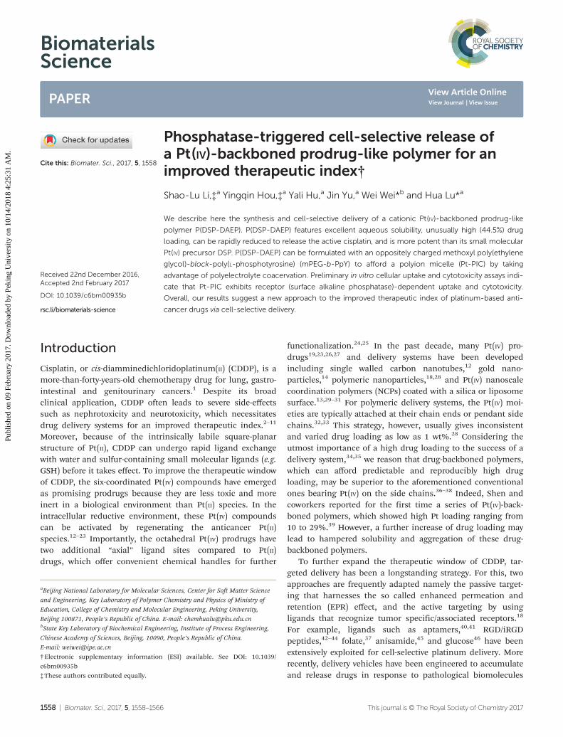
We report in the present work the synthesis of a high drug-loading (44%) Pt(IV)-backboned cationic polymer with excellentaqueous solubility, namely poly(diamminedichlorodisuccinato-platinum(IV)-diamineethylpiperazine) (P(DSP-DAEP), Fig. 1).P(DSP-DAEP) is more sensitive to reductive activation andshows enhanced in vitro anticancer activity compared to its
precursor diamminedichlorodisuccinatoplatinum(IV) (DSP).Moreover, P(DSP-DAEP), due to its cationic nature, can form aPEG-shelled polyion complex (Pt-PIC) with an anionic meth-oxyl poly(ethylene glycol)-block-poly(phosphotyrosine) diblockcopolymer (mPEG-b-PpY) (Fig. 1). Pt-PIC is designed to achieveselective internalization of P(DSP-DAEP) in ALP-overexpressingcancer cells through enzymatic cleavage of the phosphategroup of PEG-b-PpY,58 which weakens the polyion complex andconsequently accelerates the release of P(DSP-DAEP). In cellswithout surface ALP, Pt-PIC displays much less internalizationdue to the PEG-shelled polyion and negative zeta-potential.Thus, this work may provide a new approach to the cell-selec-tive delivery of Pt(IV)-backboned prodrugs via phosphatase-responsiveness.
Results and discussionSynthesis and characterization of P(DSP-DAEP)
To produce a high drug loading and yet a highly water solublePt(IV)-backboned polymer, we designed and synthesized thecationic polymer P(DSP-DAEP) as shown in Scheme 1. Briefly,the bis(carboxylic acid)-functionalized Pt(IV) intermediate DSP(Fig. S1 and S2†) was synthesized in two steps by following apreviously reported protocol,39 which was then converted toPt-NHS, the N-hydroxysuccinimide ester form of DSP, by EDCactivation in anhydrous DMF.59 Different from the Shen’sprotocol in which DSP was directly coupled to bisamines inthe presence of EDC, Pt-NHS was synthesized here because itcould be purified by a reversed phase C18 column and lyophi-lized to afford a white powder with high purity (confirmed by1H NMR, 13C NMR and HRESI-MS, Fig. S3–S5†). This purifi-cation step was essential to obtain a high-molecular-weightpolymer via step-growth condensation polymerization.Coupling of Pt-NHS with the bis(primary amine)-functiona-lized DAEP with strictly equal stoichiometry for 48 h yieldedthe Pt(IV)-backboned polymer P(DSP-DAEP) in high yield.Thanks to the cationic nature of P(DSP-DAEP), the polymerwas highly water soluble without aggregation and could be
Fig. 1 Chemical structure of mPEG-b-PpY and P(DSP-DAEP), andcartoon illustration of the phosphatase-triggered dissociation of thepolyion complex (Pt-PIC).
Scheme 1 Synthesis of P(DSP-DAEP).
Biomaterials Science Paper
This journal is © The Royal Society of Chemistry 2017 Biomater. Sci., 2017, 5, 1558–1566 | 1559
Publ
ishe
d on
09
Febr
uary
201
7. D
ownl
oade
d by
Pek
ing
Uni
vers
ity o
n 10
/14/
2018
4:2
5:31
AM
. View Article Online
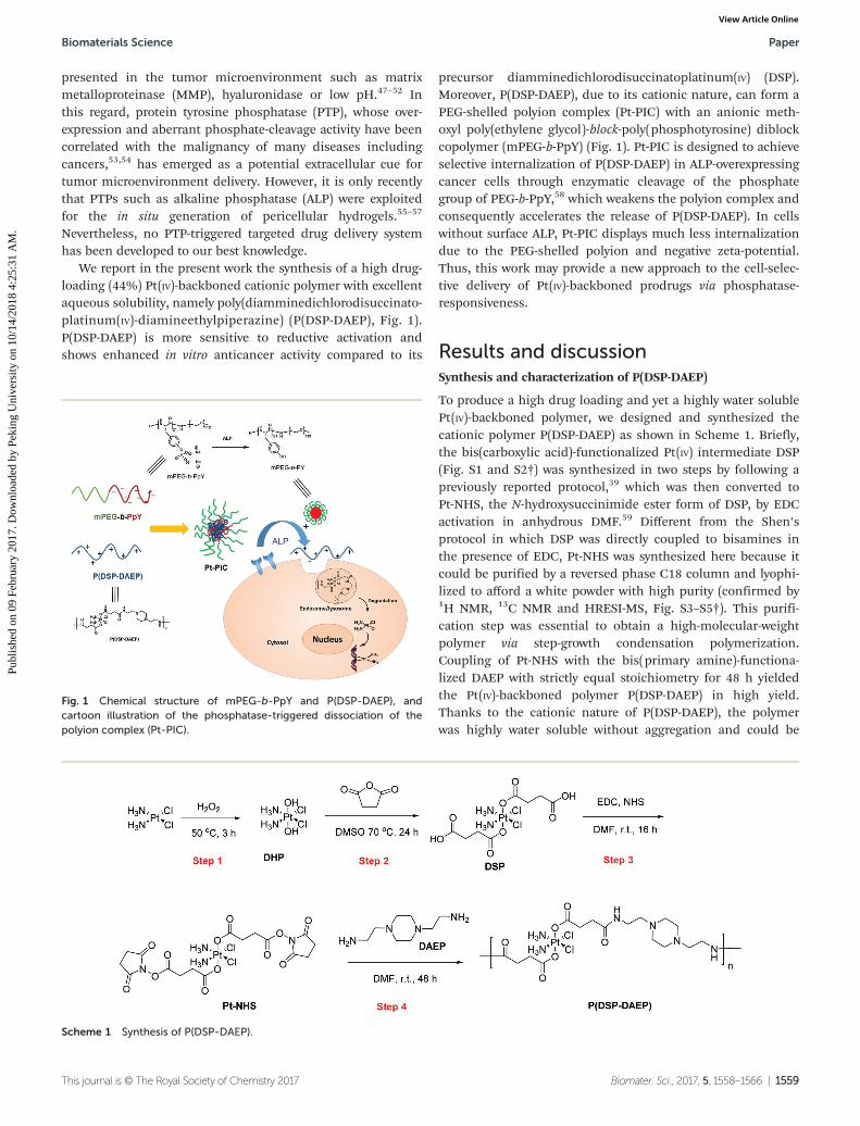
easily purified by dialysis against ultrapure water. Gel per-meation chromatography (GPC) examination of P(DSP-DAEP)revealed a MW of 55.9 kDa and a polydispersity index (PDI) of2.12 (Fig. S6†). The purity and identity of the polymer was wellcharacterized by 1H NMR spectroscopy in both DMSO-d6(Fig. 2) and D2O (Fig. S7†). The peak at 7.80 ppm was attribut-able to the newly produced amide proton. The broad peak at6.50 ppm was attributed to the protons of NH3 derived fromthe CDDP building block. The purity of P(DSP-DAEP) wasfurther confirmed by its Pt content determined by ICP-MS,which was 29.4% and in good agreement with the theoreticalvalue of 29.1%. This, in turn, gave a drug loading as high as44.5%.
Reduction of P(DSP-DAEP)
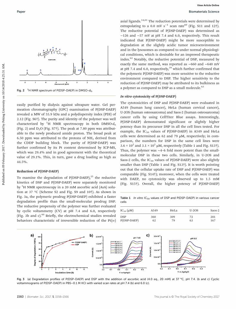
To examine the degradation of P(DSP-DAEP),25 the reductivekinetics of DSP and P(DSP-DAEP) were separately monitoredby 1H NMR spectroscopy in a 20 mM ascorbic acid (AsA) solu-tion at 37 °C (Scheme S2 and Fig. S9 and 10†). As shown inFig. 3a, the polymeric prodrug P(DSP-DAEP) exhibited a fasterdegradation profile than the small-molecular prodrug DSP.The reductive propensity of the polymer was further evaluatedby cyclic voltammetry (CV) at pH 7.4 and 6.0, respectively(Fig. 3b and c).60 Briefly, the electrochemical studies revealedbehaviors characteristic of irreversible reduction of the Pt(IV)
axial ligands.14,61 The reduction potentials were determined byextrapolating to a 0.0 mV s−1 scan rate60 (Fig. S11 and 12†).The reductive potential of P(DSP-DAEP) was determined as−126 and −57 mV at pH 7.4 and 6.0, respectively. This resultindicated that P(DSP-DAEP) might be more susceptible todegradation at the slightly acidic tumor microenvironmentand in the lysosomes as compared to under normal physiologi-cal conditions, which is desirable for an improved therapeuticindex.62 Notably, the reductive potential of DSP, measured byexactly the same method, was reported as −660 and −640 mVat pH 7.4 and 6.0, respectively,39 which further confirmed thatthe polymeric P(DSP-DAEP) was more sensitive to the reductiveenvironment compared to DSP. The higher sensitivity to thereduction of P(DSP-DAEP) may be attributed to its bulkiness asa polymer as compared to DSP as a small molecule.63
In vitro cytotoxicity of P(DSP-DAEP)
The cytotoxicities of DSP and P(DSP-DAEP) were evaluated inA549 (human lung cancer), HeLa (human cervical cancer),U-2OS (human osteosarcoma) and Saos-2 (human osteosarcoma)cancer cells by using CellTiter Blue assays. Interestingly,P(DSP-DAEP) demonstrated significant or slightly higherpotency than its precursor DSP in all the cell lines tested. Forexample, the IC50 values of P(DSP-DAEP) in A549 and HeLacells were determined as 62 and 79 µM, respectively; in com-parison, the numbers for DSP in the same cell lines were3.6 × 102 and 3.1 × 10² µM, respectively (Table 1 and Fig. S13†).Thus, the polymer was ∼4–6 fold more potent than the small-molecular DSP in these two cells. Similarly, in U-2OS andSaos-2 cells, the IC50 values of P(DSP-DAEP) were also slightlysmaller than DSP (Table 1 and Fig. S13†). It is worth pointingout that the cellular uptake rate of DSP and P(DSP-DAEP) wascomparable (Fig. S14†); moreover, when the cells were treatedwith DAEP, no cytotoxicity was observed up to 1.5 mM(Fig. S15†). Overall, the higher potency of P(DSP-DAEP)
Fig. 2 1H NMR spectrum of P(DSP-DAEP) in DMSO-d6.
Fig. 3 (a) Degradation profiles of P(DSP-DAEP) and DSP with the addition of ascorbic acid (4.0 eq., 20 mM) at 37 °C, pH 7.4. (b and c) Cyclicvoltammograms of P(DSP-DAEP) in PBS–0.1 M KCl with varied scan rates at pH 7.4 (b) and 6.0 (c).
Table 1 In vitro IC50 values of DSP and P(DSP-DAEP) in various cancercells
IC50 (µM) A549 HeLa U-2OS Saos-2
DSP 360 309 72 281P(DSP-DAEP) 62 79 63 167
Paper Biomaterials Science
1560 | Biomater. Sci., 2017, 5, 1558–1566 This journal is © The Royal Society of Chemistry 2017
Publ
ishe
d on
09
Febr
uary
201
7. D
ownl
oade
d by
Pek
ing
Uni
vers
ity o
n 10
/14/
2018
4:2
5:31
AM
. View Article Online
compared to DSP was in good agreement with the results ofprevious reduction experiments (Fig. 3).
Pt-PIC formation and ALP-responsiveness
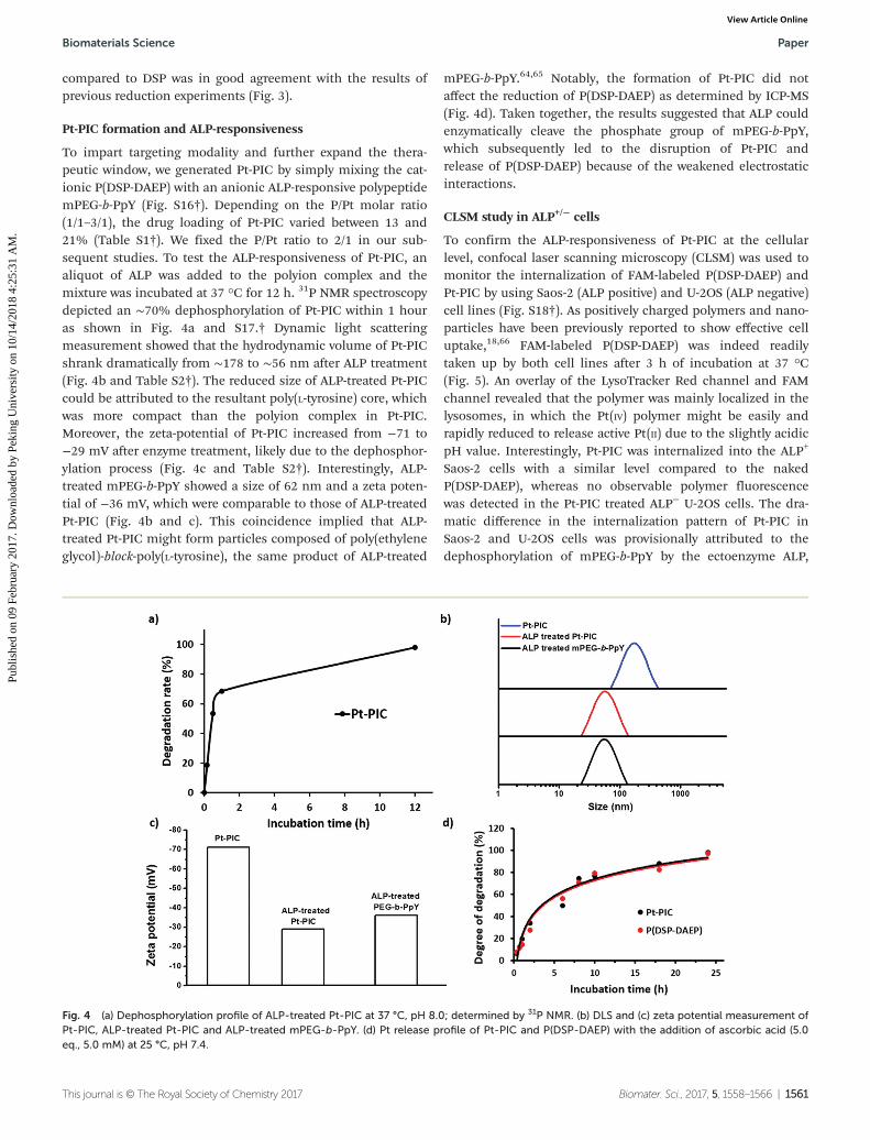
To impart targeting modality and further expand the thera-peutic window, we generated Pt-PIC by simply mixing the cat-ionic P(DSP-DAEP) with an anionic ALP-responsive polypeptidemPEG-b-PpY (Fig. S16†). Depending on the P/Pt molar ratio(1/1–3/1), the drug loading of Pt-PIC varied between 13 and21% (Table S1†). We fixed the P/Pt ratio to 2/1 in our sub-sequent studies. To test the ALP-responsiveness of Pt-PIC, analiquot of ALP was added to the polyion complex and themixture was incubated at 37 °C for 12 h. 31P NMR spectroscopydepicted an ∼70% dephosphorylation of Pt-PIC within 1 houras shown in Fig. 4a and S17.† Dynamic light scatteringmeasurement showed that the hydrodynamic volume of Pt-PICshrank dramatically from ∼178 to ∼56 nm after ALP treatment(Fig. 4b and Table S2†). The reduced size of ALP-treated Pt-PICcould be attributed to the resultant poly(L-tyrosine) core, whichwas more compact than the polyion complex in Pt-PIC.Moreover, the zeta-potential of Pt-PIC increased from −71 to−29 mV after enzyme treatment, likely due to the dephosphor-ylation process (Fig. 4c and Table S2†). Interestingly, ALP-treated mPEG-b-PpY showed a size of 62 nm and a zeta poten-tial of −36 mV, which were comparable to those of ALP-treatedPt-PIC (Fig. 4b and c). This coincidence implied that ALP-treated Pt-PIC might form particles composed of poly(ethyleneglycol)-block-poly(L-tyrosine), the same product of ALP-treated
mPEG-b-PpY.64,65 Notably, the formation of Pt-PIC did notaffect the reduction of P(DSP-DAEP) as determined by ICP-MS(Fig. 4d). Taken together, the results suggested that ALP couldenzymatically cleave the phosphate group of mPEG-b-PpY,which subsequently led to the disruption of Pt-PIC andrelease of P(DSP-DAEP) because of the weakened electrostaticinteractions.
CLSM study in ALP+/− cells
To confirm the ALP-responsiveness of Pt-PIC at the cellularlevel, confocal laser scanning microscopy (CLSM) was used tomonitor the internalization of FAM-labeled P(DSP-DAEP) andPt-PIC by using Saos-2 (ALP positive) and U-2OS (ALP negative)cell lines (Fig. S18†). As positively charged polymers and nano-particles have been previously reported to show effective celluptake,18,66 FAM-labeled P(DSP-DAEP) was indeed readilytaken up by both cell lines after 3 h of incubation at 37 °C(Fig. 5). An overlay of the LysoTracker Red channel and FAMchannel revealed that the polymer was mainly localized in thelysosomes, in which the Pt(IV) polymer might be easily andrapidly reduced to release active Pt(II) due to the slightly acidicpH value. Interestingly, Pt-PIC was internalized into the ALP+
Saos-2 cells with a similar level compared to the nakedP(DSP-DAEP), whereas no observable polymer fluorescencewas detected in the Pt-PIC treated ALP− U-2OS cells. The dra-matic difference in the internalization pattern of Pt-PIC inSaos-2 and U-2OS cells was provisionally attributed to thedephosphorylation of mPEG-b-PpY by the ectoenzyme ALP,
Fig. 4 (a) Dephosphorylation profile of ALP-treated Pt-PIC at 37 °C, pH 8.0; determined by 31P NMR. (b) DLS and (c) zeta potential measurement ofPt-PIC, ALP-treated Pt-PIC and ALP-treated mPEG-b-PpY. (d) Pt release profile of Pt-PIC and P(DSP-DAEP) with the addition of ascorbic acid (5.0eq., 5.0 mM) at 25 °C, pH 7.4.
Biomaterials Science Paper
This journal is © The Royal Society of Chemistry 2017 Biomater. Sci., 2017, 5, 1558–1566 | 1561
Publ
ishe
d on
09
Febr
uary
201
7. D
ownl
oade
d by
Pek
ing
Uni
vers
ity o
n 10
/14/
2018
4:2
5:31
AM
. View Article Online
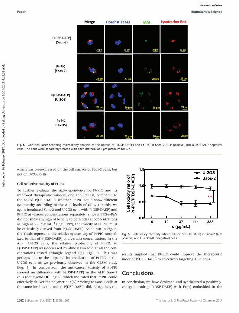
which was overexpressed on the cell surface of Saos-2 cells, butnot on U-2OS cells.
Cell selective toxicity of Pt-PIC
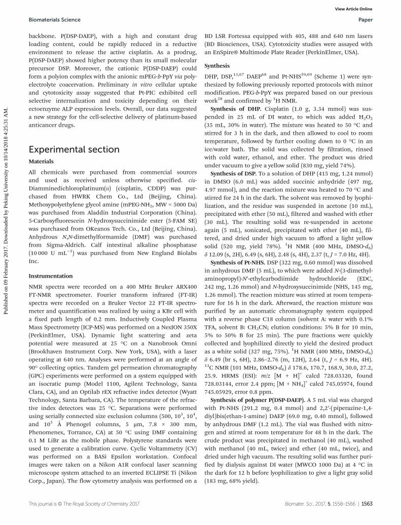
To further evaluate the ALP-dependence of Pt-PIC and itsimproved therapeutic window, one should test, compared tothe naked P(DSP-DAEP), whether Pt-PIC could show differentcytotoxicity according to the ALP levels of cells. For this, weagain incubated Saos-2 and U-2OS cells with P(DSP-DAEP) andPt-PIC at various concentrations separately. Since mPEG-b-PpYdid not show any sign of toxicity to both cells at concentrationsas high as 2.8 mg mL−1 (Fig. S19†), the toxicity of Pt-PIC mustbe exclusively derived from P(DSP-DAEP). As shown in Fig. 6,the Y axis represents the relative cytotoxicity of Pt-PIC normal-ized to that of P(DSP-DAEP) at a certain concentration. In theALP− U-2OS cells, the relative cytotoxicity of Pt-PIC toP(DSP-DAEP) was decreased by almost two fold at all the con-centrations tested (triangle legend (△), Fig. 6). This wasperhaps due to the impeded internalization of Pt-PIC in theU-2OS cells as we previously observed in the CLSM study(Fig. 5). In comparison, the anti-cancer toxicity of Pt-PICshowed no difference with P(DSP-DAEP) in the ALP+ Saos-2cells (dot legend (●), Fig. 6), which indicated that Pt-PIC couldeffectively deliver the polymeric Pt(IV)-prodrug to Saos-2 cells atthe same level as the naked P(DSP-DAEP) did. Altogether, the
results implied that Pt-PIC could improve the therapeuticindex of P(DSP-DAEP) by selectively targeting ALP+ cells.
Conclusions
In conclusion, we have designed and synthesized a positively-charged prodrug P(DSP-DAEP) with Pt(IV) embedded in the
Fig. 5 Confocal laser scanning microscopy analysis of the uptake of P(DSP-DAEP) and Pt-PIC in Saos-2 (ALP positive) and U-2OS (ALP negative)cells. The cells were separately treated with each material at 5 μM platinum for 3 h.
Fig. 6 Relative cytotoxicity ratio of Pt-PIC/P(DSP-DAEP) in Saos-2 (ALPpositive) and U-2OS (ALP negative) cells.
Paper Biomaterials Science
1562 | Biomater. Sci., 2017, 5, 1558–1566 This journal is © The Royal Society of Chemistry 2017
Publ
ishe
d on
09
Febr
uary
201
7. D
ownl
oade
d by
Pek
ing
Uni
vers
ity o
n 10
/14/
2018
4:2
5:31
AM
. View Article Online
backbone. P(DSP-DAEP), with a high and constant drugloading content, could be rapidly reduced in a reductiveenvironment to release the active cisplatin. As a prodrug,P(DSP-DAEP) showed higher potency than its small molecularprecursor DSP. Moreover, the cationic P(DSP-DAEP) couldform a polyion complex with the anionic mPEG-b-PpY via poly-electrolyte coacervation. Preliminary in vitro cellular uptakeand cytotoxicity assay suggested that Pt-PIC exhibited cellselective internalization and toxicity depending on theirectoenzyme ALP expression levels. Overall, our data suggesteda new strategy for the cell-selective delivery of platinum-basedanticancer drugs.
Experimental sectionMaterials
All chemicals were purchased from commercial sourcesand used as received unless otherwise specified. cis-Diamminedichloroplatinum(II) (cisplatin, CDDP) was pur-chased from HWRK Chem Co., Ltd (Beijing, China).Methoxypolyethylene glycol amine (mPEG-NH2, MW = 5000 Da)was purchased from Aladdin Industrial Corporation (China).5-Carboxyfluorescein N-hydroxysuccinimide ester (5-FAM SE)was purchased from OKeanos Tech. Co., Ltd (Beijing, China).Anhydrous N,N-dimethylformamide (DMF) was purchasedfrom Sigma-Aldrich. Calf intestinal alkaline phosphatase(10 000 U mL−1) was purchased from New England BiolabsInc.
Instrumentation
NMR spectra were recorded on a 400 MHz Bruker ARX400FT-NMR spectrometer. Fourier transform infrared (FT-IR)spectra were recorded on a Bruker Vector 22 FT-IR spectro-meter and quantification was realized by using a KBr cell witha fixed path length of 0.2 mm. Inductively Coupled PlasmaMass Spectrometry (ICP-MS) was performed on a NexION 350X(PerkinElmer, USA). Dynamic light scattering and zetapotential were measured at 25 °C on a Nanobrook Omni(Brookhaven Instrument Corp. New York, USA), with a laseroperating at 640 nm. Analyses were performed at an angle of90° collecting optics. Tandem gel permeation chromatography(GPC) experiments were performed on a system equipped withan isocratic pump (Model 1100, Agilent Technology, SantaClara, CA), and an Optilab rEX refractive index detector (WyattTechnology, Santa Barbara, CA). The temperature of the refrac-tive index detectors was 25 °C. Separations were performedusing serially connected size exclusion columns (500, 103, 104,and 105 Å Phenogel columns, 5 μm, 7.8 × 300 mm,Phenomenex, Torrance, CA) at 50 °C using DMF containing0.1 M LiBr as the mobile phase. Polystyrene standards wereused to generate a calibration curve. Cyclic Voltammetry (CV)was performed on a BASi Epsilon workstation. Confocalimages were taken on a Nikon A1R confocal laser scanningmicroscope system attached to an inverted ECLIPSE Ti (NikonCorp., Japan). The flow cytometry analysis was performed on a
BD LSR Fortessa equipped with 405, 488 and 640 nm lasers(BD Biosciences, USA). Cytotoxicity studies were assayed withan EnSpire® Multimode Plate Reader (PerkinElmer, USA).
Synthesis
DHP, DSP,13,67 DAEP68 and Pt-NHS59,69 (Scheme 1) were syn-thesized by following previously reported protocols with minormodification. PEG-b-PpY was prepared based on our previouswork58 and confirmed by 1H NMR.
Synthesis of DHP. Cisplatin (1.0 g, 3.34 mmol) was sus-pended in 25 mL of DI water, to which was added H2O2
(35 mL, 30% in water). The mixture was heated to 50 °C andstirred for 3 h in the dark, and then allowed to cool to roomtemperature, followed by further cooling down to 0 °C in anice/water bath. The solid was collected by filtration, rinsedwith cold water, ethanol, and ether. The product was driedunder vacuum to give a yellow solid (830 mg, yield 74%).
Synthesis of DSP. To a solution of DHP (415 mg, 1.24 mmol)in DMSO (6.0 mL) was added succinic anhydride (497 mg,4.97 mmol), and the reaction mixture was heated to 70 °C andstirred for 24 h in the dark. The solvent was removed by lyophi-lization, and the residue was suspended in acetone (10 mL),precipitated with ether (50 mL), filtered and washed with ether(30 mL). The resulting solid was re-suspended in acetoneagain (5 mL), sonicated, precipitated with ether (40 mL), fil-tered, and dried under high vacuum to afford a light yellowsolid (520 mg, yield 78%). 1H NMR (400 MHz, DMSO-d6)δ 12.09 (s, 2H), 6.49 (s, 6H), 2.48 (s, 4H), 2.37 (t, J = 7.0 Hz, 4H).
Synthesis of Pt-NHS. DSP (322 mg, 0.60 mmol) was dissolvedin anhydrous DMF (5 mL), to which were added N-(3-dimethyl-aminopropyl)-N′-ethylcarbodiimide hydrochloride (EDC,242 mg, 1.26 mmol) and N-hydroxysuccinimide (NHS, 145 mg,1.26 mmol). The reaction mixture was stirred at room tempera-ture for 16 h in the dark. Afterward, the reaction mixture waspurified by an automatic chromatography system equippedwith a reverse phase C18 column (solvent A: water with 0.1%TFA, solvent B: CH3CN; elution conditions: 5% B for 10 min,5% to 50% B for 25 min). The pure fractions were quicklycollected and lyophilized directly to yield the desired productas a white solid (327 mg, 75%). 1H NMR (400 MHz, DMSO-d6)δ 6.49 (br s, 6H), 2.86–2.76 (m, 12H), 2.64 (t, J = 6.9 Hz, 4H).13C NMR (101 MHz, DMSO-d6) δ 178.6, 170.7, 168.9, 30.0, 27.2,25.9. HRMS (ESI): m/z [M + H]+ calcd 728.03320, found728.03144, error 2.4 ppm; [M + NH4]
+ calcd 745.05974, found745.05929, error 0.8 ppm.
Synthesis of polymer P(DSP-DAEP). A 5 mL vial was chargedwith Pt-NHS (291.2 mg, 0.4 mmol) and 2,2′-(piperazine-1,4-diyl)bis(ethan-1-amine) DAEP (69.0 mg, 0.40 mmol), followedby anhydrous DMF (1.2 mL). The vial was flushed with nitro-gen and stirred at room temperature for 48 h in the dark. Thecrude product was precipitated in methanol (40 mL), washedwith methanol (40 mL, twice) and ether (40 mL, twice), anddried under high vacuum. The resulting solid was further puri-fied by dialysis against DI water (MWCO 1000 Da) at 4 °C inthe dark for 12 h before lyophilization to give a light gray solid(183 mg, 68% yield).
Biomaterials Science Paper
This journal is © The Royal Society of Chemistry 2017 Biomater. Sci., 2017, 5, 1558–1566 | 1563
Publ
ishe
d on
09
Febr
uary
201
7. D
ownl
oade
d by
Pek
ing
Uni
vers
ity o
n 10
/14/
2018
4:2
5:31
AM
. View Article Online
1H NMR (400 MHz, D2O) δ 3.44 (s), 2.92–2.81 (m), 2.71–2.66(m), 2.53–2.47 (m). 1H NMR (400 MHz, DMSO-d6) δ 7.80 (br s),6.50 (br s), 3.18 (br s), 2.67–2.35 (m), 2.29–2.21 (m). Elementalanalysis by ICP-AES: Pt (wt%) calc. 29.4%; obtained 29.1%.
Synthesis of FAM-P(DSP-DAEP). A 5 mL vial was chargedwith Pt-NHS (145.6 mg, 0.20 mmol) and 2,2′-(piperazine-1,4-diyl)bis(ethan-1-amine) DAEP (34.5 mg, 0.20 mmol), followedby anhydrous DMF (1.2 mL). The vial was flushed with nitro-gen and stirred at room temperature for 48 h in the dark. Thenan additional aliquot of DAEP (3.4 mg, 0.02 mmol, 0.10 eq.)was added and stirred at r.t. for 24 h to ensure an amine-ended polymer. The resulting polymer was continuously conju-gated to 5-FAM-SE (14.2 mg, 0.03 mmol, 0.20 eq.) and stirredfor another 24 h. The crude product was precipitated in metha-nol (40 mL), washed with methanol (40 mL, twice) and ether(40 mL, twice), and dried under high vacuum. The crudeproduct was further purified by dialysis against DI water(MWCO 1000 Da) at 4 °C in the dark for 12 h before lyophiliza-tion to give an orange solid (56 mg).
Electrochemistry
Cyclic Voltammetry (CV) was performed by using degassedphosphate-buffer (10 mM, pH 6.0 or 7.4) with 0.1 M KCl as thesupporting electrolyte. The glassy carbon electrode and plati-num wire was used as the working electrode and counterelectrode, respectively. All potentials were represented usingAg/AgCl (saturated) as the reference electrode. The platinumconcentration of the polymer was set at 1.0 mM at varyingscan rates between 50 and 350 mV s−1. The electrolyte solutionwas bubbled with nitrogen for 20 minutes before eachmeasurement.
Reductive degradation of P(DSP-DAEP)
The reductive degradation of P(DSP-DAEP) was quantitativelymonitored by 1H NMR spectroscopy. Briefly, P(DSP-DAEP)(1.7 mg, 5.0 mmol Pt, 1.0 eq.) was incubated with ascorbicacid (4.0 eq.) in 2× PBS of D2O (0.50 mL, pH 7.4) at 37 °C.1H NMR spectra were recorded at various time points. Thequantification was realized by monitoring the disappearanceof the methylene peak at δ 2.68 ppm (peak of the proton a1)and the appearance of a peak at δ 2.48 ppm (peak of theproton a2) (Scheme S2 and Fig. S10†).
Polyion complex (PIC) preparation and phosphatase-responsiveness
Pt-PIC was prepared by adding the PEG-b-PpY solution(20 mM, 60 µL) into P(DSP-DAEP) (20 mM, 30 µL) in ultrapurewater. The mixture was then diluted to 1.2 mL with ultrapurewater to give a final concentration of 0.5 mM Pt(IV). Toexamine the phosphatase-responsiveness, the pH of the PICswas carefully adjusted to slightly basic by 0.1 N NaOH (pH7.4–8.6) and the resulting complexes were incubated at roomtemperature for 30 min before the assay. Alkaline phosphatase(ALP, 10 U µL−1, 4 µL) was added to the PICs (1.0 mL) andincubated at 37 °C for 12 hours. The mixture was characterizedby DLS and zeta potential.
Reductive properties of P(DSP-DAEP) and Pt-PIC measuredby ICP-MS
1.0 mL of Pt-PIC or P(DSP-DAEP) (Pt content 1.0 mM) wasseparately placed into a pre-swelled dialysis bag (cutoff mole-cular weight of 1.0 kDa) and immersed into 99 mL of 50 mMTris·HCl buffer (pH = 7.4) containing 5.0 mM ascorbic acid.The Pt-PIC was dialyzed in a shaking culture incubator at25 °C. 1.0 mL of sample was withdrawn from the mediumoutside the dialysis bag at specified time intervals, which wasmeasured for the Pt content by ICP-MS.
Dephosphorylation process of Pt-PIC with ALP measuredby 31P NMR
To a Pt-PIC (∼10 mg PpY) solution in 100 mM Tris·DCl buffer(pH = 8.0, 0.50 mL D2O), was added ALP (10 U μL−1 × 4 μL).The mixture was incubated at 37 °C and measured by 31P NMRat intervals.
Cell culture
Human osteosarcoma cell lines Saos-2 and U-2OS wereobtained from Prof. Shuyan Li (Peking University). A549 andHeLa were obtained as generous gifts from Prof. Shu Wang(Institute of Chemistry Chinese Academy of Sciences) andProf. Peng R. Chen (Peking University) respectively. The Saos-2and U-2OS cells were cultured in DMEM (Corning, Manassas,USA) supplemented with 10% FBS (Gibco), 100 U mL−1 penicil-lin and 100 U mL−1 streptomycin (Corning, Manassas, USA)under a humidified atmosphere containing 5% CO2 at 37 °C.
In vitro cell uptake of DSP and P(DSP-DAEP)
5 × 10⁶ HeLa cells were seeded in Corning® 100 mm TC-treated culture dishes and incubated for 24 h before experi-ments. The cells were treated with DSP or P(DSP-DAEP) at37 °C for 3 h. The cells were washed with 1× PBS, heparin(1 mg mL−1) and 1× PBS, trypsinized and cell numbers werecounted. The cells were then digested by HNO3 and the intra-cellular Pt amount was measured by ICP-MS.
Flow cytometry assay
The ALP expression level of Saos-2 and U-2OS was confirmedby flow cytometry assay with a mouse PE-labeled anti-humanALP monoclonal antibody (BD Biosciences, USA) following themanufacturer’s protocol. Briefly, the Saos-2 and U-2OScells were grown in a T75 cm2 flask to 90% confluenceand detached by a cell stripper (2 mL, Corning). The cells(∼1.0 × 10⁵ cells) were resuspended and incubated on ice in aPBS buffer (pH 7.4, 50 μL) supplemented with 3% BSA and1 μL anti-ALP antibody for 30 min in the dark. The stainedcells were rinsed with PBS (500 μL) 3 times and suspended incold PBS for flow cytometry analysis.
Confocal laser scanning microscopy analysis
The Saos-2/U-2OS cells in the exponential growth phase wereseeded in glass bottomed culture chambers (20 mm, Nest) at adensity of 1 × 10⁵ cells per well. The cells were incubated at
Paper Biomaterials Science
1564 | Biomater. Sci., 2017, 5, 1558–1566 This journal is © The Royal Society of Chemistry 2017
Publ
ishe
d on
09
Febr
uary
201
7. D
ownl
oade
d by
Pek
ing
Uni
vers
ity o
n 10
/14/
2018
4:2
5:31
AM
. View Article Online

37 °C under a humidified atmosphere containing 5% CO2 for24 h. The cells were treated with fresh medium (1.0 mL)containing FAM-labeled P(DSP-DAEP) and Pt-PIC (5 μM basedon platinum) for 3 h. The cells were washed with 1× PBS(1 mL × 3) and stained with Hoechst 33342 (Life TechnologiesInc. Carlsbad, USA) and LysoTracker® Red (Life TechnologiesInc. Carlsbad, USA) for 20 min at 37 °C in the dark. The cellswere imaged in cell culture medium (1.0 mL) after repetitiverinsing with 1× PBS (1.0 mL) three times. FAM-labeledP(DSP-DAEP) was prepared by conjugating 5-FAM-SE to amine-capped P(DSP-DAEP), which was obtained by maintaining aslightly higher stoichiometry of DAEP during the step-growthpolymerization. The FAM-labeled polymer was purified bydialysis against water (MW 3500) and lyophilized to afford anorange solid.
Cell viability assay
The Saos-2/U-2OS cells in the exponential growth phase wereseeded into a black 96-well plate at a density of 5000 cells perwell 24 h prior to the assay. The culture medium was removedand the cells were treated with DSP, P(DSP-DAEP), or Pt-PIC infresh medium at gradient concentrations (n = 3). After incu-bation for a certain time, the relative cell viabilities were moni-tored by the CellTiter-Blue® cell viability assay (Promega, USA)following the manufacturer’s protocol. The IC50 values werecalculated by using GraphPad Prism version 5 using log(inhibitor)vs. the response regression method.
Acknowledgements
This work was financially supported by the National KeyResearch and Development Program of China (no.2016YFA0201400). H. L. thanks the grants from the NationalNatural Science Foundation of China (NSFC21434008), and theOpen Funding Project of the State Key Laboratory ofBiochemical Engineering (no. 2015KF-01). S.-L. was supportedin part by the China Postdoctoral Science Foundation (no.2016 M600847) and the Postdoctoral Fellowship of the Peking-Tsinghua Center for Life Sciences.
References
1 L. Kelland, Nat. Rev. Cancer, 2007, 7, 573–584.2 N. Nishiyama, S. Okazaki, H. Cabral, M. Miyamoto, Y. Kato,
Y. Sugiyama, K. Nishio, Y. Matsumura and K. Kataoka,Cancer Res., 2003, 63, 8977–8983.
3 H. Cabral, Y. Matsumoto, K. Mizuno, Q. Chen,M. Murakami, M. Kimura, Y. Terada, M. R. Kano,K. Miyazono, M. Uesaka, N. Nishiyama and K. Kataoka,Nat. Nanotechnol., 2011, 6, 815–823.
4 S. Guo, Y. Wang, L. Miao, Z. Xu, C. M. Lin, Y. Zhang andL. Huang, ACS Nano, 2013, 7, 9896–9904.
5 X. Xue, M. D. Hall, Q. Zhang, P. C. Wang, M. M. Gottesmanand X. J. Liang, ACS Nano, 2013, 7, 10452–10464.
6 M. Callari, J. R. Aldrich-Wright, P. L. de Souza andM. H. Stenzel, Prog. Polym. Sci., 2014, 39, 1614–1643.
7 L. Miao, S. Guo, J. Zhang, W. Y. Kim and L. Huang, Adv.Funct. Mater., 2014, 24, 6601–6611.
8 S. Shen, C. Y. Sun, X. J. Du, H. J. Li, Y. Liu, J. X. Xia,Y. H. Zhu and J. Wang, Biomaterials, 2015, 70, 71–83.
9 H. Y. Yu, Z. H. Tang, D. W. Zhang, W. T. Song, Y. Zhanga,Y. Yang, Z. Ahmad and X. S. Chen, J. Controlled Release,2015, 205, 89–97.
10 S. Kaur, C. Prasad, B. Balakrishnan and R. Banerjee,Biomater. Sci., 2015, 3, 955–987.
11 J. Kim, S. Pramanick, D. Lee, H. Park and W. J. Kim,Biomater. Sci., 2015, 3, 1002–1017.
12 S. Dhar, F. X. Gu, R. Langer, O. C. Farokhzad andS. J. Lippard, Proc. Natl. Acad. Sci. U. S. A., 2008, 105,17356–17361.
13 W. J. Rieter, K. M. Pott, K. M. L. Taylor and W. B. Lin, J. Am.Chem. Soc., 2008, 130, 11584–11585.
14 S. Dhar, W. L. Daniel, D. A. Giljohann, C. A. Mirkin andS. J. Lippard, J. Am. Chem. Soc., 2009, 131, 14652–14653.
15 S. Venkataraman, J. L. Hedrick, Z. Y. Ong, C. Yang,P. L. R. Ee, P. T. Hammond and Y. Y. Yang, Adv. DrugDelivery Rev., 2011, 63, 1228–1246.
16 H. Q. Song, H. H. Xiao, Y. Zhang, H. D. Cai, R. Wang,Y. H. Zheng, Y. B. Huang, Y. X. Li, Z. G. Xie, T. J. Liu andX. B. Jing, J. Mater. Chem. B, 2013, 1, 762–772.
17 R. K. Pathak, S. Marrache, J. H. Choi, T. B. Berding andS. Dhar, Angew. Chem., Int. Ed., 2014, 53, 1963–1967.
18 N. Bertrand, J. Wu, X. Xu, N. Kamaly and O. C. Farokhzad,Adv. Drug Delivery Rev., 2014, 66, 2–25.
19 Y.-R. Zheng, K. Suntharalingam, T. C. Johnstone, H. Yoo,W. Lin, J. G. Brooks and S. J. Lippard, J. Am. Chem. Soc.,2014, 136, 8790–8798.
20 C. B. He, C. Poon, C. Chan, S. D. Yamada and W. B. Lin,J. Am. Chem. Soc., 2016, 138, 6010–6019.
21 C.-H. Lin, S.-H. Cheng, W.-N. Liao, P.-R. Wei, P.-J. Sung,C.-F. Weng and C.-H. Lee, Int. J. Pharm., 2012, 429, 138–147.
22 X. Xu, K. Xie, X. Q. Zhang, E. M. Pridgen, G. Y. Park,D. S. Cui, J. Shi, J. Wu, P. W. Kantoff, S. J. Lippard,R. Langer, G. C. Walker and O. C. Farokhzad, Proc. Natl.Acad. Sci. U. S. A., 2013, 110, 18638–18643.
23 S. G. Awuah, Y. R. Zheng, P. M. Bruno, M. T. Hemannand S. J. Lippard, J. Am. Chem. Soc., 2015, 137, 14854–14857.
24 T. C. Johnstone, K. Suntharalingam and S. J. Lippard,Chem. Rev., 2016, 116, 3436–3486.
25 J. J. Wilson and S. J. Lippard, Chem. Rev., 2014, 114, 4470–4495.
26 R. K. Pathak and S. Dhar, Chem. – Eur. J., 2016, 22, 3029–3036.
27 D. Zhou, Y. Cong, Y. Qi, S. He, H. Xiong, Y. Wu, Z. Xie,X. Chen, X. Jing and Y. Huang, Biomater. Sci., 2015, 3, 182–191.
28 S. Aryal, C.-M. J. Hu and L. Zhang, ACS Nano, 2010, 4,251–258.
Biomaterials Science Paper
This journal is © The Royal Society of Chemistry 2017 Biomater. Sci., 2017, 5, 1558–1566 | 1565
Publ
ishe
d on
09
Febr
uary
201
7. D
ownl
oade
d by
Pek
ing
Uni
vers
ity o
n 10
/14/
2018
4:2
5:31
AM
. View Article Online

29 Z.-T. Cao, Z.-Y. Chen, C.-Y. Sun, H.-J. Li, H.-X. Wang,Q.-Q. Cheng, Z.-Q. Zuo, J.-L. Wang, Y.-Z. Liu, Y.-C. Wangand J. Wang, Biomaterials, 2016, 94, 9–19.
30 J. D. Rocca, M. E. Werner, S. A. Kramer, R. C. Huxford-Phillips, R. Sukumar, N. D. Cummings, J. L. Vivero-Escoto,A. Z. Wang and W. Lin, Nanomedicine, 2015, 11, 31–38.
31 D. Liu, C. Poon, K. Lu, C. He and W. Lin, Nat. Commun.,2014, 5, 4182.
32 Q. Yang, J. Cai, S. Sun, X. Kang, J. Guo, Y. Zhu, L. Yan,X. Jing and Z. Wang, Biomater. Sci., 2016, 4, 661–669.
33 H. Zhou, G. Wang, Y. Lu and Z. Pan, Biomater. Sci., 2016, 4,1212–1218.
34 P. Huang, D. Wang, Y. Su, W. Huang, Y. Zhou, D. Cui,X. Zhu and D. Yan, J. Am. Chem. Soc., 2014, 136, 11748–11756.
35 K. Cai, X. He, Z. Song, Q. Yin, Y. Zhang, F. M. Uckun,C. Jiang and J. Cheng, J. Am. Chem. Soc., 2015, 137, 3458–3461.
36 H. Tang, C. J. Murphy, B. Zhang, Y. Shen, E. A. Van Kirk,W. J. Murdoch and M. Radosz, Biomaterials, 2010, 31,7139–7149.
37 Y. Dai, X. Kang, D. Yang, X. Li, X. Zhang, C. Li, Z. Hou,Z. Cheng, P. a. Ma and J. Lin, Adv. Healthcare Mater., 2013,2, 562–567.
38 X.-D. Xu, Y.-J. Cheng, J. Wu, H. Cheng, S.-X. Cheng,R.-X. Zhuo and X.-Z. Zhang, Biomaterials, 2016, 76, 238–249.
39 J. Yang, W. W. Liu, M. H. Sui, J. B. Tang and Y. Q. Shen,Biomaterials, 2011, 32, 9136–9143.
40 Z. Cao, R. Tong, A. Mishra, W. Xu, G. C. L. Wong,J. Cheng and Y. Lu, Angew. Chem., Int. Ed., 2009, 121, 6616–6620.
41 S. Dhar, N. Kolishetti, S. J. Lippard and O. C. Farokhzad,Proc. Natl. Acad. Sci. U. S. A., 2011, 108, 1850–1855.
42 W. T. Song, M. Q. Li, Z. H. Tang, Q. S. Li, Y. Yang, H. Y. Liu,T. C. Duan, H. Hong and X. S. Chen, Macromol. Biosci.,2012, 12, 1514–1523.
43 N. Graf, D. R. Bielenberg, N. Kolishetti, C. Muus,J. Banyard, O. C. Farokhzad and S. J. Lippard, ACS Nano,2012, 6, 4530–4539.
44 J. Della Rocca, R. C. Huxford, E. Comstock-Duggan andW. Lin, Angew. Chem., Int. Ed., 2011, 123, 10514–10518.
45 S. Guo, C. M. Lin, Z. Xu, L. Miao, Y. Wang and L. Huang,ACS Nano, 2014, 8, 4996–5009.
46 M. Patra, T. C. Johnstone, K. Suntharalingam andS. J. Lippard, Angew. Chem., Int. Ed., 2016, 55, 2550–2554.
47 L. Zhu, P. Kate and V. P. Torchilin, ACS Nano, 2012, 6,3491–3498.
48 J. Li, Z. Ge and S. Liu, Chem. Commun., 2013, 49, 6974–6976.
49 L. Zhu, T. Wang, F. Perche, A. Taigind and V. P. Torchilin,Proc. Natl. Acad. Sci. U. S. A., 2013, 110, 17047–17052.
50 Y. Liu, D. Zhang, Z. Y. Qiao, G. B. Qi, X. J. Liang, X. G. Chenand H. Wang, Adv. Mater., 2015, 27, 5034–5042.
51 H. Li, J. Du, X. Du, C. Xu, C. Sun, H. Wang, Z. Cao, X. Yang,Y. Zhu, S. Nie and J. Wang, Proc. Natl. Acad. Sci. U. S. A.,2016, 113, 4164–4169.
52 R. Mo, T. Jiang, R. Disanto, W. Tai and Z. Gu, Nat.Commun., 2014, 5, 3364–3364.
53 A. Ostman, C. Hellberg and F. D. Böhmer, Nat. Rev. Cancer,2006, 6, 307–320.
54 V. V. Vintonyak, A. P. Antonchick, D. Rauh andH. Waldmann, Curr. Opin. Chem. Biol., 2009, 13, 272–283.
55 J. Zhou, X. Du, N. Yamagata and B. Xu, J. Am. Chem. Soc.,2016, 138, 3813–3823.
56 R. A. Pires, Y. M. Abul-Haija, D. S. Costa, R. Novoa-Carballal, R. L. Reis, R. V. Ulijn and I. Pashkuleva, J. Am.Chem. Soc., 2015, 137, 576–579.
57 Y. Kuang, J. Shi, J. Li, D. Yuan, K. A. Alberti, Q. Xu andB. Xu, Angew. Chem., Int. Ed., 2014, 126, 8242–8245.
58 Y. Sun, Y. Hou, X. Zhou, J. Yuan, J. Wang and H. Lu, ACSMacro Lett., 2015, 4, 1000–1003.
59 Y. Yuan, R. T. K. Kwok, B. Z. Tang and B. Liu, J. Am. Chem.Soc., 2014, 136, 2546–2554.
60 R. S. Nicholson and I. Shain, Anal. Chem., 1964, 36, 706–723.
61 S. Dhar, Z. Liu, J. Thomale, H. Dai and S. J. Lippard, J. Am.Chem. Soc., 2008, 130, 11467–11476.
62 B. Arunachalam, U. T. Phan, H. J. Geuze and P. Cresswell,Proc. Natl. Acad. Sci. U. S. A., 2000, 97, 745–750.
63 L. Ellis, H. Er and T. Hambley, Aust. J. Chem., 1995, 48,793–806.
64 R. J. Amir, S. Zhong, D. J. Pochan and C. J. Hawker, J. Am.Chem. Soc., 2009, 131, 13949–13951.
65 H. Kühnle and H. G. Börner, Angew. Chem., Int. Ed., 2009,48, 6431–6434.
66 H.-J. Li, J.-Z. Du, J. Liu, X.-J. Du, S. Shen, Y.-H. Zhu,X. Wang, X. Ye, S. Nie and J. Wang, ACS Nano, 2016, 10,6753–6761.
67 K. R. Barnes, A. Kutikov and S. J. Lippard, Chem. Biol.,2004, 11, 557–564.
68 R. Filosa, A. Peduto, S. D. Micco, P. d. Caprariis, M. Festa,A. Petrella, G. Capranico and G. Bifulco, Bioorg. Med.Chem., 2009, 17, 13–24.
69 H. Liu, Y. Li, Z. Lyu, Y. Wan, X. Li, H. Chen, H. Chen andX. Li, J. Mater. Chem. B, 2014, 2, 8303–8309.
Paper Biomaterials Science
1566 | Biomater. Sci., 2017, 5, 1558–1566 This journal is © The Royal Society of Chemistry 2017
Publ
ishe
d on
09
Febr
uary
201
7. D
ownl
oade
d by
Pek
ing
Uni
vers
ity o
n 10
/14/
2018
4:2
5:31
AM
. View Article Online
Related Documents