Nanomedicine 111 5 Biomaterial surface modification of titanium and titanium alloys for medical applications Mukta Kulkarni 1,2 , Anca Mazare 2 , Patrik Schmuki 2 , Aleš Iglič 1 1 Laboratory of Biophysics, Faculty of Electrical Engineering, University of Ljubljana, Ljubljana SI-1000, Slovenia 2 Department of Materials Science and Engineering, University of Erlangen–Nuremberg, Erlangen, Germany Outline: Introduction ……………………………………………….………………………………………………………………………………. 112 Titanium and titanium alloys in medical applications …………………………………………..…………………….. 112 Biocompatibility of medical devices and the need for surface modification ……………………………….. 114 Surface modification of titanium and its alloys ………………………………………….……………………………….. 116 Overview of surface modification methods for titanium and its alloy …………………………………………. 116 Mechanical methods ………………………………………………………………………………………………………………….. 117 Chemical methods ……………………………………….…………………………………………………………………………….. 118 Chemical treatment ……………………………………………………………………………………………………………………. 119 Sol-gel deposition ………………………………………………………………………………………………………………………..120 Chemical vapour deposition (CVD) ……………………………………………………………………………………………… 120 Electrochemical methods ……………………………………………………………………………………………………………. 120 Biochemical methods …………………………………………………………………………………………………………………. 120 Physical methods ……………………………………………………………………………………………………………………….. 121 Selectively modified method: Anodization …………………………………………………………………………………. 123 General aspects of electrochemical anodization …………………………………………………………………………. 123 Biomedical applications of tio2 nanotubes …………………………………………………………………………………. 124 Nanotube diameter and cellular response ………………………………………………………………………………….. 124 Nanotubes and protein interaction ……………………………………………………………………………………………..126 Nanotubes for orthopaedic and dental implants ………………………………………………………………………… 126 Nanotubes for bladder stents ………………………………………………………………………………………………………127 Nanotubes for blood-contacting applications …………………………………………………………………………….. 127 Nanotubes for antibacterial activity …………………………………………………………………………………………… 128 Nanotubes for drug delivery ……………………………………………………………………………………………………….. 128 Summary …………………………………………………………………………………………………………………………………… 129 Acknowledgement ……………………………………………………………………………………………………………………. 129 References……………………………………………………………………………………..…………………………………………… 130

Welcome message from author
This document is posted to help you gain knowledge. Please leave a comment to let me know what you think about it! Share it to your friends and learn new things together.
Transcript
Nanomedicine 111 111
5 Biomaterial surface modification of titanium and titanium alloys for medical applications
Mukta Kulkarni
1,2, Anca Mazare
2, Patrik Schmuki
2, Aleš Iglič
1
1Laboratory of Biophysics, Faculty of Electrical Engineering, University of Ljubljana, Ljubljana SI-1000, Slovenia 2Department of Materials Science and Engineering, University of Erlangen–Nuremberg, Erlangen, Germany
Outline: Introduction ……………………………………………….………………………………………………………………………………. 112 Titanium and titanium alloys in medical applications …………………………………………..…………………….. 112 Biocompatibility of medical devices and the need for surface modification ……………………………….. 114 Surface modification of titanium and its alloys ………………………………………….……………………………….. 116 Overview of surface modification methods for titanium and its alloy …………………………………………. 116 Mechanical methods ………………………………………………………………………………………………………………….. 117 Chemical methods ……………………………………….…………………………………………………………………………….. 118 Chemical treatment ……………………………………………………………………………………………………………………. 119 Sol-gel deposition ……………………………………………………………………………………………………………………….. 120 Chemical vapour deposition (CVD) ……………………………………………………………………………………………… 120 Electrochemical methods ……………………………………………………………………………………………………………. 120 Biochemical methods …………………………………………………………………………………………………………………. 120 Physical methods ……………………………………………………………………………………………………………………….. 121 Selectively modified method: Anodization …………………………………………………………………………………. 123 General aspects of electrochemical anodization …………………………………………………………………………. 123 Biomedical applications of tio2 nanotubes …………………………………………………………………………………. 124 Nanotube diameter and cellular response ………………………………………………………………………………….. 124 Nanotubes and protein interaction ……………………………………………………………………………………………..126 Nanotubes for orthopaedic and dental implants ………………………………………………………………………… 126 Nanotubes for bladder stents ……………………………………………………………………………………………………… 127 Nanotubes for blood-contacting applications …………………………………………………………………………….. 127 Nanotubes for antibacterial activity …………………………………………………………………………………………… 128 Nanotubes for drug delivery ……………………………………………………………………………………………………….. 128 Summary …………………………………………………………………………………………………………………………………… 129 Acknowledgement ……………………………………………………………………………………………………………………. 129 References……………………………………………………………………………………..…………………………………………… 130
Nanomedicine 112 112
Introduction The biomaterials research domain is a multidisciplinary one and includes various aspects of materials science, chemistry, physics, biology and medicine. A biomaterial is a non-viable material used in medical devices intended to interact with biological systems in order to evaluate, treat, augment or replace any tissue, organ or function of the body [1]. The performance and applications of biomaterials in biological systems are of critical importance for the development of biomedical implants and tissue engineering. There are numerous biomaterials that can be used in the human body, such as metals (e.g. stainless steel, cobalt alloys, titanium alloys), ceramics (aluminium oxide, zirconia, calcium phosphates), and synthetic and natural polymers [2]. Among these, titanium (Ti) and titanium alloys are considered to be some of the most significant biomaterials, due to their resistance to body fluid effects, great tensile strength, flexibility and high corrosion resistance and this specific combination of strength and biocompatibility [3] makes them suitable for medical applications. For example, commercially pure Ti (c.p.Ti) is the dominant material used for dental implants while for orthopaedic applications Ti-6Al-4V alloy is used [4]. Here, in this chapter various methods of surface modification of titanium and its alloys are reviewed, including promising methods of obtaining specific nanotopography (e.g. titanium nanostructures) such as electrochemical anodization, together with the latest research evaluating the use and importance of nanotubular structures on Ti and its alloys for biomedical applications, as well as future perspectives. Titanium and titanium alloys in medical applications Metallic materials have been used in medical applications (orthopaedics or dentistry) for more than 50 years. Titanium and its alloys received extensive attention in dental applications so that nowadays commercially pure Ti (c.p.Ti) is the dominant material for dental implants and is used as an alternative to Ag-Pd-Au-Cu alloy – not only because of its excellent properties but also due to the increasing cost of Pd. Other reported representative dental titanium alloys are Ti–6Al–7Nb, Ti–6Al–4V, Ti–13Cu–4.5Ni, Ti–25Pd–5Cr, Ti–20Cr–0.2Si etc. [5]. For hard tissue replacement, the low Young´s modulus of titanium and its alloys is generally viewed as a biomechanical advantage because the low elastic modulus can result in smaller stress shielding compared to other implant materials, and thus inducing healthier and faster bone regeneration [6]. Besides artificial bones, joint replacements and dental implants, titanium and titanium alloys are often used in cardiovascular implants, for example in prosthetic heart valves, protective cases for pacemakers, artificial hearts and circulatory devices. Because of their inert, strong and non-magnetic properties, some alloys like nickel-titanium alloy (Nitinol, shape memory alloy) have received more attention in magnetic resonance imaging (MRI), which is a very powerful diagnostic tool. Currently, nickel-titanium alloy stents are often used in treatment of cardiovascular disease and are usually coated with a thin carbon film to enhance blood compatibility [7]. Since the focus of biomaterials has shifted towards tissue engineering, complex medical applications and biotechnology, there is a need to better define and evaluate the specific interaction between biomaterials and tissue components. After a thorough evaluation of the biomaterials field, Williams proposed a unified concept of biocompatibility [3], which states that “Biocompatibility refers to the ability of a biomaterial to perform its desired function with respect to medical therapy, without eliciting any undesirable local or systemic effects in the recipient or beneficiary of that therapy, but generating the most appropriate beneficial cellular or tissue response in that specific situation, and optimising the clinically relevant performance of that therapy.”. Titanium and its alloys remain essentially unchanged when implanted into the body and this is a result of their excellent corrosion resistance, so that such materials are referred to as bio-stable or biologically inert [4].
Nanomedicine 113 113
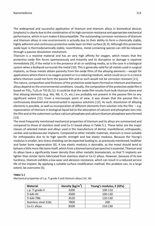
The widespread and successful application of titanium and titanium alloys in biomedical devices (implants) is clearly due to the combination of its high corrosion resistance and appropriate mechanical performance, which in turn makes it biocompatible. The outstanding corrosion resistance of titanium and titanium alloys in vivo environments is actually due to their ability to form a chemically stable, highly adherent and continuous protective oxide layer on their surface [8, 9]. Although this protective oxide layer is thermodynamically stable; nevertheless, metal containing species can still be released through a passive-dissolution mechanism. Titanium is a reactive material and has an very high affinity for oxygen, which means that the protective oxide film forms spontaneously and instantly and its disruption or damage is repaired immediately [9], if the metal is in the presence of air or oxidizing media, as is the case in a biological system when a bioliquid surrounds the metal [10]. This is generally valid for all metals used in surgical implants, as these metals obtain passivity from the oxide films of the alloying elements – except for applications where there is no oxygen present or in a reducing medium, which could occur in a crevice where titanium could not form the passive film and as such would not be corrosion resistant [11]. The nature, composition and thickness of the protective oxide layers formed on titanium and titanium alloys depend on the environmental conditions. Usually, the composition of the protective oxide film is based on TiO2, Ti2O3 or TiO [9,11]. It could be that the oxide film results from bulk titanium alone and that alloying elements (e.g. Mo, Nb, V, Cr, etc.) are probably not present in the passive film to any significant extent [11]. From a microscopic point of view, it was shown that the passive film is continuously dissolved and reconstructed in aqueous solutions [12]. As such, dissolution of alloying elements is possible, as well as incorporation of different elements from solution into the film – e.g. repassivation of titanium in biological liquid led to the adsorption of calcium and phosphate ions into the film and at the outermost surface calcium phosphate and calcium titanium phosphate were formed [13]. The most frequently mentioned mechanical properties of titanium and its alloys are summarized and compared to those of stainless steel and Co-Cr based alloys in Table 5.1. These latter are the major classes of selected metals and alloys used in the manufacture of dental, maxillofacial, orthopaedic, cardiac and cardiovascular implants. Compared to other metallic materials, titanium is more suitable for orthopaedics due to its high specific strength and low elastic modulus. Because the Young’s modulus is smaller, less stress shielding can be expected leading to, as previously mentioned, healthier and faster bone regeneration [6]. A low elastic modulus is desirable, as the metal should tend to behave a little more like bone itself, which from a biomechanical perspective is essential. Titanium and its alloys have a significantly lower density than other metallic biomaterials, so that Ti implants are lighter than similar items fabricated from stainless steel or Co-Cr alloys. However, because of its low hardness, titanium exhibits a low wear and abrasion resistance, which can result in a reduced service life of the implant. By applying a suitable surface modification method, this problem can, to a large extent, be overcome [5]. TABLE 5.1 Mechanical properties of c.p. Ti grade II and titanium alloys [14, 16]
Material Density (kg/m3) Young’s modulus, E (GPa)
c.p. Ti grade II 4200 100-110
Ti-6Al-4V 4500 100-130
Ti-6Al-7Nb 4520 110-130
Stainless steel 316L 7800 200
Co-Cr alloys 8500 210-230
Nanomedicine 114 114
As a result of the properties shown above, titanium is considered to be one of the most promising biomaterials for implants, especially in orthopaedic applications such as joint replacements and bone pins, plates and screws for repairing broken bones. One important aspect is that the fate of the implant material is not only governed by the bulk of the material (critical in determining the biological performance), but also by its surface properties (surface chemistry and structure) which are crucial factors in the interactions of the material with the surrounding tissue. The material chosen as bulk material should withstand stresses which are too high for ceramic or polymeric materials, but acceptable for metallic materials. However, the human body is able to recognise implant materials as foreign and tries to isolate them by encasing in fibrous tissues. Such is the case if the surface properties are not capable of leading to formation of a stable bonding between the surface of the implant and the surrounding tissue, but result in the formation of a fibrous layer which would undermine the load transmission between bone and implant, and would favour micro movements, eventually leading to implant failure [17].
Biocompatibility of medical devices and the need for surface modification Depending on the intended implant location, namely the desired application of the biomedical device, there are different factors to be considered. For example, if the biomedical device is intended to be a blood-contacting device (catheter, graft and stent), blood compatibility (haemocompatibility) of the biomaterials is crucial, whereas for bone applications osseointegration is the key parameter. For both types of applications, the host response and its severity are strictly related to the surface properties of the biomaterial. Biomedical devices for use in contact with blood must not activate the intrinsic blood coagulation system, nor attract or alter platelets or leucocytes. From this point of view, biocompatibility is more difficult to achieve as it covers aspects such as thrombogenicity, complement activation, leukocyte activation and changes in plasma proteins [18]. After the implantation of a blood contacting biomaterial, the first event that rapidly takes place is blood protein adsorption at the solid-liquid interface. The proteins undergo conformational changes allowing biological interactions and depending on the exposure time, the composition of the adsorbed protein layer varies and proteins with stronger adsorption are favoured. In time, a resident protein layer is formed which influences the interaction of platelets, activation of intrinsic coagulation, adhesion and aggregation of platelets and activation of the complement system [18]. Furthermore, at the implant or platelet adhesion surface, some blood coagulation factors are triggered and this could lead to formation of thrombin, converting fibrinogen into insoluble fibrin, from which a fibrin network and thrombin can be produced [19]. Several studies have reported the haemocompatibility of titanium [18,19] but less data is available on the haemocompatibility of nanobiomaterials [20,21]: the current status of research is further discussed in next sections. The clinical goal and most critical factor in the success of bone implants (orthopaedics and dentistry) is achieving osseointegration, particularly by establishing a strong and long-lasting connection between the implant surface and peri-implant bone, leading to a stable mechanical attachment of the implant at the site of the implantation [22]. In bone, titanium is integrated in close apposition to the mineralized tissues under the proper conditions. However, titanium and bone are generally separated by a thin soft-tissue layer as a result of a weak foreign body reaction that prevents titanium from being in direct contact with the bone [23]. As
Nanomedicine 115 115
soon as the implantation procedure occurs, several biological reactions take place in a specific order. Initially, there will be wetting of the implant surface and rapid adsorption of biologically active molecules (such as proteins), followed by enlisting of the osteoprogenitor cells that would regenerate the tissue [17]. It is obvious that the two factors affecting osseointegration are the mechanical properties of the implant and the biological interactions with the metal surface, of which the latter is more relevant. These interactions could lead to:
I. Successful osseointegration as a result of the osteoconductive process of healing of the peri-implant bone, when newly formed bone has direct contact with the implant surface as a result of bone cell proliferation and differentiation.
II. Rejection, due to an acute foreign body response caused by the inflammatory response reaction of the body to the implant.
III. Micromovements of the implant, favouring the formation of fibrous tissue instead of a bony interface, due to the lack of stability between the surrounding tissue and the implant surface. Micromovements can lead to implant failure.
IV. Bacterial infection at the surface of the implant that might lead to biofilm formation and thus to short-term or long-term failure of the implant.
Usually, the steps occuring in the interaction between a biomaterial and the body, i.e. the healing response, consist of acute inflammation, chronic inflammation, granulation tissue formation, foreign body reaction and fibrosis [24]. Regardless of the type of biomaterial used or of the injury location, the initial inflammation response is always present and will progress to acute inflammation (which usually lasts only a few days). If the inflammatory responses do not subside, chronic inflammation sets in followed by granulation tissue formation (the amount of granulation tissue determines the extent of fibrosis). The foreign body response is next and the most important factor at this point is the surface properties of the biomaterial as they influence the presence and magnitude of the foreign body response. The last step in the healing process is fibrosis, which consists of encapsulation of the fibrous tissue of the implant and depends on the proliferation capacity of the cells in the respective tissue [24]. It should be pointed out that for both orthopaedic and dental implants, fibrous encapsulation is not desirable as it cannot withstand the same physical stresses as bone, thus leading to micromovements. Recruitment of parenchymal cells (specifically osteosblasts) on the implant surface is desired. Considering the above-mentioned factors, it is obvious that there is still room to improve the implant surface, especially to enhance tissue engineering and to decrease implant failure or rejection. Firstly, bone regeneration is a slow process so improvements were and are currently being made in order to achieve faster osseointegration, either by morphological modifications or by various coatings, as will be discussed in detail in the following sections. Secondly, a common cause of implant failure is bacterial infection and the possibility of a bacteria-repellent surface modification is worth investigating. Currently, the most common method of achieving improvement is by modification of the implant’s surface properties, either morphologically and/or by biochemical coatings. It follows that there is a major need for surface modification of implants in order to increase tissue adhesion, implant integration, decrease bacterial adhesion and decrease inflammatory response or to avoid the foreign body response.
Nanomedicine 116 116
Surface modification of titanium and its alloys It is evident that the response of a biomaterial depends entirely on its biocompatibility and surface properties. Therefore, in order to improve the performance of biomaterials in biological systems, there is an urgent need for their surface modification [25]. The structures encountered by the cells present in the human body are not only on the micrometre scale but also on the nanometre scale since bone is made up of nanostructures. Consequently, it is necessary to produce better implant materials, i.e with nanometre roughness, especially since the influence of the surface roughness on cell attachment was evaluated mainly on the micrometre scale. There are various approaches possible to modify the surface of metallic materials (including nanophase materials) that improve their cellular activities when compared with conventional microrough materials. Generally, nanoscale surfaces possess high surface energy leading to increased initial protein adsorption which is very important in regulating the cellular interactions on the implant surface. Surface properties also have an impact on adhesion, together with charge distribution and the chemistry of the material [26,27]. It was recently observed that the roughness of titanium nanostructures alone influences the adhesion of osteoblast cells and their spreading and proliferation [28]. In the present section, surface modification methods for titanium and titanium alloys will be reviewed, including the mechanical, chemical and physical methods used for morphological modifications (increasing roughness, shifting topography from the micro to the nanoscale, tailoring the nanoscale morphology), or for obtaining different coatings on the surface of the implant. These include hydroxyapatite, biomimetic calcium phosphate coatings, biomolecule functionalized coatings, as well as a mixture of morphological changes and coatings for a combined synergistic effect. The goal is to improve bioactivity, biocompatibility, blood compatibility, and the wear and corrosion resistance of titanium and titanium alloys for their respective applications.
Overview of surface modification methods for titanium and its alloy The assumption that increasing the surface roughness of a metallic implant will result in higher micromechanical retention than for a smooth or as-machined implant was proven to be correct [29]. Nowadays, the implant surface is modified in commercial products by mechanical methods (grit blasting with various types and sizes of abrasives, attrition for obtaining nanophase materials), chemical methods (acid etching, electrochemical processes) and physical methods (plasma-sprayed titanium coatings) or by their combination [30,31]. In vitro results indicated a correlation between surface roughness and cellular attachment and osteoblast activity [32,33], as well as with other factors influencing implant osteointegration (selective protein adhesion, collagen synthesis, and chondrocyte maturation) [34, 36]. The results were conclusively supported by in vivo tests [37, 39]; namely, microstructured surfaces provided a better implant surface–bone contact and an increased mechanical retention after implantation. Material roughness modification influences other physicochemical properties, e.g. the higher the roughness, the higher the local surface electrostatic charge density [40,41] and adhesion energy. It is possible to design metallic surfaces with high surface energy and superhydrophilic properties through different methods [40,42], which are shown to accelerate the early stages of bone healing [42,43], possibly by preferential adsorption of fibronectin, osteocalcin or other growth factors and by favouring bone growth [44]; however, the mechanism of this higher osseointegration has not yet been elucidated.
Nanomedicine 117 117
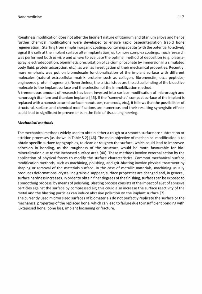
Roughness modification does not alter the bioinert nature of titanium and titanium alloys and hence further chemical modifications were developed to ensure rapid osseointegration (rapid bone regeneration). Starting from simple inorganic coatings containing apatite (with the potential to actively signal the cells at the implant surface after implantation) up to more complex coatings, much research was performed both in vitro and in vivo to evaluate the optimal method of deposition (e.g. plasma-spray, electrodeposition, biomimetic precipitation of calcium phosphate by immersion in a simulated body fluid, protein adsorption, etc.), as well as investigation of their mechanical properties. Recently, more emphasis was put on biomolecule functionalization of the implant surface with different molecules (natural extracellular matrix proteins such as collagen, fibronenctin, etc.; peptides; engineered protein fragments). Nevertheless, the critical steps are the actual binding of the bioactive molecule to the implant surface and the selection of the immobilization method. A tremendous amount of research has been invested into surface modification of microrough and nanorough titanium and titanium implants [45]. If the “somewhat” compact surface of the implant is replaced with a nanostructured surface (nanotubes, nanorods, etc.), it follows that the possibilities of structural, surface and chemical modifications are numerous and their resulting synergistic effects could lead to significant improvements in the field of tissue engineering. Mechanical methods The mechanical methods widely used to obtain either a rough or a smooth surface are subtraction or attrition processes (as shown in Table 5.2) [46]. The main objective of mechanical modification is to obtain specific surface topographies, to clean or roughen the surface, which could lead to improved adhesion in bonding, as the roughness of the structure would be more favourable for bio-mineralization due to the increased surface area [40]. These methods involve external action by the application of physical forces to modify the surface characteristics. Common mechanical surface modification methods, such as machining, polishing, and grit-blasting involve physical treatment by shaping or removal of the materials surface. In the case of metallic materials, machining usually produces deformations: crystalline grains disappear, surface properties are changed and, in general, surface hardness increases. In order to obtain finer degrees of the finishing, surfaces can be exposed to a smoothing process, by means of polishing. Blasting process consists of the impact of a jet of abrasive particles against the surface by compressed air; this could also increase the surface reactivity of the metal and the blasting particles can induce abrasive pollution on the implant surface [7]. The currently used micron sized surfaces of biomaterials do not perfectly replicate the surface or the mechanical properties of the replaced bone, which can lead to failure due to insufficient bonding with juxtaposed bone, bone loss, implant loosening or fracture.
Nanomedicine 118 118
TABLE 5.2 Overview of mechanical methods used for surface modification of Ti and Ti alloys [7]
Mechanical methods [7] Modified layer Objective
Grinding Polishing
Machining Blasting
Rough or smooth surface formed by the subtraction process
Produce specific surface topographies; Clean and roughen surface; Improve adhesion in bonding.
Attrition
To fabricate nanophase surface layers on titanium of commercial purity which improve the tensile properties and surface hardness of the titanium
Produce materials with nanometre size grains (1-100nm); To produce rough morphology and higher hydrophilicity.
Nanophase materials are unique materials that simulate the dimensions of the components constituing bone since they possess particle or grain sizes less than 100 nm [33]. Interestingly, osteoblasts were observed to adhere specifically at particle boundaries. Since a nanophase metal has a higher percentage of particle boundaries at the surface, this could explain the greater number of osteoblast cells on nanophase metals compared to conventional ones [33]. Nanophase materials possess unique surface and mechanical properties similar to bone and are thus considered to represent the future generation of orthopaedic biomaterials (human bone consists of inorganic minerals of grain size varying from 20 to 80 nm in length and 2 to 3 nm in diameter) [33]. Variations of the adhesion energy due to variation in the nanosurface roughness lead to desirable cellular responses on nanostructured titanium and other materials, resulting in high osseointegration. Studies [33-41] investigating cell adhesion on a submicron, nanometre structured titanium surface compared to a flat smooth titanium surface suggested that both nanometre and submicron surfaces have very high adhesion energy, thus resulting in a high adhesion of bone cells. Chemical methods The main objective of using chemical methods is to improve biocompatibility, bioactivity and bone conductivity, corrosion resistance and removal of contamination. These methods provide titanium with bioactive surface characteristics. Some of the commonly used chemical methods are reviewed in this section (Table 5.3). The most widely used chemical methods are acid and alkaline etching, electrochemical anodization, chemical deposition and biochemical surface coating methods [7,47,48], and these will be further discussed in what follows. The chemical methods described here include chemical treatments consisting of soaking in NaOH followed by heat treatment, or etching in HCl [49] and subsequent NaOH treatment, electrochemical treatment (anodic oxidation) [50, 53], sol–gel deposition [54,55], hydrogen peroxide treatment [
56],
chemical vapour deposition (CVD) [57,58], and biochemical modification [17,23]. During chemical treatment, electrochemical treatment or biochemical modification, chemical, electrochemical or biochemical reactions occur, respectively, at the interface between titanium and the solution.
Nanomedicine 119 119
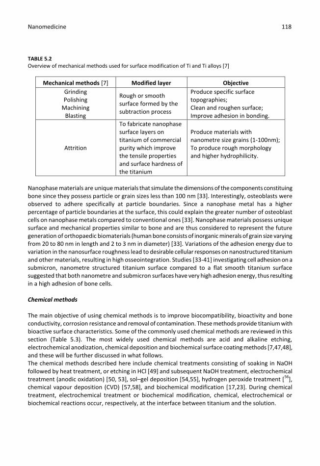
Chemical treatment Chemical treatments of titanium and its alloys are mainly based on chemical reactions occurring at the interface between titanium and the solution. Common chemical treatments include acid, alkali, H2O2, heat, and passivation treatments [56,59,60]. Such treatments are generally performed to remove the oxide scales and contamination present on the surface and can also improve biocompatibility, bioactivity or bone conductivity. TABLE 5.3 Overview of chemical methods used for surface modification of Ti and Ti alloys [7,47,48]
Chemical methods Modified layer Objective
Chemical treatment Acidic treatment [59] Alkaline treatment [60] Hydrogen peroxide treatment [56]
<10 nm of surface oxide layer ~1µm of sodium titanate gel ~5 nm of dense inner oxide and porous outer layer
Remove oxide scales and contamination. Improve biocompatibility, bioactivity or bone conductivity. Improve biocompatibility, bioactivity or bone conductivity.
Sol-gel [54]
~10µm of thin film, such as calcium phosphate, TiO2 and silica
Improve biocompatibility, bioactivity or bone conductivity
CVD [56]
~1µm of TiN, TiC, TiCN, diamond and diamond-like carbon thin film
Improve wear resistance, corrosion resistance and blood compatibility
Anodic oxidation [50-53]
~10 nm to 40µm of TiO2 layer, adsorption and incorporation of electrolyte anions
Produce specific surface topographies; improve corrosion resistance; improve biocompatibility, bioactivity or bone conductivity
Biochemical methods [23,61,62]
Coating deposition Modification through silanized titania, photochemistry, self-assembled monolayers, protein-resistance, etc.
Induce specific cell and tissue response by means of surface-immobilized peptides, proteins, or growth factors
Nanomedicine 120 120
Sol-gel deposition In the sol–gel method, chemical reactions occur not at the interface between the sample surface and solution or gel, but rather in the solution. Generally, the sol-gel method is widely used in order to deposit thin (<10µm) ceramic coatings. When compared to conventional thin film processes, the sol-gel method ensures better control regarding of the chemical composition and microstructure of the coating, preparation of homogeneous films, reduction of the densification temperature, and finally simpler equipment and lower cost [7]. Many coatings, such as titanium oxide (TiO2), calcium phosphate (CaP), TiO2–CaP composites and silica-based coatings have been prepared on titanium and its alloys for biomedical applications [7,54]. Chemical vapour deposition (CVD) On the other hand, chemical vapour deposition (CVD) is a process involving chemical reactions between chemicals in the gas phase and the surface of the substrate, resulting in the deposition of a non-volatile compound on the substrate and is quite widely used as a chemical surface modification method. Untill now, many different CVD processes have been developed [57], such as atmospheric-pressure chemical vapour deposition (APCVD) – good uniformity of the coating; low-pressure chemical vapour deposition (LPCVD) – increased hardness and corrosion resistance; laser-enhanced chemical vapour deposition (LECVD) – improved wear and corrosion resistance; plasma-enhanced chemical vapour deposition (PECVD) – improved wear and corrosion resistance; and plasma-assisted chemical vapour deposition (PACVD) – improved biocompatibility, chemical stability and corrosion resistance. More details about each of these chemical vapour deposition procedures and their advantages can be found in the literature [7,57]. Electrochemical methods Electrochemical processes are performed by connecting the metallic device to the positive pole of an electrical circuit and immersing the entire device into an electrolyte solution containg ionic substances or oxidants. This methodology can lead to incorporation of some ions on the material surface, and includes the possibility of changing the surface finish. One method that can be identified is anodization, which is obtained by using strong acids (e.g. H2SO4, H3PO4, HNO3, and HF) as the electrolyte solution. The end results of the anodization process are the thickening of the oxide layer from the usual 5-10 nm oxide layer formed due to atmospheric oxidation, or by modifications in the microstructure and the crystallinity of the titanium oxide layer [50-52]. Another electrochemical method which has been extensively used for deposition of ceramic coatings on the surface of metals is micro-arc oxidation (MAO), and high micro-hardness coatings with good adhesion, strength and wear resistance are attained [63]. Furthermore, if MAO is performed in the presence of an electrolyte containing calcium and phosphorus, a bioactive coating with reduced osseointegration of the implant is obtained [63]. Biochemical methods Some processes are based on the deposition of foreign chemical substances on the implant surface by electrodeposition, biomimetic precipitation of calcium phosphate through immersion in simulated body fluid, protein adsorption, etc. [17,62]. For example, hydroxyapatite (HA) is considered one of the most effective bioactive materials and can be easily deposited by immersion of the implant surface in different simulated body fluids.
Nanomedicine 121 121
Biochemical methods [23] provide the possibility of adding and bonding specific biomolecules at the implant surface. These treatments are focused on controlling and guiding the complex sequence of biochemical phenomena that take place at the interface between an implanted device and biological tissue, such as osseointegration. There are three major methods available for the biochemical treatment of a metallic surface: a) physico-chemical adsorption; b) covalent binding of a biomolecule on the surface and c) peptide inclusion into a carrier material. Physico-chemical adsorption of an active molecule to the surface is based on the simple process of immersing the sample into a bioactive peptide-containing solution. Despite the ease of this method, the main drawback is that it does not ensure controlled deposition, which is essential for directing the interactions with biological tissues. Furthermore, the adsorbed molecules can be displaced and diffused from the adsorption site. Covalent attachment uses the chemical properties of the material surface to covalently bind the bioactive molecule. Here different strategies are focused on increasing the number of reactive -OH groups. However, covalent binding does not result in control of the surface density of peptides, which can affect the biological response. Hence, peptide inclusion into a carrier molecule has the advantage of enabling control of the amount of bioactive peptide introduced and used to coat the implant surface. Carriers, mostly polymers, can be either simply impregnated with the active biomolecules or covalently bound to the carrier structure. An elegant method frequently used to render the metallic substrate responsive to various stimuli is based on using self-assembled monolayers (SAMs) as coatings. The advantage of SAMs lies in their structure, which is that of a bifunctional molecule with a head group that is able to interact strongly with a metal, oxide or polymer and arrange itself on the surface of the material; the attachment mechanism has been extensively discussed [64] and is based on immobilization of the biomolecule on the biomedical implant surface [65]. The biomechanical performance of an implanted device largely depends on the properties of its surface, in terms of both chemical composition and roughness. The anchored or adsorbed biomolecules are normally present on the cell membrane and extra-cellular matrix. Among other proteins, many studies have been focused on the family of beta transforming growth factors (TGF-β) and bone morphogenetic proteins (BMPs) [61]. However, the use of a protein by itself is not very reproducible since it will have both a low chemical stability and solubility in the biological environment. As an alternative, smaller biologically active sequences, namely peptides, which are part of the total aminoacid sequence of a protein have been isolated/synthesized and attached to the desired substrate [17]. One of the most investigated peptides is the Arg-Gly-Asp (RGD) amino acid sequence, which is known to be the minimal cell-recognisable sequence in many adhesive proteins [66,67]. Physical methods Physical surface modification methods include processes such as thermal spraying, physical vapour deposition, ion implantation and glow discharge plasma treatment, where chemical reactions do not occur. The surface modified layer, film or coating obtained on the titanium substrate is mainly a product of the thermal, kinetic, and electrical energy involved. Some of these methods and their objectives are listed in Table 5.4. In thermal spraying methods, the coating is obtained by thermally melting the coating materials into liquid droplets and spraying them onto the substrate at high speed – in this case the coating is due to kinetic energy. Depending on the maximum temperature used, thermal spraying may be separated into flame and plasma spraying, in which plasma spraying can provide very high temperatures. Other spraying techniques include high velocity oxy-fuel (HVOF) spraying, detonation gun (D-GUN) spraying, arc spraying and so on [7,68,69]; for the milestones achieved in thermal spray coating of functional
Nanomedicine 122 122
titanium dioxide, as well as a thorough review of their applications in bone implants, see Gardon et al. [70]. Recently, cold gas spray for deposition of titanium dioxide coatings has gained interest [70,71] Physical vapour deposition (PVD) consists of evaporating or sputtering the target materials in a vacuum to form atoms, molecules or ions that are then transported to the surface of the substrate where film growth takes place by condensation or by reaction with the surface of the material [7].
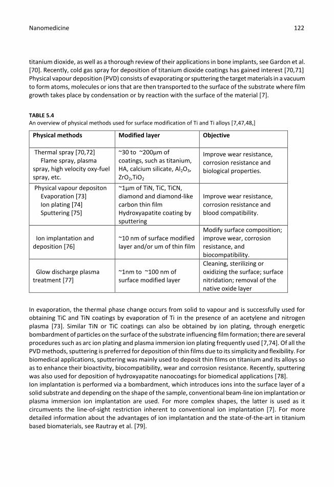
TABLE 5.4 An overview of physical methods used for surface modification of Ti and Ti alloys [7,47,48,]
Physical methods
Modified layer
Objective
Thermal spray [70,72] Flame spray, plasma spray, high velocity oxy-fuel spray, etc.
~30 to ~200µm of coatings, such as titanium, HA, calcium silicate, Al2O3, ZrO2,TiO2
Improve wear resistance, corrosion resistance and biological properties.
Physical vapour depositon Evaporation [73] Ion plating [74] Sputtering [75]
~1µm of TiN, TiC, TiCN, diamond and diamond-like carbon thin film Hydroxyapatite coating by sputtering
Improve wear resistance, corrosion resistance and blood compatibility.
Ion implantation and deposition [76]
~10 nm of surface modified layer and/or um of thin film
Modify surface composition; improve wear, corrosion resistance, and biocompatibility.
Glow discharge plasma treatment [77]
~1nm to ~100 nm of surface modified layer
Cleaning, sterilizing or oxidizing the surface; surface nitridation; removal of the native oxide layer
In evaporation, the thermal phase change occurs from solid to vapour and is successfully used for obtaining TiC and TiN coatings by evaporation of Ti in the presence of an acetylene and nitrogen plasma [73]. Similar TiN or TiC coatings can also be obtained by ion plating, through energetic bombardment of particles on the surface of the substrate influencing film formation; there are several procedures such as arc ion plating and plasma immersion ion plating frequently used [7,74]. Of all the PVD methods, sputtering is preferred for deposition of thin films due to its simplicity and flexibility. For biomedical applications, sputtering was mainly used to deposit thin films on titanium and its alloys so as to enhance their bioactivity, biocompatibility, wear and corrosion resistance. Recently, sputtering was also used for deposition of hydroxyapatite nanocoatings for biomedical applications [78]. Ion implantation is performed via a bombardment, which introduces ions into the surface layer of a solid substrate and depending on the shape of the sample, conventional beam-line ion implantation or plasma immersion ion implantation are used. For more complex shapes, the latter is used as it circumvents the line-of-sight restriction inherent to conventional ion implantation [7]. For more detailed information about the advantages of ion implantation and the state-of-the-art in titanium based biomaterials, see Rautray et al. [79].
Nanomedicine 123 123
Another physical method for implant modification is glow discharge plasma treatment, which is extensively used for surface modification of titanium or titanium alloys, so as to increase the adsorption of extracellular matrix proteins on the implant surface [80].
Selectively modified method: Anodization General aspects of electrochemical anodization
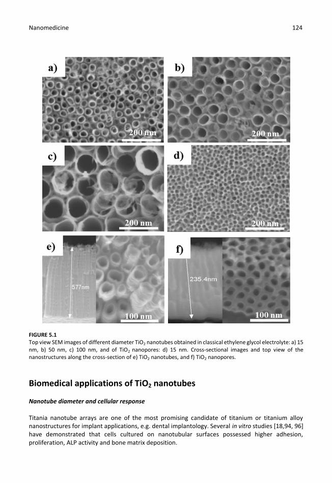
In recent years, several methods have been developed to produce nanoscale structures on titanium surfaces. While irregular nanomorphologies can be easily established by chemical methods, electrochemical anodization of titanium is one of the most popular and novel strategies to produce controlled structures (including nanotubes, pillarlike nanostructures, and nanodots) on implant surfaces. In order to improve the biological, chemical, and mechanical properties of biomaterials, significant research has been aimed at finding more suitable biomaterials with nanotopography which could offer improved bioperformance. TiO2 nanotubes can be easily grown by electrochemical anodization of Ti or its alloys. Over the last two decades, the electrochemical formation of self-organized nanotube layers in dilute fluoride-containing electrolytes has been studied intensively [81, 84]. Wei et al. [85] showed that on anodization of Ti in organic electrolytes of low water content, the formation of ordered TiO2 nanoporous structures could be observed. I.e., the water content in the electrolyte is the critical factor that determines whether self-ordered oxide nanotubes or nanopores are formed. This supports the concept that tube formation originates from ordered porous oxide by a “pore-wall-splitting” mechanism [85]. By tailoring the anodization conditions [86,87] (applied voltage, anodization time and concentrations of chemicals) TiO2 nanotubes of different diameters from 15 nm up to 300 nm and different lengths can be obtained [86]. Recently, Roy et al. [87] and Kowalski et al. [88] reviewed the mechanisms involved in anodization and the recent advances in the formation of nanostructured oxides in the form of nanotubes, nanopores with hole morphology, mesosponges, nanochannels and microcones grown on titanium or titanium alloys. The general nanotubular and nanoporous morphologies obtained for titanium in classical organic electrolytes, similar to [85], are presented in Figure 5.1 with emphasis on the ability to tailor nanotubular morphologies (e.g. diameter controlled), as well as indicating the difference between nanotubular and nanoporous structures (see Figure 5.1 e) and f)). For more details about the mechanism of anodization and all the factors involved in controlling the morphology of the nanostructures through the anodization conditions, see the comprehensive review by Roy et al. [87]. Moreover, the possibility of using amorphous or crystalline nanostructures, and the effect of crystallinity on the hydrophilic character or other morphological or structural properties should be considered. TiO2 nanotubes can be obtained on almost all titanium alloys containing valve metals (transition metals), such as Ti-6Al-7Nb [89, 91], Ti-6Al-4V [89], TiZr alloys with different Zr concentrations [92,93] or on other biomedical alloys developed or tested for biomedical applications (Ti-6Al-4Zr, etc.).
Nanomedicine 124 124
FIGURE 5.1 Top view SEM images of different diameter TiO2 nanotubes obtained in classical ethylene glycol electrolyte: a) 15 nm, b) 50 nm, c) 100 nm, and of TiO2 nanopores: d) 15 nm. Cross-sectional images and top view of the nanostructures along the cross-section of e) TiO2 nanotubes, and f) TiO2 nanopores.
Biomedical applications of TiO2 nanotubes Nanotube diameter and cellular response Titania nanotube arrays are one of the most promising candidate of titanium or titanium alloy nanostructures for implant applications, e.g. dental implantology. Several in vitro studies [18,94, 96] have demonstrated that cells cultured on nanotubular surfaces possessed higher adhesion, proliferation, ALP activity and bone matrix deposition.
Nanomedicine 125 125
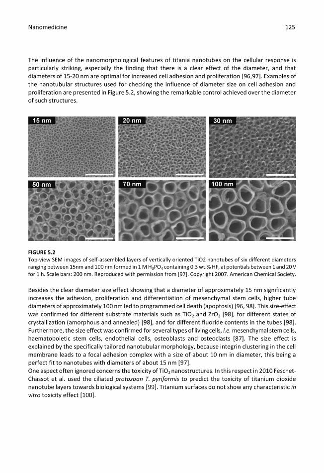
The influence of the nanomorphological features of titania nanotubes on the cellular response is particularly striking, especially the finding that there is a clear effect of the diameter, and that diameters of 15-20 nm are optimal for increased cell adhesion and proliferation [96,97]. Examples of the nanotubular structures used for checking the influence of diameter size on cell adhesion and proliferation are presented in Figure 5.2, showing the remarkable control achieved over the diameter of such structures.
FIGURE 5.2 Top-view SEM images of self-assembled layers of vertically oriented TiO2 nanotubes of six different diameters ranging between 15nm and 100 nm formed in 1 M H3PO4 containing 0.3 wt.% HF, at potentials between 1 and 20 V for 1 h. Scale bars: 200 nm. Reproduced with permission from [97]. Copyright 2007. American Chemical Society.
Besides the clear diameter size effect showing that a diameter of approximately 15 nm significantly increases the adhesion, proliferation and differentiation of mesenchymal stem cells, higher tube diameters of approximately 100 nm led to programmed cell death (apoptosis) [96, 98]. This size-effect was confirmed for different substrate materials such as TiO2 and ZrO2 [98], for different states of crystallization (amorphous and annealed) [98], and for different fluoride contents in the tubes [98]. Furthermore, the size effect was confirmed for several types of living cells, i.e. mesenchymal stem cells, haematopoietic stem cells, endothelial cells, osteoblasts and osteoclasts [87]. The size effect is explained by the specifically tailored nanotubular morphology, because integrin clustering in the cell membrane leads to a focal adhesion complex with a size of about 10 nm in diameter, this being a perfect fit to nanotubes with diameters of about 15 nm [97]. One aspect often ignored concerns the toxicity of TiO2 nanostructures. In this respect in 2010 Feschet-Chassot et al. used the ciliated protozoan T. pyriformis to predict the toxicity of titanium dioxide nanotube layers towards biological systems [99]. Titanium surfaces do not show any characteristic in vitro toxicity effect [100].
Nanomedicine 126 126
Nanotubes and protein interaction Irrespective of the location of the implant (blood-contacting, orthopaedic or dental implant) the first step taking place after implantation is the adsorption of proteins from the surrounding tissue or medium. The amount and type of protein adsorbed further influences the fate of the implant– as pointed out in section “Biocompatibilty of medical devices”. Gongadze et al. [40,41] proposed a mechanism for the adhesion of cells to a nanorough titanium implant surface with sharp edges. The basic assumption was that the attraction between the negatively charged titanium surface and a negatively charged osteoblast is mediated by charged proteins with a distinctive quadrupolar internal charge distribution. Similarly, cation-mediated attraction between fibronectin molecules (present in the extracellular matrix) and the titanium surface is expected to be more efficient for a high surface charge density, resulting in facilitated integrin-mediated osteoblast adhesion. Osteoblasts could be more strongly bound along the sharp convex edges or spikes of nanorough titanium surfaces where the magnitude of the negative surface charge density is the highest. It is therefore plausible that the nanorough regions of titanium surfaces with sharp edges and spikes could promote the adhesion of osteoblasts. A small diameter nanotube surface has on average more sharp convex edges per unit area than a large one, leading to a strong binding affinity on the surface of small diameter nanotubes [40,41]. Nanotubes for orthopaedic and dental implants
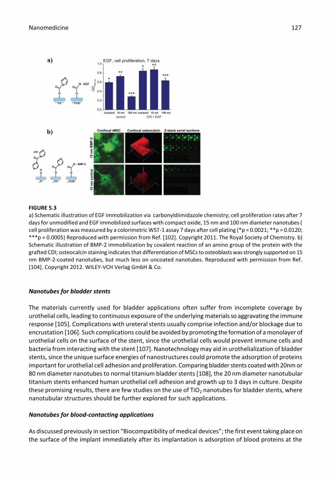
Implant topography is critical to the clinical success of bone-anchored implants, yet further research needs to be done on how nanomodified implant topography affects osseointegration. Previous in vitro studies [98] reported that the topography of TiO2 nanotubes improved osteoblast proliferation and adhesion compared to normal titanium surfaces. The increased in vitro cellular activities for titania nanotubes also translated into in vivo bone bonding [96,98]. Nanotubular surfaces significantly improved bone bonding strength by as much as nine-fold compared with grit blasted surfaces [44], and histological analysis revealed greater bone-implant contact and collagen type I expression, confirming the better in vivo behaviour of titania nanotubes. In vitro tests performed on nanotubes obtained on different alloys such as Ti-6Al-7Nb [90] or TiZr alloys [101] suggest their use in orthopaedic cellular therapy. Covalent immobilization of different biomolecules can be used as a tool for differentiation of mesenchymal stem cells (MSCs) from specific cells. For example, immobilization of epidermal growth factor (EGF) [102] on 100nm diameter TiO2 nanotubes enabled the seeded cells to avoid apoptosis and to become attached and promote proliferation (as shown in Figure 5.3a). Immobilization of bone morphogenetic protein-2 (BMP-2) [103] on nanotubes of varying diameter showed higher osteocalcin and osteopontin levels on 30 nm diameter TiO2 nanotubes. When BMP-2 was covalently immobilized via carbonyldiimidazol (CDI) [104] the differentiation of MSCs was observed to depend on diameter, namely, an enhanced osteogenic differentiation occurred on 15 nm nanotubes (see Figure 5.3b), and chondrogenic differentiation on 100 nm nanotubes.
Nanomedicine 127 127
FIGURE 5.3 a) Schematic illustration of EGF immobilization via carbonyldiimidazole chemistry; cell proliferation rates after 7 days for unmodified and EGF immobilized surfaces with compact oxide, 15 nm and 100 nm diameter nanotubes ( cell proliferation was measured by a colorimetric WST-1 assay 7 days after cell plating (*p = 0.0021; **p = 0.0120; ***p = 0.0005) Reproduced with permission from Ref. [102]. Copyright 2011. The Royal Society of Chemistry. b) Schematic illustration of BMP-2 immobilization by covalent reaction of an amino group of the protein with the grafted CDI; osteocalcin staining indicates that differentiation of MSCs to osteoblasts was strongly supported on 15 nm BMP-2-coated nanotubes, but much less on uncoated nanotubes. Reproduced with permission from Ref. [104]. Copyright 2012. WILEY-VCH Verlag GmbH & Co.
Nanotubes for bladder stents
The materials currently used for bladder applications often suffer from incomplete coverage by urothelial cells, leading to continuous exposure of the underlying materials so aggravating the immune response [105]. Complications with ureteral stents usually comprise infection and/or blockage due to encrustation [106]. Such complications could be avoided by promoting the formation of a monolayer of urothelial cells on the surface of the stent, since the urothelial cells would prevent immune cells and bacteria from interacting with the stent [107]. Nanotechnology may aid in urothelialization of bladder stents, since the unique surface energies of nanostructures could promote the adsorption of proteins important for urothelial cell adhesion and proliferation. Comparing bladder stents coated with 20nm or 80 nm diameter nanotubes to normal titanium bladder stents [108], the 20 nm diameter nanotubular titanium stents enhanced human urothelial cell adhesion and growth up to 3 days in culture. Despite these promising results, there are few studies on the use of TiO2 nanotubes for bladder stents, where nanotubular structures should be further explored for such applications. Nanotubes for blood-contacting applications As discussed previously in section “Biocompatibility of medical devices”; the first event taking place on the surface of the implant immediately after its implantation is adsorption of blood proteins at the
Nanomedicine 128 128
implant-liquid interface. What happens to the adsorbed protein layer governs the interaction of platelets and their adhesion or activation, leukocyte recruitment, activation of intrinsic coagulation and of the complement [109]; moreover, all four processes are capable of eliciting a thrombogenic response in vivo. Stepwise, first the adsorbed protein layer will lead to adhesion and activation of the platelets, which is fundamental in forming the fibrin clot and recruiting leukocytes (as monocytes and neutrophils). Second, the platelets will trigger an inflammatory immune response which will lead to either thrombosis and/ or fibrous encapsulation of the implant [109,110]. Studies [111, 113] indicated increased blood serum protein adsorption, platelet adhesion and activation and whole blood clotting kinetics on titania nanotube arrays. Furthermore, a decrease of thrombogenic effects and surface-induced fibrin clot formation was observed on nanotubes when compared to titanium surfaces. This was evident from the slightly decreased levels of complement activation and slightly increased degree of free fibrinogen on nanotubular surfaces [21,111,112]. When the structure of the nanotubes was modified from amorphous to crystalline, a decrease of the haemolytic index was observed, indicating an increase in the haemocompatibility; although amorphous nanotubes were also non-haemolytic with a haemolytic activity below 2% [114]. Overall, titania nanotubular surfaces seem a promising interfacial material for the long-term success of blood-contacting implants.
Nanotubes for antibacterial activity
Bacterial infection of in-dwelling medical devices is a growing problem that cannot be treated by traditional antibiotics due to the increasing prevalence of antimicrobial resistance and biofilm formation [115,116]. Controlled diameter nanotubes (amorphous or crystalline) displayed significantly changed responses to both Staphylococcus epidermidis (S. epidermidis) and Staphylococcus aureus (S. aureus) – pathogens relevant for orthopaedic and other medical device-related infections [115,117]. It is obvious that a similar size-effect also exists for bacteria, where use of larger diameter nanotubes decreased the number of live bacteria (S. aureus and S. epidermidis) as compared to lower diameter ones (20 nm) or Ti [115,118]. The antibacterial activity of titanium or titanium alloy nanostructures could be due to: i) the nanostructuring of the implant surface (the diameter size-effect); ii) the use of alloys with inherent antimicrobial properties or by decorating the nanostructures with antimicrobial nanoparticles; and finally iii) by the functionalization of the nanostructures with anti-inflammatory agents or by loading drugs inside the nanostructures (as will be discussed in the following subsection). When using titanium alloys containing elements which could inhibit bacteria (eg. zirconium) such as Ti50Zr alloy [119], smaller diameter nanotubes showed an increased antibacterial effect against E. coli. Other examples of such alloys include Ti-Nb-Zr-Mo [120], Ti-Al-Nb and so on [87]. Antimicrobial nanoparticles used for decorating the nanostructures include Ag [121] and Zn [122], while less investigated antimicrobial agents include copper, fluorine and calcium. Nanotubes for drug delivery
Current orthopaedic implants have functional lifetimes of only 10–15 years for a variety of reasons including infection, extensive inflammation, and overall poor osseointegration (or a lack of prolonged bonding of the implant to the juxtaposed bone) [4]. To improve properties of titanium for orthopaedic applications, it is possible to coat the nanotubular structures with infection-reducing drugs (penicillin/streptomycin) or inflammation-reducing drugs (dexamethasone) by simple physical adsorption or deposition from simulated body fluid (SBF). For example when drugs where deposited from SBF, a drug elution times of up to 3 days was registered [123].
Nanomedicine 129 129
Shrestha et al. [124] showed that TiO2 nanotubes can be filled with magnetic Fe3O4 particles and thus be magnetically guided to desired locations. Such a structure can be used directly for photocatalytic reactions with cells or tissue, such as the site-selective killing of cancer cells. UV light can also be used for killing cancer cells though the use of nanotubes but there is the disadvantage of needing direct access for the UV light to the TiO2 nanotubes. More recent work [125,126] focused on using an amphiphilic nanotubular structure consisting of nanotubes that provide a hydrophobic cap (using a monolayer coating) which does not allow body fluids to enter the nanotubes unless the cap is opened by a photocatalytic interaction. Based on the same principle, drug-loaded nanotubular layers can also be capped with biopolymers [127].
Summary Titanium and its alloys continue to be some of the most promising biomaterials used for biomedical devices. Despite their outstanding properties (good mechanical resistance, corrosion resistance, biocompatibility) since they are bioinert materials surface modification is necessary to improve osseointegration, haemocompatibility or other properties important in their respective biomedical applications. In this work, an overview of the generally used surface modification methods for improving the properties of titanium and titanium alloys for biomedical applications was presented, also taking into account the current shift of research from the micrometre to the nanometre scale. Mechanical methods (grinding, polishing, attrition, etc.), chemical methods (chemical treatments, sol-gel methods, anodic oxidation, etc.) and physical methods (thermal spray, ion implantation and deposition, etc.) were discussed with respect to their effects on the implant surface and its biocompatibility. One of the most promising recently emerging methods for obtaining nanometre scale surfaces, namely electrochemical anodizing leading to nanotubular structures with controlled diameters in the range of 15 nm up to 250 nm, was discussed. General aspects of electrochemical anodization were presented, as well as the use of such nanostructures in the biomedical field where cellular interactions and protein adhesion in orthopaedic, dental and blood-contacting applications or drug-delivery applications were discussed. With the development of current surface engineering techniques, cutting edge morphologies in the nanometre scale for implant applications, bladder stents and for specific biomedical applications. Visualizing biointerfaces and biomaterials with nanometre precision in a three-dimensional scale, could reveal new fundamental information on material properties and bone response, enabling better design of biomaterials for the future.
Acknowledgement
This work was supported by Deutsche Forschungsgemeinschaft (DFG), the DFG Cluster of Excellence (Engineering of Advanced Materials, EAM) and Slovenian Research Agency (ARRS) grants J1-4109, J1-4136, J3-4108 and P2-0232.
Nanomedicine 130 130
References
1. Williams D. On the nature of biomaterials. Biomaterials 2009; 30: 5897–5909. 2. Mihov D, Katerska B. Some biocompatible materials used in medical practice. Trakia Journal of
Sciences 2010; 8: 119–125. 3. Williams D. On the mechanisms of biocompatibility. Biomaterials 2008; 29: 2941–2953. 4. Williams D. Titanium in Medicine: Material Science, Surface Science, Engineering, Biological
Responses and Medical Applications (eds. Brunette D M, Tengvall P, Textor M, Thompson P), Springer-Verlag, Berlin and Heidelberg 2001;13–24.
5. Geetha M, Singh A, Asokamani R, Gogia A. Ti based biomaterials, the ultimate choice for orthopaedic implants – A review. Prog. Mater. Sci. 2009; 54: 397–425.
6. Ratner B. Titanium in Medicine: Material Science, Surface Science, Engineering, Biological Responses and Medical Applications (eds. Brunette D M, Tengvall P, Textor M, Thompson P), Springer-Verlag, Berlin and Heidelberg 2001; 1–12.
7. Liu X, Chu P, Ding C. Surface modification of titanium, titanium alloys, and related materials for biomedical applications. Mater. Sci. Eng. R 2004; 47: 49–121.
8. Adya M, Alam M, Ravindranath T, Mubeen A, Saluja B. Corrosion in titanium dental implants: literature review. J. Indian Prosthodont. Soc. 2005; 5(3): 126–131.
9. Donachie M.J. Titanium: A technical guide. ASM International 2000. 10. Hanawa T, Asami K, Asaoka K. Repassivation of titanium and surface oxide film regenerated in
simulated bioliquid. J. Biomed. Mater. Res. 1998; 40(4): 530–538. 11. Brown S, Lemons J. Medical Applications of Titanium and Its Alloys: The Material and
Biological Issues, ASTM special series publication 1272, 1996. 12. Kelly E. Modern aspects of electrochemistry no. 14 (eds. Bockris J O’M, Conway, B E, White R
E), Springer-US 1982; 319–424. 13. Hiromoto S, Hanawa T, Asami K. Composition of surface oxide film of titanium with culturing
murine fibroblasts L929. Biomaterials 2004; 25: 979-986. 14. Park J, Lakes R S. Biomaterials. An introduction. Springer, New York 2007; 2–13. 15. Boyer R, Welsch G, Collings E W. Materials properties handbook. Titanium Alloys, 3
rd edition,
ASM International, Ohio 2003. 16. Black J, Hastings G. Handbook of biomaterial properties, Springer US 1998. 17. Mas-Moruna C, Espanol M, Montufar E, Mestres G, Aparicio C, Javier F, Ginebra M.
Biomaterials Surface Science (eds. Taubert A, Mano J F, Rodríguez-Cabello J C), Wiley-VCH Verlag GmbH & Co. KGaA, Weinheim 2013; 337–374.
18. Bakir M. Haemocompatibility of titanium and its alloys. J. Biomater. Appl. 2012; 27: 3–15. 19. Blombäck B, Hessel B, Hogg D, Therkildsen L. A two-step fibrinogen–fibrin transition in blood
coagulation. Nature 1978; 275: 501–505. 20. Roy S, Paulose M, Grimes C. The effect of TiO2 nanotubes in the enhancement of blood
clotting for the control of hemorrhage. Biomaterials. 2007; 28(31): 4667–72. 21. Smith B, Yoriya S, Grissom L, Grimes C, Popat K. Hemocompatibility of titania nanotube arrays.
J. Biomed. Mater. Res. A. 2010; 95(2): 350–60. 22. Branemark P, Hansson B, Adell R, Breine U, Lindstrom J, Hallen O, Ohman A. Osseointegrated
implants in the treatment of edentulous jaw. Scand. J. Plast. Reconstr. Surg. Suppl. 1977; 16: 1–132.
23. Xiao S-J, Kenausis G, Textor M. Titanium in Medicine: Material Science, Surface Science, Engineering, Biological Responses and Medical Applications (eds. eds. Brunette D M, Tengvall P, Textor M, Thompson P), Springer-Verlag, Berlin and Heidelberg 2001; 417–455.
Nanomedicine 131 131
24. Anderson J M, Cristina A G, Hanson S R, Harker L A, Johnson R J, Merritt K, Naylor P T, Schoen
F J. Biomaterials Science: An Introduction to Materials in Medicine (eds. Ratner B D, Hoffman A S, Schoen F J, Lemons J E) Academic Press 1997; 165–214.
25. Vasilev K, Poh Z, Kant K, Chan J, Michelmore A, Losic D. Tailoring the surface functionalities of titania nanotube arrays. Biomaterials 2010; 31: 532–540.
26. Diebold U. The surface science of titanium dioxide. 2003; 48: 53–229. 27. Puckett S D, Taylor E, Raimondo T, Webster T J. The relationship between the nanostructure
of titanium surfaces and bacterial attachment. Biomaterials 2010; 31: 706–713. 28. Dale G R, Hamilton J W, Dunlop P S, Lemoine P, Byrne J A. Electrochemical growth of titanium
oxide nanotubes: the effect of surface roughness and applied potential. J Nanosci. Nanotechnol. 2009; 9(7): 4215–4219.
29. Anil S, Anand P S, Alghamdi H, Jansen J A. Dental Implant Surface Enhancement and Osseointegration, Implant Dentistry - A Rapidly Evolving Practice. (eds. Turkyilmaz I) InTech, 2011; Available from: http://www.intechopen.com/books/implant-dentistry-a-rapidly-evolvingpractice/dental-implant-surface-enhancement-and-osseointegration,
30. Le Guéhennec L, Soueidan A, Layrolle P, Amouriq Y. Surface treatments of titanium dental implants for rapid osseointegration. Dent Mater. 2007; 23(7): 844–854.
31. Bagno A, Bello C. Surface treatments and roughness properties of Ti-based biomaterials. J. Mater. Sci. Mater. Med. 2004; 15: 935–949.
32. Han C H, Johansson C B, Wennerberg A, Albrektsson T. Quantitative and qualitative investigations of surface enlarged titanium and titanium alloy implants. Clin. Oral Implants Res. 1998; 9(1): 1–10.
33. Webster T J, Ejiofor J U. Increased osteoblast adhesion on nanophase metals: Ti, Ti6Al4V, and CoCrMo. Biomaterials 2004; 25: 4731–4739.
34. Pegueroles M, Aparicio C, Bosio M, Engel E, Gil F J, Planell J A, Altankov G. Spatial organization of osteoblast fibronectin matrix on titanium surfaces: effects of roughness, chemical heterogeneity and surface energy. Acta Biomater. 2010; 6(1): 291–301.
35. Boyan B D, Hummert T W, Dean D D, Schwartz Z. Role of material surfaces in regulating bone and cartilage cell response. Biomaterials 1996; 17(2): 137–146.
36. Martin J Y, Schwartz Z, Hummert T, Schraub D, Simpson J, Lankford J, Dean D, Cochran D, Boyan B. Effect of titanium surface roughness on proliferation, differentiation, and protein synthesis of human osteoblast-like cells (MG63). J. Biomed. Mater. Res. 1995; 29(3): 389–401.
37. Cochran D , Schenk R, Lussi A, Higginbottom F, Buser D. Bone response to unloaded and loaded titanium implants with a sandblasted and acid-etched surface: A histometric study in the canine mandible. J. Biomed. Mater. Res. 1998; 40: 1-11.
38. Ivanoff c , Hallgren C, Widmark G, Sennerby L, Wennerberg A. Histologic evaluation of the bone integration of TiO 2 blasted and turned titanium microimplants in humans. Clin. Oral Implants Res. 2001; 12: 128-134.
39. Rupp F, Scheideier L, Olshanska N, De Wild M, Wieland M, Geis-Gerstorfer J. Enhancing surface free energy and hydrophilicity through chemical modification of microstructured titanium implant surfaces. J. Biomed. Mater. Res. A 2006; 76: 323-334.
40. Gongadze E, Kabaso D, Bauer S, Slivnik T, Schmuki P, van Rienen U, Iglič A. Adhesion of osteoblasts to a nanorough titanium implant surface. Int. J. Nanomed. 2011; 6: 1801–1816.
41. Gongadze E, Kabaso D, Bauer S, Park J, Schmuki P, Iglič A. Adhesion of osteoblasts to a vertically aligned TiO2 nanotube surface. Mini Rev. Med. Chem. 2012; 13: 94–200.
Nanomedicine 132 132
42. Ueno T, Yamada M, Suzuki T, Minamikawa H, Sato N, Hori N, Takeuchi K, Hattori M, Ogawa
T. Enhancement of bone–titanium integration profile with UV-photofunctionalized titanium in a gap healing model. Biomaterials 2010; 31: 1546-1557.
43. Schwarz F, Herten M, Sager M, Wieland M, Dard M, Becker J. Bone regeneration in dehiscence-type defects at chemically modified (SLActive) and conventional SLA titanium implants: a pilot study in dogs. J. Clin. Periodontol. 2007; 34(1): 78-86.
44. Zhao G, Schwartz Z, Wieland M, Rupp F, Geis-Gerstorfer J, Cochran D L, Boyan B D. High surface energy enhances cell response to titanium substrate microstructure. J. Biomed. Mater. Re.s A. 2005; 74(1): 49-58.
45. Mas-Moruna C, Espanol M, Montufar E, Mestres G, Aparicio C, Javier G F, Ginebra M. Biomaterials Surface Science. (eds. Taubert A, Mano J F, Rodriguez-Cabello J C), Wiley-VCH Verlag, 2013; 337-374.
46. Lausmaa J, Brunette D, Tengvall P, Textor M, Thomsen P. Titanium in Medicine: Material Science, Surface Science, Engineering, Biological Responses and Medical Applications (eds. Brunette D M, Tengvall P, Textor M, Thompson P), Springer-Verlag, Berlin and Heidelberg 2001; 231–266.
47. Bagno A, Bello C. Surface treatments and roughness properties of Ti-based biomaterials. J. Mater. Sci. Mater. Med. 2004; 15: 935-949.
48. Kim K H, Ramaswani N. Electrochemical surface modification of titanium in dentistry. Dent. Mater. J. 22 2009; 28(1): 20–36.
49. Xiao S J, Textor M, Spencer N D, Sigrist H, Langmuir 1998; 14: 5507–5516. 50. Li P, Ohtsuki C, Kokubo T, Nakanishi K, Soga N. J. Am. Ceram. Soc. 1992; 75(8): 2094–2097. 51. Cho S, Nakanishi K, Kokubo T, Soga N. J. Am. Ceram. Soc. 1995; 78: 1769–1774. 52. Yoshida K, Kamada K, Sato K, Hatada R, Baba K, Atsuta M. J. Biomed. Mater. Res. 1999; 48:
778–785. 53. Sul Y, Johansson C B, Jeong Y, Albrektsson T. Med. Eng. Phys. 2001; 23: 329–346. 54. Brinker C, Scherer G. Sol–Gel Science: The Physics and Chemistry of Sol–Gel Processing.
American Press, San Diego; 1990. 55. Advincula M, Fan X, Lemons J, Advincula R. Surface modification of surface sol-gel derived
titanium oxide films by self-assembled monolayers (SAMs) and non-specific protein adsorption studies. Colloids Surf. B Biointerfaces 2005; 42(1): 29–43.
56. Li P, Kangasniemi I, de Groot K, Kokubo T. Bonelike Hydroxyapatite Induction by a Gel-Derived Titania on a Titanium Substrate. J. Am. Ceram. Soc. 1994; 77: 1307–1312.
57. Ohring M. Materials Sciences of Thin Film: Deposition and Structure, Academic Press Ltd., Boston, 1992; 277.
58. Hampden-Smith M J, Kodas T T. Chemical Vapor Deposition of Metals: Part 2. Overview of Selective CVD of Metals. Chem. Vap. Deposition 1995; 1: 39-48.
59. Nanci A, Wuest J D, Peru L, Brunet P, Sharma V, Zalzal S, McKee M D. J. Biomed. Mater. 1998; 40: 324–335.
60. Kim K-H, Narayanan R, Rautray R T. Surface Modification of Titanium for Biomaterial Applications. Nova Publishers Science Inc, 2010.
61. Schliephake H, Scharnweber D, Dard M, Sewing A, Aref A, Roessler S. Functionalization of dental implant surfaces using adhesion molecules. J. Biomed. Mater. Res. B 2005; 73(1): 88–96.
62. Morra M. Biochemical modification of titanium surfaces: peptides and ECM proteins. Eur. Cell Mater. 2006; 12: 1–15.
Nanomedicine 133 133
63. Krzakala A, Kazek-Kesik A, Simka W. Application of plasma electrolytic oxidation to bioactive
surface formation on titanium and its alloys. RCS Adv. 2013; 3: 19725-19743. 64. Schwartz D K. Mechanisms and kinetics of self-assembled monolayer formation. Annu. Rev.
Phys. Chem. 2001; 52: 107–137. 65. Bauer S, Schmuki P, von der Mark K, Park J. Engineering biocompatible implant surface: Part I:
Materials and surfaces, Prog. Mater. Sci. 2013; 58: 261-326. 66. Ruoslahti E. RGD and other recognition sequences for integrins. Annu. Rev. Cell Dev. Biol.
1996; 12: 697–715 67. D’Souza S E, Ginsberg M H, Plow E F. Arginyl-glycyl-aspartic acid (RGD): a cell adhesion motif.
Trends Biochem. Sci. 1991; 16: 246–250 68. Li H, Khor K A, Cheang P. Titanium dioxide reinforced hydroxyapatitate coatings deposited by
high velocity oxy-fuel (HVOF) spray. Biomaterials 2002; 23: 58–91. 69. de Groot K, Geesink R, Klein C P A T, Serekian P. Plasma-sprayed coatings of hydroylapatite J.
Biomed. Mater. Res. 1987; 21: 1375–1391. 70. Gardon M, Guilemany J M. Milestones in Functional Titanium Dioxide Thermal Spray Coatings:
A Review, JTTEES 2014; 23: 577–595. 71. Hussain T. Cold Spraying of Titanium: A Review of Bonding Mechanisms, Microstructure and
Properties, Key Eng. Mater. 2012; 533: 53–90. 72. Driskell T. Early History of Calcium Ohosphate Materials and Coatings, ASTM Publication,
Philadelphia, 1994; 3. 73. LeClair P, Berera G P, Moodera J S. Titanium nitride thin films obtained by a modified physical
vapor deposition proces. Thin Solid Films 2000; 1: 376–379. 74. Chen Y M, Yu G P, Huang J H. Role of process parameters in the texture evolution of TiN films
deposited by hollow cathode discharge ion plating. Surf. Coat. Technol. 2001; 141:156–163. 75. Jung M J, Nam K H, Shaginyan L R, Han J G. Deposition of Ti thin film using the magnetron
sputtering method. Thin Solid Films 2003; 435:145–149. 76. Rautray T R, Narayan R, Kwon T Y, Kim K H. Surface modification of titanium and titanium
alloys by ion implantation. J. Biomed. Mater. Res. B 2010; 93: 581–591. 77. Shibata Y, Miyazaki T. Anode glow discharge plasma treatment enhances calcium phosphate
adsorption onto titanium plates. J. Dent. Res. 2000; 81: 841–844. 78. Mediaswanti K, Wen C, Ivanova E, Berndt C, Wang J. Titanium Alloys – Advances in Properties
Control (eds. Sieniawski J, Ziaja W), Intech, 2013; Available from: http://www.intechopen.com/books/titanium-alloys-advances-in-properties-control/sputtered-hydroxyapatite-nanocoatings-on-novel-titanium-alloys-for-biomedical-applications
79. Rautray T R, Narayanan R, Kim K-H. Ion implantation of titanium based biomaterials. Prog. Mater. Sci. 2011; 56: 1137–1177.
80. Yamamoto H, Shibata Y, Miyazaki T. Anode glow discharge plasma treatment of titanium plates facilitates adsorption of extracellular matrix proteins to the plates. J.Dent. Res. 2005; 84: 668–671.
81. Assefpour-Dezfuly M, Vlachos C, Andrews E H. Oxide morphology and adhesive bonding on titanium surfaces. J. Mater. Sci. 1984; 19: 3626–3639
82. Zwilling V, Darque-Ceretti E, Boutry-Forveille A, David D, Perrin M, Aucouturier M. Structure and Physicochemistry of Anodic Oxide Films on Titanium and TA6V Alloy. Surf. Interface Anal. 1999; 27: 629–637
Nanomedicine 134 134
83. Beranek R, Hildebrand H, Schmuki P. Self-organized porous titanium oxide prepared in
H2SO4/HF electrolytes. Electrochem. Solid-State Lett. 2003; 6: B12–B14. 84. Albu S, Ghicov A, Macak J, Schmuki P. 250 µm long anodic TiO2 nanotubes with hexagonal self-
ordering. Phys. Status Solidi RRL 2007; 1: R65–R67. 85. Wei W, Berger S, Hauser C, Meyer K, Yang M, Schmuki P. Transition of TiO2 nanotubes to
nanopores for electrolytes with very low water contents. Electrochem. Commun. 2010; 12: 1184–1186.
86. Rohani S, Ruby A. Synthesis of Titania Nanotube Arrays by Anodization. AIDIC Conference series 2009; 9: 121–129
87. Roy P, Berger S, Schmuki P. TiO2 Nanotubes: Synthesis and Applications. Angew. Chem. Int. Ed. 2011; 50: 2904–2939.
88. Kowalski D, Kim D, Schmuki P. TiO2 Nanotubes, Nanochannels and Mesosponge: Self-Organized Formation and Applications. Nano Today 2013; 8: 235–264.
89. Macak J, Tsuchiya H, Ghicov A, Yasuda K, Hahn R, Bauer S, Schmuki P. TiO2 nanotubes: Self-organized electrochemical formation, properties and applications. Curr. Opin. Solid State Mater. Sci. 2007; 11: 3–18.
90. Mazare A, Dilea M, Ionita D, Titorencu I, Trusca V, Vasile E. Changing bioperformance of TiO2
amorphous nanotubes as an effect of inducing crystallinity. Bioelectrochem. 2012; 87:124-131.
91. Mazare A, Dilea M, Ionita D, Demetrescu I. Electrochemical behaviour in simulated body fluid of TiO2 nanotubes on TiAlNb alloy elaborated in various anodizing electrolyte. Surf. Interface Anal. 2014; 46: 186-192.
92. Kim W, Choe H, Ko Y, Brantley W, Nanotube morphology changes for Ti–Zr alloys as Zr content increases. Thin Solid Films 2009; 517: 5033–5037.
93. Minagar S, Berndt C C, Gengenbach T, Wen C. Fabrication and characterization of TiO2-ZrO2-ZrTiO4 nanotubes on TiZr alloy manufactured via anodization. J. Mater. Chem. B 2014; 2: 71-83.
94. Sabrina D, Erik T, Raimondo T, Webster T J. The relationship between the nanostructure of titanium surfaces and bacterial attachment. Biomaterials 2010; 31: 706–713.
95. Ross A P, Webster T J. Anodizing color coded anodized Ti6Al4V medical devices for increasing bone cell functions. Int. J. Nanomed. 2013; 8: 109-117.
96. Bauer S , Park J , von der Mark K , Schmuki P. TiO2 nanotubes for stimulated cell response: Control of cell-surface interactions at the nanoscale. Europ. Cells Mater. 2010; 20: 16.
97. Park J, Bauer S, von der Mark K, Schmuki, P. Nanosize and vitality: TiO2 nanotube diameter directs cell fate. Nano Letters 2007; 7: 1686-1691.
98. Bauer S , Park J , Faltenbacher J, Berger S, von der Mark K, Schmuki P. Size selective behavior of mesenchymal stem cells on ZrO2 and TiO2 nanotube arrays. Integr. Biol. 2009; 1: 525–532.
99. Feschet-Chassot E, Raspal V, Sibaud Y, Awitor O K, Bonnemoy F, Bonnet J L, Bohatier J. Tunable Functionality and Toxicity Studies on Titanium Dioxide Nanotube Layers. Thin Solid Films 2011; 519: 2564–2568.
100. Lucchini J P, Aurelle J L, Therin M, Donath K, Becker W. A pilot study comparing screw-shaped implants. Surface analysis and histologic evaluation of bone healing. Clin. Oral. Implants Res. 1996; 7: 397–404.
101. Grigorescu S, Pruna V, Titorencu I, Jinga V V, Mazare A, Schmuki P, Demetrescu I. The two step nanotube formation on TiZr scaffolds for cell growth. Bioelectrochem. 2014; 98: 39-45.
Nanomedicine 135 135
102. Bauer S, Park J, Pittrof A, Song, Y, von der Mark K, Schmuki P. Covalent functionalization of
TiO2 nanotube arrays with EGF and BMP-2 for modified behavior towards mesenchymal stem cell. Integr. Biol. 2011; 3: 927–936.
103. Lai M, Cai K, Zhao L, Chen X, Hou Y, Yang Z. Surface functionalization of TiO2 nanotubes with bone morphogenetic protein 2 and its synergistic effect on the differentiation of mesenchymal stem cells. Biomacromolecules 2011; 12: 1097–1105.
104. Park J, Bauer S, Pittrof A, Killian M S, Schmuki P, von der Mark K. Synergistic control of mesenchymal stem cell differentiation by nanoscale surface geometry and immobilized growth factors on TiO2 nanotubes. Small 2012; 8: 98–107.
105. Saltzman B. Ureteral stents – indications, variations and complications: endourology update. Urol. Clin. North Am. 1988; 15: 481–491.
106. Dyer R, Chen M, Zagoria R, Regan J, Hood C, Kavanagh P. Complications of ureteral stent placement. Radiographics 2002; 22: 1005–1022.
107. Chun Y, Khang D, Haberstroh K, Webster T J. The role of polymer nanosurface roughness and submicron pores in improving bladder urothelial cell density and inhibiting calcium oxalate stone formation. Nanotechnology 2009; 20: 1–8.
108. Alpaslan E, Ercan B, Webster T J. Anodized 20nm diameter nanotubular titanium for improved bladder stent applications. Int. J. Nanomed. 2011; 6: 219–225.
109. Riehle M. Biocompatibility: Nanomaterials for cell- and tissue engineering. NanoBiotechnology 2005; 1: 308–309.
110. Gorbet M B, Sefton M V. Biomaterial-associated thrombosis: roles of coagulation factors, complement, platelets and leukocytes. Biomaterials 2004; 25: 5681–5703.
111. Roy S C, Paulose M, Grimes C A. The effect of TiO2 nanotubes in the enhancement of blood clotting for the control of hemorrhage. Biomaterials 2007; 28: 4667–4672.
112. Smith B S. PhD thesis. Titania nanotube arrays: Interfaces for implantable devices. Colorado State University. 2012.
113. Li JW, Zhu W, Liu JF, Liu X, Liu HQ. The hemocompatibility and the reabsorption function of TiO2 nanotubes biomembranes. Chin Sci Bull 2012; 57: 2022–2028.
114. Mazare A, Ionita D, Totea G, Demetrescu I. Calcination condition effect on microstructure, electrochemical and hemolytic behaviour of amorphous nanotubes on Ti6Al7Nb alloy. Surf. Coat. Tech. 2014; 252: 87–92.
115. Ercan B, Taylor E, Alpaslan E, Webster T J. Diameter of titanium nanotubes influences anti-bacterial efficacy. Nanotechnology 2011; 22: 295102.
116. Huo K, Zhang X, Wang H, Zhao L, Liu X, Chu P K. Osteogenic activity and antibacterial effects on titanium surfaces modified with Zn-incorporated nanotube arrays. Biomaterials 2013; 34: 3467–3478.
117. Puckett S D, Tayler E, Raimondo T, Webster T J. The relationship between the nanostructure of titanium surfaces and bacterial attachment. Biomaterials 2010; 31: 706–713
118. Kummer K M, Taylor E N, Durmas N G, Tarquinio K M, Ercan B, Webster T J. Effects of different sterilization techniques and varying anodized TiO2 nanotube dimensions on bacteria growth. J. Biomed. Mater. Res. B 2013; 101B: 677–688.
119. Grigorescu S, Ungureanu C, Kirchgeorg R, Schmuki P, Demetrescu I. Various sized nanotubes on TiZr for antibacterial surfaces. Appl. Surf. Sci. 2013; 270: 190–196.
120. Qi Y M, Ma B H, Geng Y J, Deng J Y, Cui C X. Nanosurface modification of biomedical β titanium alloy for dental implant and its antibacterial property. Adv. Mater. Res. 2014; 904: 142–145.
Nanomedicine 136 136
121. Zhao L, Wang H, Huo K, Cui L, Zhang W, Ni H, Zhang Y, Wu Z, Chu P K. Antibacterial nano-
structured titania coating incorporated with silver nanoparticles. Biomaterials 2011; 32: 5706–5716.
122. Huo K, Zhang X, Wang H, Zhao L, Liu H, Chu P K. Biomaterials 2013; 34: 3467–3478. 123. Aninwene II G E, Yao C, Webster T J. Enhanced osteoblast adhesion to drug-coated anodized
nanotubular titanium surfaces. Int. J. Nanomedicine 2008; 3: 257–264. 124. Shrestha N K, Macak J, Schmidt-Stein F, Hahn R, Mierke C T, Fabry B, Schmuki P. Magnetically
Guided Titania Nanotubes for Site-Selective Photocatalysis and Drug Release. Angew. Chem. Int. Ed. 2009; 48: 969 –972.
125. Song Y Y, Schmidt-Stein F, Bauer S, Schmuki P. Amphiphilic TiO2 Nanotube Arrays: An Actively Controllable Drug Delivery System. J. Am. Chem. Soc. 2009; 131: 4230–4232.
126. Song Y Y, Schmuki P. Modulated TiO2 nanotube stacks and their use in interference sensors. Electrochem. Commun. 2010; 12: 579–582.
127. Moom S, Karan G, Dusan L. Controlling Drug Release from Titania Nanotube Arrays Using Polymer Nanocarriers and Biopolymer Coating. JBNB 2011; 2: 477-484.
Related Documents