ADAPTATION TO ENVIRONMENTAL STRESS Atrophy, hypoplasia and aplasia Hypertrophy Hyperplasia Metaplasia . Maha Arafah sistant Professor partment of Pathology ng Khalid University Hospital and King Saud University ail: [email protected] marafah @hotmail.com Saturday, Oct 9, 201 9:00 to 10:00 am

Welcome message from author
This document is posted to help you gain knowledge. Please leave a comment to let me know what you think about it! Share it to your friends and learn new things together.
Transcript
ADAPTATION TO ENVIRONMENTAL STRESS
Atrophy, hypoplasia and aplasiaHypertrophyHyperplasiaMetaplasia
Dr. Maha ArafahAssistant ProfessorDepartment of PathologyKing Khalid University Hospital and King Saud UniversityEmail: [email protected] marafah @hotmail.com
Saturday, Oct 9, 20109:00 to 10:00 am
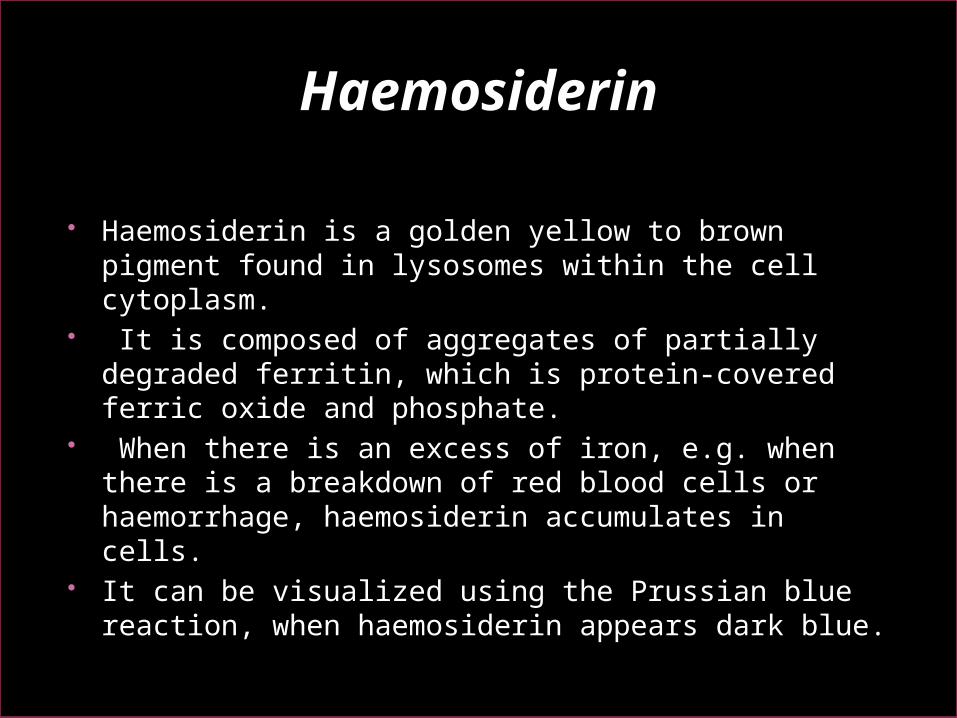
Haemosiderin
Haemosiderin is a golden yellow to brown pigment found in lysosomes within the cell cytoplasm.
It is composed of aggregates of partially degraded ferritin, which is protein-covered ferric oxide and phosphate.
When there is an excess of iron, e.g. when there is a breakdown of red blood cells or haemorrhage, haemosiderin accumulates in cells.
It can be visualized using the Prussian blue reaction, when haemosiderin appears dark blue.
Haemosiderin
Primary haemochromatosis is an inherited disease in which there is excessive accumulation of iron and widespread deposition of haemosiderin in the tissues, especially the liver, pancreas and skin.
The iron is toxic to the tissues and leads to fibrosis of the liver (cirrhosis) and pancreas (leading to diabetes mellitus).
Melanin
Melanin is the brown/black pigment which is normally present in the cytoplasm of cells in the basal cell layer of the epidermis, called melanocytes.
Melanin is derived from tyrosine, stored in melanosomes and distributed to the other epidermal cells.
The function of melanin is to block harmful UV rays from the epidermal nuclei.
Melanin
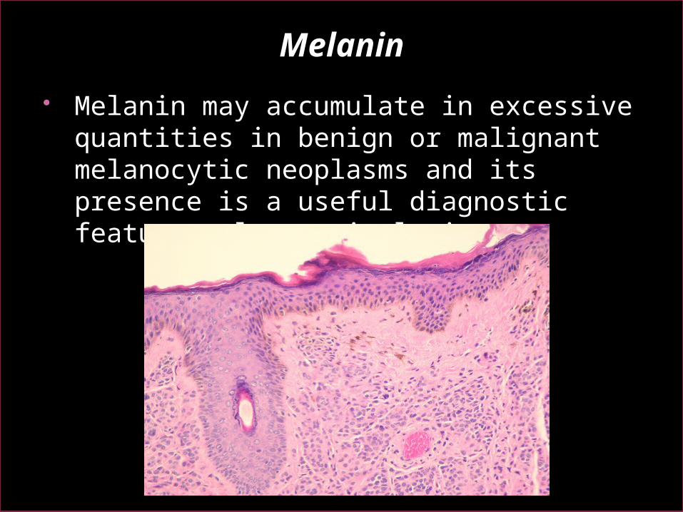
Melanin may accumulate in excessive quantities in benign or malignant melanocytic neoplasms and its presence is a useful diagnostic feature melanocytic lesions.
Melanin
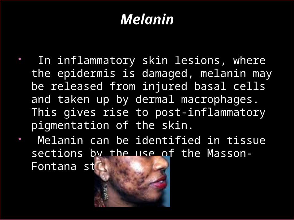
In inflammatory skin lesions, where the epidermis is damaged, melanin may be released from injured basal cells and taken up by dermal macrophages. This gives rise to post-inflammatory pigmentation of the skin.
Melanin can be identified in tissue sections by the use of the Masson-Fontana stain.
Lipofuscin
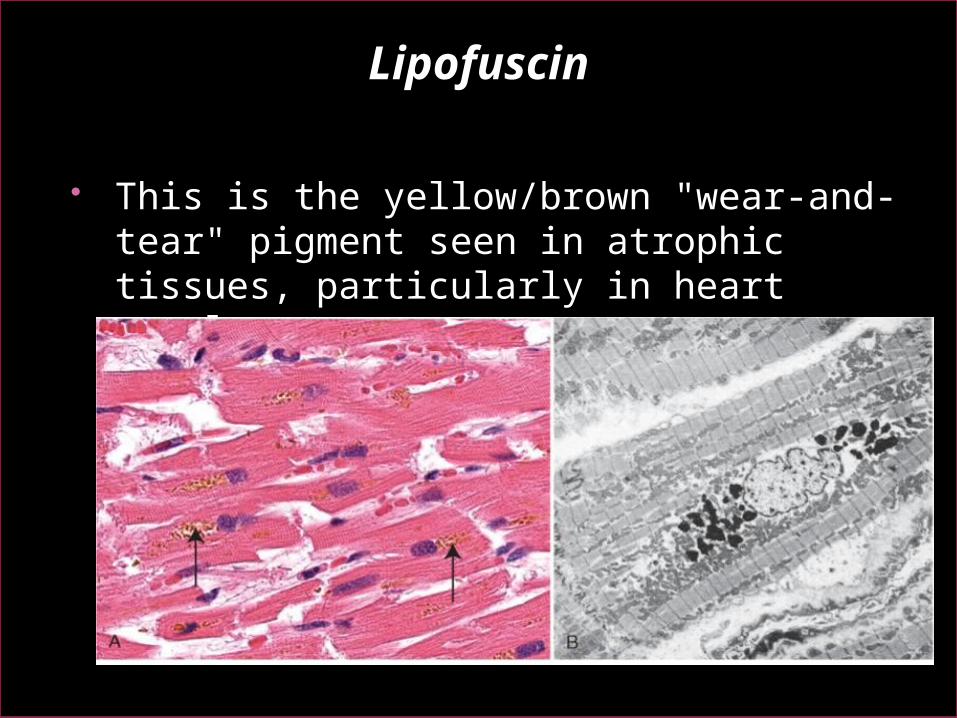
This is the yellow/brown "wear-and-tear" pigment seen in atrophic tissues, particularly in heart muscle.
Other pigments and dyes
Pigments and insoluble substances may enter the body from a variety of sources. They may be toxic and produce inflammatory tissue reactions or they may be relatively inert. Indian ink pigments produce effective tattoos
because they are engulfed by dermal macrophages which become immobilized and permanently deposited.
Inhaled substances such as coal dust and silica are engulfed by pulmonary macrophages. Although pure carbon is inert, silica and coal dust are toxic and ultimately cause serious lung fibrosis (pneumoconiosis).

Calcification
There are two main types of calcification: Dystrophic. Metastatic.
Dystrophic calcification
This type of calcification occurs within diseased tissues. The plasma calcium and phosphate levels are normal.
The exact mechanism by which dystrophic calcification occurs is not known.
The best examples include calcification within foci of old tuberculosis and in atheromatous plaques.
In both these cases, the calcification can often be seen, incidentally, on radiographs.
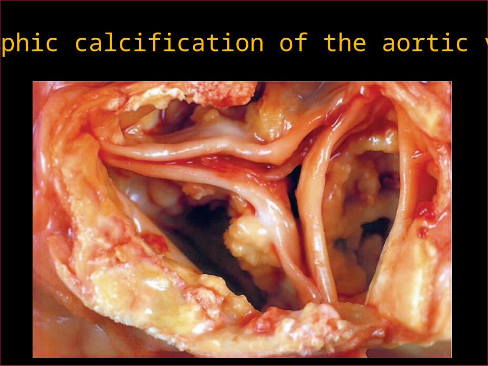
Dystrophic calcification of the aortic valves
Metastatic calcification
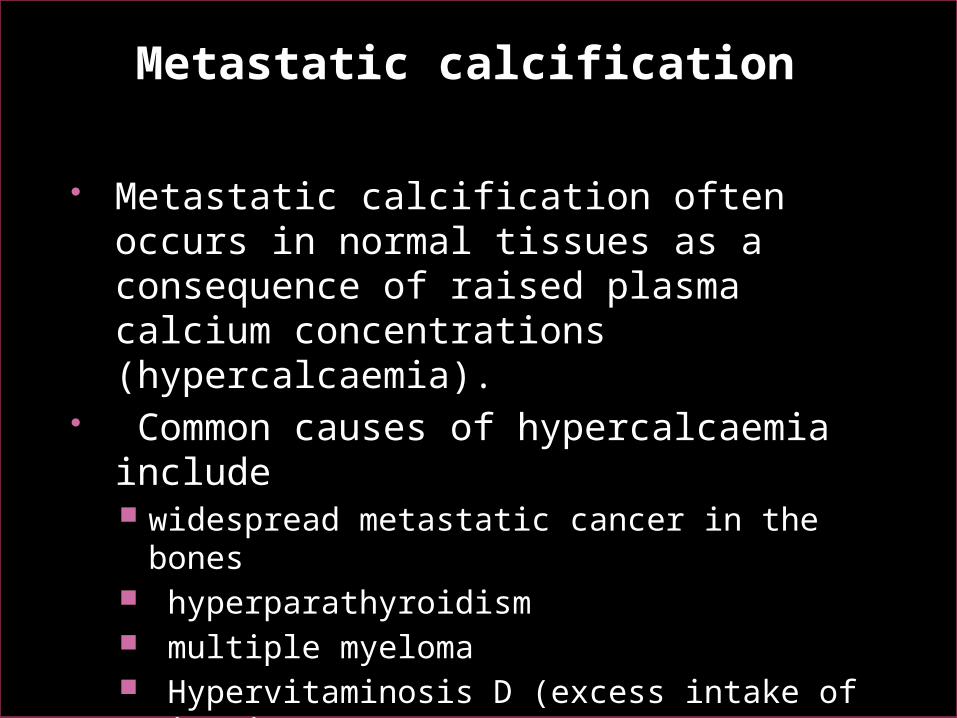
Metastatic calcification often occurs in normal tissues as a consequence of raised plasma calcium concentrations (hypercalcaemia).
Common causes of hypercalcaemia include widespread metastatic cancer in the bones hyperparathyroidism multiple myeloma Hypervitaminosis D (excess intake of vitamin
D)
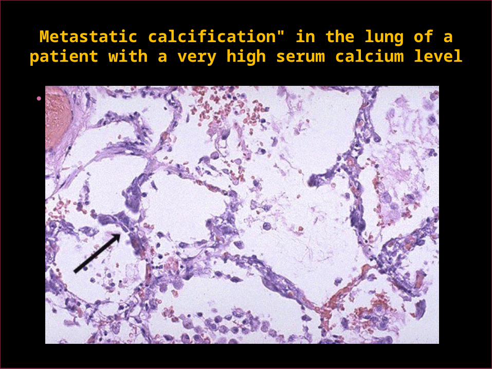
Metastatic calcification" in the lung of a patient with a very high serum calcium level

ADAPTATION TO ENVIRONMENTAL STRESS
Atrophy, hypoplasia and aplasiaHypertrophyHyperplasiaMetaplasia
Dr. Maha ArafahAssistant ProfessorDepartment of PathologyKing Khalid University Hospital and King Saud UniversityEmail: [email protected] marafah @hotmail.com
Saturday, Oct 9, 20109:00 to 10:00 am
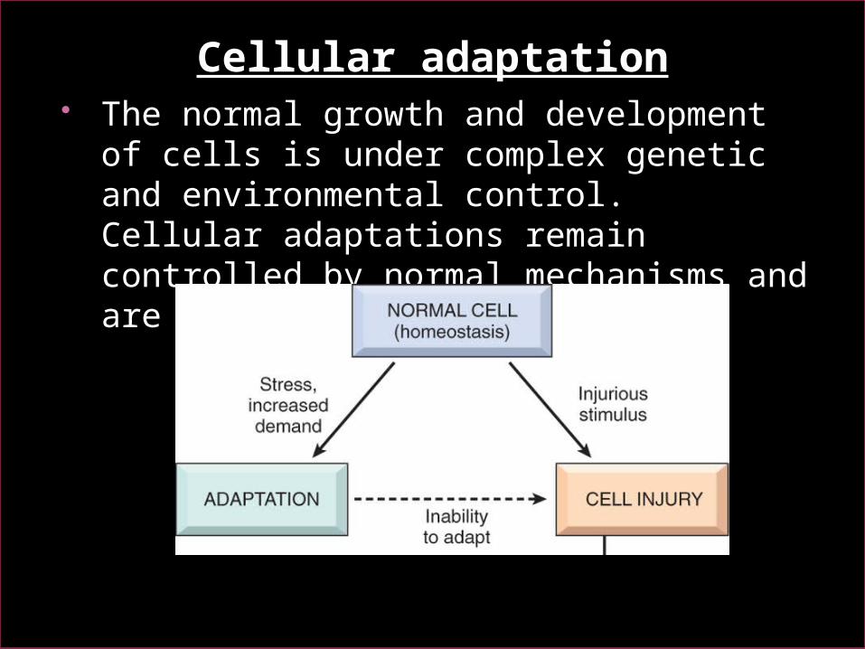
Cellular adaptation The normal growth and development of
cells is under complex genetic and environmental control. Cellular adaptations remain controlled by normal mechanisms and are reversible.
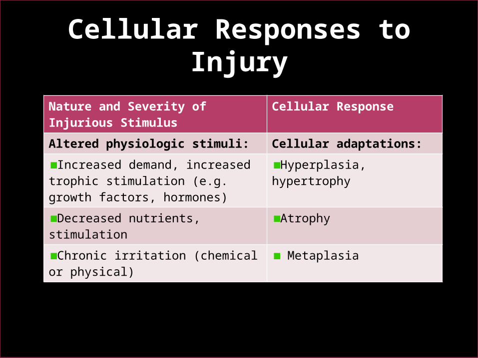
Cellular Responses to Injury
Nature and Severity of Injurious Stimulus
Cellular Response
Altered physiologic stimuli: Cellular adaptations:
Increased demand, increased trophic stimulation (e.g. growth factors, hormones)
Hyperplasia, hypertrophy
Decreased nutrients, stimulation Atrophy
Chronic irritation (chemical or physical)
Metaplasia
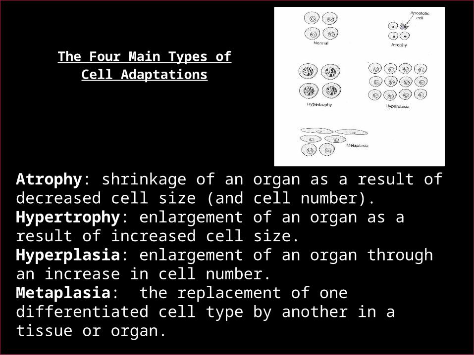
The Four Main Types of Cell Adaptations
Atrophy: shrinkage of an organ as a result of decreased cell size (and cell number).Hypertrophy: enlargement of an organ as a result of increased cell size.Hyperplasia: enlargement of an organ through an increase in cell number.Metaplasia: the replacement of one differentiated cell type by another in a tissue or organ.
Atrophy Atrophy is defined as the shrinkage of a
cell by loss of the cell substance, which leads to a reduction in size of the whole organ.
The cell retreats to a smaller size at which survival is still possible and sequesters its internal structures.
In many cases of atrophy, individual cells may undergo apoptosis.
Atrophy Atrophy is accompanied by increased autophagy,
(increases in the number of autophagic vacuoles) Autophagy ("self-eating") is the process in which
the starved cell eats its own components in an attempt to find nutrients and survive
Lipofuscin granules are yellow/brown in color and represent non-digestible fragments of lipids and phospholipids combined with protein within autophagic vacuoles. They are commonly seen in ageing liver and myocardial cells.
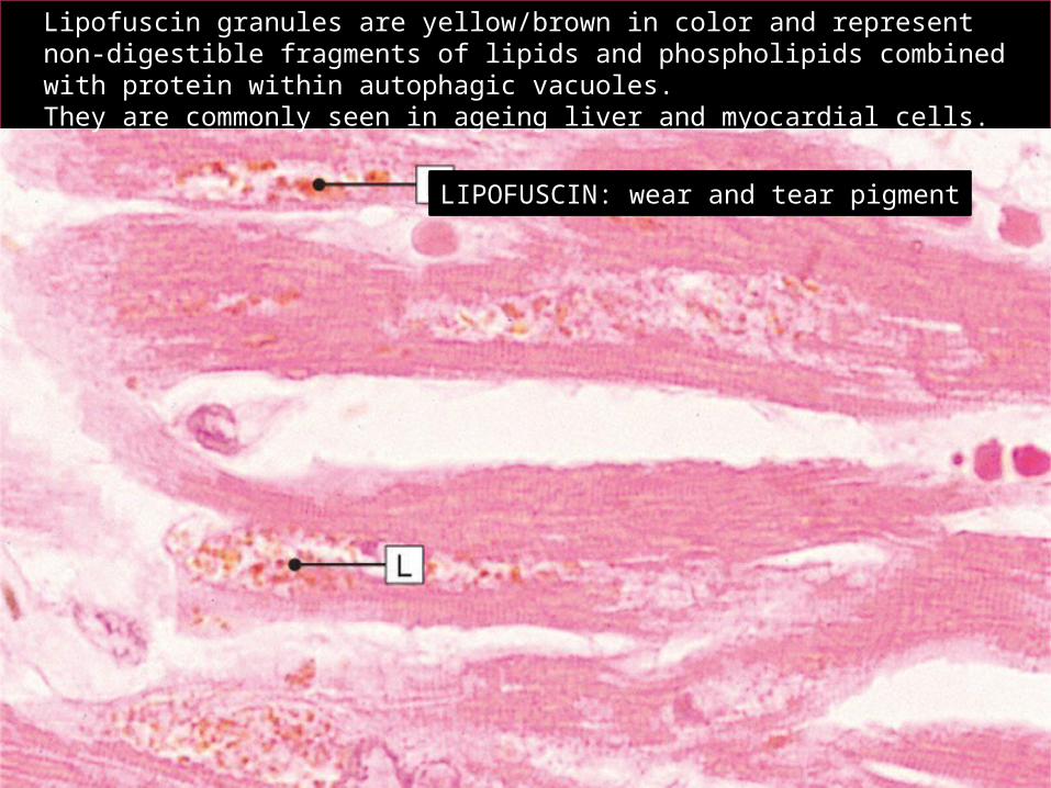
LIPOFUSCIN: wear and tear pigment
Lipofuscin granules are yellow/brown in color and represent non-digestible fragments of lipids and phospholipids combined with protein within autophagic vacuoles. They are commonly seen in ageing liver and myocardial cells.
Cell structural components are reduced in atrophy: Less mitochondria. Less endoplasmic reticulum. Fewer myofilaments.
The metabolic rate is reduced: Less amino acid uptake. Less oxygen consumption. Less protein synthesis.
In atrophy, there is an increase in the number of autophagic vacuoles (intracellular dustbins) which contain fragments of intracellular debris awaiting destruction.
Physiological situations leading to atrophy
Reduction/loss of endocrine stimulation, e.g. shrinkage of testes and ovaries with age.
Pathological causes of atrophy:
1. Denervation, e.g. wasting of muscle caused by a lack of nerve stimulation, for example in poliomyelitis.
2. Reduced blood supply, e.g. shrinkage of brain caused by atherosclerosis of carotid arteries.
3. Inadequate nutrition, e.g. wasting of muscles and major organs in starvation.
4. Decreased workload (disuse) e.g. wasting of muscles and bone (osteoporosis) after immobilization of a bone fracture in a plaster cast.
Hypertrophy
Individual cells increase in size as a result of an increase in their structural components leading to an overall increase in the size of the organ.
Protein synthesis increases and a decrease in protein degradation.
Hypertrophy is mainly seen in cells which have no capacity for mitotic division, e.g. cardiac and skeletal muscle cells (permanent cells).
The limiting factor for the eventual muscle size is the nutrient and blood supply available for oxidative phosphorylation.
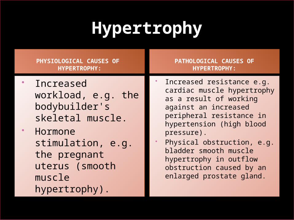
Hypertrophy
PHYSIOLOGICAL CAUSES OF HYPERTROPHY:
PATHOLOGICAL CAUSES OF HYPERTROPHY:
Increased workload, e.g. the bodybuilder's skeletal muscle.
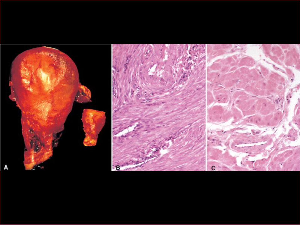
Hormone stimulation, e.g. the pregnant uterus (smooth muscle hypertrophy).
Increased resistance e.g. cardiac muscle hypertrophy as a result of working against an increased peripheral resistance in hypertension (high blood pressure).
Physical obstruction, e.g. bladder smooth muscle hypertrophy in outflow obstruction caused by an enlarged prostate gland.
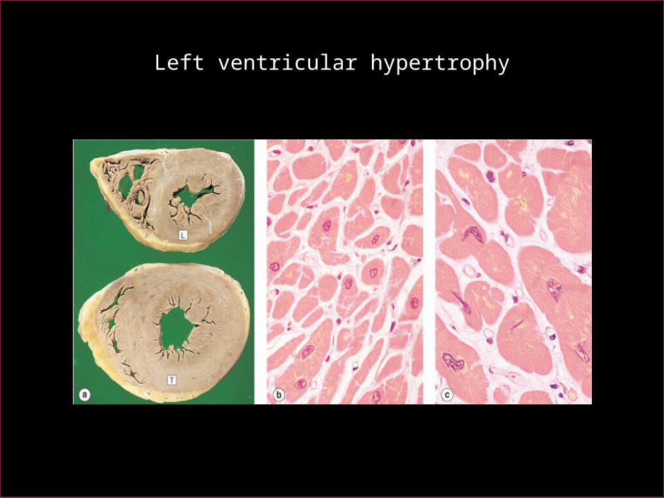
Left ventricular hypertrophy
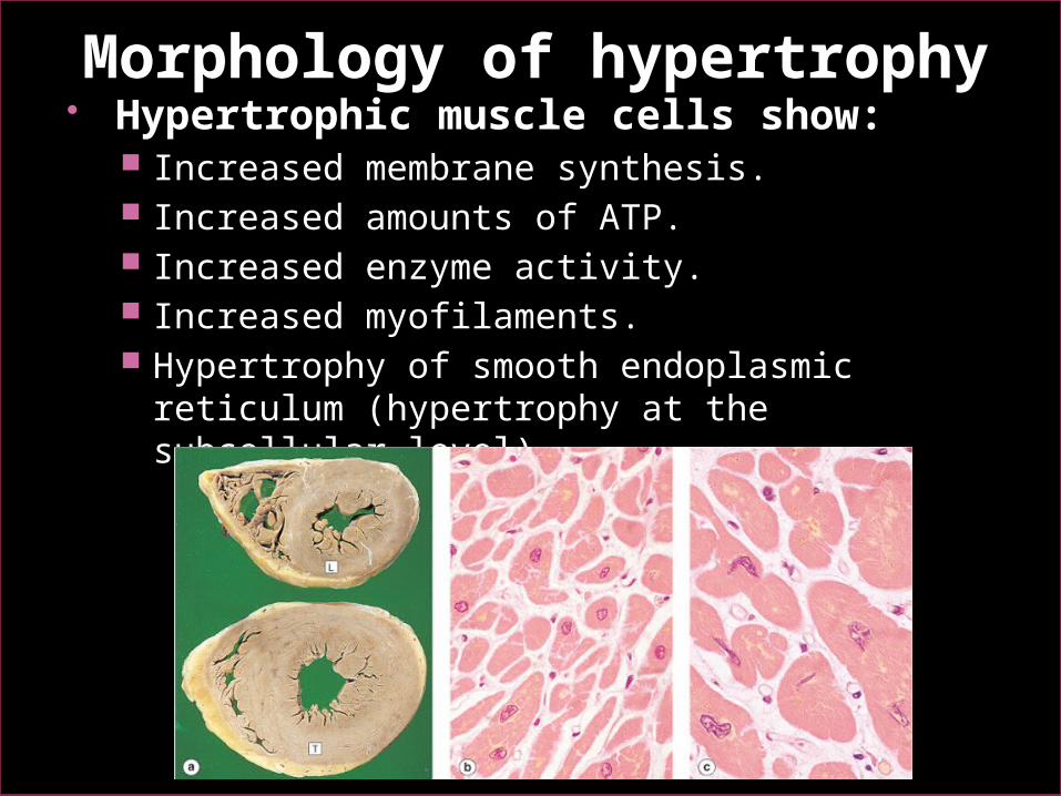
Morphology of hypertrophy Hypertrophic muscle cells show:
Increased membrane synthesis. Increased amounts of ATP. Increased enzyme activity. Increased myofilaments. Hypertrophy of smooth endoplasmic reticulum
(hypertrophy at the subcellular level)
Effects of hypertrophy
Drugs such as phenobarbatone cause an increase in the activity of the mixed function oxidase system of liver cells, which results in increased metabolism of other agents.
In some instances, this is therapeutically beneficial, in others, it can lead to poisoning of the liver cells by toxic metabolites.
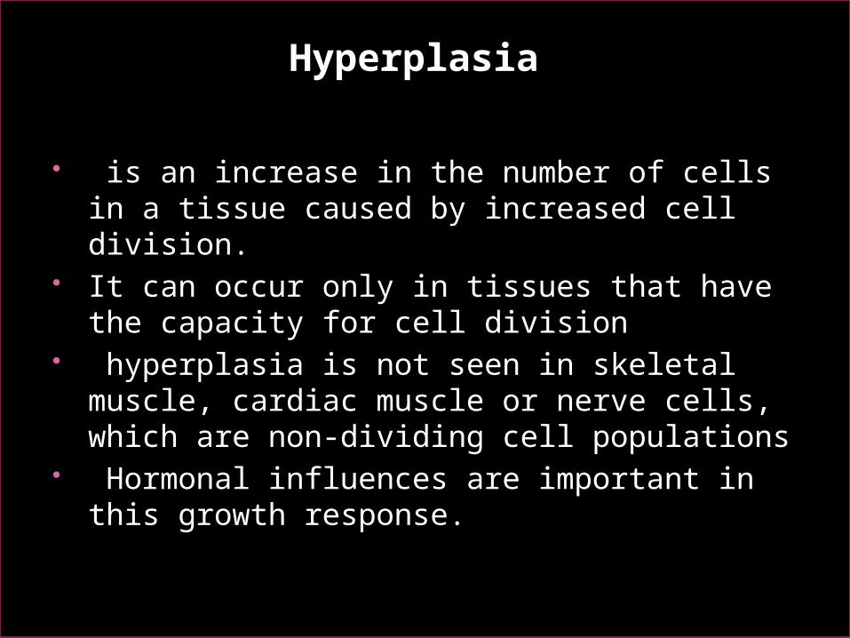
Hyperplasia
is an increase in the number of cells in a tissue caused by increased cell division.
It can occur only in tissues that have the capacity for cell division
hyperplasia is not seen in skeletal muscle, cardiac muscle or nerve cells, which are non-dividing cell populations
Hormonal influences are important in this growth response.
Causes of Hyperplasia
Physiologic Pathologic
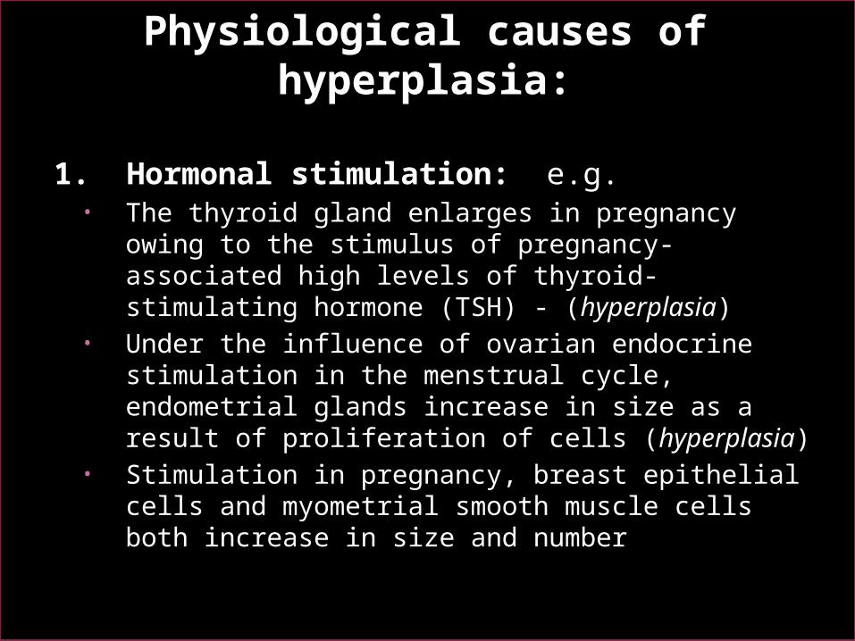
Physiological causes of hyperplasia:
1. Hormonal stimulation: e.g.• The thyroid gland enlarges in pregnancy owing
to the stimulus of pregnancy-associated high levels of thyroid-stimulating hormone (TSH) - (hyperplasia)
• Under the influence of ovarian endocrine stimulation in the menstrual cycle, endometrial glands increase in size as a result of proliferation of cells (hyperplasia)
• Stimulation in pregnancy, breast epithelial cells and myometrial smooth muscle cells both increase in size and number
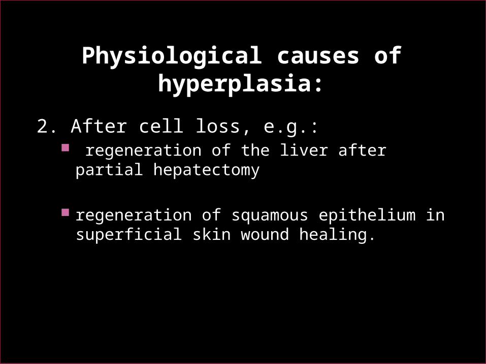
Physiological causes of hyperplasia:
2. After cell loss, e.g.: regeneration of the liver after partial
hepatectomy
regeneration of squamous epithelium in superficial skin wound healing.
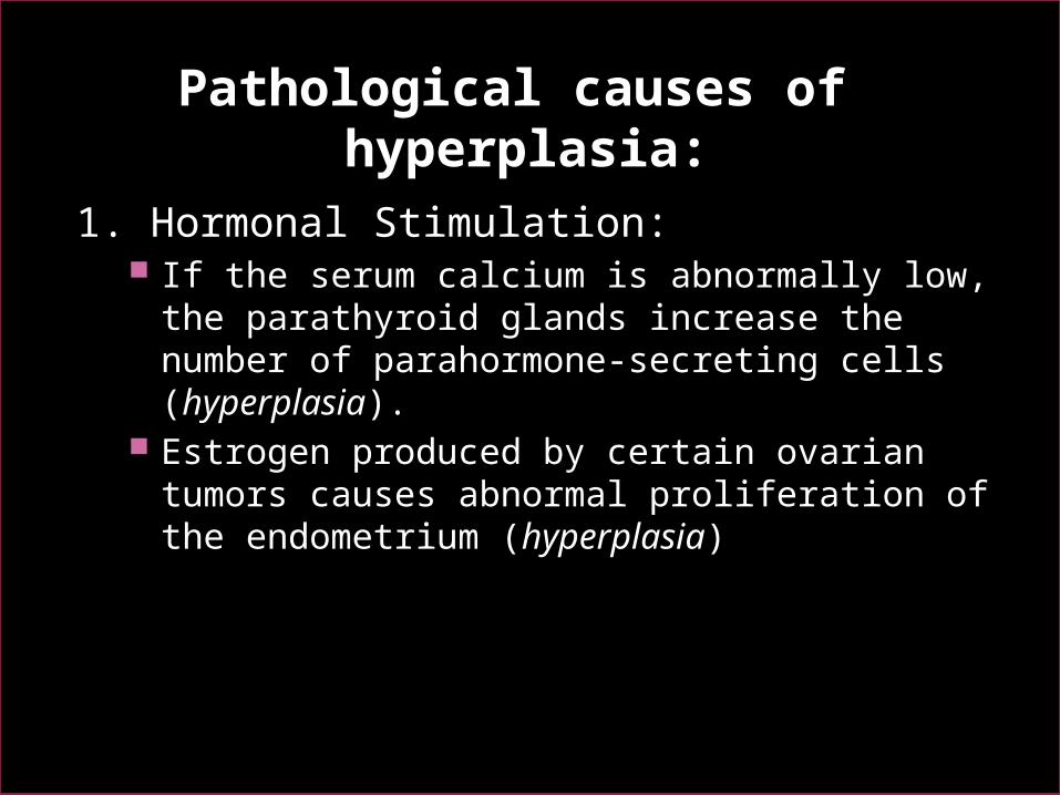
Pathological causes of hyperplasia:
1. Hormonal Stimulation: If the serum calcium is abnormally low, the
parathyroid glands increase the number of parahormone-secreting cells (hyperplasia).
Estrogen produced by certain ovarian tumors causes abnormal proliferation of the endometrium (hyperplasia)
35
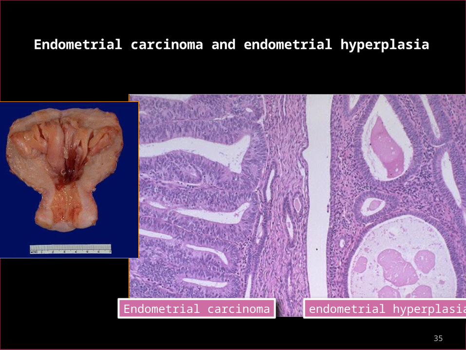
Endometrial carcinoma and endometrial hyperplasia
Endometrial carcinoma endometrial hyperplasia
Pathological causes of hyperplasia:
Cell destruction e.g. ulcerative colitis the
colonic mucosa undergoes repeated ulceration (destruction), regeneration and hyperplasia.
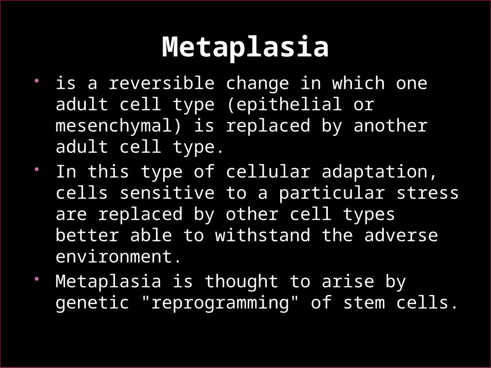
Metaplasia is a reversible change in which one adult
cell type (epithelial or mesenchymal) is replaced by another adult cell type.
In this type of cellular adaptation, cells sensitive to a particular stress are replaced by other cell types better able to withstand the adverse environment.
Metaplasia is thought to arise by genetic "reprogramming" of stem cells.
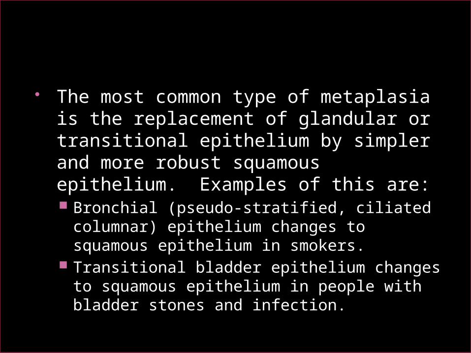
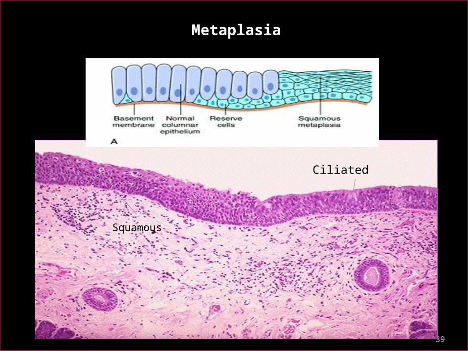
The most common type of metaplasia is the replacement of glandular or transitional epithelium by simpler and more robust squamous epithelium. Examples of this are: Bronchial (pseudo-stratified, ciliated
columnar) epithelium changes to squamous epithelium in smokers.
Transitional bladder epithelium changes to squamous epithelium in people with bladder stones and infection.
39
Ciliated
Squamous
Metaplasia
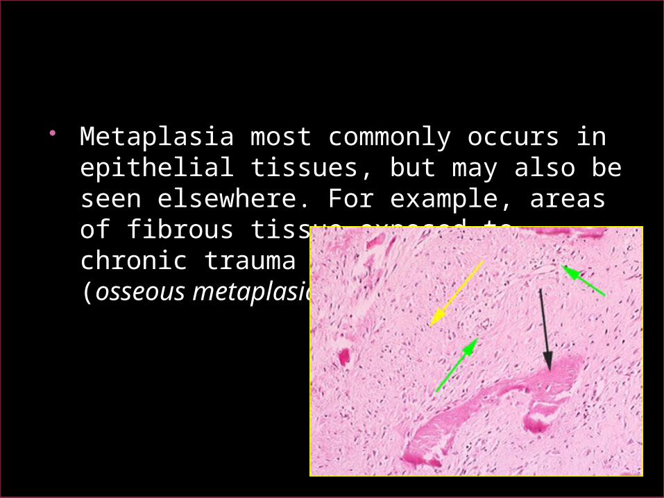
Metaplasia most commonly occurs in epithelial tissues, but may also be seen elsewhere. For example, areas of fibrous tissue exposed to chronic trauma may form bone (osseous metaplasia).
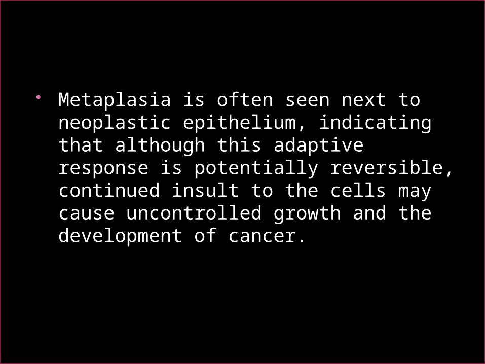
Metaplasia is often seen next to neoplastic epithelium, indicating that although this adaptive response is potentially reversible, continued insult to the cells may cause uncontrolled growth and the development of cancer.
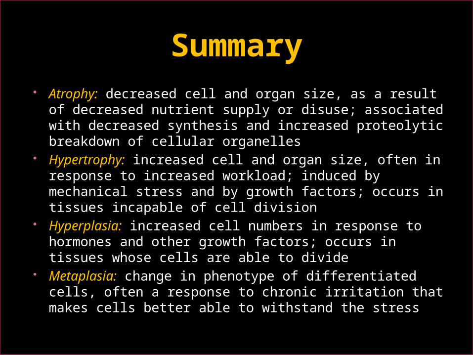
Summary Atrophy: decreased cell and organ size, as a result of
decreased nutrient supply or disuse; associated with decreased synthesis and increased proteolytic breakdown of cellular organelles
Hypertrophy: increased cell and organ size, often in response to increased workload; induced by mechanical stress and by growth factors; occurs in tissues incapable of cell division
Hyperplasia: increased cell numbers in response to hormones and other growth factors; occurs in tissues whose cells are able to divide
Metaplasia: change in phenotype of differentiated cells, often a response to chronic irritation that makes cells better able to withstand the stress
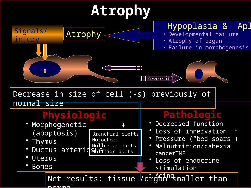
Atrophy Hypoplasia & Aplasia• Developmental failure• Atrophy of organ• Failure in morphogenesis
Reversible
Decrease in size of cell (-s) previously of normal size
Physiologic • Morphogenetic (apoptosis)• Thymus• Ductus arteriosus• Uterus• Bones
Pathologic • Decreased function• Loss of innervation• Pressure (“bed soars”)• Malnutrition/cahexia
cancerTNF• Loss of endocrine stimulation• Aging
Branchial cleftsNotochordMullerian ductsWolffian ducts
Net results: tissue /organ smaller than normal
Signals/injury
Atrophy
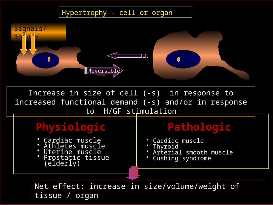
Hypertrophy – cell or organ
Reversible
Increase in size of cell (-s) in response to increased functional demand (-s) and/or in response to H/GF stimulation
Physiologic
• Cardiac muscle• Athletes muscle• Uterine muscle• Prostatic tissue (elderly)
Pathologic • Cardiac muscle• Thyroid• Arterial smooth muscle• Cushing syndrome
Net effect: increase in size/volume/weight of tissue / organ
Signals/injury
45
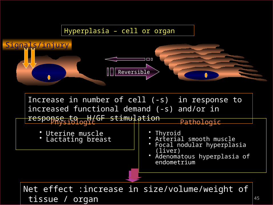
Hyperplasia – cell or organ
Reversible
Increase in number of cell (-s) in response to increased functional demand (-s) and/or in response to H/GF stimulationPhysiologic
• Uterine muscle• Lactating breast
Pathologic • Thyroid• Arterial smooth muscle• Focal nodular hyperplasia (liver)• Adenomatous hyperplasia of
endometrium
Net effect :increase in size/volume/weight of tissue / organ
Signals/injury
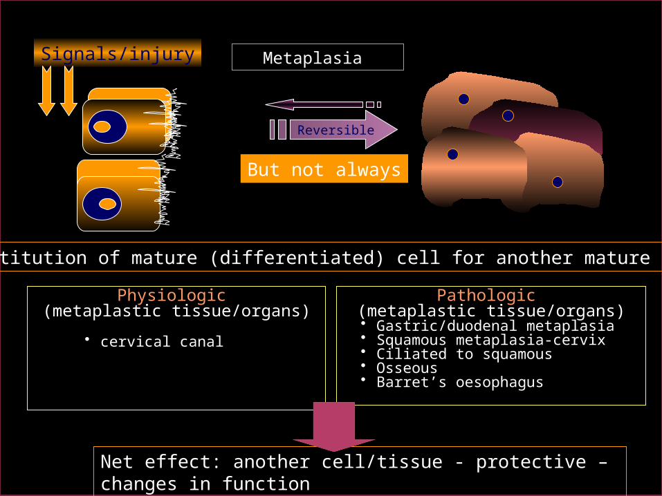
Signals/injury
Reversible
But not always
Metaplasia
Substitution of mature (differentiated) cell for another mature cell
Physiologic (metaplastic tissue/organs)
• cervical canal
Pathologic (metaplastic tissue/organs)
• Gastric/duodenal metaplasia• Squamous metaplasia-cervix• Ciliated to squamous• Osseous• Barret’s oesophagus
Net effect: another cell/tissue - protective – changes in function
Related Documents