Assessing threats from coral and crustose coralline algae disease on the reefs of New Caledonia Greta Aeby A,E , Aline Tribollet B , Gregory Lasne C and Thierry Work D A Hawaii Institute of Marine Biology, PO Box 1346, Kaneohe, HI 96744, USA. B IRD-Sorbonne Universite ´s (UPMC, Univ Paris 6)-CNRS-MNHN, LOCEAN Laboratory, IRD France Nord, 32 Avenida Henri Varagnat, F-93143 Bondy Cedex, France. C BIOCENOSE MARINE Sarl, Centre IRD de Noume ´a, 98848 Noume ´a Cedex, New Caledonia. D US Geological Survey, National Wildlife Health Center, Honolulu Field Station, PO Box 50187, Honolulu, HI 96850, USA. E Corresponding author. Email: [email protected] Abstract. The present study reports the results of the first quantitative survey of lesions on coral and crustose coralline algae (CCA) on reefs in the lagoon of New Caledonia. Surveys on inshore and offshore reefs were conducted at 13 sites in 2010, with 12 sites resurveyed in 2013. Thirty coral diseases affecting 15 coral genera were found, with low overall disease prevalence (,1%). This study extends the known distribution of growth anomalies to the coral genera Platygyra and Hydnophora, endolithic hypermycosis to Platygyra, Leptoria and Goniastrea and extends the geographic range of three CCA diseases. We found the first trematode infection in Porites outside of Hawaii. Disease prevalence differed among coral genera, with Porites having more lesions, and Acropora and Montipora fewer lesions, than expected on the basis of field abundance. Inshore reefs had a lower coral-colony density, species diversity and reduced CCA cover than did the offshore reefs. Disease prevalence was significantly higher on inshore reefs in 2013 than in 2010, but did not change on offshore reefs. The potential ecological impact of individual coral diseases was assessed using an integrative-scoring and relative-ranking scheme based on average frequency of occurrence, prevalence and estimated degree of virulence. The top-five ranked diseases were all tissue-loss diseases. Additional keywords: CCA disease, coral disease, ecological impact of coral disease, endolithic hypermycosis, growth anomalies, trematode infection, white syndrome. Received 12 June 2014, accepted 27 February 2015, published online 9 July 2015 Introduction Recently, coral disease has emerged as a serious threat to coral reefs worldwide and the numbers of diseases, species affected and the distributions of diseases have all increased within the past decade (Green and Bruckner 2000; Sutherland et al. 2004). Diseases have already dramatically affected the reefs of the Caribbean (Gardner et al. 2003; Sutherland et al. 2004; Schutte et al. 2010) and are now being reported with increasing fre- quency from across the Indo-Pacific (Willis et al. 2004; Vargas- A ´ ngel and Wheeler 2009; Aeby et al. 2011a). Increased anthropogenic stress on near-shore environments, overfishing and environmental conditions associated with global climate change have all been implicated as factors contributing to increases in observed disease levels (Colwell 2004; Harvell et al. 2007). The Indo-Pacific region poses a serious challenge for the management of emerging coral diseases because it is much larger than the Caribbean and supports entire communities that depend on reefs for their livelihood (Hughes et al. 2003). Baseline disease surveys provide a basis to assess status and trends of coral-reef health over time, yet numerous regions throughout the Indo-Pacific have not been surveyed. New Caledonia forms a large group of islands in the South-west Pacific that has one of the longest continuous barrier reefs and the largest lagoon in the world, including fringing reefs, patch reefs and barrier reefs (Laboute and Richer de Forges 2004; Andre ´foue ¨t et al. 2006). The reefs of New Caledonia have a fairly high diversity of corals (Payri and Richer de Forges 2006; Fenner and Muir 2009; Fenner 2011), and this unique resource has received international recognition and has been designated a UNESCO World Heritage site. However, reefs of New Caledonia are prone to both natural and man-made threats. Cyclones have been known to severely reduce coral cover (Wilkinson 2008), and New Caledonia has active open-surface nickel mining (1/4 of the global nickel resources are located in New Caledonia) and mining pits are not re-vegetated. They also have agricultural industries that result in extensive runoff, laden with pollutants that are parti- cularly severe during the wet season (Fernandez et al. 2006; Bonvallot et al. 2012). In addition, some reefs could be subject CSIRO PUBLISHING Marine and Freshwater Research http://dx.doi.org/10.1071/MF14151 Journal compilation Ó CSIRO 2015 www.publish.csiro.au/journals/mfr

Welcome message from author
This document is posted to help you gain knowledge. Please leave a comment to let me know what you think about it! Share it to your friends and learn new things together.
Transcript

Assessing threats from coral and crustose corallinealgae disease on the reefs of New Caledonia
Greta AebyA,E, Aline TribolletB, Gregory LasneC and Thierry WorkD
AHawaii Institute of Marine Biology, PO Box 1346, Kaneohe, HI 96744, USA.BIRD-Sorbonne Universites (UPMC, Univ Paris 6)-CNRS-MNHN, LOCEAN Laboratory,
IRD France Nord, 32 Avenida Henri Varagnat, F-93143 Bondy Cedex, France.CBIOCENOSE MARINE Sarl, Centre IRD de Noumea, 98848 Noumea Cedex, New Caledonia.DUS Geological Survey, NationalWildlife Health Center, Honolulu Field Station, PO Box 50187,
Honolulu, HI 96850, USA.ECorresponding author. Email: [email protected]
Abstract. The present study reports the results of the first quantitative survey of lesions on coral and crustose coralline
algae (CCA) on reefs in the lagoon of New Caledonia. Surveys on inshore and offshore reefs were conducted at 13 sites in2010, with 12 sites resurveyed in 2013. Thirty coral diseases affecting 15 coral genera were found, with low overall diseaseprevalence (,1%). This study extends the known distribution of growth anomalies to the coral genera Platygyra and
Hydnophora, endolithic hypermycosis to Platygyra, Leptoria and Goniastrea and extends the geographic range of threeCCA diseases. We found the first trematode infection in Porites outside of Hawaii. Disease prevalence differed amongcoral genera, with Porites having more lesions, and Acropora andMontipora fewer lesions, than expected on the basis of
field abundance. Inshore reefs had a lower coral-colony density, species diversity and reduced CCA cover than did theoffshore reefs. Disease prevalence was significantly higher on inshore reefs in 2013 than in 2010, but did not change onoffshore reefs. The potential ecological impact of individual coral diseases was assessed using an integrative-scoring andrelative-ranking scheme based on average frequency of occurrence, prevalence and estimated degree of virulence. The
top-five ranked diseases were all tissue-loss diseases.
Additional keywords: CCA disease, coral disease, ecological impact of coral disease, endolithic hypermycosis, growthanomalies, trematode infection, white syndrome.
Received 12 June 2014, accepted 27 February 2015, published online 9 July 2015
Introduction
Recently, coral disease has emerged as a serious threat to coralreefs worldwide and the numbers of diseases, species affectedand the distributions of diseases have all increased within the
past decade (Green and Bruckner 2000; Sutherland et al. 2004).Diseases have already dramatically affected the reefs of theCaribbean (Gardner et al. 2003; Sutherland et al. 2004; Schutteet al. 2010) and are now being reported with increasing fre-
quency from across the Indo-Pacific (Willis et al. 2004; Vargas-Angel and Wheeler 2009; Aeby et al. 2011a). Increasedanthropogenic stress on near-shore environments, overfishing
and environmental conditions associated with global climatechange have all been implicated as factors contributing toincreases in observed disease levels (Colwell 2004; Harvell
et al. 2007).The Indo-Pacific region poses a serious challenge for the
management of emerging coral diseases because it is much
larger than the Caribbean and supports entire communities thatdepend on reefs for their livelihood (Hughes et al. 2003).Baseline disease surveys provide a basis to assess status and
trends of coral-reef health over time, yet numerous regions
throughout the Indo-Pacific have not been surveyed. NewCaledonia forms a large group of islands in the South-westPacific that has one of the longest continuous barrier reefs and
the largest lagoon in the world, including fringing reefs, patchreefs and barrier reefs (Laboute and Richer de Forges 2004;Andrefouet et al. 2006). The reefs of New Caledonia have afairly high diversity of corals (Payri and Richer de Forges 2006;
Fenner and Muir 2009; Fenner 2011), and this unique resourcehas received international recognition and has been designated aUNESCO World Heritage site.
However, reefs of New Caledonia are prone to both naturaland man-made threats. Cyclones have been known to severelyreduce coral cover (Wilkinson 2008), and New Caledonia has
active open-surface nickel mining (1/4 of the global nickelresources are located in New Caledonia) and mining pits arenot re-vegetated. They also have agricultural industries that
result in extensive runoff, laden with pollutants that are parti-cularly severe during the wet season (Fernandez et al. 2006;Bonvallot et al. 2012). In addition, some reefs could be subject
CSIRO PUBLISHING
Marine and Freshwater Research
http://dx.doi.org/10.1071/MF14151
Journal compilation � CSIRO 2015 www.publish.csiro.au/journals/mfr
to episodic industrial chemical spills and sewage because ofinsufficient treatment infrastructures. A few coral diseases have
been reported from the reefs of NewCaledonia (McKenna 2009,2011), yet no quantitative baseline disease data exist for thesereefs. The objectives of the present study were to determine thehealth status of corals and crustose coralline algae in the lagoon
of New Caledonia, in nearshore reefs subject to terrigenousinputs and anthropogenic activities, comparedwithmore remoteoffshore barrier reefs in a 2-year time period.
Materials and methods
Study site
New Caledonia is a special collectivity (semi-independent sta-tus) of France located in the Melanesian region of the south-
western Pacific Ocean, 1210 km east of Australia (Fig. 1). Thearchipelago includes the main island of Grande Terre, theLoyalty Islands, the Chesterfield Islands, the Belep archipelago,
the Isle of Pines and other assorted islets (Lasne 2007).
Disease surveys
Thirteen reefs were surveyed in the lagoon surrounding themain
island of Grande Terre, including one mid-lagoonal reef, sixinshore and six offshore reefs (Fig. 1). Owing to logisticalconstraints and difficulties in accessing eastern Grande Terre,
most surveys were conducted on the western coast of GrandTerre, with only two sites on the north-eastern coast. Inshore–offshore comparisons were made to examine potential anthro-pogenic effects on coral reef health. The locations of inshore reef
sites ranged between 1- and 8-m depth and were situated 1–3 kmfrom the shore. Outer barrier reefs were located ,10–16 km
offshore on the outer reef slope at depths between 6 and 19mandthe lagoonal reef was located at 1–2-m depth, a few kilometersnorth of the city of Noumea (south lagoon). The baseline levelsof coral and crustose coralline disease at each of the selected
sites were documented using two 25-m belt transects laid end-to-end along depth contours, separated by ,5 m. All coralcolonies within each 1-m-wide belt (25� 1 m) were identified
to genus and enumerated. Substrate was quantified by point-intercept method, whereby the substratum underlying the tapemeasure was recorded at 50-cm intervals. The average per-
centage coral cover and colony density per square metre weredetermined from the diver surveys. Lesions in corals and CCAwere quantified along wider 25� 6 m transects (total of 300-m2
area of reef). All lesions encountered during the surveys werephotographed and representative samples collected for histo-pathology and surveys of microboring organisms, which can beinvolved in coral diseases (e.g. Work et al. 2008a; see also
Tribollet 2008). Surveys were conducted in the summer season,January 2010 (n¼ 13 sites) and February 2013 (n¼ 12 sites).The GPS coordinates were recorded for each site in 2010 and
were used to relocate sites in 2013.Gross lesions were classified into three broad categories
including tissue loss, discoloration and growth anomaly (Work
andAeby 2006). Nomenclature for lesions was based on the hostaffected and lesion type (e.g.Porites growth anomaly;Work andAeby 2006). Tissue-loss lesions can be caused by biologicalfactors, such as predation or sedimentation, as well as infectious
Fig. 1. Sites surveyed for coral and crustose coralline algae (CCA) disease in 2010 and 2013 around the
largest island of New Caledonia, Grande Terre. Six inshore–offshore pairs of reefs were surveyed
(n¼ 12 sites) and one lagoonal reef. 1, Ilot Casy (inshore reef); 2, Gue reef (offshore barrier reef); 3, Baie
des Citrons (inshore reef); 4, Seche Croissant reef (lagoonal patch reef); 5, Mbere reef (offshore barrier
reef); 6, Chenal de Teremba (inshore reef); 7, Passe de Ouaraı (offshore barrier reef); 8, Kreliat reef
(inshore reef); 9, Passe deKone (offshore barrier reef); 10, Bouerabate reef (inshore reef); 11, Passe de la
Gazelle (offshore barrier reef); 12, Neongaon reef* (inshore reef); 13, Balade reef (offshore reef in front
of the Col d’Amos). Sites with asterisks (*) were surveyed in 2010 only.
B Marine and Freshwater Research G. Aeby et al.

disease, and it can be challenging to discriminate between theseprocesses in the field. However, valuable clues can be gained
from looking at the lesion size, shape, presence of predators,knowledge of what common predation marks look like andevidence of lesion progression based on degree of algal coloni-
sation onto the bare coral skeleton. We conducted our diseasesurveys using the aforementioned criteria but also collectedsamples for follow-up histological analyses. Histology allows
you to further differentiate between potentially infectious versusnon-infection processes (Work et al. 2012). After histologicalexamination of lesions, tissue-loss lesions not found associatedwith an obvious pathogenic organism, usually ciliates or
cyanobacteria, were termed ‘white syndrome’. If ciliates orcyanobacteria were found microscopically invading coral tissuein the lesion, then they were termed ciliate or cyanobacterial
infections. Results of histological analyses for cnidaria arereported in Work et al. (2014). Most disease surveys have notincluded histological findings in the interpretation of their
survey results.
Data analysis
For corals, prevalence of lesions was calculated by extrapo-
lating colony counts within the 25� 1 m transect to the wider25� 6 m disease survey area and by using this as the denomi-nator of prevalence calculations (e.g. (number of colonieswith lesions C total number of estimated colonies)� 100).
CCA substrate cover was estimated within each survey areaby determining the percentage cover by point-intercept andextrapolating that out to the spatial area of the belt transects.
Abundance of CCA disease was then determined by calculatingthe number of CCA lesions per estimated square metre of CCAsurveyed. The frequency of disease occurrence (FOC) was
defined as the number of sites having corals or CCA withlesions divided by total number of sites surveyed. FOC reflectsthe spatial distribution of diseases on reefs.
Data were not normally distributed, even with transforma-
tion, so non-parametric analyses were used. A matched-pairssigned-ranks test was used to examine differences in coraldisease prevalence and CCA disease abundance within sites
between years. A Wilcoxon two-group test was used to test fordifferences between regions (offshore vs inshore) in coral cover,CCA cover, coral disease prevalence, number of coral genera
and colony density (number of coral colonies per square metre).For each survey period (2010 and 2013), a chi-square goodness-of-fit test examined differences in the distribution of the number
of diseased versus healthy colonies among the scleractiniangenera affected by disease. Non-parametric statistics wereperformed using JMP statistical software (v. 10.0.2, SAS Insti-tute Inc., Buckinghamshire, UK).
To examine differences in coral community structurebetween regions (offshore vs inshore), the data were square-root transformed and an analysis of similarity (ANOSIM), a
non-parametric permutation procedure, was performed. A non-metric multi-dimensional scaling (nMDS) plot based on Bray–Curtis similarity was used to illustrate the differences in coral
communities among sites and a SIMPER analysis examined thepercentage contribution of each genus towards the groupings(PRIMER v. 6.1.12, Ivybridge, UK).
The potential for damage to coral reefs from diseases woulddepend on the spatial distribution (frequency of occurrence),
prevalence (proportion of colonies surveyed that were affected)and degree of virulence (harm to host) of the disease. These threemetrics were used to develop an integrative-scoring and
relative-ranking scheme to compare the potential ecologicalimpact of each disease. Ranking scales have been used success-fully in other studies to examine coral-ecosystem health (Jokiel
and Rodgers 2007). For each disease, the average prevalenceand frequency of occurrence was calculated across both years.On the basis of what is known about the ecology of each disease,potential virulence was categorised as low (reduced growth or
reproduction), medium (chronic tissue loss) or high (acute orsubacute tissue loss). The scores were scaled from 0 (notharmful) to 10 (very harmful). To get each metric under the
same 0–10 scale, the average frequency of occurrence data weredivided by 10, prevalence data were multiplied by 10 and theprevalence categories were assigned scores of 1 for low viru-
lence, 5 for medium virulence and 10 for high virulence.A mean score for each disease was calculated as the averageof the three metrics.
Results
Coral-reef characteristics
We identified 44 coral genera, within transects, and four of thosecoral genera were numerically dominant. These were Acropora
(2010:,43% of the coral community; 2013:,31.9%), Porites(2010: ,11.7%; 2013: ,16.1%) Montipora (2010: ,11.0%;2013:,16.0%) and Pocillopora (2010:,6.8%; 2013:,4.5%).
Coral cover ranged from 7.8–83.3% (,35.3%) in 2010 and13.9–63.4% (,44.6%) in 2013. CCA cover ranged from 0%to 38.2% (,9.9%) in 2010 and from 0% to 24.7% (,8.6%)
in 2013.
Spatial and temporal occurrence of disease
Coral diseases
We examined a total of 47 166 scleractinian corals from
3780-m2 reef area in 2010 and 38 251 corals from 3600-m2 reefarea in 2013. Thirty different coral diseases were found, affect-ing 15 coral genera on the reefs of the New Caledonian lagoon.Ten diseases were observed in both years surveyed, 11 and 9
diseases exclusively in 2010 and 2013 respectively (Table 1).Average prevalence of disease differed among disease types,but was overall low (,1%) in both years surveyed (Table 2).
Average disease prevalence was 0.165% (s.e. �0.05%) in 2010v. 0.287% (s.e.�0.1%) in 2013 (matched-pairs sign test, t¼ 2.1,n¼ 12, P¼ 0.06). Frequency of occurrence (FOC) of the differ-
ent diseases varied between years (Table 1) but the mostcommon diseases (.15% of sites in both years) were Acroporawhite syndrome, Acropora growth anomalies, Porites multi-focal pink spot, Porites growth anomalies, Montipora growth
anomalies and Pavona dark spot.
CCA disease
The following three CCA diseases were documented: CCAwhite syndrome, CCA concentric white discoloration (targetphenomenon) and coralline lethal orange disease (CLOD). CCA
Coral and CCA disease on the reefs of New Caledonia Marine and Freshwater Research C
diseases were found predominantly on offshore reefs, withfrequency of occurrence of CCA white syndrome 46.2% in
2010 and 33.3% in 2013; CCA concentric white discolorationwas 0% in 2010 and 8.3% in 2013 andCLODwas 30.8% in 2010and 16.7% in 2013. The number of CCA lesions ranged fromzero to 13 per site. At sites where CCA disease occurred, the
average number of lesions was 0.11 m�2 CCA (s.e. �0.05) in2010 and 0.09 m�2 CCA (s.e. �0.03) in 2013.
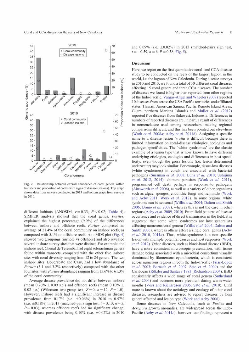
Differences in disease prevalence among coral genera
Out of 44 genera surveyed, only 15 showed signs of coral dis-ease. Prevalence of disease differed among the coral generawithin each year surveyed (2010:x2¼ 411.9 d.f.¼ 9,P, 0.001;
2013: x2¼ 174.5, d.f.¼ 12, P, 0.001). Of the four mostabundant scleractinian coral genera found within transects,Porites had the highest overall prevalence (2010: 0.73%; 2013:
1.5%), followed by Montipora (2010: 0.07%; 2013: 0.1%),Acropora (2010: 0.06%; 2013: 0.1%) and Pocillopora (0% bothyears). Of the main coral genera affected by disease, Acroporaand Montipora had fewer diseased colonies than was expected
on the basis of their abundance in surveys, and Porites had more
diseased colonies than expected (Fig. 2).
Ecological rank of potential damage from coral diseases
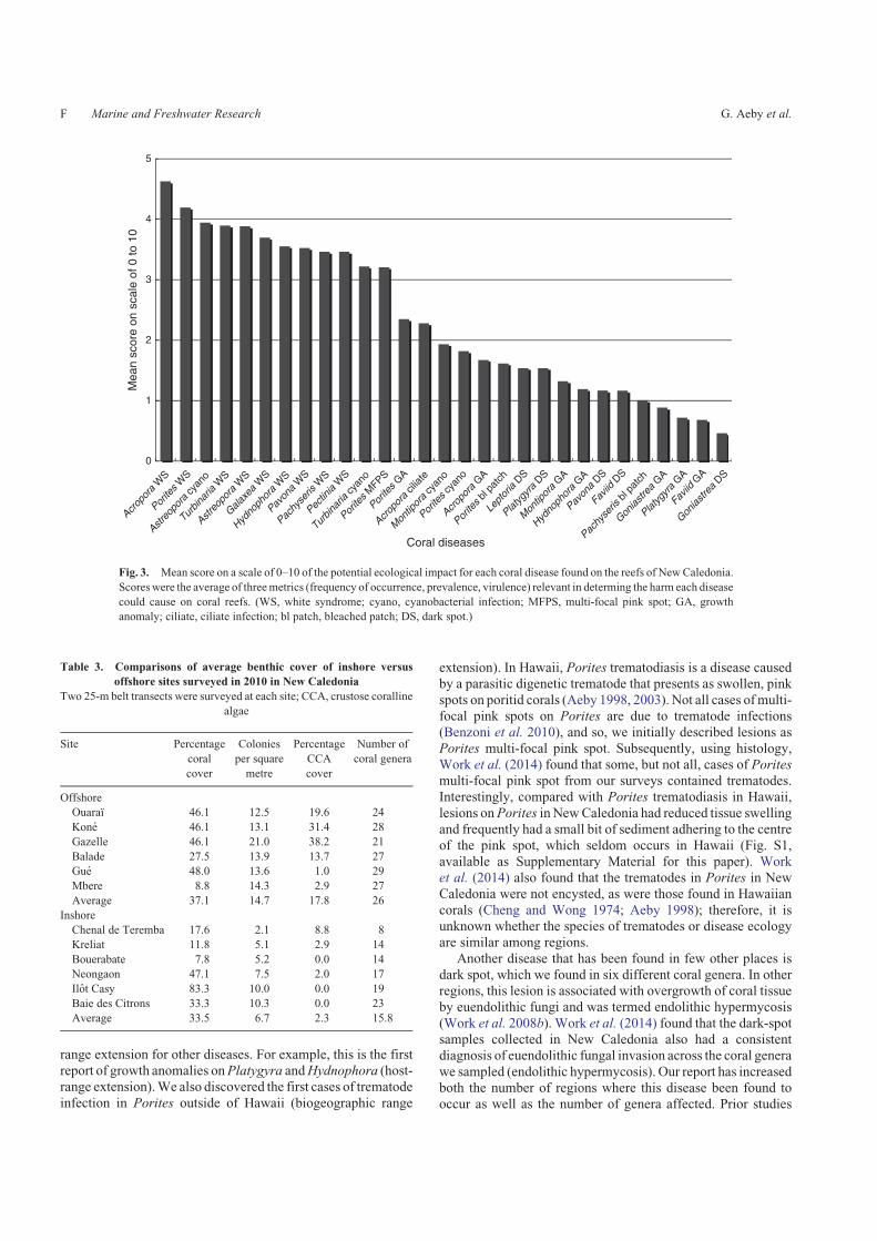
On the basis of the average score from the threemetrics (averageFOC, average prevalence and virulence category) the top-five
ranked diseases were all tissue-loss diseases and included whitesyndrome inAcropora, Porites,Astreopora and Turbinaria, andcyanobacterial infection in Turbinaria (Fig. 3). FOC and prev-
alence of diseases were low in surveys and none of the averagescores was above 5 in this 0–10 ranking scale.
Coral-reef health state of offshore versus inshore reefs
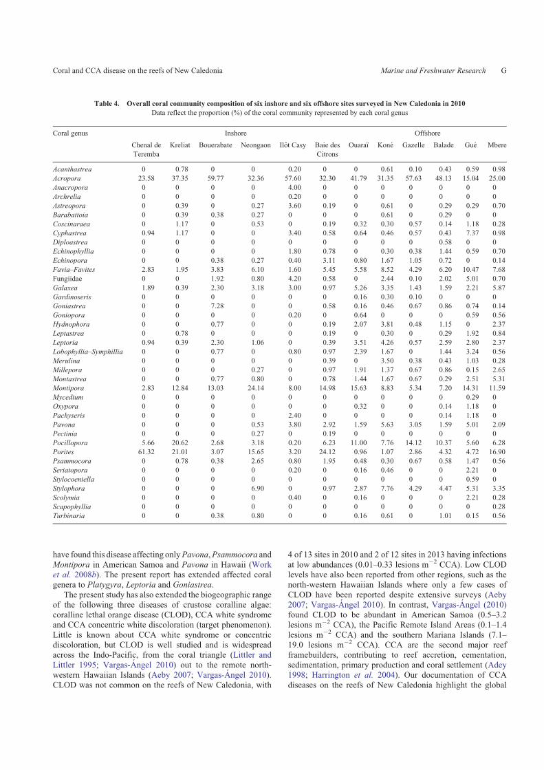
In 2010, we surveyed six inshore–offshore profiles and foundthat average coral cover did not differ significantly betweenoffshore and inshore reefs (Wilcoxon two-group test, Z¼ 0.40,
n¼ 12, P¼ 0.7; Table 3). However, offshore reefs had a sig-nificantly higher average colony density (Wilcoxon two-grouptest, Z¼ 2.8, n¼ 12, P¼ 0.005), number of coral genera
(Wilcoxon two-group test, Z¼ 2.7, n¼ 12, P¼ 0.008) andcrustose coralline algal cover (Wilcoxon two-group test,Z¼ 2.1, n¼ 12, P¼ 0.04; Table 3). There was a shift in theabundance and composition of coral genera found in the
Table 2. Mean (±s.e.) disease prevalence of coral diseases surveyed in
the Lagoon of New Caledonia in 2010 and 2013
Thirteen sites were surveyed in 2010 and 12 sites in 2013. Data include
diseased colonies only within transects and so will differ from frequency of
disease-occurrence data. White syndrome indicates a tissue-loss disease not
found associated with an obvious pathogen on histological analysis
Disease Average prevalence (%)
2010 2013
Acropora growth anomalies 0.02 (0.01) 0.06 (0.03)
Acropora white syndrome 0.04 (0.02) 0.03 (0.02)
Acropora ciliate infection 0.004 (0.004) 0.04 (0.03)
Astreopora white syndrome 0.25 (0.25) 0
Astreopora cyanobacterial infection 0 1.39 (1.39)
Faviid dark spot 0.21 (0.14) 0
Faviid growth anomalies 0.03 (0.03) 0.02 (0.02)
Galaxea white syndrome 0.04 (0.04) 0.02 (0.02)
Goniastrea dark spot 7.69 (7.69) 0
Goniastrea growth anomalies 0.25 (0.25) 0
Hydnophora growth anomalies 0 0.46 (0.46)
Hydnophora white syndrome 0 0.05 (0.05)
Leptoria dark spot 0.32 (0.32) 0
Montipora growth anomalies 0.04 (0.03) 0.06 (0.04)
Pachyseris bleached patch 0 0.35 (0.35)
Pavona dark spot 0.12 (0.09) 0.06 (0.04)
Pavona white syndrome 0.04 (0.04) 0
Platygyra dark spot 0.32 (0.32) 0
Porites bleached patch 0 0.65 (0.64)
Porites growth anomalies 0.27 (0.10) 0.23 (0.14)
Porites multi-focal pink spot 0.3 (0.13) 0.4 (0.19)
Porites white syndrome 0 0.29 (0.20)
Porites cyanobacterial infection 0 0.01 (0.01)
Turbinaria cyanobacterial infection 0.43 (0.43) 0
Turbinaria white syndrome 0.19 (0.19) 0
Table 1. Frequency of occurrence (FOC,%) of coral diseases surveyed
in New Caledonia in 2010 and 2013
Thirteen sites were surveyed in 2010 and 12 sites in 2013. White syndrome
indicates a tissue-loss disease not found associatedwith an obvious pathogen
on histological analysis
Disease FOC (%)
2010 2013
Acropora growth anomalies 23.1 50
Acropora white syndrome 46.2 25
Acropora ciliate infection 7.7 25
Astreopora white syndrome 7.7 0
Astreopora cyanobacterial infection 0 8.3
Faviid dark spot 23.1 0
Faviid growth anomalies 7.7 8.3
Galaxea white syndrome 7.7 8.3
Goniastrea dark spot 7.7 0
Goniastrea growth anomalies 7.7 0
Hydnophora growth anomalies 0 8.3
Hydnophora white syndrome 0 8.3
Leptoria dark spot 7.7 0
Montipora growth anomalies 23.1 25
Montipora cyanobacterial infection 7.7 8.3
Pachyseris bleached patch 0 8.3
Pachyseris white syndrome 7.7 0
Pavona dark spot 15.4 16.7
Pavona white syndrome 7.7 0
Pectinia white syndrome 7.7 0
Platygyra growth anomalies 0 16.7
Platygyra dark spot 7.7 0
Porites bleached patch 0 16.7
Porites growth anomalies 38.5 25
Porites multi-focal pink spot 53.8 41.7
Porites white syndrome 0 25
Porites cyanobacterial infection 0 8.3
Symphyllia dark spot 7.7 0
Turbinaria cyanobacterial infection 7.7 0
Turbinaria white syndrome 15.4 0
D Marine and Freshwater Research G. Aeby et al.
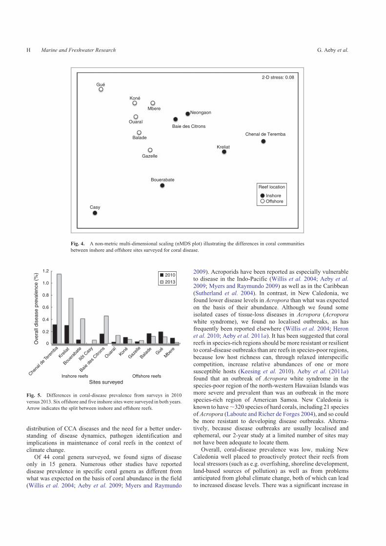
different habitats (ANOSIM, r¼ 0.33, P, 0.02; Table 4).SIMPER analysis showed that the coral genus, Porites,
explained the highest percentage (9.9%) of the differencesbetween inshore and offshore reefs. Porites comprised anaverage of 21.4% of the coral community on inshore reefs, ascompared with 5.1% on offshore reefs. An nMDS plot (Fig. 4)
showed two groupings (inshore vs offshore) and also revealedseveral inshore survey sites that were distinct. For example, theinshore reef, Chenal de Teremba, had eight scleractinian genera
found within transects, compared with the other five inshoresites with coral diversity ranging from 12 to 24 genera. The twoinshore sites, Bouerabate and Casy, had a low abundance of
Porites (3.1 and 3.2% respectively) compared with the otherfour sites, with Porites abundance ranging from 15.6% to 61.3%of the coral community.
Average disease prevalence did not differ between inshore
(mean 0.26% � 0.09 s.e.) and offshore reefs (mean 0.10% �0.02 s.e.) (Wilcoxon two-group test, Z¼ 0, n¼ 12, P¼ 1.0).However, inshore reefs had a significant increase in disease
prevalence from 0.17% (s.e. �0.06%) in 2010 to 0.57%(s.e. �0.18%) in 2013 (matched-pairs sign test, t¼ 3.13, n¼ 5,P¼ 0.03), whereas offshore reefs had no significant change,
with disease prevalence being 0.10% (s.e. �0.02%) in 2010
and 0.09% (s.e. �0.02%) in 2013 (matched-pairs sign test,t¼�0.59, n¼ 6, P¼ 0.58; Fig. 5).
Discussion
Here, we report on the first quantitative coral- and CCA-diseasestudy to be conducted on the reefs of the largest lagoon in the
world, i.e. the lagoon of NewCaledonia. During disease surveysin 2010 and 2013, we found a total of 30 different coral diseasesaffecting 15 coral genera and three CCA diseases. The number
of diseases we found is higher than reported from other regionsof the Indo-Pacific. Vargas-Angel and Wheeler (2009) reported10 diseases from across the USAPacific territories and affiliatedstates (Hawaii, American Samoa, Pacific Remote Island Areas,
Guam, northern Mariana Islands) and Muller et al. (2012)reported five diseases from Sulawesi, Indonesia. Differences innumbers of reported diseases are, in part, a result of differences
in nomenclature used among researchers, making regionalcomparisons difficult, and this has been pointed out elsewhere(Work et al. 2008a; Aeby et al. 2011b). Assigning a specific
name to a disease lesion in situ is difficult because there islimited information on coral-disease etiologies, ecologies andpathogen specificities. The ‘white syndromes’ are the classic
example of a lesion type that is now known to have differentunderlying etiologies, ecologies and differences in host speci-ficity, even though the gross lesions (i.e. lesion determinedunderwater) may look similar. For example, tissue-loss diseases
(white syndromes) in corals are associated with bacterialpathogens (Sussman et al. 2008; Luna et al. 2010; Ushijimaet al. 2012, 2014), chimera parasites (Work et al. 2011),
programmed cell death perhaps in response to pathogens(Ainsworth et al. 2006), as well as a variety of other organismssuch as algae, sponges, endolithic fungi and helminths (Work
and Aeby 2011; Work et al. 2012). In some regions, whitesyndrome can be seasonal (Willis et al. 2004; Dalton and Smith2006; Bruno et al. 2007), whereas this is not the case in otherregions (Aeby et al. 2009, 2010). From field patterns of disease
occurrence and evidence of direct transmission in the field, it issuggested that some white syndromes are host generalistsaffecting numerous coral genera (Willis et al. 2004; Dalton and
Smith 2006), whereas others affect a single coral genus (Aebyet al. 2010, 2011a). Thus, white syndrome is a non-specificlesion with multiple potential causes and host responses (Work
et al. 2012). Other diseases, such as black-band disease (BBD),have a more consistent microscopic presentation, with tissuedamage being associated with a microbial consortium visually
dominated by filamentous cyanobacteria, which is consistentacross numerous regions in both the Indo-Pacific (Frias-Lopezet al. 2003; Barneah et al. 2007; Sato et al. 2009) and theCaribbean (Rutzler and Santavy 1983; Richardson 2004). BBD
consistently affects a wide range of coral genera (Sutherlandet al. 2004) and becomes more prevalent during warm-watermonths (Voss and Richardson 2006; Sato et al. 2010). Until
more is known about the aetiology and ecology of other coraldiseases, researchers are advised to report diseases by hostgenera affected and lesion type (Work and Aeby 2006).
Some diseases in New Caledonia, such as Porites andAcropora growth anomalies, are widespread across the Indo-Pacific (Aeby et al. 2011c); however, our findings represent a
0
10
20
30
40
50
60
70
0
5
10
15
20
25
30
35
40
45
Gen
eric
abu
ndan
ce (
%)
Coral genera
Acropora
Acropora
Astreopora
CoscinareaFavia
Favia
Galaxea
Galaxea
Hydnophora
Oxypora
Pachyseris
Goniastrea
Leptoria
Montipora
Montipora
Pavona
Pavona
Platygyra
Porites
Porites
Turbinaria
2010
0
10
20
30
40
50
60
70
0
5
10
15
20
25
30
35
40
45
Pro
port
ion
of d
isea
se le
sion
s (%
)
2013
Coral communityDisease lesions
Coral communityDisease lesions
Fig. 2. Relationship between overall abundance of coral genera within
transects and proportion of corals with signs of disease (lesions). Top graph
shows data from surveys conducted in 2013 and bottom graph from surveys
in 2010.
Coral and CCA disease on the reefs of New Caledonia Marine and Freshwater Research E
range extension for other diseases. For example, this is the firstreport of growth anomalies onPlatygyra andHydnophora (host-range extension).We also discovered the first cases of trematode
infection in Porites outside of Hawaii (biogeographic range
extension). In Hawaii, Porites trematodiasis is a disease causedby a parasitic digenetic trematode that presents as swollen, pink
spots on poritid corals (Aeby 1998, 2003). Not all cases ofmulti-focal pink spots on Porites are due to trematode infections(Benzoni et al. 2010), and so, we initially described lesions as
Porites multi-focal pink spot. Subsequently, using histology,Work et al. (2014) found that some, but not all, cases of Poritesmulti-focal pink spot from our surveys contained trematodes.Interestingly, compared with Porites trematodiasis in Hawaii,
lesions onPorites in NewCaledonia had reduced tissue swellingand frequently had a small bit of sediment adhering to the centreof the pink spot, which seldom occurs in Hawaii (Fig. S1,
available as Supplementary Material for this paper). Worket al. (2014) also found that the trematodes in Porites in NewCaledonia were not encysted, as were those found in Hawaiian
corals (Cheng and Wong 1974; Aeby 1998); therefore, it isunknown whether the species of trematodes or disease ecologyare similar among regions.
Another disease that has been found in few other places isdark spot, which we found in six different coral genera. In otherregions, this lesion is associated with overgrowth of coral tissueby euendolithic fungi and was termed endolithic hypermycosis
(Work et al. 2008b). Work et al. (2014) found that the dark-spotsamples collected in New Caledonia also had a consistentdiagnosis of euendolithic fungal invasion across the coral genera
we sampled (endolithic hypermycosis). Our report has increasedboth the number of regions where this disease been found tooccur as well as the number of genera affected. Prior studies
0
1
2
3
4
5
Mea
n sc
ore
on s
cale
of 0
to 1
0
Coral diseases
Acropora
WS
Porites
WS
Astreopora
cyan
o
Turbinaria
WS
Astreopora
WS
Galaxea
WS
Hydnophora
WS
Pavona
WS
Pachyseris
WS
Pectinia
WS
Turbinaria
cyan
o
Porites
MFPS
Porites
GA
Acropora
cilia
te
Montipora
cyan
o
Porites
cyan
o
Acropora
GA
Porites
bl p
atch
Leptoria
DS
Platygyra
DS
Montipora
GA
Hydnophora
GA
Pavona
DS
Faviid
DS
Pachyseris
bl pa
tch
Goniastrea
GA
Platygyra
GAFaviid
GA
Goniastrea
DS
Fig. 3. Mean score on a scale of 0–10 of the potential ecological impact for each coral disease found on the reefs of NewCaledonia.
Scoreswere the average of threemetrics (frequency of occurrence, prevalence, virulence) relevant in determing the harm each disease
could cause on coral reefs. (WS, white syndrome; cyano, cyanobacterial infection; MFPS, multi-focal pink spot; GA, growth
anomaly; ciliate, ciliate infection; bl patch, bleached patch; DS, dark spot.)
Table 3. Comparisons of average benthic cover of inshore versus
offshore sites surveyed in 2010 in New Caledonia
Two 25-m belt transects were surveyed at each site; CCA, crustose coralline
algae
Site Percentage
coral
cover
Colonies
per square
metre
Percentage
CCA
cover
Number of
coral genera
Offshore
Ouaraı 46.1 12.5 19.6 24
Kone 46.1 13.1 31.4 28
Gazelle 46.1 21.0 38.2 21
Balade 27.5 13.9 13.7 27
Gue 48.0 13.6 1.0 29
Mbere 8.8 14.3 2.9 27
Average 37.1 14.7 17.8 26
Inshore
Chenal de Teremba 17.6 2.1 8.8 8
Kreliat 11.8 5.1 2.9 14
Bouerabate 7.8 5.2 0.0 14
Neongaon 47.1 7.5 2.0 17
Ilot Casy 83.3 10.0 0.0 19
Baie des Citrons 33.3 10.3 0.0 23
Average 33.5 6.7 2.3 15.8
F Marine and Freshwater Research G. Aeby et al.
have found this disease affecting onlyPavona,Psammocora and
Montipora in American Samoa and Pavona in Hawaii (Worket al. 2008b). The present report has extended affected coralgenera to Platygyra, Leptoria and Goniastrea.
The present study has also extended the biogeographic rangeof the following three diseases of crustose coralline algae:coralline lethal orange disease (CLOD), CCA white syndromeand CCA concentric white discoloration (target phenomenon).
Little is known about CCA white syndrome or concentricdiscoloration, but CLOD is well studied and is widespreadacross the Indo-Pacific, from the coral triangle (Littler and
Littler 1995; Vargas-Angel 2010) out to the remote north-western Hawaiian Islands (Aeby 2007; Vargas-Angel 2010).CLOD was not common on the reefs of New Caledonia, with
4 of 13 sites in 2010 and 2 of 12 sites in 2013 having infections
at low abundances (0.01–0.33 lesions m�2 CCA). Low CLODlevels have also been reported from other regions, such as thenorth-western Hawaiian Islands where only a few cases of
CLOD have been reported despite extensive surveys (Aeby2007; Vargas-Angel 2010). In contrast, Vargas-Angel (2010)found CLOD to be abundant in American Samoa (0.5–3.2lesions m�2 CCA), the Pacific Remote Island Areas (0.1–1.4
lesions m�2 CCA) and the southern Mariana Islands (7.1–19.0 lesions m�2 CCA). CCA are the second major reefframebuilders, contributing to reef accretion, cementation,
sedimentation, primary production and coral settlement (Adey1998; Harrington et al. 2004). Our documentation of CCAdiseases on the reefs of New Caledonia highlight the global
Table 4. Overall coral community composition of six inshore and six offshore sites surveyed in New Caledonia in 2010
Data reflect the proportion (%) of the coral community represented by each coral genus
Coral genus Inshore Offshore
Chenal de
Teremba
Kreliat Bouerabate Neongaon Ilot Casy Baie des
Citrons
Ouaraı Kone Gazelle Balade Gue Mbere
Acanthastrea 0 0.78 0 0 0.20 0 0 0.61 0.10 0.43 0.59 0.98
Acropora 23.58 37.35 59.77 32.36 57.60 32.30 41.79 31.35 57.63 48.13 15.04 25.00
Anacropora 0 0 0 0 4.00 0 0 0 0 0 0 0
Archrelia 0 0 0 0 0.20 0 0 0 0 0 0 0
Astreopora 0 0.39 0 0.27 3.60 0.19 0 0.61 0 0.29 0.29 0.70
Barabattoia 0 0.39 0.38 0.27 0 0 0 0.61 0 0.29 0 0
Coscinaraea 0 1.17 0 0.53 0 0.19 0.32 0.30 0.57 0.14 1.18 0.28
Cyphastrea 0.94 1.17 0 0 3.40 0.58 0.64 0.46 0.57 0.43 7.37 0.98
Diploastrea 0 0 0 0 0 0 0 0 0 0.58 0 0
Echinophyllia 0 0 0 0 1.80 0.78 0 0.30 0.38 1.44 0.59 0.70
Echinopora 0 0 0.38 0.27 0.40 3.11 0.80 1.67 1.05 0.72 0 0.14
Favia–Favites 2.83 1.95 3.83 6.10 1.60 5.45 5.58 8.52 4.29 6.20 10.47 7.68
Fungiidae 0 0 1.92 0.80 4.20 0.58 0 2.44 0.10 2.02 5.01 0.70
Galaxea 1.89 0.39 2.30 3.18 3.00 0.97 5.26 3.35 1.43 1.59 2.21 5.87
Gardinoseris 0 0 0 0 0 0 0.16 0.30 0.10 0 0 0
Goniastrea 0 0 7.28 0 0 0.58 0.16 0.46 0.67 0.86 0.74 0.14
Goniopora 0 0 0 0 0.20 0 0.64 0 0 0 0.59 0.56
Hydnophora 0 0 0.77 0 0 0.19 2.07 3.81 0.48 1.15 0 2.37
Leptastrea 0 0.78 0 0 0 0.19 0 0.30 0 0.29 1.92 0.84
Leptoria 0.94 0.39 2.30 1.06 0 0.39 3.51 4.26 0.57 2.59 2.80 2.37
Lobophyllia–Symphillia 0 0 0.77 0 0.80 0.97 2.39 1.67 0 1.44 3.24 0.56
Merulina 0 0 0 0 0 0.39 0 3.50 0.38 0.43 1.03 0.28
Millepora 0 0 0 0.27 0 0.97 1.91 1.37 0.67 0.86 0.15 2.65
Montastrea 0 0 0.77 0.80 0 0.78 1.44 1.67 0.67 0.29 2.51 5.31
Montipora 2.83 12.84 13.03 24.14 8.00 14.98 15.63 8.83 5.34 7.20 14.31 11.59
Mycedium 0 0 0 0 0 0 0 0 0 0 0.29 0
Oxypora 0 0 0 0 0 0 0.32 0 0 0.14 1.18 0
Pachyseris 0 0 0 0 2.40 0 0 0 0 0.14 1.18 0
Pavona 0 0 0 0.53 3.80 2.92 1.59 5.63 3.05 1.59 5.01 2.09
Pectinia 0 0 0 0.27 0 0.19 0 0 0 0 0 0
Pocillopora 5.66 20.62 2.68 3.18 0.20 6.23 11.00 7.76 14.12 10.37 5.60 6.28
Porites 61.32 21.01 3.07 15.65 3.20 24.12 0.96 1.07 2.86 4.32 4.72 16.90
Psammocora 0 0.78 0.38 2.65 0.80 1.95 0.48 0.30 0.67 0.58 1.47 0.56
Seriatopora 0 0 0 0 0.20 0 0.16 0.46 0 0 2.21 0
Stylocoeniella 0 0 0 0 0 0 0 0 0 0 0.59 0
Stylophora 0 0 0 6.90 0 0.97 2.87 7.76 4.29 4.47 5.31 3.35
Scolymia 0 0 0 0 0.40 0 0.16 0 0 0 2.21 0.28
Scapophyllia 0 0 0 0 0 0 0 0 0 0 0 0.28
Turbinaria 0 0 0.38 0.80 0 0 0.16 0.61 0 1.01 0.15 0.56
Coral and CCA disease on the reefs of New Caledonia Marine and Freshwater Research G
distribution of CCA diseases and the need for a better under-standing of disease dynamics, pathogen identification and
implications in maintenance of coral reefs in the context ofclimate change.
Of 44 coral genera surveyed, we found signs of diseaseonly in 15 genera. Numerous other studies have reported
disease prevalence in specific coral genera as different fromwhat was expected on the basis of coral abundance in the field(Willis et al. 2004; Aeby et al. 2009; Myers and Raymundo
2009). Acroporids have been reported as especially vulnerableto disease in the Indo-Pacific (Willis et al. 2004; Aeby et al.
2009; Myers and Raymundo 2009) as well as in the Caribbean(Sutherland et al. 2004). In contrast, in New Caledonia, wefound lower disease levels in Acropora than what was expected
on the basis of their abundance. Although we found someisolated cases of tissue-loss diseases in Acropora (Acroporawhite syndrome), we found no localised outbreaks, as hasfrequently been reported elsewhere (Willis et al. 2004; Heron
et al. 2010; Aeby et al. 2011a). It has been suggested that coralreefs in species-rich regions should be more resistant or resilientto coral-disease outbreaks than are reefs in species-poor regions,
because low host richness can, through relaxed interspecificcompetition, increase relative abundances of one or moresusceptible hosts (Keesing et al. 2010). Aeby et al. (2011a)
found that an outbreak of Acropora white syndrome in thespecies-poor region of the north-western Hawaiian Islands wasmore severe and prevalent than was an outbreak in the more
species-rich region of American Samoa. New Caledonia isknown to have,320 species of hard corals, including 21 speciesof Acropora (Laboute and Richer de Forges 2004), and so couldbe more resistant to developing disease outbreaks. Alterna-
tively, because disease outbreaks are usually localised andephemeral, our 2-year study at a limited number of sites maynot have been adequate to locate them.
Overall, coral-disease prevalence was low, making NewCaledonia well placed to proactively protect their reefs fromlocal stressors (such as e.g. overfishing, shoreline development,
land-based sources of pollution) as well as from problemsanticipated from global climate change, both of which can leadto increased disease levels. There was a significant increase in
Gué
Koné
Mbere
Ouaraï
Balade
Neongaon
Baie des Citrons
Gazelle
Bouerabate
Casy
Kreliat
Chenal de Teremba
Reef location
InshoreOffshore
2-D stress: 0.08
Fig. 4. A non-metric multi-dimensional scaling (nMDS plot) illustrating the differences in coral communities
between inshore and offshore sites surveyed for coral disease.
0
Sites surveyedInshore reefs
2010
2013
Offshore reefsChena
l de T
erem
ba
Krelia
t
Bouer
abat
e
Ilôt C
asy
Baie d
es C
itron
s
Ouara
ï
Koné
Gazell
e
Balade Gué
Mbe
re
0.2
0.4
0.6
Ove
rall
dise
ase
prev
alen
ce (
%)
0.8
1.0
1.2
Fig. 5. Differences in coral-disease prevalence from surveys in 2010
versus 2013. Six offshore and five inshore sites were surveyed in both years.
Arrow indicates the split between inshore and offshore reefs.
H Marine and Freshwater Research G. Aeby et al.
disease on inshore reefs between surveys in 2010 and 2013;however, it is unclear whether this increase could be ascribed to
background temporal or spatial variation, because surveys werenot on permanently marked transects. The consistent increase indisease between 2010 and 2013 across numerous sites was
noticeable, and it would be useful if disease surveys wereincorporated as part of current or future monitoring efforts toget a more concrete assessment of longer-term trends. This also
points out the critical need to document baseline disease levelson reefs before changes occur from chronic anthropogenic stressand climate change, with the latter imposing its own set ofimpacts on coral reefs (Hoegh-Guldberg et al. 2007; Pandolfi
et al. 2011). For example, Willis et al. (2004) found that thenumber of cases of tissue-loss diseases (white syndrome) atmonitoring sites on the Great Barrier Reef increased 20-fold
between 1998 and 2002–2003. Hence, the definition of ‘normal’disease levels on those reefs would be very different had theyconducted their very first disease survey in 2002, and this
highlights the problem of ‘shifting baselines’. Numerousregions throughout the Indo-Pacific still lack baseline diseasestudies and this is problematic.
The potential ecological impact of disease on coral reefs
would depend on the spatial distribution, prevalence and thedegree of virulence of specific diseases. Chronic diseases, suchas growth anomalies orPorites trematodiasis, can bewidespread
and prevalent, but result only in reduced growth or reproduction(Aeby 1993; Stimson 2011), whereas other diseases, such asblack-band disease, result in slow but recurrent tissue loss
(Kuta and Richardson 1996; Sato et al. 2009). Acute andsubacute tissue-loss diseases are the most virulent, resulting insignificant colonymortality, reported aswhite syndromes across
the Indo-Pacific (Aeby 2005; Dalton and Smith 2006; Sussmanet al. 2008) and white plague or white band from the Caribbean(Richardson 1998; Aronson and Precht 2001). The reefs of theCaribbean have already lost significant coral cover (Gardner
et al. 2003) in large part from coral disease (Sutherland et al.
2004), and a similar pattern of coral loss is arising in the Indo-Pacific (Bruno and Selig 2007; De’ath et al. 2012). Knowing
which coral diseasesmay bemost damaging to reefs would be animportant first step in focussing limited resources and directingfuture studies. We used integrative scoring of three disease
metrics (frequency of occurrence, prevalence, virulence) and arelative ranking scheme to compare the potential ecologicalimpact of the diseases encountered in New Caledonia. On thebasis of this metric, the top five diseases of concern were white
syndrome in Acropora, Porites, Astreopora and Turbinaria andcyanobacterial infection in Turbinaria. Not surprising, all fiveare tissue-loss diseases, resulting in partial to total colony
mortality. Use of a ranking scheme to communicate relativethreat from different diseases facilitates reporting threats toresource managers and allows cross-regional comparisons.
We found that inshore reefs had lower densities of coralcolonies, lower species diversity and a reduced CCA cover.Fenner (2011) also found lower coral-species richness on
inshore reefs than on outer barrier reefs in New Caledonia.Inshore reefs also showed a shift in the structure of coralcommunities, with sediment-tolerant corals such as Porites
found to be more abundant. Evidence of sediment stress was
common in inshore reefs, along with reduced water quality
(poor visibility and layers of sediments accumulated on the topof coral colonies), but was never observed on the offshore reefs.
The serious threat that sedimentation poses to New Caledonia’sinshore reefs has been mentioned elsewhere (Lasne 2007;Fenner and Muir 2009; McKenna 2011), with open-surface
nickel mining being identified as one of the major sources ofsediment (Fenner and Muir 2009). Many studies have shownthat land-based pollution, such as sedimentation, nutrient
enrichment and turbidity, can severely degrade coral reefs atthe local scale (reviewed by Fabricius 2005). Improvement inwater quality may be an effective way to improve overall coral-reef integrity and should be considered a priority for reef
management.The degree of environmental stress to inshore reefs would
vary depending on, for example, their proximity to rivermouths,
adjacent shoreline development, density of human populationsin adjacent watersheds. Accordingly, we found that not allinshore reefs appeared to be affected by terrestrial runoff and
this was reflected in the higher variability in the structure ofcoral communities found among the inshore reefs than amongthe offshore reefs. However, inshore reefs, by spatial proximity,would also be more prone to other types of anthropogenic
damage. For example, one of our survey sites, Casy, showedfew signs of sediment stress and was dominated by branchingacroporids. Disease levels were also low; however, in 2013, we
found a large area of the reef reduced to a pile of rubble, fromwhat appeared to be mechanical breakage (probably anchordamage). Coral cover declined at Casy from 83.3% in 2010 to
63.4% in 2013.
Acknowledgements
The South Pacific Center (CPS) through the program CRISP (Coral Reef
Initiative in the Pacific) funded this study in 2010, and the FrenchMinistry of
Ecology through the program IFRECOR (Initiative Francaise pour les Recifs
Coralliens) in 2013. We also deeply thank the Institut de Recherche pour le
Developpement (IRD) for its financial support in 2013.We thank the Center
of IRD of Noumea for allowing us to use IRD boat and diving facilities.
We thank Miguel Clarque, Philippe Naudin, Samuel Tereua, Eric Folcher,
Bertrand Bourgeois and Armelle Renaud for their help. We also thank the
local authorities for delivering permits of collection and the Aquarium de
Noumea (Richard Farmann) and the Centre d’Initiation a l’Environnement
for their support and help. Mention of products or trade names do not imply
endorsement by the US Government.
References
Adey, W. H. (1998). Coral reefs: algal structured and mediated ecosystems
in shallow, turbulent, alkaline waters. Journal of Phycology 34,
393–406. doi:10.1046/J.1529-8817.1998.340393.X
Aeby, G. S. (1993). The potential effect the ability of a coral intermediate
host to regenerate may have had on the evolution of its associationwith a
marine parasite. In ‘Proceedings of the 7th International Coral Reef
Symposium, 22–27 June 1992, Guam’, Vol. 2, pp. 809–815. (University
of Guam Press, Guam.)
Aeby, G. S. (1998). A digenean metacercaria from the reef coral, Porites
compressa, experimentally identified as Podocotyloides stenometra.
The Journal of Parasitology 84, 1259–1261. doi:10.2307/3284684
Aeby, G. (2003). Corals in the genusPorites are susceptible to infection by a
larval trematode. Coral Reefs 22, 216. doi:10.1007/S00338-003-0310-9
Aeby, G. S. (2005). Outbreak of coral disease in the northwestern Hawaiian
Islands. Coral Reefs 24, 481. doi:10.1007/S00338-005-0493-3
Coral and CCA disease on the reefs of New Caledonia Marine and Freshwater Research I
Aeby, G. S. (2007). First record of coralline lethal orange disease (CLOD) in
the northwestern Hawaiian Islands. Coral Reefs 26, 385. doi:10.1007/
S00338-007-0223-0
Aeby, G.,Work, T., Fenner, D., andDiDonato, E. (2009). Coral and crustose
coralline algae disease on the reefs of American Samoa. In ‘Proceedings
of the 11th International Coral Reef Symposium, 7–11 July 2008’, pp.
197–201. (National Coral Reef Institute (NCRI), Nova Southeastern
University Oceanographic Center, Florida.)
Aeby, G. S., Ross,M.,Williams, G. J., Lewis, T. D., andWork, T.M. (2010).
Disease dynamics of Montipora white syndrome within Kaneohe Bay,
Oahu, Hawaii: distribution, seasonality, virulence, and transmission.
Diseases of Aquatic Organisms 91, 1–8. doi:10.3354/DAO02247
Aeby, G. S., Bourne, D. G., Wilson, B., and Work, T. M. (2011a). Coral
diversity and the severity of disease outbreaks: a cross-regional compar-
ison of Acropora white syndrome in a species-rich region (American
Samoa) with a species-poor region (northwestern Hawaiian Islands).
Journal of Marine Biology 2011, 490198. doi:10.1155/2011/490198
Aeby,G. S.,Williams, G. J., Franklin, E. C., Kenyon, J., Cox, E. F., Coles, S.,
and Work, T. M. (2011b). Patterns of coral disease across the Hawaiian
archipelago: relating disease to environment. PLoS One 6(5), e20370.
doi:10.1371/JOURNAL.PONE.0020370
Aeby,G. S.,Williams,G. J., Franklin, E., Haapkyla, J., Harvell, C. D.,Neale,
S., Page, C., Raymundo, L., Vargas-Angel, B., Willis, B., Work, T., and
Davy, S. (2011c). Growth anomalies on the coral genera Acropora and
Porites are strongly associated with host density and human population
size. PLoS One 6(2), e16887. doi:10.1371/JOURNAL.PONE.0016887
Ainsworth, T., Kvennefors, E., Blackall, L., Fine, M., and Hoegh-Guldberg,
O. (2006). Disease and cell death in white syndrome of acroporid corals
on the Great Barrier Reef. Marine Biology 151, 19–29. doi:10.1007/
S00227-006-0449-3
Andrefouet, S., Cabioch, G., Flamand, B., and Pelletier, B. (2006). The
diversity of New Caledonia coral reef geomorphology and genetic
processes: a synthesis from optical remote sensing, coring and acoustic
multi-beamobservations. In’ CompendiumofMarine Species fromNew
Caledonia. Scientific Papers and Techniques ll7’. (Eds C. E. Payri and B.
Richer de Forges.) pp. 33–49. (Institute of Research for Development:
Noumea.)
Aronson, R. B., and Precht, W. F. (2001). White-band disease and the
changing face of Caribbean coral reefs. In ‘The Ecology and Etiology of
Newly EmergingMarineDiseases’. (Ed. J. Porter.) pp. 25–38. (Springer:
Dordrecht, The Netherlands.)
Barneah, O., Ben-Dov, E., Kramarsky-Winter, E., andKushmaro,A. (2007).
Characterization of black band disease in Red Sea stony corals. Envi-
ronmental Microbiology 9, 1995–2006. doi:10.1111/J.1462-2920.2007.
01315.X
Benzoni, F., Galli, P., and Pichon, M. (2010). Pink spots on Porites: not
always a coral disease. Coral Reefs 29, 153. doi:10.1007/S00338-009-
0571-Z
Bonvallot, J., Gay, J. C., and Habert, E. (2012). ‘Atlas of New Caledonia.’
(Marseille-Noumea, IRD – Congress New Caledonia: IRD (Institut de
Recherche pour le Developpement), Location: Marseille, France.)
Bruno, J. F., and Selig, E. R. (2007). Regional decline of coral cover in the
Indo-Pacific: timing, extent, and subregional comparisons. PLoS One
2(8), e711. doi:10.1371/JOURNAL.PONE.0000711
Bruno, J. F., Selig, E. R., Casey, K. S., Page, C. A., Willis, B. L., Harvell,
C. D., Sweatman, H., and Melendy, A. M. (2007). Thermal stress and
coral cover as drivers of coral disease outbreaks. PLoS Biology 5(6),
e124. doi:10.1371/JOURNAL.PBIO.0050124
Cheng, T. C., and Wong, A. K. (1974). Chemical, histochemical, and
histopathological studies on corals, Porites spp., parasitized by trema-
tode metacercariae. Journal of Invertebrate Pathology 23, 303–317.
doi:10.1016/0022-2011(74)90095-0
Colwell, R. (2004). Infectious disease and environment: cholera as a para-
digm for waterborne disease. International Microbiology 7, 285–289.
Dalton, S., and Smith, S. (2006). Coral disease dynamics at a subtropical
location, Solitary IslandsMarine Park, easternAustralia.Coral Reefs 25,
37–45. doi:10.1007/S00338-005-0039-8
De’ath, G., Fabricius, K. E., Sweatman, H., and Puotinen, M. (2012). The
27-year decline of coral cover on the Great Barrier Reef and its
causes. Proceedings of the National Academy of Sciences of the
United States of America 109(44), 17 995–17 999. doi:10.1073/
PNAS.1208909109
Fabricius, K. E. (2005). Effects of terrestrial runoff on the ecology of corals
and coral reefs: review and synthesis. Marine Pollution Bulletin 50,
125–146. doi:10.1016/J.MARPOLBUL.2004.11.028
Fenner, D. (2011). Reef corals of the eastern lagoon (Touho–Ponerihouen)
of Grande-Terre, New Caledonia. In ‘A Rapid Marine Biodiversity
Assessment of the Northeastern Lagoon from Touhou to Ponerihouen,
ProvinceNord,NewCaledonia’. (Eds S.A.McKenna,M.A.Hosken and
N. Baillon.) pp. 141–160. RAP Bulletin of Biological Assessment 62.
(Conservation International: Arlington, VA.)
Fenner, D., and Muir, P. (2009). Reef corals of the northwestern lagoon of
Grande-Terre, New Caledonia. In ‘Rapid Marine Biodiversity Assess-
ment of the Coral Reefs of the Northwest Lagoon, between Yandee and
Koumac, ProvinceNord,NewCaledonia’. (Ed. S.McKenna.) pp. 18–32.
RAPBulletin of Biological Assessment 53. (Conservation International:
Arlington, VA.)
Fernandez, J. M., Ouillon, S., Chevillon, C., Douillet, P., Fichez, R., and
Gendre, R. L. (2006). A combined modeling and geochemical study of
the fate of terrigenous inputs from mixed natural and mining sources in
a coral reef lagoon (New Caledonia). Marine Pollution Bulletin 52,
320–331. doi:10.1016/J.MARPOLBUL.2005.09.010
Frias-Lopez, J., Bonheyo, G., Jin, Q., and Fouke, B. W. (2003). Cyanobac-
teria associated with coral black band disease in Caribbean and Indo-
Pacific reefs. Applied and Environmental Microbiology 69, 2409–2413.
doi:10.1128/AEM.69.4.2409-2413.2003
Gardner, T. A., Cote, I. M., Gill, J. A., Grant, A., and Watkinson, A. R.
(2003). Long-term region-wide declines in Caribbean corals. Science
301, 958–960. doi:10.1126/SCIENCE.1086050
Green, E. P., and Bruckner, A. W. (2000). The significance of coral disease
epizootiology for coral reef conservation. Biological Conservation 96,
347–361. doi:10.1016/S0006-3207(00)00073-2
Harrington, L., Fabricius, K., De’ath, G., and Negri, A. (2004). Recognition
and selection of settlement substrata determine post-settlement survival
in corals. Ecology 85, 3428–3437. doi:10.1890/04-0298
Harvell, C. D., Jordan-Dahlgren, E., Merkel, S., Rosenberg, E., Raymundo,
L., Smith, G., Weil, E., and Willis, B. (2007). Coral disease, environ-
mental drivers, and the balance between coral and microbial associates.
Oceanography 20, 172–195. doi:10.5670/OCEANOG.2007.91
Heron, S. F., Willis, B. L., Skirving, W. J., Eakin, C. M., Page, C. A., and
Miller, I. R. (2010). Summer hot snaps and winter conditions: modeling
white syndrome outbreaks on Great Barrier Reef Corals. PLoS One 5(8),
e12210. doi:10.1371/JOURNAL.PONE.0012210
Hoegh-Guldberg, O., Mumby, P. J., Hooten, A. J., Steneck, R. S., Green-
field, P., Gomez, E., Harvell, C. D., Sale, P. F., Edwards, A. J., Caldeira,
K., Knowlton, N., Eakin, C. M., Iglesias-Prieto, R., Muthiga, N., Brad-
bury, R. H., Dubi, A., and Hatziolos, M. E. (2007). Coral reefs under
rapid climate change and ocean acidification. Science 318, 1737–1742.
doi:10.1126/SCIENCE.1152509
Hughes, T. P., Baird, A. H., Bellwood, D. R., Card, M., Connolly, S. R.,
Folke, C., Grosberg, R., Hoegh-Guldberg, O., Jackson, J., Kleypas, J.,
Lough, J.,Marshall, P., Nystrom,M., Palumbi, S., Pandolfi, J., Rosen, B.,
and Roughgarden, J. (2003). Climate change, human impacts, and the
resilience of coral reefs. Science 301, 929–933. doi:10.1126/SCIENCE.
1085046
Jokiel, P. L., andRodgers,K. S. (2007). Ranking coral ecosystem ‘health and
value’ for the Islands of the Hawaiian archipelago.Pacific Conservation
Biology 13, 60.
J Marine and Freshwater Research G. Aeby et al.
Keesing, F., Belden, L. K., Daszak, P., Dobson, A., Harvell, C. D., Holt,
R. D., Hudson, P., Jolles, A., Jones, K., Mitchell, C., Myers, S., Bogich,
T., and Ostfeld, R. S. (2010). Impacts of biodiversity on the emergence
and transmission of infectious diseases. Nature 468, 647–652.
doi:10.1038/NATURE09575
Kuta, K., and Richardson, L. (1996). Abundance and distribution of black
band disease on coral reefs in the northern Florida Keys. Coral Reefs
15, 219–223. doi:10.1007/BF01787455
Laboute, P., and Richer de Forges, B. (2004). ‘Lagoons and Reefs of New
Caledonia. Editions Catherine Ledru Noumea.’ (Research Institute for
Development: Noumea, New Caledonia.)
Lasne, G. (2007). ‘Les Coraux de la Nouvelle-Caldonie: Synthese Biblio-
graphique. Coral Reef Initiative for the South Pacific (CRISP).’ (Institut
de Recherche pour le Developpement (IRD): Noumea.)
Littler,M., andLittler, D. (1995). Impact of CLODpathogen on Pacific coral
reefs. Science 267, 1356–1360. doi:10.1126/SCIENCE.267.5202.1356
Luna, G. M., Bongiorni, L., Gili, C., Biavasco, F., and Danovaro, R. (2010).
Vibrio harveyi as a causative agent of the white syndrome in tropical
stony corals. Environmental Microbiology Reports 2, 120–127.
doi:10.1111/J.1758-2229.2009.00114.X
McKenna, S. A. (2009). Reef condition. In ‘Rapid Marine Biodiversity
Assessment of the Coral Reefs of the Northwest Lagoon, between
Yandee and Koumac, ProvinceNord, NewCaledonia’. In ‘RAPBulletin
of Biological Assessment 53’. (Ed. S. McKenna.) pp. 61–69. (Conser-
vation International: Arlington, VA.)
McKenna, S. A. (2011). Reef condition: Touho, Poindimie, and Poneri-
houen. In ‘A RapidMarine Biodiversity Assessment of the Northeastern
Lagoon from Touho to Ponerihouen, Province Nord, New Caledonia’.
(Eds S. A. McKenna, M. A. Hosken and N. Baillon.) pp. 194–204. RAP
Bulletin of Biological Assessment 62. (Conservation International:
Arlington, VA.)
Muller, E. M., Raymundo, L. J., Willis, B. L., Haapkyla, J., Yusuf, S.,
Wilson, J. R., and Harvell, D. C. (2012). Coral health and disease in the
Spermonde Archipelago and Wakatobi, Sulawesi. Journal of Indonesia
Coral Reefs 1, 147–159.
Myers, R., and Raymundo, L. (2009). Coral disease in Micronesian reefs: a
link between disease prevalence and host abundance. Diseases of
Aquatic Organisms 87, 97–104. doi:10.3354/DAO02139
Pandolfi, J. M., Connolly, S. R., Marshall, D. J., and Cohen, A. L. (2011).
Projecting coral reef futures under global warming and ocean acidifica-
tion. Science 333, 418–422. doi:10.1126/SCIENCE.1204794
Payri, C. E., and Richer de Forges, B. (2006). ‘Compendium of Marine
Species from New Caledonia. Scientific Papers and Techniques II7.’
(Institute of Research for Development: Noumea.)
Richardson, L. L. (1998). Coral diseases: what is really known? Trends
in Ecology & Evolution 13, 438–443. doi:10.1016/S0169-5347(98)
01460-8
Richardson, L. L. (2004). Black band disease. In ‘Coral Health andDisease’.
(Eds E. Rosenberg and Y. Loya.) pp. 325–336. (Springer-Verlag:
Berlin.)
Rutzler, K., and Santavy, D. L. (1983). The black band disease of Atlantic
reef corals.Marine Ecology 4, 301–319. doi:10.1111/J.1439-0485.1983.
TB00116.X
Sato, Y., Bourne, D. G., and Willis, B. L. (2009). Dynamics of seasonal
outbreaks of black band disease in an assemblage ofMontipora species
at Pelorus Island (Great Barrier Reef, Australia). Proceedings of the
Royal Society B: Biological Sciences 276, 2795–2803.
Sato, Y., Willis, B. L., and Bourne, D. G. (2010). Successional changes in
bacterial communities during the development of black band disease
on the reef coral, Montipora hispida. The ISME Journal 4, 203–214.
doi:10.1038/ISMEJ.2009.103
Schutte, V. G., Selig, E. R., and Bruno, J. F. (2010). Regional spatio-
temporal trends in Caribbean coral reef benthic communities. Marine
Ecology Progress Series 402, 115–122. doi:10.3354/MEPS08438
Stimson, J. (2011). Ecological characterization of coral growth anomalies on
Porites compressa in Hawai‘i. Coral Reefs 30, 133–142. doi:10.1007/
S00338-010-0672-8
Sussman, M., Willis, B., Victor, S., and Bourne, D. (2008). Coral pathogens
identified for white syndrome (WS) epizootic in the Indo-Pacific. PLoS
One 3(6), e2393. doi:10.1371/JOURNAL.PONE.0002393
Sutherland, K., Porter, J., and Torres, C. (2004). Disease and immunity in
Caribbean and Indo-Pacific zooxanthellate corals. Marine Ecology
Progress Series 266, 273–302. doi:10.3354/MEPS266273
Tribollet, A. (2008). The boringmicroflora inmodern coral reef ecosystems:
a review of its roles. In ‘Current Developments in Bioerosion’. (Eds
M. Wisshak and L. Tapanila.) pp. 67–94. (Springer-Verlag: Berlin.)
Ushijima, B., Smith, A., Aeby, G. S., and Callahan, S. M. (2012). Vibrio
owensii strain OCN002 induces the tissue loss diseaseMontipora white
syndrome in the Hawaiian rice coral Montipora capitata. PLoS One
7(10), e46717. doi:10.1371/JOURNAL.PONE.0046717
Ushijima, B., Videau, P., Burger, A., Shore-Maggio, A., Runyon, C., Sudek,
M., Aeby, G., and Callahan, S. (2014). Vibrio coralliilyticus strain
OCN008 is an etiological agent of acute Montipora white syndrome.
Applied and Environmental Microbiology. doi:10.1128/AEM.03463-13
Vargas-Angel, B. (2010). Crustose coralline algal diseases in the US-
affiliated Pacific Islands. Coral Reefs 29, 943–956. doi:10.1007/
S00338-010-0646-X
Vargas-Angel, B., and Wheeler, B. (2009). Coral health and disease
assessment in the US Pacific territories and affiliated states. In ‘Proceed-
ings of the 11th International Coral Reef Symposium, 7–11 July 2008’.
pp. 175–179. (National Coral Reef Institute (NCRI), Nova Southeastern
University Oceanographic Center, Florida.)
Voss, J. D., and Richardson, L. L. (2006). Nutrient enrichment enhances
black band disease progression in corals. Coral Reefs 25, 569–576.
doi:10.1007/S00338-006-0131-8
Wilkinson, C. (2008). ‘Status of Coral Reefs of the World.’ (Australian
Institute of Marine Science: Townsville, Qld.)
Willis, B., Page, C., and Dinsdale, E. (2004). Coral disease on the Great
Barrier Reef. In ‘Coral Health and Disease’. (Eds E. Rosenberg and
Y. Loya.) pp. 69–104. (Springer-Verlag: Germany.)
Work, T., and Aeby, G. (2006). Systematically describing gross lesions in
corals. Diseases of Aquatic Organisms 70, 155–160. doi:10.3354/
DAO070155
Work, T. M., and Aeby, G. S. (2011). Pathology of tissue loss (white
syndrome) in Acropora sp. corals from the central Pacific. Journal of
Invertebrate Pathology 107, 127–131. doi:10.1016/J.JIP.2011.03.009
Work, T., Richardson, L., Reynolds, T., and Willis, B. (2008a). Biomedical
and veterinary science can increase our understanding of coral disease.
Journal of Experimental Marine Biology and Ecology 362, 63–70.
doi:10.1016/J.JEMBE.2008.05.011
Work, T., Aeby, G., Stanton, F., and Fenner, D. (2008b). Overgrowth of
fungi (endolithic hypermycosis) associated with multifocal to diffuse
distinct amorphous dark discoloration of corals in the Indo-Pacific.
Coral Reefs 27, 663. doi:10.1007/S00338-008-0374-7
Work, T. M., Forsman, Z., Szabo, Z., Lewis, T., Aeby, G., and Toonen, R.
(2011). Inter-specific coral chimerism: genetically distinct metazoa
associated with tissue loss in Montipora capitata. PLoS One 6(7),
e22869. doi:10.1371/JOURNAL.PONE.0022869
Work, T. M., Russell, R., and Aeby, G. S. (2012). Tissue loss (white
syndrome) in the coral Montipora capitata is a dynamic disease with
multiple host responses and potential causes. Proceedings of the Royal
Society of London – B. Biological Sciences 279, 4334–4341.
doi:10.1098/RSPB.2012.1827
Work, T., Aeby, G., Lasne, G., and Tribollet, A. (2014). Gross and
microscopic pathology of hard and soft corals in New Caledonia.
Journal of Invertebrate Pathology. doi:10.1016/J.JIP.2014.05.007
www.publish.csiro.au/journals/mfr
Coral and CCA disease on the reefs of New Caledonia Marine and Freshwater Research K
Marine and Freshwater Research © CSIRO 2015 © CSIRO 2015
http://dx.doi.org/10.1071/MF14151_AC
Page 1 of 2
Supplementary material
Assessing threats from coral and crustose coralline algae disease on the reefs of New
Caledonia
AHawaii Institute of Marine Biology, PO Box 1346, Kaneohe, HI 96744, USA.
BIRD-Sorbonne Universités (UPMC, Univ Paris 6)-CNRS-MNHN, LOCEAN Laboratory, IRD
France Nord, 32 Avenida Henri Varagnat, F-93143 Bondy Cedex, France.
CBIOCENOSE MARINE Sarl, Centre IRD de Nouméa, 98848 Nouméa Cedex, New Caledonia.
DUS Geological Survey, National Wildlife Health Center, Honolulu Field Station, PO Box 50187,
Honolulu, HI 96850, USA.
ECorresponding author. Email: [email protected]

Marine and Freshwater Research © CSIRO 2015 © CSIRO 2015
http://dx.doi.org/10.1071/MF14151_AC
Page 2 of 2
Fig. S1. (a) Photograph of
multi-focal pink spot on
Porites in New Caledonia.
Note the reduced swelling
and sediment adhering to
the pink spots. (b)
Photograph of Porites
trematodiasis in Hawaii.
Note the protruberant
swelling of the pink spots.
A
B
Related Documents