Apoptosis Induction by MEK Inhibition in Human Lung Cancer Cells Is Mediated by Bim Jieru Meng 1 *, Bingliang Fang 1 , Yong Liao 2 , Christine M. Chresta 3 , Paul D. Smith 3 , Jack A. Roth 1 1 Department of Thoracic and Cardiovascular Surgery, The University of Texas M. D. Anderson Cancer Center, Houston, Texas, United States of America, 2 Department of Experimental Therapeutics, The University of Texas M. D. Anderson Cancer Center, Houston, Texas, United States of America, 3 Cancer and Infection Research, Astrazeneca, Macclesfield, United Kingdom Abstract AZD6244 (ARRY-142886) is an inhibitor of MEK1/2 and can inhibit cell proliferation or induce apoptosis in a cell-type dependent manner. The precise molecular mechanism of AZD6244-induced apoptosis is not clear. To investigate mechanisms of AZD6244 induced apoptosis in human lung cancer, we determined the molecular changes of two subgroups of human lung cancer cell lines that are either sensitive or resistant to AZD6244 treatment. We found that AZD6244 elicited a large increase of Bim proteins and a smaller increase of PUMA and NOXA proteins, and induced cell death in sensitive lung cancer cell lines, but had no effect on other Bcl-2 related proteins in those cell lines. Knockdown of Bim by siRNA greatly increased the IC 50 and reduced apoptosis for AZD6244 treated cells. We also found that levels of endogenous p-Thr32-FOXO3a and p-Ser253-FOXO3a were lower in AZD6244-sensitive cells than in AZD6244-resistant cells. In the sensitive cells, AZD6244 induced FOXO3a nuclear translocation required for Bim activation. Moreover, the silencing of FOXO3a by siRNA abrogated AZD6244-induced cell apoptosis. In addition, we found that transfection of constitutively active AKT up-regulated p-Thr32-FOXO3a and p-Ser253-FOXO3a expression and inhibited AZD6244-induced Bim expression in sensitive cells. These results show that Bim plays an important role in AZD6244-induced apoptosis in lung cancer cells and that the PI3K/AKT/FOXO3a pathway is involved in Bim regulation and susceptibility of lung cancer cells to AZD6244. These results have implications in the development of strategies to overcome resistance to MEK inhibitors. Citation: Meng J, Fang B, Liao Y, Chresta CM, Smith PD, et al. (2010) Apoptosis Induction by MEK Inhibition in Human Lung Cancer Cells Is Mediated by Bim. PLoS ONE 5(9): e13026. doi:10.1371/journal.pone.0013026 Editor: Gen Sheng Wu, Wayne State University, United States of America Received May 23, 2010; Accepted August 31, 2010; Published September 27, 2010 Copyright: ß 2010 Meng et al. This is an open-access article distributed under the terms of the Creative Commons Attribution License, which permits unrestricted use, distribution, and reproduction in any medium, provided the original author and source are credited. Funding: This work was supported by the National Cancer Institute Specialized Program of Research Excellence (SPORE) Grant CA-70907 (J. Minna and J. Roth), R01 Grant CA-092487 (B. Fang), and Cancer Center Support Grant CA-16672. The funders had no role in study design, data collection and analysis, decision to publish, or preparation of the manuscript. This work was also supported by a Sponsored Research Grant from AstraZeneca Pharmaceutics who did have a role in study design, data collection and analysis, decision to publish, or preparation of the manuscript. Competing Interests: This work was supported by a Sponsored Research Grant from AstraZeneca Pharmaceutics. The funder AstraZeneca had a role in either the study design, data collection and analysis, decision to publish, or preparation of the manuscript. This does not alter the authors’ adherence to all the PLoS ONE policies on sharing data and materials. * E-mail: [email protected] Introduction Activation of the Ras/Raf/MEK/MAP kinase pathway has been implicated in uncontrolled cell proliferation and tumor growth. AZD6244 (ARRY-142886), a novel, selective, ATP-uncompetitive inhibitor of mitogen-activated protein kinase kinase 1/2 (MEK1/2), has shown activity in nanomolar concentrations against isolated MEK enzyme and numerous cancer cell lines [1]. In vitro studies showed that AZD6244 down-regulated levels of p-ERK efficiently. AZD6244 has shown activity in several tumor xenograft models of human cancer [2–4]. In clinical trials, whilst patients from several tumor types have shown responses to MEK inhibitor monotherapy, other patients’ tumors, particularly non-small cell lung cancers, are inherently resistant to MEK inhibition. Therefore it is important to understand the underlying mechanisms responsible for resistance to MEK inhibition in the event it becomes important therapeutic modality in this very common cancer. Our previous study [5] showed that the MEK inhibitor AZD6244 potently inhibited proliferation at nanomolar concentrations in Calu- 6, H2347, and H3122 lung cancer cell lines but had little effect on H196, Calu-3, H522, or HCC2450 cell lines. In addition, we found that following sub-G1 cell cycle arrest, 20–40% of AZD6244-sensitive cells underwent apoptosis, we observed no apoptosis in AZD6244- resistant cells. We previously showed that p-AKT expression is low in AZD6244-sensitive lung cancer cell lines but high in resistant cells, suggesting that p-AKT is a mediator of resistance to AZD6244 treatment. In this paper we investigate downstream mediators in AZD6244-induced apoptosis in human lung cancer cells. Apoptosis could be regulated via extrinsic (death receptor) or intrinsic (mitochondrial) cell death pathways. Intrinsic apoptosis is mediated by the Bcl-2 family proteins, consisting of three subfamilies: the pro-survival members, such as Bcl-2 or Mcl-1, the pro-apoptotic Bax/Bak subgroup, and the pro-apoptotic Bcl-2 homology 3-only (BH3-only) proteins. Apoptotic stimuli trigger activation of specific BH3-only proteins, which then engage the pro-survival Bcl-2 family members and liberate the downstream effectors, Bax and Bak, to elicit mitochondrial outer membrane permeabilization, unleashing the caspase cascade and culminating in cell death. Bim, p53-up- regulated modulator of apoptosis (PUMA) and NOXA have been recently reported to play an important role in chemotherapy and targeted therapy induced apoptosis in breast cancer [6], leukemia [7], myeloma [8] and NSCLC [9] cells. PLoS ONE | www.plosone.org 1 September 2010 | Volume 5 | Issue 9 | e13026

Welcome message from author
This document is posted to help you gain knowledge. Please leave a comment to let me know what you think about it! Share it to your friends and learn new things together.
Transcript
Apoptosis Induction by MEK Inhibition in Human LungCancer Cells Is Mediated by BimJieru Meng1*, Bingliang Fang1, Yong Liao2, Christine M. Chresta3, Paul D. Smith3, Jack A. Roth1
1 Department of Thoracic and Cardiovascular Surgery, The University of Texas M. D. Anderson Cancer Center, Houston, Texas, United States of America, 2 Department of
Experimental Therapeutics, The University of Texas M. D. Anderson Cancer Center, Houston, Texas, United States of America, 3 Cancer and Infection Research, Astrazeneca,
Macclesfield, United Kingdom
Abstract
AZD6244 (ARRY-142886) is an inhibitor of MEK1/2 and can inhibit cell proliferation or induce apoptosis in a cell-typedependent manner. The precise molecular mechanism of AZD6244-induced apoptosis is not clear. To investigatemechanisms of AZD6244 induced apoptosis in human lung cancer, we determined the molecular changes of twosubgroups of human lung cancer cell lines that are either sensitive or resistant to AZD6244 treatment. We found thatAZD6244 elicited a large increase of Bim proteins and a smaller increase of PUMA and NOXA proteins, and induced celldeath in sensitive lung cancer cell lines, but had no effect on other Bcl-2 related proteins in those cell lines. Knockdown ofBim by siRNA greatly increased the IC50 and reduced apoptosis for AZD6244 treated cells. We also found that levels ofendogenous p-Thr32-FOXO3a and p-Ser253-FOXO3a were lower in AZD6244-sensitive cells than in AZD6244-resistant cells.In the sensitive cells, AZD6244 induced FOXO3a nuclear translocation required for Bim activation. Moreover, the silencing ofFOXO3a by siRNA abrogated AZD6244-induced cell apoptosis. In addition, we found that transfection of constitutivelyactive AKT up-regulated p-Thr32-FOXO3a and p-Ser253-FOXO3a expression and inhibited AZD6244-induced Bim expressionin sensitive cells. These results show that Bim plays an important role in AZD6244-induced apoptosis in lung cancer cells andthat the PI3K/AKT/FOXO3a pathway is involved in Bim regulation and susceptibility of lung cancer cells to AZD6244. Theseresults have implications in the development of strategies to overcome resistance to MEK inhibitors.
Citation: Meng J, Fang B, Liao Y, Chresta CM, Smith PD, et al. (2010) Apoptosis Induction by MEK Inhibition in Human Lung Cancer Cells Is Mediated by Bim. PLoSONE 5(9): e13026. doi:10.1371/journal.pone.0013026
Editor: Gen Sheng Wu, Wayne State University, United States of America
Received May 23, 2010; Accepted August 31, 2010; Published September 27, 2010
Copyright: � 2010 Meng et al. This is an open-access article distributed under the terms of the Creative Commons Attribution License, which permitsunrestricted use, distribution, and reproduction in any medium, provided the original author and source are credited.
Funding: This work was supported by the National Cancer Institute Specialized Program of Research Excellence (SPORE) Grant CA-70907 (J. Minna and J. Roth),R01 Grant CA-092487 (B. Fang), and Cancer Center Support Grant CA-16672. The funders had no role in study design, data collection and analysis, decision topublish, or preparation of the manuscript. This work was also supported by a Sponsored Research Grant from AstraZeneca Pharmaceutics who did have a role instudy design, data collection and analysis, decision to publish, or preparation of the manuscript.
Competing Interests: This work was supported by a Sponsored Research Grant from AstraZeneca Pharmaceutics. The funder AstraZeneca had a role in eitherthe study design, data collection and analysis, decision to publish, or preparation of the manuscript. This does not alter the authors’ adherence to all the PLoS ONEpolicies on sharing data and materials.
* E-mail: [email protected]
Introduction
Activation of the Ras/Raf/MEK/MAP kinase pathway has been
implicated in uncontrolled cell proliferation and tumor growth.
AZD6244 (ARRY-142886), a novel, selective, ATP-uncompetitive
inhibitor of mitogen-activated protein kinase kinase 1/2 (MEK1/2),
has shown activity in nanomolar concentrations against isolated
MEK enzyme and numerous cancer cell lines [1]. In vitro studies
showed that AZD6244 down-regulated levels of p-ERK efficiently.
AZD6244 has shown activity in several tumor xenograft models of
human cancer [2–4]. In clinical trials, whilst patients from several
tumor types have shown responses to MEK inhibitor monotherapy,
other patients’ tumors, particularly non-small cell lung cancers, are
inherently resistant to MEK inhibition. Therefore it is important to
understand the underlying mechanisms responsible for resistance to
MEK inhibition in the event it becomes important therapeutic
modality in this very common cancer.
Our previous study [5] showed that the MEK inhibitor AZD6244
potently inhibited proliferation at nanomolar concentrations in Calu-
6, H2347, and H3122 lung cancer cell lines but had little effect on
H196, Calu-3, H522, or HCC2450 cell lines. In addition, we found
that following sub-G1 cell cycle arrest, 20–40% of AZD6244-sensitive
cells underwent apoptosis, we observed no apoptosis in AZD6244-
resistant cells. We previously showed that p-AKT expression is low in
AZD6244-sensitive lung cancer cell lines but high in resistant cells,
suggesting that p-AKT is a mediator of resistance to AZD6244
treatment. In this paper we investigate downstream mediators in
AZD6244-induced apoptosis in human lung cancer cells.
Apoptosis could be regulated via extrinsic (death receptor) or
intrinsic (mitochondrial) cell death pathways. Intrinsic apoptosis is
mediated by the Bcl-2 family proteins, consisting of three subfamilies:
the pro-survival members, such as Bcl-2 or Mcl-1, the pro-apoptotic
Bax/Bak subgroup, and the pro-apoptotic Bcl-2 homology 3-only
(BH3-only) proteins. Apoptotic stimuli trigger activation of specific
BH3-only proteins, which then engage the pro-survival Bcl-2 family
members and liberate the downstream effectors, Bax and Bak, to
elicit mitochondrial outer membrane permeabilization, unleashing
the caspase cascade and culminating in cell death. Bim, p53-up-
regulated modulator of apoptosis (PUMA) and NOXA have been
recently reported to play an important role in chemotherapy and
targeted therapy induced apoptosis in breast cancer [6], leukemia [7],
myeloma [8] and NSCLC [9] cells.
PLoS ONE | www.plosone.org 1 September 2010 | Volume 5 | Issue 9 | e13026
The FOXO transcription factor members promote or inactivate
multiple target genes involved in tumor suppression, such as Bim,
FasL, and TRAIL genes for inducing apoptosis [10,11], p27kip1,
cyclin D15 for cell cycle regulation [12], and GADD45a for DNA
damage repair [13]. FOXO3a is one of the most important
FOXO family of transcription factors that have a wide range of
cellular functions. FOXO3a are phosphorylated and inactivated
by AKT through phosphorylation at Thr32, Ser253, and Ser315
which results in nuclear export and inhibition of its transcription
activity [14,15]. FOXO3a has also been shown to be regulated by
the oncoprotein ERK [16] at three ERK phosphorylation sites,
Ser 294, Ser 344, and Ser 425. As with AKT, phosphorylation of
these serine residues with ERK increased FOXO3a cytoplasmic
distribution and nuclear export.
Because the balance between antiapoptotic and proapoptotic
proteins is critical to drug-induced apoptosis, we evaluated
changes in Bcl-2 family proteins in AZD6244 sensitive and
resistant lung cancer cell lines and found that the MEK inhibitor
AZD6244 up-regulates the proapoptotic BH3-only proteins Bim,
PUMA and NOXA, a process associated with subsequent cell
death. We also found that silencing either FOXO3a, a
transcriptional regulator of Bim, or Bim with small interfering
RNA (siRNA) greatly inhibited apoptosis. Furthermore, expression
of constitutively active AKT (caAKT) in sensitive cells inhibited
AZD6244-induced Bim over-expression and led to AZD6244
resistance. In contrast, stable transfection of dominant-negative
AKT into resistant cells enhanced AZD6244-induced Bim over-
exrpession.
Materials and Methods
MaterialsAZD6244, provided by AstraZeneca Pharmaceuticals (Maccles-
field, UK), was dissolved to 25 mM in dimethyl sulfoxide (DMSO)
and stored at –80uC. Antibody against Bim was purchased from
Calbiochem (San Diego, CA). Antibodies against p-ERK,
FOXO3a, p-FOXO3a (Thr32), p-FOXO3a (Ser253), Bad, PARP,
NOXA and PUMA, and the AKT kinase assay kit were purchased
from Cell Signaling Technology (Danvers, MA). Antibodies
against Bak, Bcl-xl and Caspase-9 were purchased from Santa
Cruz Biotechnology (Santa Cruz, CA). Predesigned FOXO3a
siRNA were purchased from Santa Cruz Biotechnology (Santa
Cruz, CA), and Bim and control siRNA were from Qiagene
(Valencia, CA). The full-length human BimEL cDNA, which is
cloned into the expression vector pCMV6-XL4, was purchased
from OriGene Technologies (Rockville, MD). Protease inhibitor
cocktail, b-actin antibody, and sulforhodamine B (SRB) were from
Sigma Chemical Corporation (St. Louis, MO). Protein assay
materials and SYBR Green Supermix were purchased from Bio-
Rad Laboratories (Hercules, CA), and Geneticin was from Life
Technologies Corporation (Carlsbad, CA). Lipofactamin 2000
and Trizol reagent were purchased from Invitrogen Corporation
(Carlsbad, CA), and reverse transcription reagents were from
Applied Biosystems Inc. (Foster City, CA). DeadEndTM Fluro-
metic TUNEL System was purchased from Promega (Madison,
WI).
Cell cultureCell lines H2347, H3122, H196, HCC2450, and H522 were
provided by Drs. A. Gazdar and J. Minna, Hamon Center for
Therapeutic Oncology Research, The University of Texas
Southwestern Medical Center, Dallas, TX. All lung cancer cell
lines were maintained in high-glucose Dulbecco’s modified Eagle’s
medium (DMEM) supplemented with 10% fetal bovine serum
(FBS), 100 mg/mL ampicillin, and 0.1 mg/mL streptomycin; the
cells were cultured at 37uC in a humidified atmosphere containing
5% CO2 and 95% air.
Cell viability assayCell viability was determined by using the SRB assay, and each
assay was carried out in quadruplicate. Lung cancer cells were
seeded at about 3,000 per well in 96-well plates and incubated for
24 hours in DMEM supplemented with 10% FBS. The cells were
then treated with AZD6244 at the indicated concentrations which
were equivalent to serum concentrations achieved in patients after
oral administration. Cells treated with DMSO were used as
controls. Cells were fixed 96 hours after treatment by adding
50 mL of 10% trichloroacetic acid at 4uC for 1 hour. They were
then stained with 70 mL of 0.4% SRB for 60 minutes and washed
with 1% acetic acid; 200 mL of Tris base (10 mmol/L; pH, 10.5)
was added. Absorbance readings at 570 nm were determined by
using a microplate analyzer. The relative survival rate (%) was
calculated by the equation ODT/ODC 6 100% (with ODT
representing the absorbance of treatment groups, and ODC the
absorbance of control groups). Median inhibitory concentrations
(IC50 values) were determined by using CurveExpert 1.3 software
and plotted in dose-response curves. Experiments were repeated at
least three times.
Western blot analysisWhole-cell lysates were prepared by washing the cells with
phosphate-buffered saline (PBS) and subjecting them to lysis with
Laemmli sample buffer supplemented with protease inhibitor
cocktail. After the lysates were sonicated for 15 seconds, the
protein concentrations were quantified by using the Bio-Rad
protein assay kit. Equivalent proteins were loaded, separated by
10% or 12% sodium dodecyl sulfate–polyacrylamide (SDS-PAGE)
gel electrophoresis, and then transferred to nitrocellulose mem-
branes at 80 V for 2 hours. The membranes were blocked for
1 hour with 5% nonfat dried milk in Tris buffer containing 0.1%
Tween (TBST) and probed with diluted primary antibody at 4uCovernight. The membranes were then washed three times in
TBST buffer and probed with infrared dye–labeled secondary
antibodies; the immunoreactive bands were visualized with use of
the OdysseyH Imager (Li-COR Biosciences, Lincoln, NE).
Cell cycle and apoptosis assayCells were harvested by trypsinization, washed twice in cold
PBS, fixed with ice-cold 70% methanol, and incubated at 4uCovernight. Cells were then washed with PBS and incubated with
25 mg/mL propidium iodide containing 30 mg/mL ribonuclease
for 30 minutes at room temperature. Cells were analyzed on an
EPICS Profile II flow cytometer (Coulter Corp., Hialeah, FL) with
the Multicycle Phoenix Flow Systems program (Phoenix Flow
Systems, San Diego, CA). Experiments were repeated at least
three times.
Measurement of apoptosis by TUNEL (terminaldeoxynucleotidyl transferase mediated nick-end labeling)assay
The TUNEL assay was performed following the instructions
provided by the manufacturer of a commercially available kit
(DeadEndTM Fluorometric TUNEL System) from Promega.
Apoptotic cells exhibit a strong nuclear green fluorescence that
could be detected using a standard fluorescein filter. All cells
stained with DAPI exhibit a strong blue nuclear fluorescence. The
slides were observed under fluorescence microscopy with relative
Bim in AZD6244 Treatment
PLoS ONE | www.plosone.org 2 September 2010 | Volume 5 | Issue 9 | e13026
apoptotic cells determined by counting TUNEL-positive cells in
five random fields (at 6100 magnification) for each sample.
Real-time PCRTotal RNA was isolated by using Trizol reagent and reverse
transcribed to cDNA. As previously described, we used Bim
primers [6] in our study. Quantitative polymerase chain reaction
(PCR) was performed in 25 mL of mixture, with 12.5 mL of 26SYBR Green Supermix, 1 mM of each forward and reverse
primer, and 4 to 12 ng of template, using the CFX96 real-time
PCR detection system (Bio-Rad). PCR was performed for an
initial denaturation of 10 minutes at 95uC followed by 39 cycles of
15 seconds at 95uC, 30 seconds at 58uC, and 30 seconds at 72uC.
All samples were analyzed in triplicates, and human glyceralde-
hyde 3-phosphate dehydrogenase (GAPDH) was used as an
endogenous control. Relative expression was calculated by using
the 2–ddCt method.
siRNA and Bim cDNA transfectionCells were cultured in 6-well plates until 70% confluent and
transfected with 200 nmol/L of control nonspecific siRNA, Bim-
targeted siRNA, or FOXO3a-targeted siRNA by using Lipofecta-
mineTM 2000 according to the manufacturer’s instructions.
Twenty-four hours after transfection, the cells were treated with
DMSO (control) or AZD6244 at indicated doses and time points.
The cells were then collected and processed for immunoblotting or
propidium iodide staining for the cell cycle assay.
For Bim cDNA transfection, cells were also cultured in 6-well
plates until 70% confluent and transfected with control vector or
BimEL expression vector, at a concentration of 4 mg in 250 ml
medium, using Lipofectamine 2000. Forty-eight hours after
transfection, the cells were harvested for immunoblotting or fixed
with 4% formaldehyde for TUNEL assay.
AKT kinase activity assayCell were washed twice with PBS, subjected to lysis in cell lysis
buffer, and sonicated for 15 seconds. The extracts were
centrifuged to remove cellular debris, and the protein concentra-
tions of the supernatants were determined by using Bio-Rad
protein assay reagent. A 200- mL cell lysate sample was incubated
with 20 mL of immobilized anti-AKT antibody at 4uC overnight
with gentle rocking. The resulting immunoprecipitates were
washed three times with lysis buffer and twice with AKT kinase
buffer. Kinase assays were performed for 30 minutes at 30uCunder continuous agitation in kinase buffer containing 200 mM
ATP and 1 mg of GSK-3 fusion protein. Reaction products were
resolved by 10% SDS-PGAE, followed by Western blotting with
an anti-phospho-GSK-3a/b antibody according to the manufac-
turer’s instructions for the nonradioactive AKT kinase assay.
Experiments were repeated at least three times.
Immunofluorescence stainingCells were cultured on CultureSlides (BD Biosciences, CA). The
medium was aspirated, and the cells were washed three times with
PBS and then fixed with freshly prepared 4% paraformaldehyde
for 30 minutes at room temperature. After another washing step
with PBS, cells were permeabilized for 20 minutes at room
temperature by using PBS buffer containing 0.2% Triton X-100
and 0.1% sodium citrate. Then the cells were incubated in PBS
containing 5% nonfat dry milk at room temperature for 1 hour.
Primary antibody incubation was carried out with anti-FOXO3a
(1:100 dilutions) at 4uC overnight. After another washing step with
PBS, the cells were incubated with the secondary antibody, FITC-
conjugated anti-rabbit antibody (1:100; Jackson ImmunoResearch
Laboratories, Inc., West Grove, PA) for 30 minutes at room
temperature. All antibodies were diluted in PBS plus 5% nonfat
dry milk. The slides were then stained with Prolong antifade
solution (Molecular Probes, Inc., Eugene, OR) for 5 minutes at
room temperature followed by washing three times in PBS. Images
were acquired by fluorescence microscopy with an inverted Zeiss
laser-scanning microscope. Individual nuclei were outlined by
using DAPI fluorescence, and the nuclear fluorescence of Cy3 was
quantified by using Zeiss KS400 image analysis software (Carl
Zeiss, Inc., Oberkochen, Germany). Experiments were repeated at
least three times.
Statistical analysisData were expressed as the mean 6 SD and calculated as the
mean values with 95% confidence intervals. Statistical comparison
between experimental groups was performed by two-way ANOVA
test by using Microsoft Excel software. Values of P,0.05 were
considered statistically significant.
Results
AZD6244 increases Bim expression in lung cancer celllines
Our previous study [5] showed that the AZD6244 inhibited
proliferation in Calu-6, H2347, and H3122 lung cancer cell lines
but had little effect on H196, Calu-3, H522, or HCC2450 cell
lines. In addition, we found that following sub-G1 cell cycle arrest,
20–40% of AZD6244-sensitive cells underwent apoptosis, but we
observed no apoptosis in AZD6244-resistant cells. In this study, we
used these same cell lines to further determine the mechanisms of
AZD6244-induced apoptosis.
The mitochondrial apoptotic pathway is known to play a critical
role in tyrosine kinase inhibitor–induced apoptosis [6–9]. To
evaluate which Bcl-2 family members are critically affected by
AZD6244 treatment, we determined their protein levels in the
three sensitive lung cancer cell lines after treatment with 3 mM
AZD6244, the concentration reached in the serum of patients
receiving oral AZD6244. Calu-6 has a mutant KRAS and
wildtype BRAF while H2347 in mutant NRAS and H3122 [17]
have both wildtype KRAS and BRAF. Western blot analysis
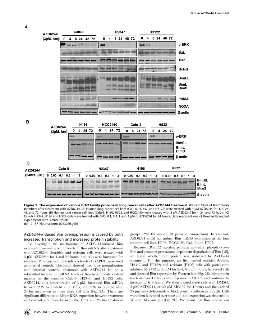
showed that treatment with AZD6244 induced rapid and
sustained increases in levels of BimEL and, to a lesser extent, of
BimL and BimS, in all sensitive cell lines (Fig. 1A). Furthermore,
treatment with sub-micromolar concentrations (0.03, 0.1, 0.3, 1,
and 3 mM) of AZD6244 for 24 hrs induced marked increase in
levels of Bim (Fig. 1C). These findings indicated that AZD6244
induced its effects on Bim expression in a concentration- and time-
dependent manner. However, in these cells, the levels of other Bcl-
2 family members (Bax, Bak, and Bcl-xL) did not change
noticeably at any concentration of AZD6244 or at any time point
(Fig. 1A). In contrast, AZD6244 did not induce obvious changes in
Bim expression in resistant cell lines (Fig. 1B). The resistant cell
lines are all wild type for BRAF and KRAS. We also detected
suppression of p-ERK expression with AZD6244 in both sensitive
and resistant cells (Fig. 1A and 1B). We also investigated the
expression of BH3-only proteins PUMA and NOXA following
3 mM AZD6244 treatment. PUMA and NOXA expressions were
increased upon AZD6244 treatment, however, the levels of the
increase were much less than that observed with Bim (Fig. 1A).
Since the upregulation of Bim is much more dramatic than PUMA
and NOXA in AZD6244-treated cells, we focused on the role of
Bim in subsequent studies.
Bim in AZD6244 Treatment
PLoS ONE | www.plosone.org 3 September 2010 | Volume 5 | Issue 9 | e13026
AZD6244-induced Bim overexpression is caused by bothincreased transcription and increased protein stability
To investigate the mechanisms of AZD6244-induced Bim
expression, we analyzed the levels of Bim mRNA after treatment
with AZD6244. Sensitive and resistant cells were treated with
3 mM AZD6244 for 4 and 24 hours, and cells were harvested for
real-time PCR analysis. The mRNA levels of GAPDH were used
as internal controls. The result showed that, after normalization
with internal controls, treatment with AZD6244 led to a
substantial increase in mRNA levels of Bim in a time-dependent
manner in the sensitive Calu-6, H2347, and H3122 cells.
AZD6244, at a concentration of 3 mM, increased Bim mRNA
between 2.2- to 2.5-fold after 4 hrs, and 2.9- to 3.8-fold after
24 hrs incubation in these three cell lines (Fig. 2A). There are
significant difference in Bim mRNA expression between treatment
and control groups or between the 4 hrs and 24 hrs treatment
groups (P,0.05 among all pairwise comparison). In contrast,
AZD6244 could not induce Bim mRNA expression in the four
resistant cell lines H196, HCC2450, Calu-3 and H522.
Because ERK1/2 signaling pathway activation phosphorylates
Bim and promotes proteasome-dependent degradation of Bim [18],
we tested whether Bim protein was stabilized by AZD6244
treatment. For this purpose, we first treated sensitive (Calu-6,
H2347 and H3122) and resistant (H196) cells with proteosome
inhibitor MG132 at 30 mM for 2, 4, 6 and 8 hours, harvested cells
and detected Bim expression by Western blot (Fig. 2B). Bim protein
levels increased 2 hours after exposure to MG132 and continued to
increase at 6–8 hours. We then treated these cells with DMSO,
3 mM AZD6244, or 30 mM MG132 for 4 hours and then added
25 mg/ml cycloheximide to block protein synthesis in the cells. Cells
were then harvested over time and Bim expression was detected by
Western blot analysis (Fig. 2C). We found that Bim protein was
Figure 1. The expression of various Bcl-2 family proteins in lung cancer cells after AZD6244 treatment. Western blots of Bcl-2 familymembers after treatment with AZD6244. (A) Human lung cancer cell lines (Calu-6, H2347, and H3122) were treated with 3 mM AZD6244 for 4, 8, 24,48, and 72 hours. (B) Human lung cancer cell lines (Calu-3, H196, H522, and HCC2450) were treated with 3 mM AZD6244 for 4, 24, and 72 hours. (C)Calu-6, H2347, H196 and H522 cells were treated with 0.03, 0.1, 0.3, 1 and 3 mM of AZD6244 for 24 hours. Data represent one of three independentexperiments with similar results.doi:10.1371/journal.pone.0013026.g001
Bim in AZD6244 Treatment
PLoS ONE | www.plosone.org 4 September 2010 | Volume 5 | Issue 9 | e13026
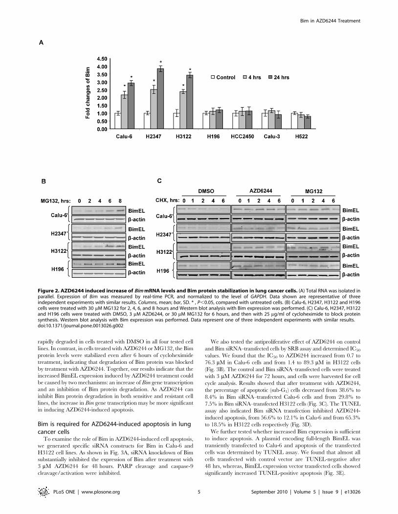
rapidly degraded in cells treated with DMSO in all four tested cell
lines. In contrast, in cells treated with AZD6244 or MG132, the Bim
protein levels were stabilized even after 6 hours of cycloheximide
treatment, indicating that degradation of Bim protein was blocked
by treatment with AZD6244. Together, our results indicate that the
increased BimEL expression induced by AZD6244 treatment could
be caused by two mechanisms: an increase of Bim gene transcription
and an inhibition of Bim protein degradation. As AZD6244 can
inhibit Bim protein degradation in both sensitive and resistant cell
lines, the increase in Bim gene transcription may be more significant
in inducing AZD6244-induced apoptosis.
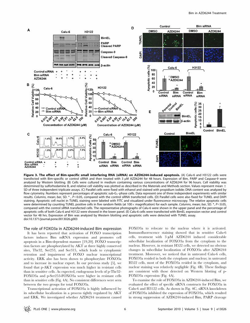
Bim is required for AZD6244-induced apoptosis in lungcancer cells
To examine the role of Bim in AZD6244-induced cell apoptosis,
we generated specific siRNA constructs for Bim in Calu-6 and
H3122 cell lines. As shown in Fig. 3A, siRNA knockdown of Bim
substantially inhibited the expression of Bim after treatment with
3 mM AZD6244 for 48 hours. PARP cleavage and caspase-9
cleavage/activation were inhibited.
We also tested the antiproliferative effect of AZD6244 on control
and Bim siRNA–transfected cells by SRB assay and determined IC50
values. We found that the IC50 to AZD6244 increased from 0.7 to
76.3 mM in Calu-6 cells and from 1.4 to 89.3 mM in H3122 cells
(Fig. 3B). The control and Bim siRNA–transfected cells were treated
with 3 mM AZD6244 for 72 hours, and cells were harvested for cell
cycle analysis. Results showed that after treatment with AZD6244,
the percentage of apoptotic (sub-G1) cells decreased from 38.6% to
8.4% in Bim siRNA–transfected Calu-6 cells and from 29.8% to
7.5% in Bim siRNA–transfected H3122 cells (Fig. 3C). The TUNEL
assay also indicated Bim siRNA transfection inhibited AZD6244-
induced apoptosis, from 56.6% to 12.1% in Calu-6 and from 65.3%
to 18.5% in H3122 cells respectively (Fig. 3D).
We further tested whether increased Bim expression is sufficient
to induce apoptosis. A plasmid encoding full-length BimEL was
transiently transfected to Calu-6 and apoptosis of the transfected
cells was determined by TUNEL assay. We found that almost all
cells transfected with control vector are TUNEL-negative after
48 hrs, whereas, BimEL expression vector transfected cells showed
significantly increased TUNEL-positive apoptosis (Fig. 3E).
Figure 2. AZD6244 induced increase of Bim mRNA levels and Bim protein stabilization in lung cancer cells. (A) Total RNA was isolated inparallel. Expression of Bim was measured by real-time PCR, and normalized to the level of GAPDH. Data shown are representative of threeindependent experiments with similar results. Columns, mean; bar, SD. * , P,0.05, compared with untreated cells. (B) Calu-6, H2347, H3122 and H196cells were treated with 30 mM MG132 for 2, 4, 6, and 8 hours and Western blot analysis with Bim expression was performed. (C) Calu-6, H2347, H3122and H196 cells were treated with DMSO, 3 mM AZD6244, or 30 mM MG132 for 6 hours, and then with 25 mg/ml of cycloheximide to block proteinsynthesis. Western blot analysis with Bim expression was performed. Data represent one of three independent experiments with similar results.doi:10.1371/journal.pone.0013026.g002
Bim in AZD6244 Treatment
PLoS ONE | www.plosone.org 5 September 2010 | Volume 5 | Issue 9 | e13026
The role of FOXO3a in AZD6244-induced Bim expressionIt has been reported that activation of FOXO transcription
factors induces Bim mRNA expression and promotes cell
apoptosis in a Bim-dependent manner [19,20]. FOXO transcrip-
tion factors are phosphorylated by AKT at three highly conserved
sites, Thr32, Ser253, and Ser315, which leads to cytoplasmic
retention and impairment of FOXO nuclear transcriptional
activity. ERK also has been shown to phosphorylate FOXO3a
and to increase its nuclear export. In our previous study [5], we
found that p-AKT expression was much higher in resistant cells
than in sensitive cells. As expected, endogenous levels of p-Thr32-
FOXO3a and p-Ser253-FOXO3a were higher in resistant cells
than in sensitive cells (Fig. 4A). No consistent differences were seen
between the two groups for total FOXO3a.
Transcriptional activation of FOXO3a is highly influenced by
its subcellular localization in a process tightly regulated by AKT
and ERK. We investigated whether AZD6244 treatment caused
FOXO3a to relocate to the nucleus where it is activated.
Immunofluorescence staining showed that in sensitive Calu-6
cells, treatment with 3 mM AZD6244 induced considerable
subcellular localization of FOXO3a from the cytoplasm to the
nucleus. However, in resistant H522 cells, we detected no obvious
changes in subcellular localization of FOXO3a after AZD6244
treatment. Moreover, we noticed that in untreated Calu-6 cells,
FOXO3a resided in both the cytoplasm and nucleus; in untreated
H522 cells, most of the FOXO3a resided in the cytoplasm, and
nuclear staining was relatively negligible (Fig. 4B). These findings
are consistent with those detected on Western blotting of p-
FOXO3a expression (Fig. 4A).
To examine the role of FOXO3a in AZD6244-induced Bim, we
evaluated the effect of specific siRNA constructs for FOXO3a in
Calu-6 and H3122 cells. As shown in Fig. 4C, siRNA knockdown
of FOXO3a inhibited the expression of FOXO3a, which resulted
in strong suppression of AZD6244-induced Bim, PARP cleavage
Figure 3. The effect of Bim-specific small interfering RNA (siRNA) on AZD6244-induced apoptosis. (A) Calu-6 and H3122 cells weretransfected with Bim-specific or control siRNA and then treated with 3 mM AZD6244 for 48 hours. Expression of Bim, PARP and Caspase-9 wereanalyzed by Western blotting. (B) Cells were cultured in medium containing various concentrations of AZD6244 for 96 hours. Cell viability wasdetermined by sulforhodamine B, and relative cell viability was plotted as described in the Materials and Methods section. Values represent mean 6SD of three independent triplicate assays. (C) Parallel cells were fixed with ethanol and stained with propidium iodide; DNA content was analyzed byflow cytometry. Numbers represent percentages of apoptotic sub-G1–phase cells. Data represent one of three independent experiments with similarresults. Columns, mean; bar, SD. * , P,0.05, compared with the control siRNA transfected cells. (D) Parallel cells were also fixed for TUNEL and DAPIstaining. Apoptotic cell nuclei in TUNEL staining were labeled with FITC and visualized under fluorescence microscopy. The relative apoptotic cellswere determined by counting TUNEL positive cells in five random fields (at 1006magnification) for each sample. Columns, mean; bar, SD. *, P,0.05,compared with the control siRNA transfected cells. The representative photographs of Calu-6 were shown in the upper panel and the percentage ofapoptotic cells of both Calu-6 and H3122 were showed in the lower panel. (E) Calu-6 cells were transfected with BimEL expression vector and controlvector for 48 hrs. Expression of Bim was analyzed by Western blotting and apoptotic cells were detected with TUNEL assay.doi:10.1371/journal.pone.0013026.g003
Bim in AZD6244 Treatment
PLoS ONE | www.plosone.org 6 September 2010 | Volume 5 | Issue 9 | e13026
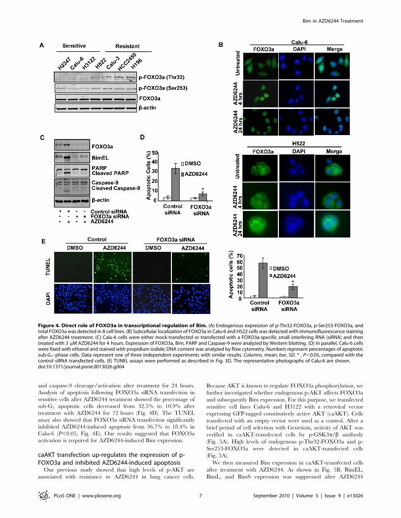
and caspase-9 cleavage/activation after treatment for 24 hours.
Analysis of apoptosis following FOXO3a siRNA transfection in
sensitive cells after AZD6244 treatment showed the percentage of
sub-G1 apoptotic cells decreased from 32.5% to 10.9% after
treatment with AZD6244 for 72 hours (Fig. 4D). The TUNEL
assay also showed that FOXO3a siRNA transfection significantly
inhibited AZD6244-induced apoptosis from 56.7% to 18.4% in
Calu-6 (P,0.05, Fig. 4E). Our results suggested that FOXO3a
activation is required for AZD6244-induced Bim expression.
caAKT transfection up-regulates the expression of p-FOXO3a and inhibited AZD6244-induced apoptosis
Our previous study showed that high levels of p-AKT are
associated with resistance to AZD6244 in lung cancer cells.
Because AKT is known to regulate FOXO3a phosphorylation, we
further investigated whether endogenous p-AKT affects FOXO3a
and subsequently Bim expression. For this purpose, we transfected
sensitive cell lines Calu-6 and H3122 with a retroviral vector
expressing GFP-tagged constitutively active AKT (caAKT). Cells
transfected with an empty vector were used as a control. After a
brief period of cell selection with Geneticin, activity of AKT was
verified in caAKT-transfected cells by p-GSK3a/b antibody
(Fig. 5A). High levels of endogenous p-Thr32-FOXO3a and p-
Ser253-FOXO3a were detected in caAKT-transfected cells
(Fig. 5A).
We then measured Bim expression in caAKT-transfected cells
after treatment with AZD6244. As shown in Fig. 5B, BimEL,
BimL, and BimS expression was suppressed after AZD6244
Figure 4. Direct role of FOXO3a in transcriptional regulation of Bim. (A) Endogenous expression of p-Thr32-FOXO3a, p-Ser253-FOXO3a, andtotal FOXO3a was detected in 8 cell lines. (B) Subcellular localization of FOXO3a in Calu-6 and H522 cells was detected with immunofluorescence stainingafter AZD6244 treatment. (C) Calu-6 cells were either mock-transfected or transfected with a FOXO3a-specific small interfering RNA (siRNA) and thentreated with 3 mM AZD6244 for 4 hours. Expression of FOXO3a, Bim, PARP and Caspase-9 were analyzed by Western blotting. (D) In parallel, Calu-6 cellswere fixed with ethanol and stained with propidium iodide; DNA content was analyzed by flow cytometry. Numbers represent percentages of apoptoticsub-G1–phase cells. Data represent one of three independent experiments with similar results. Columns, mean; bar, SD. * , P,0.05, compared with thecontrol siRNA transfected cells. (E) TUNEL assays were performed as described in Fig. 3D. The representative photographs of Calu-6 are shown.doi:10.1371/journal.pone.0013026.g004
Bim in AZD6244 Treatment
PLoS ONE | www.plosone.org 7 September 2010 | Volume 5 | Issue 9 | e13026
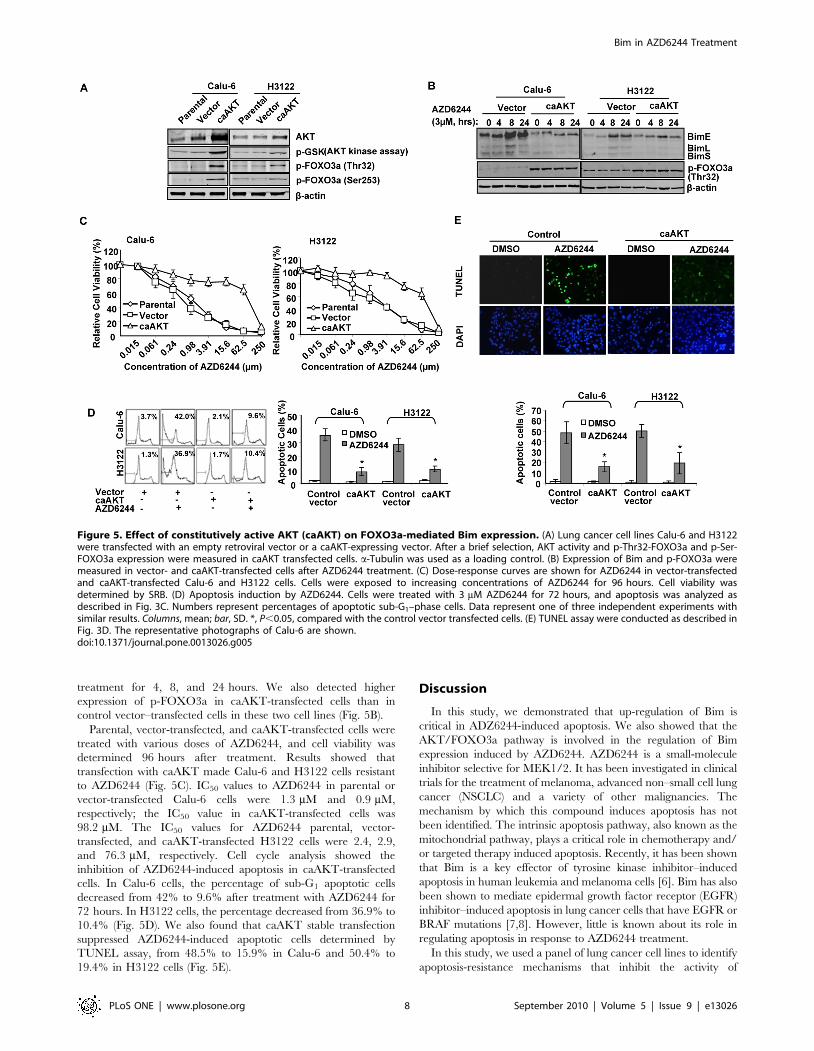
treatment for 4, 8, and 24 hours. We also detected higher
expression of p-FOXO3a in caAKT-transfected cells than in
control vector–transfected cells in these two cell lines (Fig. 5B).
Parental, vector-transfected, and caAKT-transfected cells were
treated with various doses of AZD6244, and cell viability was
determined 96 hours after treatment. Results showed that
transfection with caAKT made Calu-6 and H3122 cells resistant
to AZD6244 (Fig. 5C). IC50 values to AZD6244 in parental or
vector-transfected Calu-6 cells were 1.3 mM and 0.9 mM,
respectively; the IC50 value in caAKT-transfected cells was
98.2 mM. The IC50 values for AZD6244 parental, vector-
transfected, and caAKT-transfected H3122 cells were 2.4, 2.9,
and 76.3 mM, respectively. Cell cycle analysis showed the
inhibition of AZD6244-induced apoptosis in caAKT-transfected
cells. In Calu-6 cells, the percentage of sub-G1 apoptotic cells
decreased from 42% to 9.6% after treatment with AZD6244 for
72 hours. In H3122 cells, the percentage decreased from 36.9% to
10.4% (Fig. 5D). We also found that caAKT stable transfection
suppressed AZD6244-induced apoptotic cells determined by
TUNEL assay, from 48.5% to 15.9% in Calu-6 and 50.4% to
19.4% in H3122 cells (Fig. 5E).
Discussion
In this study, we demonstrated that up-regulation of Bim is
critical in ADZ6244-induced apoptosis. We also showed that the
AKT/FOXO3a pathway is involved in the regulation of Bim
expression induced by AZD6244. AZD6244 is a small-molecule
inhibitor selective for MEK1/2. It has been investigated in clinical
trials for the treatment of melanoma, advanced non–small cell lung
cancer (NSCLC) and a variety of other malignancies. The
mechanism by which this compound induces apoptosis has not
been identified. The intrinsic apoptosis pathway, also known as the
mitochondrial pathway, plays a critical role in chemotherapy and/
or targeted therapy induced apoptosis. Recently, it has been shown
that Bim is a key effector of tyrosine kinase inhibitor–induced
apoptosis in human leukemia and melanoma cells [6]. Bim has also
been shown to mediate epidermal growth factor receptor (EGFR)
inhibitor–induced apoptosis in lung cancer cells that have EGFR or
BRAF mutations [7,8]. However, little is known about its role in
regulating apoptosis in response to AZD6244 treatment.
In this study, we used a panel of lung cancer cell lines to identify
apoptosis-resistance mechanisms that inhibit the activity of
Figure 5. Effect of constitutively active AKT (caAKT) on FOXO3a-mediated Bim expression. (A) Lung cancer cell lines Calu-6 and H3122were transfected with an empty retroviral vector or a caAKT-expressing vector. After a brief selection, AKT activity and p-Thr32-FOXO3a and p-Ser-FOXO3a expression were measured in caAKT transfected cells. a-Tubulin was used as a loading control. (B) Expression of Bim and p-FOXO3a weremeasured in vector- and caAKT-transfected cells after AZD6244 treatment. (C) Dose-response curves are shown for AZD6244 in vector-transfectedand caAKT-transfected Calu-6 and H3122 cells. Cells were exposed to increasing concentrations of AZD6244 for 96 hours. Cell viability wasdetermined by SRB. (D) Apoptosis induction by AZD6244. Cells were treated with 3 mM AZD6244 for 72 hours, and apoptosis was analyzed asdescribed in Fig. 3C. Numbers represent percentages of apoptotic sub-G1–phase cells. Data represent one of three independent experiments withsimilar results. Columns, mean; bar, SD. *, P,0.05, compared with the control vector transfected cells. (E) TUNEL assay were conducted as described inFig. 3D. The representative photographs of Calu-6 are shown.doi:10.1371/journal.pone.0013026.g005
Bim in AZD6244 Treatment
PLoS ONE | www.plosone.org 8 September 2010 | Volume 5 | Issue 9 | e13026
AZD6244 in lung cancer cells. First, our results showed that Bim is
critical in apoptosis induced by the MEK inhibitor AZD6244.
Second, FOXO3a, regulated by p-AKT and p-ERK, is a direct
transcriptional regulator of Bim. Induction of apoptosis by the
MEK inhibitor AZD6244 required a low level of endogenous p-
FOXO3a (Thr32 and Ser253). Third, expression of constitutively
active AKT could up-regulate p-FOXO3a (Thr32 and Ser253)
and induce resistance to MEK inhibition.
We have previously shown that p-AKT expression is low in
AZD6244-sensitive lung cancer cell lines but high in resistant cells,
suggesting that p-AKT is a potential biomarker of sensitivity to
AZD6244 treatment. Moreover, the down-regulation of p-AKT
with transfected dominant-negative AKT sensitized resistant cells
to AZD6244. In this study, we determined that AZD6244
treatment can strongly induce Bim expression in all three sensitive
cell lines but not in resistant cells. Increased Bim levels in both
protein and mRNA expression were detected with Western
blotting and real-time PCR, respectively in sensitive cells.
Knockdown of Bim with siRNA in the sensitive Calu-6 and
H3122 cell lines increased the IC50 value to AZD6244 and
substantially decreased apoptosis. This data clearly demonstrates
that Bim is an important intermediary in AZD6244-induced
apoptosis.
Both the Ras/Raf/MEK/ERK pathway and the PI3K/AKT
pathway mediate signals from various growth factor receptors, and
these two pathways regulate several common downstream
molecules that are critical in cell survival and cell cycle progression
such as forkhead transcription factors [21], cyclin D1 [22], Bad
[23] and caspase-9 [24]. In our study, we determined endogenous
expression levels of total FOXO3a, p-Thr32-FOXO3a, and p-
Ser253-FOXO3a in all sensitive and resistant cell lines. Except for
the sensitive H2347 cell line, which showed lower expression, the
expression of total FOXO3a was not noticeably different between
the sensitive and resistant cell lines. As we expected, basal levels of
p-Thr32-FOXO3a and p-Ser253-FOXO3a were higher in
resistant cells, which was consistent with higher levels of p-AKT
expression shown in our previous study. Moreover, AZD6244
treatment did not alter the expression of p-Thr32-FOXO3a and
p-Ser253-FOXO3a in any of the cell lines.
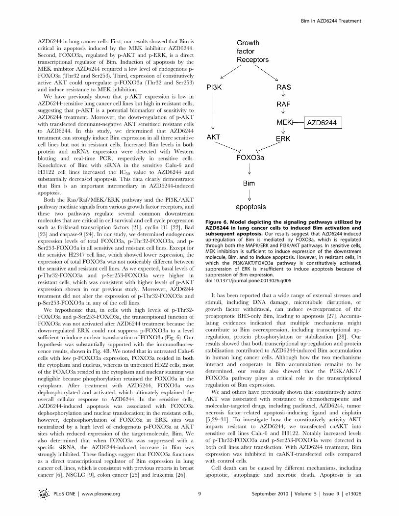
We hypothesize that, in cells with high levels of p-Thr32-
FOXO3a and p-Ser253-FOXO3a, the transcriptional function of
FOXO3a was not activated after AZD6244 treatment because the
down-regulated ERK could not suppress p-FOXO3a to a level
sufficient to induce nuclear translocation of FOXO3a (Fig. 6). Our
hypothesis was substantially supported with the immunofluores-
cence results, shown in Fig. 4B. We noted that in untreated Calu-6
cells with low p-FOXO3a expression, FOXO3a resided in both
the cytoplasm and nucleus, whereas in untreated H522 cells, most
of the FOXO3a resided in the cytoplasm and nuclear staining was
negligible because phosphorylation retained the FOXO3a in the
cytoplasm. After treatment with AZD6244, FOXO3a was
dephosphorylated and activated, which ultimately explained the
overall cellular response to AZD6244. In the sensitive cells,
AZD6244-induced apoptosis was associated with FOXO3a
dephosphorylation and nuclear translocation; in the resistant cells,
however, dephosphorylation of FOXO3a at ERK sites was
neutralized by a high level of endogenous p-FOXO3a at AKT
sites which reduced expression of the target-molecule, Bim. We
also determined that when FOXO3a was suppressed with a
specific siRNA, the AZD6244-induced increase in Bim was
strongly inhibited. These findings suggest that FOXO3a functions
as a direct transcriptional regulator of Bim expression in lung
cancer cell lines, which is consistent with previous reports in breast
cancer [6], NSCLC [9], colon cancer [25] and leukemia [26].
It has been reported that a wide range of external stresses and
stimuli, including DNA damage, microtubule disruption, or
growth factor withdrawal, can induce overexpression of the
proapoptotic BH3-only Bim, leading to apoptosis [27]. Accumu-
lating evidences indicated that multiple mechanisms might
contribute to Bim overexpression, including transcriptional up-
regulation, protein phosphorylation or stabilization [28]. Our
results showed that both transcriptional up-regulation and protein
stabilization contributed to AZD6244-induced Bim accumulation
in human lung cancer cells. Although how the two mechanisms
interact and cooperate in Bim accumulation remains to be
determined, our results also showed that the PI3K/AKT/
FOXO3a pathway plays a critical role in the transcriptional
regulation of Bim expression.
We and others have previously shown that constitutively active
AKT was associated with resistance to chemotherapeutic and
molecular-targeted drugs, including paclitaxel, AZD6244, tumor
necrosis factor–related apoptosis-inducing ligand and cisplatin
[5,29–31]. To investigate how the constitutively activity AKT
imparts resistant to AZD6244, we transfected caAKT into
sensitive cell lines Calu-6 and H3122. Notably increased levels
of p-Thr32-FOXO3a and p-Ser253-FOXO3a were detected in
both cell lines after transfection. With AZD6244 treatment, Bim
expression was inhibited in caAKT-transfected cells compared
with control cells.
Cell death can be caused by different mechanisms, including
apoptotic, autophagic and necrotic death. Apoptosis is an
Figure 6. Model depicting the signaling pathways utilized byAZD6244 in lung cancer cells to induced Bim activation andsubsequent apoptosis. Our results suggest that AZD6244-inducedup-regulation of Bim is mediated by FOXO3a, which is regulatedthrough both the MAPK/ERK and PI3K/AKT pathways. In sensitive cells,MEK inhibition is sufficient to induce expression of the downstreammolecule, Bim, and to induce apoptosis. However, in resistant cells, inwhich the PI3K/AKT/FOXO3a pathway is constitutively activated,suppression of ERK is insufficient to induce apoptosis because ofsuppression of Bim expression.doi:10.1371/journal.pone.0013026.g006
Bim in AZD6244 Treatment
PLoS ONE | www.plosone.org 9 September 2010 | Volume 5 | Issue 9 | e13026
intracellular programmed cell death involving activation of the
cysteine proteases (caspases) cascade [32]. The markers of
apoptosis include cleavage of PARP1, release of cytochrome c
from mitochondrial cleavage of chromosomal DNA, and activa-
tion of caspases [33,34]; autophagic death involves a process of
self-digestion of cellular material through formation of lysosome-
like autophagosomes [35,36]; and necrosis is a passive death
process caused by external factors and involves loss of cellular
homeostasis [37]. In this study, the western blot and TUNEL assay
results showed that AZD6244 induced apoptosis after 4–48 h
treatment. It is not clear if AZD6244 still induced cell apoptosis
after 96 h treatment, although our anti-proliferation assay showed
AZD6244 induced cell death after this treatment duration. It is
possible that a relatively long-term treatment may cause an
apoptosis-independent cell death or a mixture of apoptotic and
non-apoptotic cell death.
In summary, our results indicated that FOXO3a is important to
the antiproliferative effect of AZD6244 and induces mitochondrial
apoptosis mediated by Bim. On the basis of our observations, we
plan to focus on the PI3K/AKT/FOXO3a pathway and BH3-
only proteins in the development of strategies to overcome
resistance to AZD6244 in lung cancer cells.
Author Contributions
Conceived and designed the experiments: JM BF JAR. Performed the
experiments: JM. Analyzed the data: JM. Contributed reagents/materials/
analysis tools: YL CMC PDS. Wrote the paper: JM BF JAR.
References
1. Yeh TC, Marsh V, Bernat BA, Ballard J, Colwell H, et al. (2007) Biological
characterization of ARRY-142886 (AZD6244), a potent, highly selectivemitogen-activated protein kinase kinase 1/2 inhibitor. Clin Cancer Res 13:
1576–83.
2. Haass NK, Sproesser K, Nguyen TK, Contractor R, Medina CA, et al. (2008)The mitogen-activated protein/extracellular signal-regulated kinase kinase
inhibitor AZD6244 (ARRY-142886) induces growth arrest in melanoma cellsand tumor regression when combined with docetaxel. Clin Cancer Res 14:
230–9.
3. Davies BR, Logie A, McKay JS, Martin P, Steele S, et al. (2007) AZD6244(ARRY-142886), a potent inhibitor of mitogen-activated protein kinase/
extracellular signal-regulated kinase kinase 1/2 kinases: mechanism of actionin vivo, pharmacokinetic/pharmacodynamic relationship, and potential for
combination in preclinical models. Mol Cancer Ther 8: 2209–19.4. Huynh H, Chow PK, Soo KC (2007) AZD6244 and doxorubicin induce growth
suppression and apoptosis in mouse models of hepatocellular carcinoma. Mol
Cancer Ther 6: 2468–76.5. Meng J, Peng H, Dai B, Guo W, Wang L, et al. (2009) High level of AKT
activity is associated with resistance to MEK inhibitor AZD6244 (ARRY-142886). Cancer Biol Ther 8: 2073–80.
6. Nordigarden A, Kraft M, Eliasson P, Labi V, Lam EW, et al. (2009) BH3-only
protein Bim more critical than Puma in tyrosine kinase inhibitor-inducedapoptosis of human leukemic cells and transduced hematopoietic progenitors
carrying oncogenic FLT3. Blood 113: 2302–11.7. Yang JY, Chang CJ, Xia W, Wang Y, Wong KK, et al. (2010) Activation of
FOXO3a Is Sufficient to Reverse Mitogen-Activated Protein/ExtracellularSignal-Regulated Kinase Kinase Inhibitor Chemoresistance in Human Cancer.
Cancer Res 70: 4709–18.
8. Sunters A, Fernandez de Mattos S, Stahl M, Brosens JJ, Zoumpoulidou G, et al.(2003) FOXO3a transcriptional regulation of Bim control apoptosis in
paclitaxel-treated breast cancer cell lines. J Biol Chem 278: 49795–805.9. Cragg MS, Kuroda J, Puthalakath H, Huang DC, Strasser A (2007) Gefitinib-
induced killing of NSCLC cell lines expressing mutant EGFR requires BIM and
can be enhanced by BH3 mimetics. PLoS Med 4: 1681–89.10. Pei XY, Dai Y, Tenorio S, Lu J, Harada H, et al. (2007) MEK1/2 inhibitors
potentiate UCN-01 lethality in human multiple myeloma cells through a Bim-dependent mechanism. Blood 110: 2092–101.
11. Finnberg N, El-Deiry WS (2004) Activating FOXO3a, NF-kappaB and p53 by
targeting IKKs: an effective multi-faceted targeting of the tumor-cell phenotype?Cancer Biol Ther 3: 614–6.
12. Yang JY, Xia W, Hu MC (2006) Ionizing radiation activates expression ofFOXO3a, Fas ligand, and Bim, and induces cell apoptosis. Int J Oncol 29:
643–48.13. Schmidt M, Fernandez de Mattos S, van der Horst A, Klompmaker R, Kops GJ,
et al. (2002) Cell cycle inhibition by FoxO forkhead transcription factors involves
downregulation of cyclin D. Mol Cell Biol 22: 7842–52.14. Medema RH, Kops GJ, Bos JL, Burgering BM (2000) AFX-like Forkhead
transcription factors mediate cell-cycle regulation by Ras and PKB throughp27kip1. Nature 404: 782–7.
15. Tran H, Brunet A, Grenier JM, Datta SR, Fornace AJ, Jr., et al. (2002) DNA
repair pathway stimulated by the forkhead transcription factor FOXO3athrough the Gadd45 protein. Science 296: 530–4.
16. Brunet A, Bonni A, Zigmond MJ, Lin MZ, Juo P, et al. (1999) AKT promotescell survival by phosphorylating and inhibiting a Forkhead transcription factor.
Cell 96: 857–68.17. Kops GJ, de Runiter ND, de Vires-Smits AM, Powell DR, Bos JL, et al. (1999)
Direct control of the Forkhead transcription factor AFX by protein kinase B.
Nature 398: 630–4.
18. Yang JY, Zong CS, Xia W, Yamaguchi H, Ding Q, et al. (2008) ERK promotes
tumorigenesis by inhibiting FOXO3a via MDM2-mediated degradation. NatCell Biol 10: 138–48.
19. Ley R, Balmanno K, Hadfield K, Weston C, Cook SJ, et al. (2003) Activation of
the ERK1/2 signaling pathway promotes phosphorylation and proteasome-dependent degradation of the BH3-only protein, Bim. J Biol Chem 278:
18811–6.20. Dijkers PF, Medema RH, Lammers JW, Koenderman L, Coffer PJ (2000)
Expression of the pro-apoptotic Bcl-2 family member Bim is regulated by the
forkhead transcription factor FKHR-L1. Curr Biol 10: 1201–4.21. Gilley J, Coffer PJ, Ham J (2003) FOXO transcription factors directly activate
bim gene expression and promote apoptosis in sympathetic neurons. J Cell Biol162: 613–22.
22. Diehl JA, Cheng M, Roussel M F, Sherr CJ (1998) Glycogen synthase kinase-3beta regulates cyclin D1 proteolysis and subcellular localization. Genes Dev 12:
3499–511.
23. Datta SR, Dudek H, Tao X, Masters S, Fu H, et al. (1997) Akt phosphorylationof BAD couples survival signals to the cell-intrinsic death machinery. Cell 91:
231–41.24. Cardone MH, Roy N, Stennicke HR, Salvesen GS, Franke TF, et al. (1998)
Regulation of cell death protease caspase-9 by phosphorylation. Science 282:
1318–21.25. Fernandez de Mattos S, Villalonga P, Clardy J, Lam EW (2008) FOXO3a
mediates the cytotoxic effects of cisplatin in colon cancer cell. Mol Cancer Ther7: 3237–46.
26. Leung KT, Li KK, Sun SS, Chan PK, Ooi VE, et al. (2008) Activation of theJNK pathway promotes phosphorylation and degradation of BimEL—a novel
mechanism of chemoresistance in T-cell acute lymphoblastic leukemia.
Carcinogenesis 29: 544–51.27. Kelekar A, Thompson CB (1998) Bcl-2-family proteins: the role of the BH3
domain in apoptosis. Trends Cell Biol 8: 324–30.28. Puthalakath H, Strasser A (2002) Keeping killers on a tight leash: transcriptional
and post-translational control of the pro-apoptotic activity of BH3-only proteins.
Cell Death Differ 9: 505–12.29. Chen X, Thakkar H, Tyan F, Gim S, Robinson H, et al. (2001) Constitutively
active Akt is an important regulator of TRAIL sensitivity in prostate cancer.Oncogene 20: 6073–83.
30. Yuan ZQ, Feldman RI, Sussman GE, Coppola D, Nicosia SV, et al. (2003)
AKT2 inhibition of cisplatin-induced JNK/p38 and Bax activation byphosphorylation of ASK1: implication of AKT2 in chemoresistance. J Biol
Chem 278: 23432–40.31. Clark AS, West K, Streicher S, Dennis PA (2002) Constitutive and inducible Akt
activity promotes resistance to chemotherapy, trastuzumab, or tamoxifen inbreast cancer cells. Mol Cancer Ther 1: 707–17.
32. Hengartner MO (2000) The biochemistry of apoptosis. Nature 407: 770–6.
33. Janicke RU, Sprengart ML, Wati MR, Porter AG (1998) Caspase-3 is requiredfor DNA fragmentation and morphological changes associated with apoptosis.
J Biol Chem 273: 9357–60.34. Boulares AH, Yakovlev AG, Ivanova V, Stoica BA, Wang G, et al. (1999) Role
of poly(ADP-ribose) polymerase (PARP) cleavage in apoptosis. Caspase 3-
resistant PARP mutant increases rates of apoptosis in transfected cells. J BiolChem 274: 22932–40.
35. Bursch W (2001) The autophagosomal-lysosomal compartment in programmedcell death. Cell Death Differ 8: 569–81.
36. Gozuacik D, Kimchi A (2004) Autophagy as a cell death and tumor suppressormechanism. Oncogene 23: 2891–906.
37. Zong WX, Thompson CB (2006) Necrotic death as a cell fate. Genes Dev 20:
1–15.
Bim in AZD6244 Treatment
PLoS ONE | www.plosone.org 10 September 2010 | Volume 5 | Issue 9 | e13026
Related Documents