Aortic intramural haematoma: pathogenesis, clinical features and imaging evaluation Department of Cardiology, Radiology, Heart of, Birmingham, UK, England Postgrad Med J 2012

Aortic intramural haematoma: pathogenesis, clinical features and imaging evaluation Department of Cardiology, Radiology, Heart of, Birmingham, UK, England.
Dec 15, 2015
Welcome message from author
This document is posted to help you gain knowledge. Please leave a comment to let me know what you think about it! Share it to your friends and learn new things together.
Transcript
Aortic intramural haematoma: pathogenesis, clinical
features and imaging evaluation
Department of Cardiology, Radiology, Heart of, Birmingham, UK, England
Postgrad Med J 2012
INTRODUCTION
• Intramural haematoma (IMH) is a localised haemorrhage within the aortic wall.
• It accounts for 10–20% of cases of acute aortic syndrome (AAS) (aortic dissection (AD) and penetrating atherosclerotic ulcer).
• It similar clinical manifestations and prognosis to AD. carries a similarly high mortality rate.
INTRODUCTION
• Imaging plays a central role in diagnosing IMH, differentiating it from AD and assessing for complications.
• Imaging provides lesion location and extent, which is vital for clinical decision making.
• Key tool: Multidetector CT (MDCT) and MRI.
This article reviews the IMH from • pathogenesis• clinical features • complications• the role of advanced imaging techniques in its
evaluation including differentiation from other pathological conditions of the thoracic aorta.
PATHOGENESIS OF IMH
• In most cases IMH is thought to begin with spontaneous rupture of the vasa vasorum.
• it can also occur secondary to aortic trauma or a penetrating aortic ulcer.
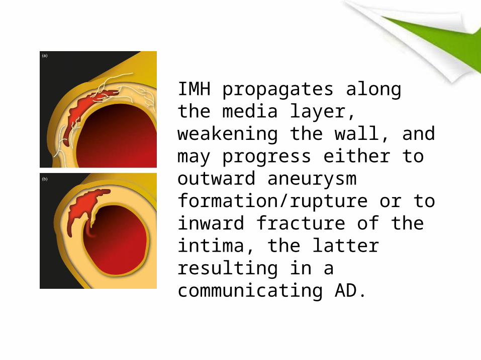
IMH propagates along the media layer, weakening the wall, and may progress either to outward aneurysm formation/rupture or to inward fracture of the intima, the latter resulting in a communicating AD.
• Dissection has been reported as a complication of IMH in 12–47% of cases.
• a recent study of 300 patients regression in 34% over a mean follow-up period of 3 years.
• Some authors have recently questioned the classical mechanism of IMH formation.
• They have postulated that small intimal tears may be the initiating pathology in some cases.
• It is generally agreed that the same Stanford classification system as is used to classify AD be used for IMH.
CLINICAL FEATURES
• IMH median age at presentation of 68 years.
• male predominance (61%).
• It exhibits nearly identical clinical signs, symptoms and risk factor profile to classic AD, and systemic hypertension recognised as the most common predisposing factor.
• Physical examination is frequently normal.
auxiliary examination
• More often than not the radiograph will have a normal appearance.
• It may occasionally mediastinum may be Widened.
• It will not be reliably detected with catheter-directed angiography.
ultrasound
• Transthoracic echocardiography (TTE) is a valuable means to assess complicating features of type A IMH.
• Transoesophageal echocardiography (TOE) gives improved spatial resolution, with a reported sensitivity and specificity of 98% and 95%.
MDCT
• Definite distinction between IMH and normal findings with TOE often requires a second imaging modality such as MDCT or MRI to confirm or refute the diagnosis.
• In recent years MDCT has become established as the leading technique for diagnosis and classification of IMH.
MRI
• MRI is rarely used to investigate the initial presentation of suspected AAS due to prolonged examination times (20–30 min for a typical aortic protocol) and incompatible life support and monitoring equipment which is usually required for critically ill patients.
• MRI can also be used to estimate the age of a haematoma based on differing signa characteristics .
MDCT technique
• Scan coverage should be from above the aortic arch to below the aortic bifurcation.
• The application of ECG gating reduces cardiac motion-related artefact. however, it carries a significant additional radiation burden.
• an initial unenhanced study must be performed as this is crucial for diagnosing IMH and for the detection of any mediastinal haemorrhage or haemorrhagic pericardial effusion.
MDCT technique
• this does not require ECG gating and a slice thickness of 2–3 mm is adequate.
• Injection rate of 3–4 ml/s is usually adequate and a slice thickness of 1–2 mm is used to provide isotropic spatial resolution.

Findings
• The hallmark non-contrast MDCT finding of IMH is high attenuation (60–80 HU) eccentric crescent-shaped thickening of the aortic wall.
• Thickening is nearly always >7 mm• IMH does not enhance.
• In contrast to dissection, the aortic thickening does not spiral around the opacified aortic lumen which is rarely compromised.
• A combined unenhanced and contrast-enhanced MDCT protocol has been shown to have a sensitivity approaching 100% for the detection of IMH.
MRI technique
• Breath-hold ECG-gated fast spin echo images acquired with T1 and T2 weighting are used to assess aortic calibre, aortic wall thickness and aortic wall signal change.
• These are acquired as contiguous transverse sections from the apices to the infrarenal abdominal aorta and also as sections parallelling the long axis of the aortic arch.
• Dynamic steady state free precession (SSFP) sequences are used to assess aortic valve function and the calibre of the aortic root. These are acquired in both transverse and coronal planes through the LVOT.
• If AR is present, its severity can be quantified via flow sensitive phase contrast sequences.
• Finally, a gadolinium-enhanced aortic angiography study is performed.

FindingsIMH appears as a focal area of mural thickening with signal characteristics that depend upon its acuity and relate to the slow conversion of oxyhaemoglobin to methaemoglobin and their differing paramagnetic properties.
• Acute IMH (0–7 days) demonstrates intermediate
signal intensity on T1-weighted spin-echo images caused by a predominance of oxyhaemoglobin making it sometimes difficult to distinguish from the normal aortic wall.
• Oxyhaemoglobin exhibits high T2 signal in the acute phase, making it appear conspicuous.
• With increasing methaemoglobin content, both T1 and T2 signals increase within IMH.
• In the absence of expected signal intensity evolution, rebleeding should be considered.
DIFFERENTIAL DIAGNOSES
• other causes of aortic wall thickening atherosclerosis, mural thrombus, thrombosed dissection and aortitis.
• Atherosclerotic wall thickening and mural thrombus has an typically irregular contour of inner margin. IMH which is usually smooth.
• However ,it is a distinction challenging IMH develops within an area of atherosclerosis. In such situations early interval repeat scanning may be the only means of discrimination.
• AD comprises a laceration of the intima and inner layer with resultant intimomedial flap and formation of a double-channel aortawith communication between true and false lumens.
• Evidence of an intimal tear is the key discriminating feature from IMH.
• Another important distinguishing feature is the spiral course of AD in contrast to the constant crescent-shaped appearance of IMH.
• Aortitis is most often caused by vasculitides such as Takayasu arteritis and giant cell arteritis.
• Active aortitis manifests on imaging as diffuse or segmental thickening of the vessel wall. often with accompanying inflammatory changes in the periaortic fat.
• Key discriminating features : Aortitis are circumferential mural thickening, mural enhancement and a patchy distribution with normal intervening segments.
COMPLICATIONS AND ADVERSE PROGNOSTIC INDICATORS
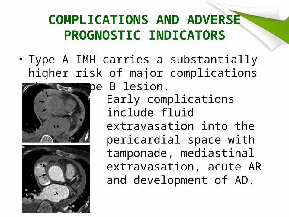
• Type A IMH carries a substantially higher risk of major complications than a type B lesion.
Early complications include fluid extravasation into the pericardial space with tamponade, mediastinalextravasation, acute AR and development of AD.
clinical outcome of IMH
• It may be spontaneous regression over time.• It may be complicated by saccular or fusiform
aneurysmal dilatation, rupture or late progression to AD.
• main adverse clinical feature is patient age >70 years carrying the highest risk of progression.
• imaging characteristics be correlated with increased mortality rates.
Imaging findings associatied with increased mortality in intramural haematoma
• Stanford type A lesion• Mural thickening >10 mm• Aortic diameter >5 cm• Coexistent penetrating ulcer• Evidence of rebleeding on serial imaging• Extension of thrombus on serial imaging
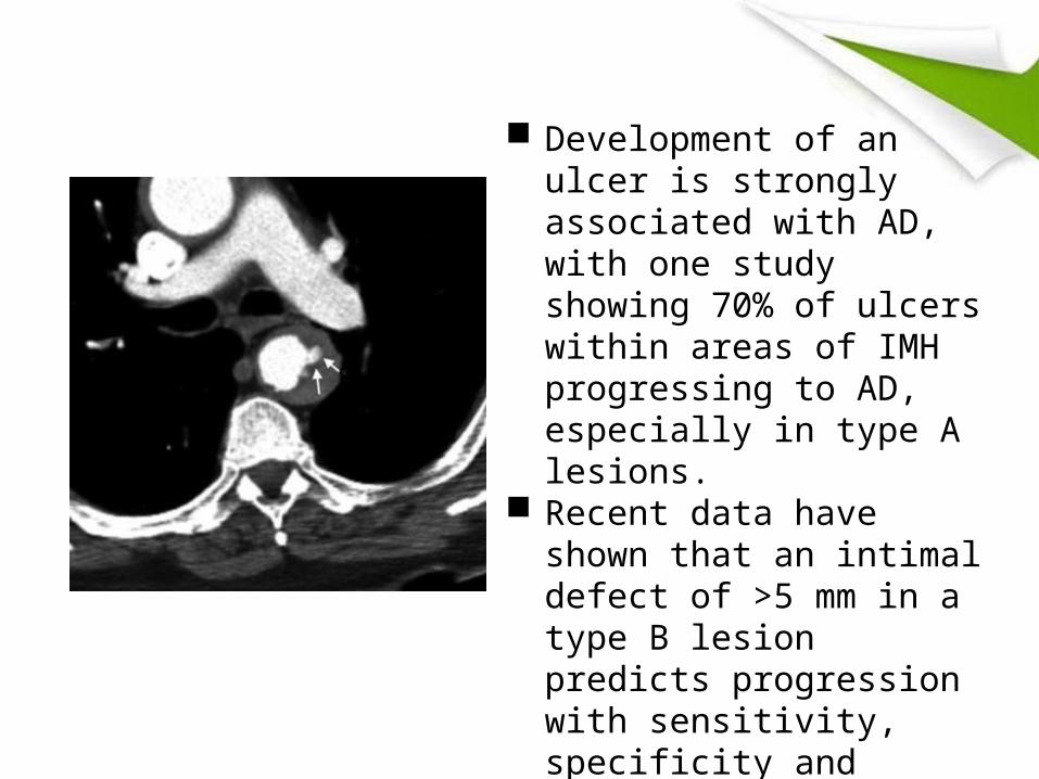
Development of an ulcer is
strongly associated with AD, with one study showing 70% of ulcers within areas of IMH progressing to AD, especially in type A lesions.
Recent data have shown that an intimal defect of >5 mm in a type B lesion predicts progression with sensitivity, specificity and postive and negative predictive values of 84%, 95%, 94% and 86%, respectively.
• Given the propensity for complications, close follow-up imaging during the 30 days after initial presentation is recommended for all patients being treated medically.
• If there is longitudinal progression of aortic involvement, progressive luminal dilation, penetrating ulcer, enlarging IMH or overt dissection surgical or endovascular treatment should be strongly considered.
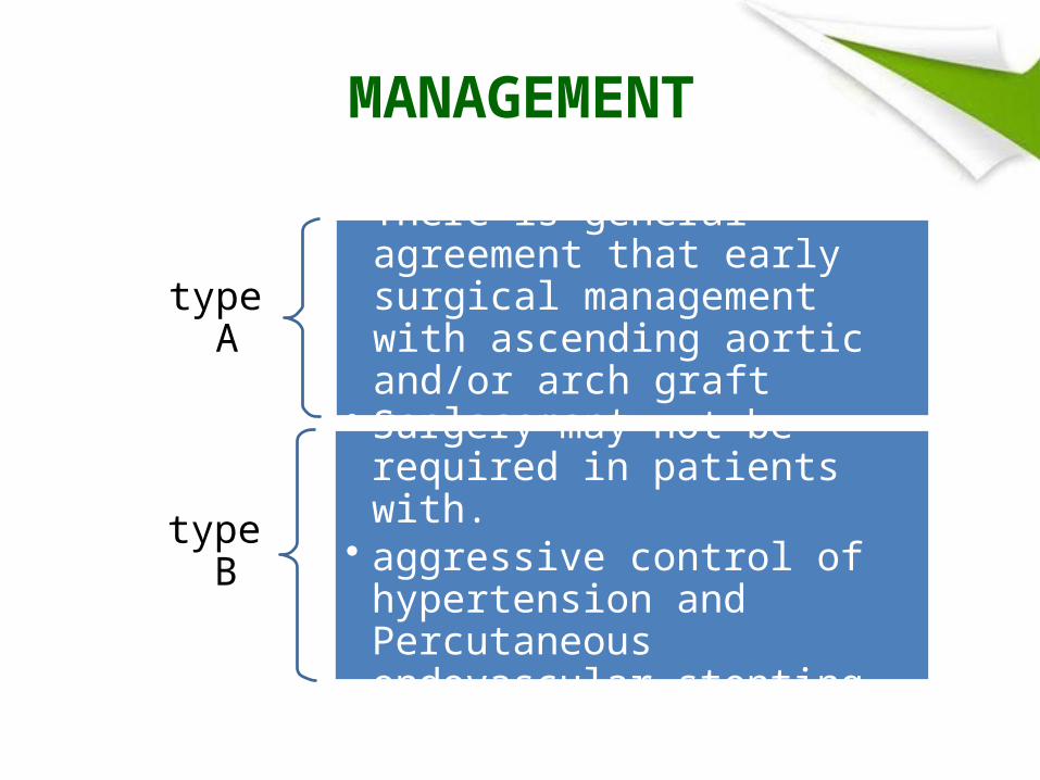
MANAGEMENT
type A
• There is general agreement that early surgical management with ascending aortic and/or arch graft replacement.
type B
• Surgery may not be required in patients with.
• aggressive control of hypertension and Percutaneous endovascular stenting.
IMAGING FOLLOW-UP
• The decision to use MDCTor MRI is guided by the patient’s age and renal function.
• with MRI preferred in young patients• and those with renal impairment• Imaging at 1, 3, 6, 9 and 12 months from diagnosis
has been suggested by Evangelista
CONCLUSIONS
• IMH is a localised haemorrhage within the aortic wall.
• It has overlapping clinical manifestations with AD and carries a similarly high mortality rate.
• Imaging is central to the diagnosis of IMH and its differentiation from AD and other causes of aortic wall thickening.
• Imaging can also provide crucial information concerning lesion location and extent which is essential for treatment planning.

• MDCT is the modality of choice for diagnosis and classification of IMH.
• imaging findings are considered adverse prognostic indicators: mural thickness >10 mm and aortic diameter >5 cm.
• Different type need different management.

Thanks for attention!
Related Documents