CHAPTER 5 Antibody Detection KENNETH L. HERRMANN Subject: Serodiagnosis (use of known antigens as reagents to detect antibodies in patient sera for diagnosing recent or past viral infection). Serodiagnostic Principles: Mechanism of antibody production, response patterns to pri- mary and recurrent infections, and significance of antibody to immunity in viral dis- eases. Laboratory Methods: Neutralization, complement fixation, hemagglutination inhibition, immune adherence hemagglutination, passive agglutination, hemolysis in gel, radioim- munoassay, enzyme immunoassay, indirect immunoftuorescencelftuoroimmunoassay, and Western immunoblotting. Interpretation of Test Results: Significance of presence of antibody or change in antibody level. Value of immunoglobulin type-specific assays in viral serodiagnosis. Problems and Pitfalls: Insensitive and nonspecific serodiagnostic tests resulting from heter- ologous cross-reactions, immunologic interference, substandard reagents, or improper test performance. Introduction Antibody detection plays an important role in the investigation of viruses as causes of human illness. Viral serology provides both clinicians and epidemi- ologists with a powerful tool for diagnosing infection in an individual patient, for determining the suscepti- bility of a person or population to a specific virus, and for assessing the prevalence of a particular virus in a community. Technologic advances during the past several years have led to more rapid, sensitive, and accurate tests for detecting and measuring anti- viral antibodies. The development of automated technology has made these tests easier to perform and has encouraged more clinical laboratories to use them. The term serodiagnosis denotes diagnosis based on procedures that include known antigens as re- agents to detect and measure antibodies in patient serum. To use such tests to diagnose an infectious disease, one must suspect a specific causative agent based on clinical or epidemiologic data. One also must have the proper antigen to detect antibody to this agent. Immunoglobulin Specificity The most basic feature of the immune response of the host to a foreign antigen is its specificity for that particular antigen. Our understanding of antibody specificity at the structural and the genetic level has been enhanced greatly during the past few years with the application of monoclonal antibodies to the anti- genic and structural characterization of viral proteins and with immunochemical or genetic mapping of the antibody-combining sites on the immunoglobulin (Ig) molecule. The mechanism by which antibodies are formed has been debated for years. It is now generally ac- cepted that each person is endowed with a large pool of B lymphocytes that possess surface receptors ca- pable of responding to or being triggered by an anti- gen (Mims and White, 1984). The initial step in anti- E. H. Lennette et al., Laboratory Diagnosis of Infectious Diseases Principles and Practice © Springer-Verlag New York Inc. 1988

Welcome message from author
This document is posted to help you gain knowledge. Please leave a comment to let me know what you think about it! Share it to your friends and learn new things together.
Transcript
CHAPTER 5
Antibody Detection KENNETH L. HERRMANN
Subject: Serodiagnosis (use of known antigens as reagents to detect antibodies in patient sera for diagnosing recent or past viral infection).
Serodiagnostic Principles: Mechanism of antibody production, response patterns to primary and recurrent infections, and significance of antibody to immunity in viral diseases.
Laboratory Methods: Neutralization, complement fixation, hemagglutination inhibition, immune adherence hemagglutination, passive agglutination, hemolysis in gel, radioimmunoassay, enzyme immunoassay, indirect immunoftuorescencelftuoroimmunoassay, and Western immunoblotting.
Interpretation of Test Results: Significance of presence of antibody or change in antibody level. Value of immunoglobulin type-specific assays in viral serodiagnosis.
Problems and Pitfalls: Insensitive and nonspecific serodiagnostic tests resulting from heterologous cross-reactions, immunologic interference, substandard reagents, or improper test performance.
Introduction
Antibody detection plays an important role in the investigation of viruses as causes of human illness. Viral serology provides both clinicians and epidemiologists with a powerful tool for diagnosing infection in an individual patient, for determining the susceptibility of a person or population to a specific virus, and for assessing the prevalence of a particular virus in a community. Technologic advances during the past several years have led to more rapid, sensitive, and accurate tests for detecting and measuring antiviral antibodies. The development of automated technology has made these tests easier to perform and has encouraged more clinical laboratories to use them.
The term serodiagnosis denotes diagnosis based on procedures that include known antigens as reagents to detect and measure antibodies in patient serum. To use such tests to diagnose an infectious disease, one must suspect a specific causative agent based on clinical or epidemiologic data. One also
must have the proper antigen to detect antibody to this agent.
Immunoglobulin Specificity
The most basic feature of the immune response of the host to a foreign antigen is its specificity for that particular antigen. Our understanding of antibody specificity at the structural and the genetic level has been enhanced greatly during the past few years with the application of monoclonal antibodies to the antigenic and structural characterization of viral proteins and with immunochemical or genetic mapping of the antibody-combining sites on the immunoglobulin (Ig) molecule.
The mechanism by which antibodies are formed has been debated for years. It is now generally accepted that each person is endowed with a large pool of B lymphocytes that possess surface receptors capable of responding to or being triggered by an antigen (Mims and White, 1984). The initial step in anti-E. H. Lennette et al., Laboratory Diagnosis of Infectious Diseases Principles and Practice
© Springer-Verlag New York Inc. 1988
body formation is phagocytosis of the invading pathogen, the viral particle, by macrophages and the presentation of specific viral antigens in some form to B cells. When a B cell encounters the antigen, it binds the antigens with receptors complementary to any of the several antigenic determinants (epitopes) on that antigen. After receiving the appropriate antigen-specific and nonspecific signals from helper T cells, the B cell responds by dividing and differentiating into antibody-secreting plasma cells. Antibodies are either secreted into the blood or lymphatic fluid by plasma cells or remain attached to the surface of lymphocytes or other effector cells. Each clone of plasma cells secretes antibody of only a single Ig class corresponding to the particular receptors it expresses and specific to the epitope that triggered it. The binding strength of a specific antibody receptor with its corresponding epitope is referred to as the affinity of the reaction.
A single virus may contain many different antigens that the host will recognize as foreign, so infection with one virus may cause many different antibodies to appear. In addition, some antigenic determinants of a virus may not be available for recognition by the host until the virus has been disrupted by enzymatic action. Certain internal, or core, antigens of a virus may not be "visible" to the host immune system until the virion has been broken down to reveal these internal components.
When large amounts of antigen are present, as in the early stages of an acute infection, the antigenreactive B cells may be triggered even if their receptors fit the epitope with relatively poor binding strength. Antibody secreted by the resulting plasma cell clones are thus likely to have low affinity for the antigen. Later, when only small amounts of antigen are present, B cells with receptors of high antigen binding strength are selected; hence the affinity of the antibody secreted increases correspondingly, sometimes loo-fold.
Each antibody-producing cell makes only one specificity of antibody. The spleen and lymph nodes receive foreign antigens via the blood or lymphatic system and synthesize humoral antibodies, mainly of the IgM and IgG classes. On the other hand, the interstitial lymphoid follicles of the respiratory and alimentary tracts, such as the tonsils and Peyer's patches, receive antigens directly from the overlying epithelial cell and secrete local antibodies mainly of the IgA class. Viremic infections lead to the production of both specific IgM and IgG antibodies in most persons. Localized virus infections, however, often stimulate little or no increase of humoral IgM or IgG antibody levels. The larger number of antigen binding sites on the IgM molecules may help to clear the invading pathogen more quickly, even though each individual antigen-binding site may not be the most
5. Antibody Detection 77
efficient for attaching the antigen. Over time, the cells producing IgM switch to producing IgG. The mechanism for this switch involves genes controlling the variable regions of the heavy chain of the IgM molecule; these genes recombine with genes controlling the constant regions of IgG so that the IgG produced is of the same specificity as the IgM. The IgG antibody often has greater avidity for the antigen and is able to bind complement.
Antibody Responses to Viral Infections
Traditional serodiagnosis is based on the detection of specific antibody in the serum of the infected person. Circulating antiviral antibodies, particularly neutralizing antibodies, are widely accepted as prima facie evidence of past infection with the particular virus. Current or very recent infection is usually detected by an initial antibody response or an increase in the level of specific antibody. Most viruses induce detectable levels of specific antibodies after a primary infection and may boost the titer after reinfection or reactivation of latent infection. Serodiagnosis of a viral infection generally requires paired acute- and convalescent-phase serum samples. Confirmation of a suspected virus infection usually requires demonstrating a significant rise of specific antibody, not merely the presence of antibody to the viral agent.
In some situations, however, an antibody rise may not be detected in conjunction with an acute viral infection. Such exceptions include 1) cases in which antibodies had reached peak levels before the acute-phase ·serum sample was collected, 2) infections in immunocompromised hosts, 3) superficial infections, such as respiratory infections, that fail to induce a humoral antibody response despite significant illness, and 4) acute infections acquired in the presence of passively transferred antibody. Congenital and neonatal infections may be difficult to diagnose by antibody rise because high levels of placentally transferred maternal antibody are often present in the newborn's serum and may mask the development of specific antibodies by the newborn infant.
As variables inherent in serologic procedures can result in substantial differences in the results obtained from one sample tested at different times, acute- and convalescent-phase samples must be tested together in the same run for differences in antibody levels to be meaningfully interpreted. In tests that use doubling dilutions of the serum, a fourfold or greater rise in antibody titer has traditionally been considered significant, whereas a twofold difference is considered within the technical error of the technique (Hall and Felker, 1970). In tests using other methods of quantitating antibody, significant
78 K. L. Herrmann
differences in antibody levels must be established independently (Wood and Durham, 1980). Frequently, a critical ratio of convalescent- to acutephase antibody levels is used to define a significant antibody rise.
As mentioned previously, each virus generally possesses several antigens, some of which are specific for the particular strain of virus and others that are shared by related viruses. Infection with a particular virus commonly stimulates antibody responses to several viral antigens, and these responses may occur at different times. Such temporal differences in antibody responses may be of considerable diagnostic value for certain viral infections such as hepatitis A and B.
Primary Infection
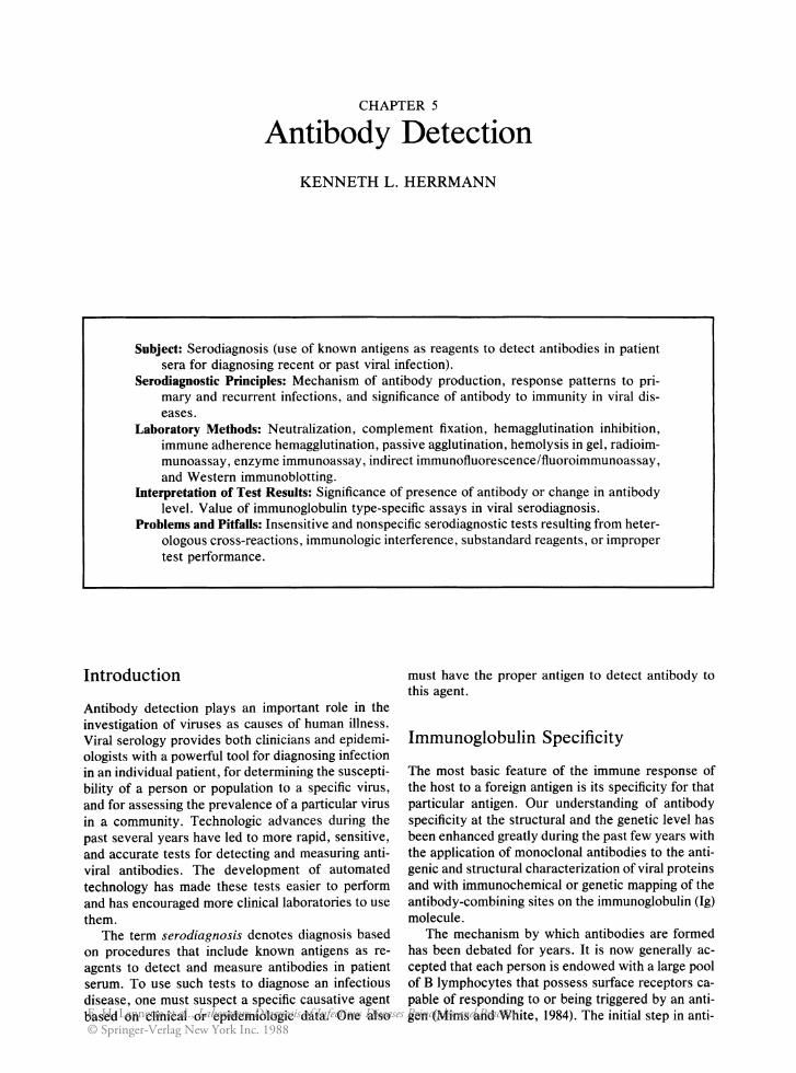
There are considerable differences between the humoral immune response following a primary, or initial, infection with a particular virus and that following a secondary infection or reinfection with the virus. In general, IgM antibodies appear earlier than IgG antibodies in the primary response (Fig. 1). In most primary viral infections, specific antibodies appear in the blood within several days, then rise in titer, and reach a plateau usually within a few weeks. Specific IgM has a half-life of only about 5 days and generally does not persist longer than 2 to 3 months after the acute viral infection. IgG antibodies, on the other hand, usually persist for many years, often for life. Because of the transient nature of the IgM response, the detection of specific IgM antibodies has often been used as an indication of recent or active infection with the virus. Some investigators, however, have reported the persistence of IgM antibody for several months or even years after the onset of primary infection (AI-Nakib et aI., 1975; Pass et aI., 1983). Such reports are more common for viruses that characteristically produce chronic or persistent infections, such as cytomegalovirus. Others have reported the appearance of specific IgM antibody to
IgG
Onset 4 8 12
Weeks postonset
FIG. l. Immune response in primary virus infection. Note early appearance and rapid decline of IgM compared with the appearance and persistence of IgG antibody.
agents other than the one responsible for the current infection (Morgan-Capner et aI., 1983). Such true heterologous IgM responses are probably rare, but should be considered when the results of IgM tests are used in determining clinical management.
Exogenous Reinfection
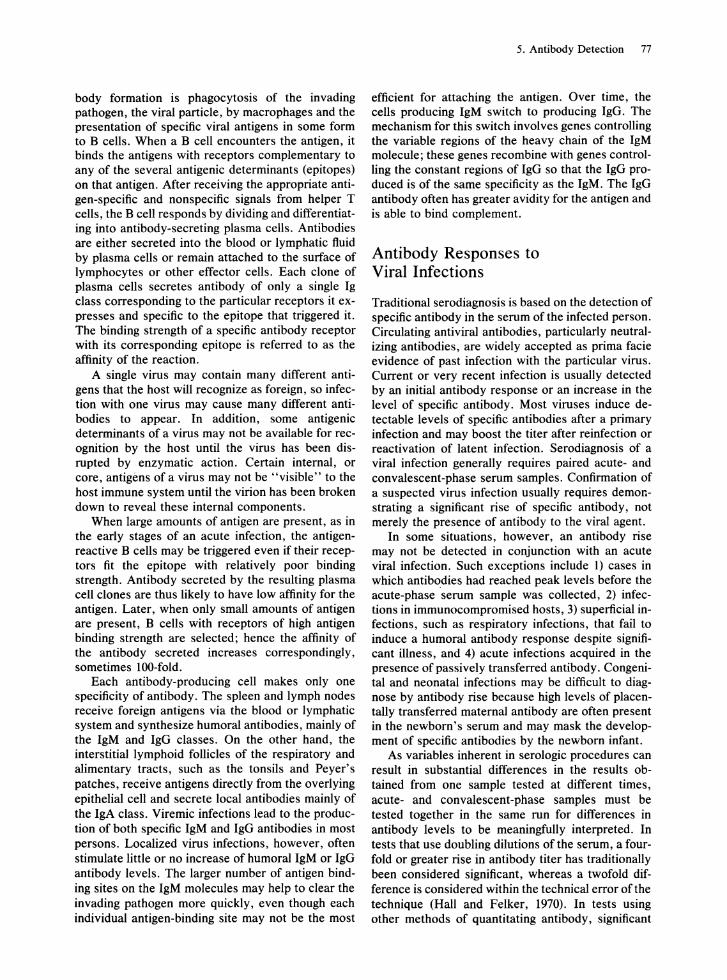
The antibody response to viral reinfection is often quite brisk and usually begins within a day or two after onset of the infection (Fig. 2). Antigen-reactive B lymphocytes, on reexposure to the same antigen, produce larger amounts of specific antibody, mainly IgG, after a delay of only a day or two. Little, if any, specific IgM antibody response is detected following most viral reinfections.
Reactivation of Latent Infection
The antibody response following reactivation of a latent viral infection is less predictable than that following either a primary acute infection or an exogenous reinfection. Examples of reactivated infections are herpes zoster (or shingles) and herpes labialis (or fever blisters). In some instances, antibody increases are brisk. However, antibody levels may not change significantly in others; thus, serodiagnostic results may be misleading.
Measurement of Viral Antibodies
Methods used to detect antiviral antibodies can generally be separated into three groups: 1) methods that measure the ability of an antibody to interfere with some special viral function; 2) methods that depend on the antibody, when reacted with antigen, to perform some non-virus-related function; and 3) methods that directly detect the interaction of the antibody with antigen. The first group includes neutralization and hemagglutination inhibition as-
." ... ____ .!.:.M Onset 4 8 12
Weeks postrelnfectlon
FIG. 2. Immune response in virus reinfection. Note the near absence of an IgM antibody response compared with the rapid booster response of IgG antibody.
says; the second includes complement fixation, passive (or indirect) hemagglutination, immune adherence hemagglutination, and latex agglutination tests; and the third includes enzyme immunoassays, radioimmunoassays, immunofluorescence tests (including fluoroimmunoassays), and Western immunoblotting.
An antibody to a particular antigen may be measurable by a number of different assays. However, different methods for the same agent may measure a different antibody; thus, the results of an antibody assay for a particular virus as measured by one method may not correlate with the results from another method. Each assay may show quantitative as well as temporal differences in antibody response following infection, especially when the assay is run with many different serum samples, representing several variables.
A general overview of the basic principles of the more commonly used methods for detecting and quantifying antiviral antibodies follows in the next sections. Immunodiagnostic tests commonly used for specific viral diseases are listed in Table 1. See the chapters dealing with each virus for additional information regarding the application, performance, and interpretation of the serodiagnostic tests.
Neutralizing Antibody Assay
The neutralization (Nt) test is used to measure antibody to a wide variety of viral agents and represents one of the most sensitive, specific, and clinically important serologic tests available. Nt antibodies persist well beyond the initial illness, and when detected in the absence of symptoms of recent infection with a specific virus, are widely accepted as evidence of immunity to the virus. As Nt antibody can persist in the host for many years, Nt tests are useful in seroepidemiologic studies to determine which viral agents have infected a given population in the past.
Classic viral neutralization occurs when antibody binds to the virion and prevents infection of a susceptible host cell. The precise mechanism of neutralization is not fully understood. Nt antibodies are directed against particular epitopes on accessible surface proteins of the outer capsid or envelope of the virion. Nt antibodies may interfere with viral infectivity by either blocking attachment of the virus to the host cell receptors or by preventing penetration or uncoating of the virion (Mims and White, 1984). The avidity of Nt antibodies may be substantially influenced by the presence of complement and by the degree of glycosylation of the viral glycoprotein antigens (Alexander and Elder, 1984; Sis sons and Oldstone, 1980).
5. Antibody Detection 79
The basic procedure for assaying the virus-neutralizing activity of a serum consists of mixing the serum with known virus, incubating the mixture under appropriate conditions, and injecting the mixture into a susceptible host system in which the presence of residual unneutralized virus can be detected. Most virus Nt antibody assays are carried out in cell cultures in vitro, although in vivo hosts, such as mice or embryonated eggs, may be required for some viruses that do not grow well in cell culture.
Serum specimens are generally assayed for Nt antibody content by testing serial dilutions of the serum against a standard dose (usually 100 TCIDso) of the virus. This is known as the "constant virus-varying serum" technique; it is more useful for demonstrating significant increases in Nt antibody than is the "constant serum-varying virus" procedure, in which a fixed dilution of serum is tested against serial dilutions of virus. In the constant virus-varying serum system, the antibody titer is expressed as the highest serum dilution that neutralizes the test dose of virus. Results of the constant serum-varying virus Nt test are expressed logarithmically as a neutralization index (NI). The NI is the difference in virus neutralizing capacity (in 10gIO LDso) between the unknown and normal control serum samples or between acuteand convalescent-phase serum samples. A log NI of 1.7 is generally accepted as the lower limit for a "positive" result.
The following general procedures can be adapted for detecting Nt antibodies to many human viral pathogens.
NEUTRALIZING ANTIBODY ASSAYS IN TUBE
MONOLA YER CELL CULTURES
1. The serum specimen (either known immune serum or unknown serum to be assayed for Nt antibody) is inactivated at 56°C for 30 min to destroy heat-labile, nonspecific viral inhibitory substances. As some Nt reactions are complement dependent, a source of complement (usually fresh guinea pig serum) is added to the maintenance medium for the cultures.
2. Appropriate serial serum dilutions are prepared in either Hanks basic salt solution (BSS) or the maintenance medium to be used on the cell cultures.
3. The virus is diluted to contain 100 TCIDso in a volume of 0.1 ml (as determined by previous titration of the virus in the selected cell system). Virus endpoint titers are calculated by the methods of Reed and Muench (1938) or Karber (1931). Virus dilutions are prepared in the same diluent used for the serum dilutions. Filtering the test virus before titration will remove aggregated virus and may substantially enhance virus neutralization.
80 K. L. Herrmann
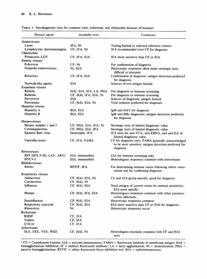
TABLE 1. Serodiagnostic tests for common viral, rickettsial, and chlamydial diseases of humans·
Disease agent
Arenaviruses Lassa Lymphocytic choriomeningitis
Chlamydiae Psittacosis-LGV
Enteric viruses Poliovirus Nonpolio enteroviruses
Rotavirus
Norwalk-like agents Exanthem viruses
Rubella Rubeola Parvovirus Poxviruses
Hepatitis viruses Hepatitis A Hepatitis B
Herpesviruses Herpes simplex I and 2 Cytomegalovirus Epstein-Barr virus
Varicella-zoster
Retroviruses HIV (HTLV-III, LAV, ARV) HTLV-I
Rhabdoviruses Rabies
Respiratory viruses Adenovirus Coronavirus Influenza
Mumps
Parainfluenza Respiratory syncytial Rhinovirus
Rickettsiae RMSF Typhus Q fever
Arboviruses SLE, EEE, VEE, WEE
Available tests
IFA, Nt CF, IFA, Nt
CF, IFA, EIA
CF, Nt Nt, EIA
CF, IFA, EIA
EIA
HAl, EIA, IFA, LA, PHA CF, HAl, IFA, EIA, Nt EIA CF, HAl, EIA, Nt
RIA,EIA RIA, EIA
CF, PHA, EIA, IFA, Nt CF, PHA, EIA, IF A Heterophil,IFA
CF, IFA, FAMA
EIA, immunoblot EIA, immunoblot
RFFIT,IFA
CF, HAl, EIA, Nt CF, HAl, Nt CF, HAl, EIA
CF, HAl, IFA, EIA
CF, HAl, EIA CF, HAl, EIA Nt
CF,IFA CF,IFA CF,IFA
CF, HAl, Nt
Comments
Testing limited to selected reference centers IF A recommended over CF for diagnosis
IF A more sensitive than CF or EIA
For confirmation of diagnosis Heterotypic responses often make serologic tests
difficult to interpret Confirmation of diagnosis; antigen detection preferred
for diagnosis Sources of test antigen limited
For diagnosis or immune screening For diagnosis or immune screening Sources of diagnostic antigen limited Viral isolation preferred for diagnosis
IgM anti-HAY for diagnosis IgM anti-HBc diagnostic; antigen detection preferred
for diagnosis
Serologic tests of limited diagnostic value Serologic tests of limited diagnostic value IFA tests for anti-VCA, anti-EBNA, and anti-EA of
limited diagnostic value CF for diagnosis only; FAMA generally acknowledged
to be most sensitive; antigen detection preferred for diagnosis
EIA for immune screening only Heterologous responses common with retroviruses
For determining immune status following rabies vaccination and for confirming diagnosis
CF and EIA group-specific; good for diagnosis
Need antigen of current strain for optimal sensitivity; EIA most specific
Heterologous responses common with other paramyx-ovirus infections
Heterotypic responses common EIA more sensitive than CF or HAl for diagnosis Heterotypic responses occur
Heterologous reactions common with CF and HAl tests
• CF = Complement fixation; EIA = enzyme immunoassay; FAMA = fluorescent antibody to membrane antigen; HAl = hemagglutination inhibition; IF = indirect fluorescent antibody; LA = latex agglutination; Nt = neutralization; PHA = passive hemagglutination; RFFIT = rabies fluorescent focus inhibition test; RIA = radioimmunoassay.
4. Equal volumes of the serum pilutions and the test virus dilution are mixed. The volume of serumvirus prepared depends on the number of tube cell cultures to be inoculated (0.1 ml each x number of tubes). For a "virus control," the test virus dilution is mixed with an equal volume of diluent, or known "normal" serum ofthe same species as the test serum, and incubated under the same conditions as the serum-virus mixtures. For Nt antibody assays, concurrent titration of the virus is necessary to confirm that the test dose actually contains approximately 100 TCIDso•
5. The conditions recommended for incubation of serum-virus mixtures vary widely. For certain viruses, preliminary incubation increases the neutralizing capacity of the serum. However, it is important to avoid incubation conditions under which a labile virus might be destroyed. For most Nt tests, serum-virus mixtures are incubated for 0.5 to 1 h at 37°C.
6. After the incubation period, the serum-virus mixtures and virus controls are inoculated in a volume of 0.2 ml into monolayer tube cultures of appropriate susceptible cells. At least two cultures are employed for each serum-virus mixture.
7. The inoculated cultures are incubated under conditions most suitable for optimal growth of the virus and examined microscopically to see if the serum inhibits the cytopathic effect (CPE) of the virus. Final readings are usually made when the virus control shows that 100 TCIDso are present in the test. Alternatively, the cell cultures may be examined for virus growth by direct immunofluorescence, enzyme-linked immunosorbent assay (EIA), or immunoperoxidase staining using labeled anti-viral antibody conjugates. In the case of noncytopathic orthomyxoviruses or paramyxoviruses, the cultures may be tested for hemad sorption after a suitable incubation period; neutralization of the virus is evidenced by failure of the cultures inoculated with serum-virus mixtures to hemadsorb the erythrocytes. The antibody titer is expressed as the reciprocal of the highest serum dilution that neutralizes the virus.
MICRO NEUTRALIZATION TESTS IN
CELL CULTURES
Neutralization tests performed in flat-bottomed wells of disposable plastic microtitration plates are more economical than tube Nt tests; smaller volumes of cell cultures and reagents are needed and there is no loss of sensitivity or reproducibility. Microtitration plates processed specially for cell culture growth are commercially available.
Cell monolayers may be prepared in the microtitration plate wells before they are inoculated with the
5. Antibody Detection 81
serum-virus mixtures, or more conveniently, the cells, serum, and virus may be added at the same time. In the latter method, unneutralized virus can infect and destroy the cells before attachment, and monolayers will not be formed, whereas in cultures containing neutralized virus, the cells may attach and proliferate into a confluent monolayer. Endpoints of microneutralization tests may be based on microscopic observation of viral CPE or hemad sorption or on colorimetrically determined metabolic inhibition, direct immunofluorescence or immunoperoxidase staining, or EIA using specific antiviral conjugates to detect unneutralized virus. As with the tube test, the endpoint titer is expressed as the reciprocal of the highest serum dilution that neutralizes the virus.
PLAQUE REDUCTION NEUTRALIZATION TESTS
Plaque reduction Nt tests are very sensitive for virus Nt antibodies. Enumerating virus plaques in monolayer cell cultures is a more precise means of quantifying viral infectivity than TCIDso endpoints in tube or microtitration plate cultures; therefore, demonstrating reduction in virus plaque counts provides a very sensitive means for measuring virus Nt capacity of a serum.
1. The serum specimen is heat inactivated, and appropriate serum dilutions are prepared as described for the tube Nt test.
2. The test virus is diluted to contain sufficient plaque-forming units (pfu) in the inoculum volume to allow precise plaque counting in the monolayer culture.
3. Equal volumes of the diluted test virus and each serum dilution are mixed.
4. After an appropriate incubation period, monolayer cell cultures in bottles or wells of polystyrene cell culture plates are inoculated with serum-virus mixtures, and the cultures are incubated for 1 to 1.5 h at 37°C to permit virus adsorption. The monolayers are then washed with Hanks BSS and covered with an appropriate nutrient overlay containing a solidifying agent such as purified agar or agarose.
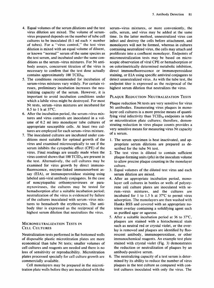
5. After a suitable incubation period at 36 to 37°C, plaques are stained with a histochemical stain such as neutral red or crystal violet, or the overlay is removed and plaques are identified by fluorescent antibody, immunoperoxidase, or other immunochemical reagents. An example test plate stained with crystal violet (Fig. 3) demonstrates the reduction or neutralization of plaques by an antibody-positive serum.
6. The neutralizing capacity of a test serum is determined by its ability to reduce the number of virus plaques in the test culture as compared with control cultures inoculated with only the virus. The
82 K. L. Herrmann
POSITIVE SERUM CONTROL 1110 1140 Vl60 1/640
1/160 NEG. SERUM CONTROL
(C<l'JTINUEO)
FIG. 3. Results of a plaque reduction neutralization test for poxvirus antibody performed using 24-well polystyrene cell culture trays and LLC-MK2 cell monolayers. Note the gradual increase in plaque breakthrough for the positive serum, showing none at 1 : 10, very few at 1: 40, few at
reciprocal of the highest dilution of test serum that reduces the plaque count by 50% or more is the plaque-neutralizing (PNt) antibody titer. The 50% plaque reduction endpoint is most frequently calculated using the pro bit transformation method (Finney, 1971). The percentage of plaque breakthrough for each serum dilution is calculated and plotted on pro bit graph paper, showing the percentages of plaque breakthrough on the ordinate and the serum dilutions in negative 10gIO on the abscissa. A best-fitting straight line is drawn through the points, and a vertical line is dropped from the intersection of the plot line; the 50% probit graph line determines the serum dilution endpoint that gives the 50% plaque reduction.
Neutralizing antibody endpoint titers can be de-termined for almost any virus that can be grown in cell culture by detecting breakthrough virus with direct immunofluorescent staining of the cell monolayer rather than by CPE. This procedure is referred to as thefiuorescentfocus inhibition test. It is particularly useful for viruses that do not produce recognizable CPEs or plaques.
Neutralization testing for serum antibody has been applied to almost all known human viral patho-
VIRUS CONTROL
1 : 160, and about 26 plaques per 3 wells at 1: 640. The 50% plaque reduction neutralization titer for this positive serum is greater than 640. The negative serum shows no plaque reduction with dilutions from 1: 10 through 1: 160.
gens. Virus Nt tests, although relatively simple to conduct, are expensive in time and materials and may be difficult to interpret because of variability in the titration endpoint and difficulties in standardizing the test method. References to protocols giving details and conditions for specific Nt antibody assays may be found in other chapters of this volume. Most viral antibodies, however, can be more easily and relatively accurately assayed by using other less expensive and less cumbersome methods.
Complement Fixation Test
One of the classic methods for demonstrating the presence of antiviral antibody in a patient's serum is the complement fixation (CF) test. The complex complement system plays an important role in mediating and amplifying immune and inflammatory reactions as part of the natural immunopathologic process. Complement is activated after combining with antigen-antibody complexes. This activation sequence is used in the complement fixation test for antibody.
The conventional CF test consists of two stages. In the first stage, serum (antibody) and known anti-
gen are mixed in the presence of a measured amount of complement (usually a pretitrated dilution of guinea pig serum). If the serum antibody and antigen react specifically and form antigen-antibody complexes, the complement will be fixed and depleted. If the serum antibody and antigen do not form antigenantibody complexes, the complement will not be fixed and will remain free in the reaction mixture. In the second stage, sheep erythrocytes sensitized (coated) with anti-sheep erythrocyte antibody (hemolysin) are added to the reacted mixture. As complement is a lytic agent, if any active complement remains in the test system, the sheep erythrocytes will be lysed (hemolyzed). Conversely, the absence of hemolysis indicates that the complement had been depleted or "fixed" and therefore an antigen-antibody reaction had occurred in the first stage.
For the successful performance of this assay, all reagents, with the exception of the unknown serum, must be carefully titrated and used in precisely measured amounts. Interpretation of the test depends on complete fixation of complement in the presence of the antibody in the unknown serum sample and complete hemolysis in the absence of the unknown antibody. Protocols for preparing and standardizing all reagents for performing the standard diagnostic complement fixation test have been published (Hawkes, 1979; Palmer and Casey, 1981; U.S. Public Health Service, 1965). The following is a condensed version of the standardized Laboratory Branch Complement Fixation (LBCF) procedure.
LABORATORY BRANCH COMPLEMENT
FIXATION TEST PROCEDURE
Serum samples may be tested with one or more antigens. Each test must include positive control serum of known reactivity. The titer of an antiserum is defined as the reciprocal of the highest dilution of serum showing 30% or less hemolysis with the optimal antigen dilution. The serum is considered anticomplementary if less than 75% hemolysis occurs in the serum control of any dilution greater than two dilutions below the titer.
Hemolysin Titration
Sheep red blood cells (RBCs) are sensitized with seven dilutions of hemolysin: 1: 1000, 1: 1500, 1 : 2000, 1: 2500, 1: 3000, 1: 4000, and 1 : 8000. Following addition of complement (C'), the hemolysincell mixtures are incubated at 37°C for 1 h, centrifuged, and read with color standards for percent hemolysis. Readings are plotted on arithmetic graph paper, and the graph is examined for a plateau. The second dilution on the plateau is considered the optimal dilution for use in the LBCF.
5. Antibody Detection 83
Complement Titration
Based on the hemolysin titration results, sheep RBCs are sensitized using the optimal dilution of hemolysin. The C' titration is performed in duplicate using the volumes shown in Table 2. Tube titration, rather than microplate titration, is recommended for the C' titration to permit greater precision in estimating the percentage of hemolysis for each dilution in the assay. After the tubes are incubated for 30 min at 37°C, they are centrifuged and read for percentage of hemolysis, and the readings are plotted using log-log graph paper. Working dilutions of C' for the LBCF antigen titration and serum assay are calculated and expressed as the dilution needed to contain 5 C'Hso in 0.4 m!. Complement titers are calculated and expressed as the number of C'Hso units per m!. One C'Hso is the amount of complement that produces 50% lysis in the indicator system.
Antigen Titration (Micro Method)
Antigens are evaluated using at least one reference animal antiserum for specificity and one human reference antiserum (if available) for reactivity. The potency or optimal antigen dilution is normally defined as the dilution that gives the greatest amount of fixation at the highest (or expected) serum antibody titer.
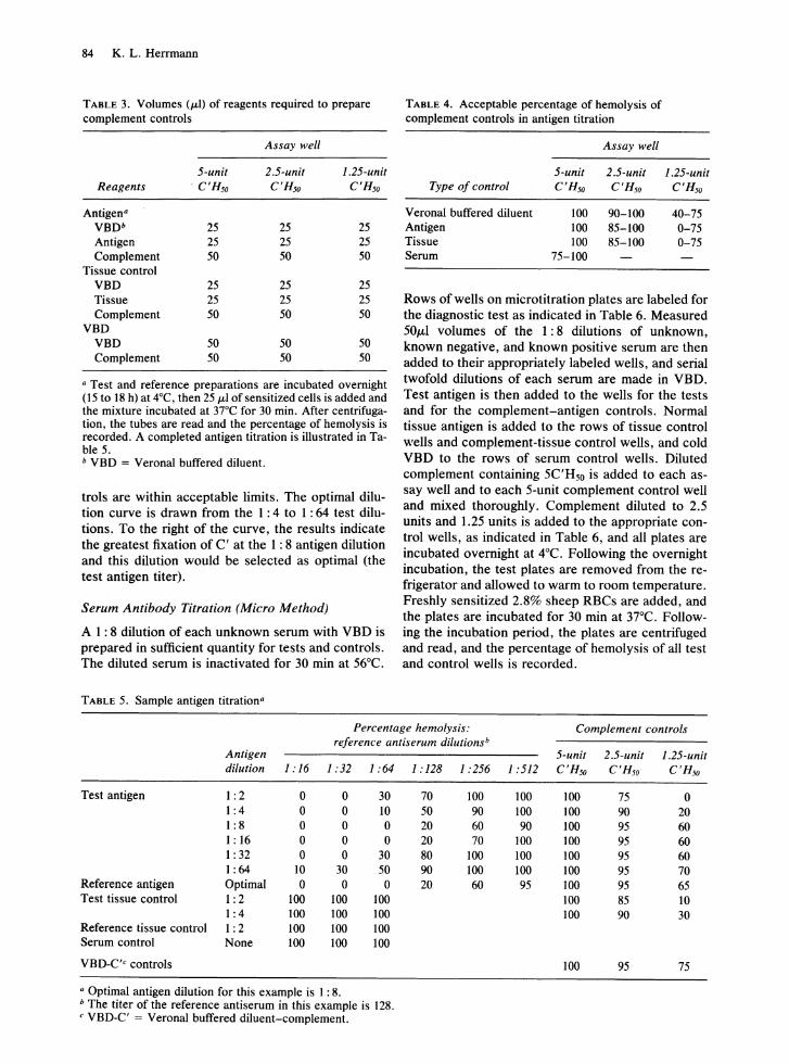
Box titrations are performed in microtitration plates using 25 ILl of serially diluted antiserum, 25 ILl of serially diluted antigen, and 50 ILl C' containing 5 C'Hso units. Complement controls for each dilution of test antigen, reference antigen, tissue control antigen, and Veronal-buffered diluent (VBD) are included. The volumes used to prepare the controls are given in Table 3. Acceptable limits of reactivity for the C' controls are given in Table 4. If the VBD controls are not within acceptable limits, the entire test is invalid. Antigen and tissue dilutions that give less than 85% hemolysis in the 2.5-unit control are considered anticomplementary (AC).
In interpreting each antigen titration, it is convenient to draw a line, known as the optimal dilution curve, through the 30% hemolysis endpoints (interpolated if necessary) of each antigen dilution that is not AC. In the sample titration (Table 5), the 1: 2 dilution of test antigen is AC, and all other C' con-
TABLE 2. Volumes (m!) of reagents required for complement titration
Tube no.
Reagent 2 3
Veronal buffered diluent 0.60 0.55 0.50 1 : 400 dilution of complement 0.20 0.25 0.30 Sensitized cells 0.20 0.20 0.20
4
0.40 0.40 0.20
84 K. L. Herrmann
TABLE 3. Volumes (pol) of reagents required to prepare complement controls
Assay well
5-unit 2.5-unit 1.25-unit Reagents ' C'Rso C'Rso C'Rso
Antigen" VBOb 25 25 25 Antigen 25 25 25 Complement 50 50 50
Tissue control VBO 25 25 25 Tissue 25 25 25 Complement 50 50 50
VBO VBO 50 50 50 Complement 50 50 50
" Test and reference preparations are incubated overnight (15 to 18 h) at 4°C, then 25 pol of sensitized cells is added and the mixture incubated at 37°C for 30 min. After centrifugation, the tubes are read and the percentage of hemolysis is recorded. A completed antigen titration is illustrated in Table 5. b VBO = Verona! buffered diluent.
troIs are within acceptable limits. The optimal dilution curve is drawn from the 1: 4 to 1 : 64 test dilutions. To the right of the curve, the results indicate the greatest fixation of C' at the 1: 8 antigen dilution and this dilution would be selected as optimal (the test antigen titer).
Serum Antibody Titration (Micro Method)
AI: 8 dilution of each unknown serum with VBD is prepared in sufficient quantity for tests and controls. The diluted serum is inactivated for 30 min at 56°C.
TABLE 5. Sample antigen titration"
TABLE 4. Acceptable percentage of hemolysis of complement controls in antigen titration
Assay well
5-unit 2.5-unit Type of control C'Rso C'Rso
Verona! buffered diluent 100 90-100 Antigen 100 85-100 Tissue 100 85-100 Serum 75-100
1.25-unit C'Rso
40-75 0-75 0-75
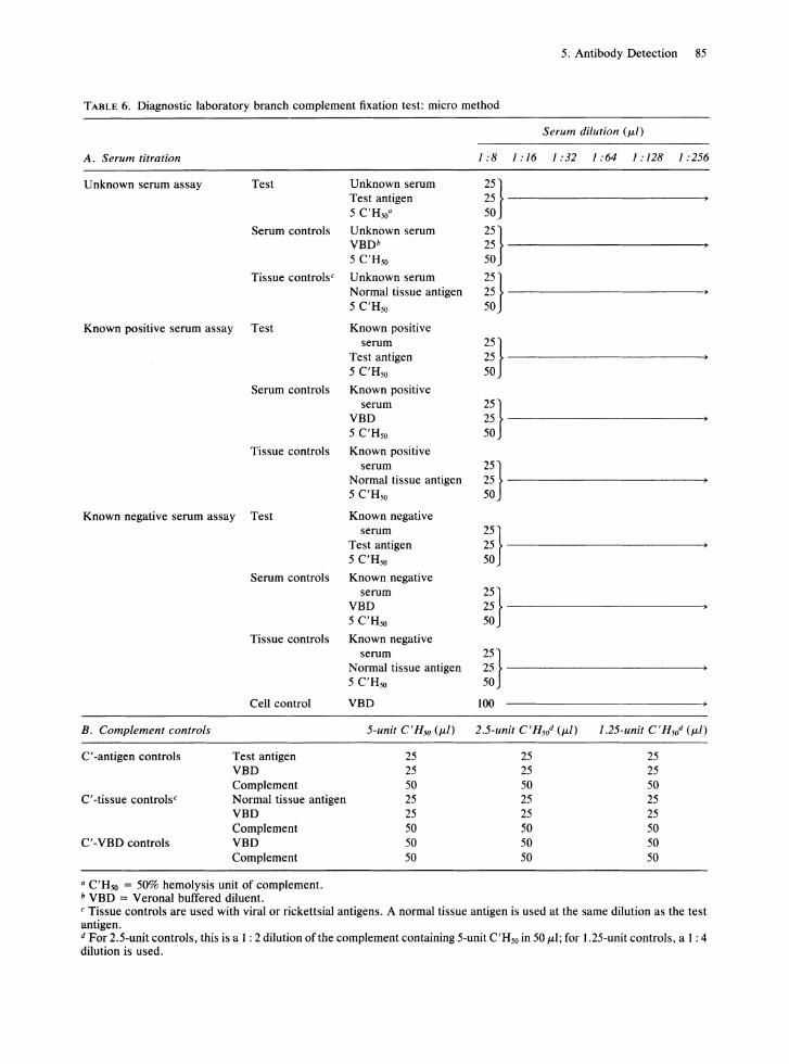
Rows of wells on microtitration plates are labeled for the diagnostic test as indicated in Table 6. Measured 50#LI volumes of the 1: 8 dilutions of unknown, known negative, and known positive serum are then added to their appropriately labeled wells, and serial twofold dilutions of each serum are made in VBD. Test antigen is then added to the wells for the tests and for the complement-antigen controls. Normal tissue antigen is added to the rows of tissue control wells and complement-tissue control wells, and cold VBD to the rows of serum control wells. Diluted complement containing 5C'H50 is added to each assay well and to each 5-unit complement control well and mixed thoroughly. Complement diluted to 2.5 units and 1.25 units is added to the appropriate control wells, as indicated in Table 6, and all plates are incubated overnight at 4°C. Following the overnight incubation, the test plates are removed from the refrigerator and allowed to warm to room temperature. Freshly sensitized 2.8% sheep RBCs are added, and the plates are incubated for 30 min at 37°C. Following the incubation period, the plates are centrifuged and read, and the percentage of hemolysis of all test and control wells is recorded.
Percentage hemolysis: Complement controls reference antiserum dilutionsb
Test antigen
Reference antigen Test tissue control
Reference tissue control Serum control
VBO-C'c controls
Antigen dilution
1: 2 1: 4 1: 8 1:16 1: 32 1 :64 Optima! 1: 2 1: 4 I: 2 None
1:16 1 :32
0 0 0 0 0 0 0 0 0 0
10 30 0 0
100 100 100 100 100 100 100 100
" Optima! antigen dilution for this example is I : 8.
1 :64
30 10 0 0
30 50 0
100 100 100 100
b The titer of the reference antiserum in this example is 128. c VBO-C' = Verona! buffered diluent-complement.
1:128 1:256
70 100 50 90 20 60 20 70 80 100 90 100 20 60
5-unit 2.5-unit 1.25-unit 1 :512 C'Rso C'Rso C'Rso
100 100 75 0 100 100 90 20 90 100 95 60
100 100 95 60 100 100 95 60 100 100 95 70 95 100 95 65
100 85 10 100 90 30
100 95 75
5. Antibody Detection 85
TABLE 6. Diagnostic laboratory branch complement fixation test: micro method
A. Serum titration
Unknown serum assay Test Unknown serum Test antigen 5 C'H50"
Serum controls Unknown serum VBDb 5 C'H50
Tissue controls c Unknown serum Normal tissue antigen 5 C'H50
Serum dilution (ILl)
1:81:161:321:641:1281:256
;;}-------------------------50
;;}------------------------50
;;}------------------------50
Known positive serum assay Test Known positive
Serum controls
Tissue controls
Known negative serum assay Test
B. Complement controls
C' -antigen controls
C' -tissue controls c
C' -VBD controls
Serum controls
Tissue controls
Cell control
Test antigen VBD Complement Normal tissue antigen VBD Complement VBD Complement
a C'H50 = 50% hemolysis unit of complement. b VBD = Verona! buffered diluent.
serum Test antigen 5 C'H50
Known positive serum
VBD 5 C'H50
Known positive
;;}------------------------50
;;}------------------------50
serum 25} Normal tissue antigen 25 ----------------5 C'H50 50
Known negative serum
Test antigen 5 C'H50
Known negative serum
VBD 5 C'H50
Known negative
;;}-------------------------50
;;}-------------------------50
serum 25} Normal tissue antigen 25 --------------~ 5 C'H50 50
VBD
25 25 50 25 25 50 50 50
100
25 25 50 25 25 50 50 50
25 25 50 25 25 50 50 50
C Tissue controls are used with viral or rickettsial antigens. A normal tissue antigen is used at the same dilution as the test antigen. d For 2.5-unit controls, this is a 1 : 2 dilution of the complement containing 5-unit C'H50 in 50 ILl; for 1.25-unit controls, a 1 : 4 dilution is used.
86 K. L. Herrmann
Interpretation of Results
In serum antibody assays, the complement control readings must meet the same criteria as outlined for antigen titrations in Table 4. If the VBn controls are not within acceptable limits, the test results are not valid. The serum titer is the reciprocal of the highest dilution showing 30% or less hemolysis with the optimal dilution of antigen. A serum is AC if the lowest dilution of the serum without antigen indicates less than 75% hemolysis. When serum-AC activity is encountered, usually another serum sample from the patient is requested. AC reactions are usually limited to the lower serum dilutions (i.e., 1 : 8 or 1 : 16); thus high titers (64 or greater) may be demonstrable by CF even in serum with low levels of AC activity.
The CF test has been used widely over the years to detect many antiviral antibodies. In large laboratories where CF is performed routinely, this assay
serves as an accurate diagnostic method for many viral infections. The procedure however, is complex, involves a relatively large number of reagents, requires rigid standardization, and takes 18 to 24 h to be completed. For most viral diseases, alternative test systems have been developed that are less technically demanding, more automated, quicker, and more sensitive than the CF test. Laboratories without experience with CF tests should not adopt this method for routine diagnostic testing when other less demanding procedures are available.
Hemagglutination Inhibition Test
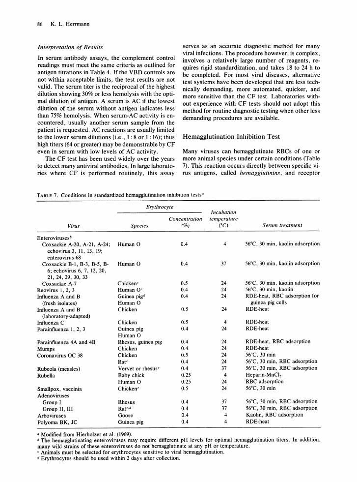
Many viruses can hemagglutinate RBCs of one or more animal species under certain conditions (Table 7). This reaction occurs directly between specific virus antigens, called hemagglutinins, and receptor
TABLE 7. Conditions in standardized hemagglutination inhibition tests·
Erythrocyte Incubation
Concentration temperature Virus Species (%) ("C) Serum treatment
Enterovirusesb
Coxsackie A-20, A-21, A-24; Human 0 0.4 4 56°C, 30 min, kaolin adsorption echovirus 3, 11, 13, 19; enterovirus 68
Coxsackie B-1, B-3, B-5, B- Human 0 0.4 37 56°C, 30 min, kaolin adsorption 6; echovirus 6, 7, 12, 20, 21,24,29,30,33
Coxsackie A-7 Chickene 0.5 24 56°C, 30 min, kaolin adsorption Reovirus 1, 2, 3 Human Oe 0.4 24 56°C, 30 min, kaolin Influenza A and B Guinea pigd 0.4 24 RDE-heat, RBC adsorption for
(fresh isolates) Human 0 guinea pig cells Influenza A and B Chicken 0.5 24 RDE-heat
Oaboratory-adapted) Influenza C Chicken 0.5 4 RDE-heat Parainfluenza 1, 2, 3 Guinea pig 0.4 24 RDE-heat
Human 0 Parainfluenza 4A and 4B Rhesus, guinea pig 0.4 24 RDE-heat, RBC adsorption Mumps Chicken 0.4 24 RDE-heat Coronavirus OC 38 Chicken 0.5 24 56°C, 30 min
Rate 0.4 24 56°C, 30 min, RBC adsorption Rubeola (measles) Vervet or rhesuse 0.4 37 56°C, 30 min, RBC adsorption Rubella Baby chick 0.25 4 Heparin-MnCIz
Human 0 0.25 24 RBC adsorption Smallpox, vaccinia Chickene 0.5 24 56°C, 30 min Adenoviruses
Group I Rhesus 0.4 37 56°C, 30 min, RBC adsorption Group II, III Ratc·d 0.4 37 56°C, 30 min, RBC adsorption
Arboviruses Goose 0.4 4 Kaolin, RBC adsorption Polyoma BK, JC Guinea pig 0.4 4 RDE-heat
• Modified from Hierholzer et al. (1969). b The hemagglutinating enteroviruses may require different pH levels for optimal hemagglutination titers. In addition, many wild strains of these enteroviruses do not hemagglutinate at any pH or temperature. e Animals must be selected for erythrocytes sensitive to viral hemagglutination. d Erythrocytes should be used within 2 days after collection.
sites on the red cell surface and results in visible clumping of the cells. Antibodies to these virus hemagglutinins react with the virus and prevent the hemagglutination. This principle forms the basis of the hemagglutination inhibition (HAl) test.
The HAl test has been widely used for diagnosing current infection and determining immune status for viruses with a hemagglutinin component. The test is relatively easy to perform. However, because most serum contains nonspecific viral inhibitors of hemagglutination, diagnostic serum specimens must be treated before testing for specific antibody. Failure to remove the nonspecific inhibitors may result in false-positive results. Natural agglutinins to the RBCs, if present, must also be removed before assaying for specific antibody.
The sensitivity and specificity of HAl tests depend on many variables, and tests must be carefully controlled if accurate and reproducible results are to be expected. The efforts to standardize viral HAl test procedures are reflected in the following section.
STANDARDIZED HEMAGGLUTINATION AND HEMAGGLUTINATION INHIBITION TESTS
Standardized micro titration methods for the HA and HAl tests have been described (Hierholzer and Suggs, 1969; Hierholzer et al., 1969). These techniques have been used for detecting and titrating antibody to adenoviruses, myxoviruses, paramyxoviruses, rubeola virus, reoviruses, and vaccinia. The following is a condensed version of this procedure.
Preparation of Red Blood Cells
Red blood cells should be obtained from the proper animal species (see Table 7). They should be washed and standardized on the day of the test.
Titration of Hemagglutinin
The antigens used are titrated by HA in microtitration plates to determine the proper dilution needed to give 4 HA units per 25 JLl. Table 7 lists the major variations in the standardized HAl tests. The plates are read for hemagglutination when the cells in the control wells have formed compact buttons or rings of nonagglutinated cells (usually after 1 to 2 h).
To facilitate reading exact endpoints, the plate may be slanted for approximately 30 s so that nonagglutinated cells will flow in a teardrop pattern. Because many viruses elute and give falsely low titers, the agglutination patterns should be read immediately. The last well that shows complete agglutination is considered the endpoint and equals 1 HA unit.
Hemagglutination Inhibition Test
The virus antigen is diluted to contain 4 HA units per 25 JLl. To ensure that the proper test dilution is used,
5. Antibody Detection 87
a back titration should be performed before and during the test. If the test dilution is accurate, agglutination will occur in the first three wells but not in the remainder of the back titration series. If too few or too many positive wells are observed, the test antigen concentration may be adjusted by adding more virus or more diluent.
The serum treatments necessary for the HAl tests are given in Table 7. The RBC adsorption is done with 50% RBC in phosphate-buffered saline (PBS) by mixing 0.1 ml ofRBC suspension with 1 ml of 1: 10 dilution of serum and incubating at 4°C for 1 h. For kaolin adsorption, equal volumes of a 1 : 5 dilution of serum and a 25% suspension (wt/vol) acid-washed kaolin in PBS are incubated at room temperature for 20 min. Other serum treatment methods have been recommended in the literature for specific HAl tests.
The HAl test is performed in a total volume of 100 JLI (25 JLI of serum, 25 JLI of antigen, and 50 JLI of RBC suspension) with suitable reference controls and is read when the control wells show compact buttons or rings (usually after 1 to 2 h). As in the HA test, to facilitate reading, plates may be slanted for 30 s so that nonagglutinated (inhibited) cells will "tear." Cell control wells and serum control wells should show no agglutination. The HAl titer of each serum is defined as the reciprocal of the highest dilution of serum that completely inhibits hemagglutination. If nonspecific agglutination is seen in the 1 : 20 dilution or above, the serum should be readsorbed with a 50% RBC suspension and the HAl repeated.
Unique HAl test protocols have been developed for some viruses such as rubella and other togaviruses. See other chapters for details and references for these special methods.
Immune Adherence Hemagglutination
Immune adherence hemagglutination (IAHA) is a variation of the CF test in which agglutination rather than lysis of erythrocytes is measured (Lennette and Lennette, 1978; Nelson, 1963). Both methods detect antibodies by demonstrating bound complement to indicate the presence of antigen-antibody complexes. In IAHA, complement that has been bound by antigen-antibody complexes is then bound to the C'3b receptors located on the surface of primate erythrocytes, resulting in hemagglutination. This technique has been used to detect antibodies to influenza, herpes simplex, Epstein-Barr, varicella-zoster, hepatitis B, and rubeola viruses, cytomegalovirus, human rotavirus, and is reportedly up to 20 times more sensitive than CF tests.
Although IAHA offers advantages of increased sensitivity for antibody detection, reduced performance time, and less sensitivity to anticomplementary activity compared with CF tests, few clinical
88 K. L. Herrmann
laboratories have adopted this method in place of CF tests for routine viral diagnosis.
Reagents and equipment required for IAHA are similar to those needed for the CF test. The following brief outline for the IAHA procedure has been summarized from that published by Lennette and Lennette (1986).
IMMUNE ADHERENCE HEMAGGLUTINATION
MICROTITRATION PROCEDURE
1. Each test serum is pretreated by diluting the serum 1 : 4 with Veronal-buffered saline (VBS) and incubating it at 56°C for 30 min to inactivate natural complement in the serum.
2. VBS (25 ,oOtI) containing 0.1% bovine serum albumin is added to each well of the plate.
3. Then 25 ILl of each inactivated serum is added to wells 1 and 8 of the corresponding row on the test plate.
4. Using micro diluters , serial twofold dilutions (in sets of seven and five wells) are made for each test serum.
5. Optimally diluted test antigen (25 ILl) is added to wells 1 through 7 of each row; 25 ILl of identically diluted negative (control) antigen is added to wells 8 through 12. The plates are shaken to mix contents of wells. The optimal antigen dilution is determined by prior block titration with control reference serum.
6. The plates are covered and incubated for 30 min at 37°C.
7. Diluted guinea pig complement (25 ILl of 1 : 100 C' in VBS) is added to all test wells. The plates are again shaken and incubated for 40 min at 3rC.
8. Following incubation, the reaction is stopped by adding 25 ILl of VBS containing disodium ethylenediaminetetraacetic acid (EDTA) and dithiothreitol (DTT) to each well, and 25 ILl of 0.4% human 0 erythrocytes is added to the mixture.
9. The hemagglutination patterns are read after 1 h. A positive agglutination reaction (Le., granular or coarse pattern of the settled cells) indicates the presence of antibody; a negative reaction appears as a smooth compact cell button. The reciprocal of the highest dilution of serum resulting in a clearly positive agglutination pattern is considered the endpoint titer.
Passive Agglutination and Passive Hemagglutination
Measuring the agglutination of a particulate antigen by its specific antibody (direct agglutination) is the simplest way to estimate the quantity of that antibody in serum. Very small amounts of antibody can be detected by this method, because only a small
number of antibody molecules are necessary to form the antigen-antibody lattice.
The agglutination reaction has been extended to include a wide variety of antigens by attaching soluble antigens to the surface of inert particles, such as latex, bentonite, or RBCs. The role of these particles, once coated or sensitized, is passive; they react as if they themselves possess the antigenic specificity of the coating antigen.
RBCs are extremely convenient carriers of antigen. When specific antibody is added to antigencoated RBCs, antibody bridges are formed between neighboring cells, and large aggregates of RBCs are produced that are visible to the naked eye. This agglutination is designated passive hemagglutination (PHA) or indirect hemagglutination. The following PHA procedure is adopted and summarized from that recommended by the Centers for Disease Control for toxoplasma, herpes simplex, and cytomegalovirus antibody assay (Palmer et ai., 1977).
PASSIVE HEMAGGLUTINATION
TEST PROCEDURE
Preparation of 2.5% Sheep Red Blood Cells
Sheep blood is washed three times in PBS (pH 7.2), centrifuging for 5 min at 900 x g between each wash. After the third wash, the cells are packed for 10 min. A 2.5% sheep RBC suspension is then prepared from the packed cells (39 volumes of PBS, pH 7.2, to 1 vol of packed cells).
Tanning of Standardized Cells
Fresh tannic acid solution is prepared by dissolving tannic acid in PBS (pH 7.2) to yield a final dilution of 1: 1000 (e.g., for each milligram of tannic acid, add 1 ml of PBS, pH 7.2). This 1: 1000 stock solution may be further diluted 20-fold to obtain the 1 : 20,000 dilution for use in the test. Five milliliters of 2.5% SRBC is mixed with 5 ml of the 1 : 20,000 tannic acid solution in a 15-ml centrifuge tube, and the tube is incubated in a 37°C water bath for 10 min. The mixture is then centrifuged for 5 min, the supernatant fluid is removed, and the cells are resuspended in 10 ml of PBS (pH 7.2). Following a final centrifugation, the supernatant fluid is removed and the cells are reconstituted to a 2.5% suspension by adding PBS (pH 6.4).
Sensitization of Cells
Antigen is diluted to its optimal dilution in PBS, pH 6.4 (the optimal dilution as determined by block titrations is the lowest dilution of antigen yielding the highest specific serum titer). One milliliter of the 2.5% tanned cells is mixed with 1.0 ml of optimal diluted antigen and incubated for 15 min at 37°C.
Control cells are prepared in parallel by mixing 1.0 ml of 2.5% tanned cells with 1.0 ml of PBS (pH 6.4) and incubating for 15 min at 37°C. After centrifugation and removal of the supernatant, the cells are washed several times in rabbit serum diluent (PBS, pH 7.2, containing 1% normal rabbit serum). Following the washings, the cells are adjusted to a 1.0% suspension by adding normal rabbit serum diluent. The 1.0% suspensions of tanned, sensitized cells and tanned, unsensitized cells are now ready for use in the test.
Serum Antibody Assay (Micro titration Adaptation)
Each test serum is inactivated for 30 min at 56°C and diluted 1 : 10 in the rabbit serum diluent. Two rows of eight wells marked for each serum on microtitration U plates, and two wells are marked for the cell controls (one for sensitized and one for unsensitized cells). With a multichannel pipetter or microdropper, 50 ILl of normal rabbit serum diluent is added to all wells in which serum dilutions will be made except for the first well in each row, and to the two wells that will serve as the cell controls. One-tenth milliter of the 1 : 10 dilution of each serum is pipetted into the first wells of the two rows, and twofold dilutions of serum are made with 50-ILl microtitration loops. Tanned, sensitized sheep RBCs are added to the wells of the first row of each serum and to the well marked for the sensitized cell control; tanned, un sensitized sheep RBCs are added to wells of the second row of each serum and to the well marked for the un sensitized cell control. The plates are covered and incubated in a 37°C water bath for 2 to 3 h or left at room temperature until the controls show wellformed buttons.
The reciprocal of the highest initial dilution yielding a clearly positive agglutination of sensitized red cells is considered an endpoint. If agglutination of control cells in the second row is equal to that of the sensitized cells, the serum must be adsorbed as follows and the test repeated: 1) Inactivate serum and dilute 1: 10. 2) Adsorb two or more times with 0.1 ml of 50% sheep RBC per ml of diluted serum for 30 min at 4°C. If this treatment fails to remove nonspecific agglutination, the serum must be adsorbed in the same manner with sheep RBCs sensitized with rabbit serum.
Only in the past few years have passive agglutination and PHA become widely accepted in viral serology. Latex agglutination and PHA tests are currently the most widely used procedures in the United States for rubella immunity screening.
Hemolysis in Gel
Hemolysis in gel (HIG) , sometimes referred to as single radial hemolysis, is a serologic method based
5. Antibody Detection 89
on lysis of antigen-sensitized RBCs in a gel by antibody in the presence of guinea pig complement. This technique has been widely accepted in some countries to detect or measure antibodies to several hemagglutinating viruses, including rubella, influenza, mumps, and parainfluenza 3 viruses.
The reaction is carried out on RBCs to which viral antigen has been bound and takes place with the cells suspended in an agarose gel containing complement. The serum specimen, previously heated to inactivate native complement, is allowed to diffuse into the gel from a well, and if specific antibody is present, causes a zone of hemolysis around the well. The diameter of the concentric zone of hemolysis is proportional to the concentration of specific antibody in the serum.
Several procedures for performing HIG tests have been reported (Neumann and Weber, 1983; Russell et al., 1975). The method described here has been adapted from the protocol recommended by Public Health Laboratory Service of Great Britain for rubella antibody testing (Pattison, 1982).
HEMOLYSIS IN GEL PROCEDURE
Gel Preparation
One test and one control gel can be prepared in 100-by l00-mm trays or petri dishes as follows. Two 15-ml quantities of 1 % agarose in veronal-buffered saline (VBS), pH 7.3, are melted in a 44°C water bath. Into each of two 30-ml screw-cap centrifuge bottles, one marked "test" and the other "control," 0.3 ml of a 15% (vol/vol) suspension of sheep RBCs in VBS is pipetted. The "test" cells are combined with 0.3 ml of the test antigen (hemagglutinin) diluted to contain approximately 128 HA units (see Titration of Hemagglutinin). After 30 min of incubation at room temperature, both bottles are filled with VBS and centrifuged at 900 x g for 10 min. The supernatants are discarded, and the cells are resuspended in 0.5 ml of VBS. One-half milliter of undiluted guinea pig complement is then added to each bottle, both bottles are placed in a 44°C water bath, and 15 ml of the melted agarose is immediately added to each. After mixing, the contents of each are poured into appropriately labeled 100-mm2 dishes or trays placed on a level surface. After the gels have thoroughly set at room temperature, they may be stored at 4°C in a humid chamber for up to 5 days before use.
Test Procedure and Controls
Serum to be tested must be inactivated by heating for 20 min at 60°C. Wells with diameters of 2 to 3 mm (capacity, 8 to 11 ILl) are cut in the gels using a template, maintaining a minimum of 10 mm between wells. The agar cylinders are sucked out with a Pasteur pipette attached to a vacuum line and trap. Each
90 K. L. Herrmann
inactivated serum is added to the appropriate well in the test and control gels. The wells should not be overfilled. Known positive and negative control sera must be tested on each gel tray. If antibody levels are to be quantitated by this method, serial dilutions of a reference "calibrator" serum of known titer must be included. One positive control should be adjusted to have a minimal (breakpoint) potency to define positive (immune) from negative (nonimmune). The gels are incubated in a humid chamber at 37°C overnight. Zones of hemolysis are best read by transilluminating the gels against a black background. The diameter of the zones of hemolysis are measured to the nearest 0.1 mm and compared with the controls.
Interpretation
Test zones larger than that of the minimal potency (breakpoint) control may be reported positive. Test zones smaller than that of the breakpoint control should be reported negative for purposes of immune screening.
Although the HIG test has primarily been used as a qualitative or semiquantitative antibody assay, when accurately calibrated, it can be used in place of other quantitative tests for determining antibody titers for diagnostic purposes.
HIG has several theoretical and practical advantages over other viral antibody assays. It is simple to perform. It is unaffected by most nonspecific inhibitors in human serum that interfere with the virus hemagglutinins, and is therefore more specific than corresponding HI tests, which require special serum treatment to remove nonspecific HA inhibitors before testing. On the other hand, the limited "shelf life" of prepared gels requires that clinicallaboratories in countries where the time between kit production and delivery is greater than 2 weeks prepare their own gels.
Radioimmunoassay
Radioimmunoassay (RIA) offers a very sensItIve technique for detecting and quantitating viral antibodies and is currently one of the most widely applied of all immunoassays. During the past decade, many RiA systems have been developed (Mushahwar and Brawner, 1986; Mushahwar and Overby, 1983). However, because of the relative instability of the labeled reagents used and the special conditions and equipment required for handling radioisotopes, RIA systems in virology have been limited primarily to the research laboratory, and only a few of these procedures, notably hepatitis antigen and antibody assays, have successfully made the transition from research procedures to commercially available diagnostic tests.
Most radioimmunoassays for detecting anti-viral antibodies are based on either the traditional indirect solil;l-phase RIA (SPRIA), solid-phase competitive binding RIA, or radioimmunoprecipitation (RIP) systems.
INDIRECT SOLID-PHASE RADIOIMMUNOASSAY
PROCEDURES
The indirect SPRIA for antibody detection uses a purified or semi-purified viral antigen adsorbed to plastic beads, tubes, or microtitration wells to create a solid surface capable of capturing and binding specific antibody in the test serum sample. Removable "strips" or rows of microtitration wells are commonly used as the solid phase for RIA and EIA test systems, are available commercially (Titertek FB strips, Flow Laboratories, McLean, Va.), and allow greater economy in use of antigen coated microwells when a full 96-well plate is not needed for an assay. Although a variety of commercially available viral antigens have been used successfully in indirect SPRIAs, the specificity of the assay depends primarily on the quality and purity of the antigen preparation, that is, freedom from contaminating nonviral proteins (Parratt et al., 1982). Control antigen (i.e., antigen that contains all components in the test antigen except the virus) must be available and used whenever possible to detect nonspecific reactivity.
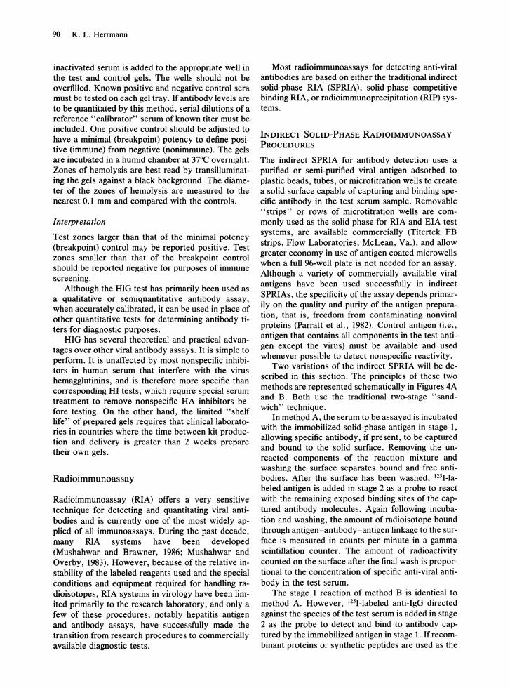
Two variations of the indirect SPRIA will be described in this section. The principles of these two methods are represented schematically in Figures 4A and B. Both use the traditional two-stage "sandwich" technique.
In method A, the serum to be assayed is incubated with the immobilized solid-phase antigen in stage 1, allowing specific antibody, if present, to be captured and bound to the solid surface. Removing the unreacted components of the reaction mixture and washing the surface separates bound and free antibodies. After the surface has been washed, 125I-Iabeled antigen is added in stage 2 as a probe to react with the remaining exposed binding sites of the captured antibody molecules. Again following incubation and washing, the amount of radioisotope bound through antigen-antibody-antigen linkage to the surface is measured in counts per minute in a gamma scintillation counter. The amount of radioactivity counted on the surface after the final wash is proportional to the concentration of specific anti-viral antibody in the test serum.
The stage 1 reaction of method B is identical to method A. However, 125I-Iabeled anti-IgG directed against the species of the test serum is added in stage 2 as the probe to detect and bind to antibody captured by the immobilized antigen in stage 1. If recombinant proteins or synthetic peptides are used as the
5. Antibody Detection 91
FIG. 4. Indirect solid-phase radioimmunoassay for detecting antibody. Known antigen bound to the solid surface is reacted with the sample containing antibody. Unbound proteins are washed away and the antibody is detected by a second radiolabeled antigen probe (A) or by a radiolabeled anti-human immunoglobulin probe (B).
R. Using Radial.baled Rntigen Praba
Incubat.~ ;.\~ Incubat. ~. ~ and and
~ o~ 0 Immablllzid
Spioili. + Sirum ADtiglD
125I_Laba ... + SpI.ili.
B.Il.I.
B. Using Radialabalad Rnti-Ig Praba
Incubate t-t. and
Wash
==> I--r! InCUbat.~ ;.\~ and .
Wash
==>
ImlDabilizld Splcific Ballgl.
+ Sirum 1251_Labilld
BDIi-I.
capture antigen and labeled monoclonal anti-human immunoglobulin as the probe, method B may be simplified to one stage by allowing the addition of the probe immediately following the test serum without the need for an intervening wash step. Again as described for method A, the amount of radioactivity binding to the solid phase through the antigen-antibody-anti-IgG complex is proportional to the concentration of specific anti-viral antibody in the test serum.
These indirect SPRIA procedures have been used in the development of commercial test kits for detecting antibody to hepatitis B surface antigen.
COMPETITIVE BINDING SOLID-PHASE
RADIOIMMUNOASSA YS
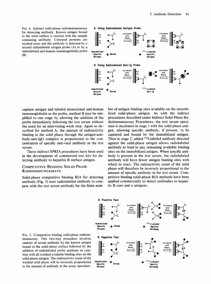
Solid-phase competitive binding RIA for detecting antibody (Fig. 5) uses radiolabeled antibody to compete with the test serum antibody for the finite num-
ber of antigen binding sites available on the immobilized solid-phase antigen. As with the indirect procedures described under Indirect Solid-Phase Radioimmunoassay Procedures, the test serum specimen is incubated in stage 1 with the solid-phase antigen, allowing specific antibody, if present, to be captured and bound by the immobilized antigen. Then in stage 2, added 125I-Iabeled antibody directed against the solid-phase antigen allows radiolabeled antibody to bind to any remaining available binding sites on the immobilized antigen. When specific antibody is present in the test serum, the radiolabeled antibody will have fewer antigen binding sites with which to react. The radioactivity count of the solid phase will therefore be inversely proportional to the amount of specific antibody in the test serum. Competitive binding solid-phase RIA methods have been applied commercially to detect antibodies to hepatitis B core and e antigens.
R. Pasitin Ta.t
FIG. 5. Competitive binding solid-phase radioimmunoassay. This two-step procedure involves capture of serum antibody by the known antigen bound to the solid-phase surface followed by the addition of radiolabeled probe antibody to combine with all residual available binding sites on the solid-phase antigen. The radioactivity count of the washed solid phase will be inversely proportional to the amount of antibody in the assay specimen.
lmmabUilid SpI.llle + Sirum RDIi.ID
B. Dagatin Tlst
JllllDabililld Splcific RDligl.
+ Sirum
Incubat.~;.\ ~ '-"* Incubat. Iq.' and ~ and Wash Wash
==> X==> 1251_Llbilld
+ Spleilie BDlibad.
Incubat. ~. and
Wash
==>
+ '25I-Labllld Splcific BDlibadg
Incubat. tt· and
~i.
92 K. L. Herrmann
RADIOIMMUNOPRECIPIT AnON
The radioimmunoprecipitation (RIP) assay for antibody is a competitive assay carried out in a liquid phase system. The test is usually performed using single, purified proteins as antigen and thus is highly specific. For this test, the viral protein antigen is radiolabeled, added to the test serum, and after incubation, anti-IgG directed against the species of the test serum is added. The anti-Ig cross-links the antigen-antibody complexes already in solution and precipitates the complexes. Radiolabeled viral protein is included in the precipitate if the test serum contains homologous viral antibodies. The precipitate is washed, and the level of radioactivity in the precipitate counted. The amount of radioactivity in the precipitate is proportional to the concentration of specific antibody in the serum.
The accuracy of the RIP test for viral antibody depends on the antigen used. The extreme sensitivity and potential specificity of RIP can be a disadvantage as well as an advantage. Theoretically, the test can detect very low levels of specific antibody; however, it may also detect unwanted cross-reacting antibodies or antibodies to nonviral components of the antigen. For many other types of antibody assays, satisfactory antigens can be purchased or obtained from colleagues without much difficulty. However, the investigator must frequently prepare his own test antigens for an RIP assay. Purifying the antigen, which may be specific viral protein, is often tedious work. RIP tests, therefore, have had only limited success outside the research laboratory.
Enzyme Immunoassay
Among the most useful and widely applied methods of detecting antiviral antibodies are the indirect enzyme immunoassays (EIA or ELISA). The basic principles of EIA are similar to the indirect solidphase RIA methods described under Indirect SolidPhase Radioimmunoassay Procedures.
The EIA incorporates many of the desirable features of the RIA and provides the added advantages of greater reagent stability and potential safety (for example, no handling of radioisotope-labeled reagents). Theoretically these methods can be used to detect and titrate antibody directed against any virus antigen that can be grown in cell culture or in an animal host. The degree of purity required for EIA antigens depends on the information being sought (Kenny and Dunsmoor, 1983). Lysates of virus infected cells are often satisfactory as EIA antigens, especially if a broad-reacting antigen is needed. Cell lysate antigens may be prepared from infected cell cultures by harvesting and washing the infected
cells, disrupting them by sonication or freeze-thawing, clarifying the suspension by low-speed centrifugation, and semipurifying the antigen by pelleting the viral proteins at high speed centrifugation followed by resuspending the pellets in PBS or buffer. When preparing cell lysate antigens, it is important that the lysate be as free as possible of serum or other protein supplements from the cell culture medium that may contribute adversely to nonspecific reactivity of the antigen. Such cell lysate antigens should always be used together with control antigens consisting of uninfected cells processed in the same way as the virusinfected cells.
If greater specificity is required, gradient-purified virus or viral subunits may be prepared and used as antigen. Control antigens for these more purified materials may not always be available because uninfected cell cultures processed in the same manner as the antigen often contain no residual protein to bind to the solid phase.
The solid phase used in the EIA is critical to the precision and accuracy of the assay. Although polystyrene microtitration plates or strips and tubes have been the most commonly used, other supports such as beads, disks, sticks, and cuvettes have also been used. Reproducibility of the assay depends on wellto-well consistency in the coating of the solid phase with the antigen.
The sensitivity of any enzyme immunoassay is directly related to the properties of the enzyme label used. A suitable enzyme for EIA is one that can be readily linked to the detector antibody without losing functional activity. To provide sensitivity, the enzyme must have a sufficiently high turnover rate to permit adequate amplification of the serologic reaction within a desirable time. The enzyme should have substrates that are degradable to stable and easily measured products. Finally, the enzyme should be relatively inexpensive, nontoxic, and readily available in purified form. To date, horseradish peroxidase and alkaline phosphatase have been the two most widely used enzymes for EIA systems. Methods for conjugating these enzymes with antihuman gamma globulin have been thoroughly described elsewhere (Avrameas, 1969; Shekarchi and Sever, 1986; Wilson and Nakane, 1978).
Indirect EIAs have been used to measure specific antibodies in a wide variety of viral infections, and the number of applications of the test continues to expand rapidly. In the process, many different variations in both the procedure and in the reagents used have developed. All indirect EIA systems, however, have certain common features. Indirect EIA is similar to the indirect SPRIA (method B) described under Indirect Solid-Phase Radioimmunoassay Procedures. All EIA systems require careful washing be-
tween steps to remove unbound reactants, which, if not completely removed, might result in nonspecific or false-positive results. A variety of automated and semiautomated devices and readers are available for EIA. These include pipetting devices, plate washers, and spectrophotometric plate readers.
As with test methodology, many systems have been developed for reporting EIA test results. At one extreme, a single serum dilution is tested, and antibody is measured quantitatively in a spectrophotometer, absorbance is relative to the quantity of antibody present (Voller and Bidwell, 1979). Others have used an "index" or ratio of a spectrophotometric reading for the unknown specimen compared with that of a reference calibrator reading. At the other extreme is the use of serial twofold dilutions with a visual or spectrophotometric reading of the test endpoint (Bullock and Walls, 1977).
EIA technology continues to evolve at a rapid rate. New solid-phase carriers, more sensitive and specific reagents, and improved instruments and readers continue to be introduced. Over the past several years, EIA has replaced other traditional test methods for the serodiagnosis of many common viral infections.
Indirect Immunofluorescence Assays
Indirect fluorescent antibody (IF A) tests and indirect solid-phase fluoroimmunoassays (PIA) have been widely used in many clinical and research laboratories for detecting and quantitating antiviral antibodies. The IF A test is a two-step procedure similar in principle to the indirect EIA and RIA (see sections on radioimmunoassay and enzyme immunoassay and Fig. 4) involving application of patient serum to the antigen preparation (usually virus-infected cells fixed on glass microscope slides) followed by incubation of the slide with fluorochrome-labeled anti-human globulin. Although the IF A procedure is rapid and may be useful in detecting antibodies that are difficult or even impossible to demonstrate by any other means, nonspecific fluorescence frequently occurs and results are open to subjective interpretation. This has resulted in less than optimum sensitivity and specificity in many laboratories.
INDIRECT FLUORESCENT ANTIBODY METHOD
IF A test procedures vary slightly according to the specific antigen and test system used. Specific IF A test protocols and references may be found in some of the other chapters of this book. In general, however, the following procedure can be used to identify antibodies to almost any virus that can be grown in cell culture.
5. Antibody Detection 93
Preparation of Antigen Slides
Slides may be prepared using infected cell monolayers showing 2+ to 3+ CPE. The infected cells are suspended and mixed with an equal number of cells from an uninfected culture. Mter thorough rinsing in PBS, the cells are sedimented by centrifugation and resuspended in a small amount of PBS containing I to 2% fetal bovine serum or 0.05% gelatin. The cell suspension is then spotted on glass slides, allowed to air-dry at room temperature, and fixed in acetone for 5 to 10 min. In addition, uninfected cells are prepared separately to serve as an uninfected cell control. Slides with 10 pre-etched wells are routinely used for antigen spots for IF A testing. After fixation, the antigen slides may be stored frozen in airtight envelopes until needed for testing. Antigen and control slides for viral antibody IF A assays are available commercially.
Test Procedure and Controls
Serial dilutions (beginning at 1 : 2) of each test serum specimen and of the positive control serum are prepared in PBS. One drop of each serum dilution is placed on a corresponding antigen spot on the slide. One drop of undiluted negative control serum and one drop of PBS are placed on corresponding antigen spots as a negative serum control and a nonspecific staining control for the test. The slides are then incubated in a covered moist chamber for 30 min at 35°C. Following the incubation, each slide is gently rinsed and emersed in a staining dish with PBS for 10 min. The slides are then dipped in distilled water, blotted gently, and air-dried for 10 min at 35°C. A drop of fluorescein-labeled anti-human globulin (conjugate) is placed on each antigen spot, and the slides are again incubated in a moist chamber for 30 min at 35°C. After incubating, the slides are again rinsed thoroughly and air-dried, and a cover slip is added using several drops of buffered glycerol (pH 9.0) or Elvanol mounting medium on the slide.
Interpretation
The stained spots are examined as soon as possible using a fluorescence microscope. Fluoresence of infected cells is recorded as follows: 3 to 4+, bright green; 2+, dull yellow-green; 1+, dim yellow-green; Neg, no yellow-green. The controls are also examined and results recorded. The positive control serum should give the expected endpoint titer and the negative control serum should be negative. The endpoint titer of each patient serum is recorded as the highest dilution showing a 1 + or greater fluorescence.
Commercial reagents and kits are available for IF A testing for some viral antibodies. These kits pro-
94 K. L. Herrmann
vide antigen slides (for the desired virus), positive and negative control sera, buffered diluent, and fluorescein-labeled anti-globulin conjugate.
ANTICOMPLEMENT IMMUNOFLUORESCENCE
METHOD
The anticomplement immunofluorescence (ACIF) test first described by Goldwasser and Shepard (1958) is a modification of the traditional IFA test that involves incubating patient serum on the antigen slide and adding human complement and then fluorescein-labeled anti-human C'3. The ACIF test avoids the nonspecific staining in the IF A test that results from Fc receptors binding human immunoglobulin nonspecifically (Rao et ai., 1977). The ACIF assay also gives brighter staining than the IFA test does because of the amplifying effect resulting from the many C'3 molecules bound by each IgG molecule.
The ACIF test is performed by reacting the diluted immune serum with the virus antigen on a slide for 1 h at 35°C or for 30 min at 35°C followed by overnight refrigeration at 4°C. After washing, the appropriately diluted complement is added and allowed to react for 45 min at 35°C. After further washing, fluorescein-labeled anti-human C'3 is added and allowed to react for 45 min. at 35°C. After washing once more, the preparation is rinsed, air-dried, mounted, and examined as described for the IF A test.
SOLID-PHASE FLUORESCENCE IMMUNOASSAY
The principle of the solid-phase fluorescence immunoassay (FIA) is similar to the IF A, except that the viral antigen is immobilized on an opaque solidphase surface rather than on a glass slide and fluorescence is measured by a fluorometer rather than by a fluorescence microscope. A commercially available assay system using this principle is the FlAX system marketed by M.A. Bioproducts (Walkersville, Md.). The FlAX test has been used to detect and quantitate antibodies to rubella, measles, mumps, and herpes simplex viruses and cytomegalovirus, as well as nonviral antibodies.
Western Immunoblotting
Western immunoblotting provides an extremely sensitive and specific analytical tool for characterizing antiviral antibodies. Gel electrophoresis is used to separate antigens and identify antibodies to specific viral proteins (Bers and Gartin, 1985). The process of transferring proteins from polyacrylamide gels to an immobilizing matrix, usually nitrocellulose paper, is commonly referred to as blotting. Protein blots can
then be subjected to immunologic or biochemical analysis.
Western blotting was originally described by Towbin and colleagues (1979) to detect specific antibodies against ribosomal proteins of Escherichia coli. The procedure has since been modified to allow sensitive and specific confirmation of other viral antibodies including human immunodeficiency virus (HTL V-III/LA V) antibodies in serum samples (Sarngadharan et ai., 1984; Schupback et ai., 1985). Figure 6 summarizes the principal steps of the assay.
1. The proteins from semipurified (density gradientbanded) virus are separated by electrophoresis on a 12% polyacrylamide gel in the presence of sodium dedecyl sulfate as described by Laemmli (1970).
2. The protein bands are then transferred electrophoretically to a nitrocellulose sheet. Proteins can be readily electroeluted out of the gel onto the nitrocellulose matrix. A wet nitrocellulose filter is placed on the gel, and the filter and gel are then sandwiched between supportive porous pads. The supported "gel and filter sandwich" is submerged in a tank containing transfer buffer and placed between two electrodes. Johnson et ai. (1984) recommend a transfer buffer consisting of phosphate buffered saline (PBS) containing 5% non-fat dry milk and 0.001% merthiolate. The choice of transfer buffer depends on the type of gel, the particular immobilizing matrix, and the physical characteristics of the proteins of interest.
3. After completing the electrophoretic transfer, the nitrocellulose filter is removed, rinsed, placed between two layers of parafilm, and cut into 3- to 5-mm wide strips; these can be used immediately or stored at -20°C.
4. For testing, the nitrocellulose strips are freed from their parafilm covers, placed in tubes with 2.4 ml of transfer buffer containing 100 ml of normal goat serum, and incubated for 1 h. Test serum is then added in a dilution of 1 : 50 (50 ILl) or 1 : 100 (25 ILl), and the strips are incubated overnight at 4°C; the strips are then thoroughly washed in PBS containing 0.05% Tween 20 to remove all unreacted serum proteins.
5. To detect the bound serum antibodies, a second labeled anti-human immunoglobulin probe, appropriately diluted in transfer buffer, is incubated with the strips. After final washings, the strips are examined for bound labeled probe. Excellent results have been obtained with affinity-purified and 125I-Iabeled anti-IgG antibody as well as with biotinylated antibody; with the latter, additional incubations with avidin D-horseradish peroxidase are followed by an appropriate substrate. Both detection systems are very sensitive. Direct
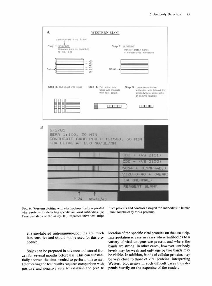
5. Antibody Detection 95
WE. TER~ BLOT
Seml-PUflhed Virus EIC1r3Cl
Step 1. SDS-PAGE Separate protems according 10 their size
_j~~~~~~~ _ p U - "" " - g pO , - pl2
Ge l _ pH
t:t::======:l - p 17
Step 2. "SlOntN(;"
Trans'er prote.n bands 10 nitrocellulose membrane
Step 3. Cut sheet ,nto sthpS Step 4. Put strops ,nto Step 5. locate bound human tubas and ,neubate anbbcd,e. WIth baled 2nd With test sel1.lm anllbody1autoradlography
or enzyme reaction
B 6 / 2 / 85 SERA 1:100. ~O MIN CONJUGATE G HG-POD-H 1:1500 FD LOT 2 AT 8.0 NG/UL/MM
FIG. 6. Western blotting with electrophoretically separated viral proteins for detecting specific antiviral antibodies . (A) Principal steps of the assay. (B) Representative test strips
enzyme-labeled anti-immunoglobulins are much less sensitive and should not be used for this procedure.
Strips can be prepared in advance and stored frozen for several months before use. This can substantially shorten the time needed to perform this assay. Interpreting the test results requires comparison with positive and negative sera to establish the precise
from patients and controls assayed for antibodies to human immunodeficiency virus proteins.
location of the specific viral proteins on the test strip. Interpretation is easy in cases where antibodies to a variety of viral antigens are present and where the bands are strong. In other cases, however, antibody levels may be weak and only one or two bands may be visible. In addition, bands of cellular proteins may be very close to those of viral proteins. Interpreting Western blot assays in such difficult cases thus depends heavily on the expertise of the reader.
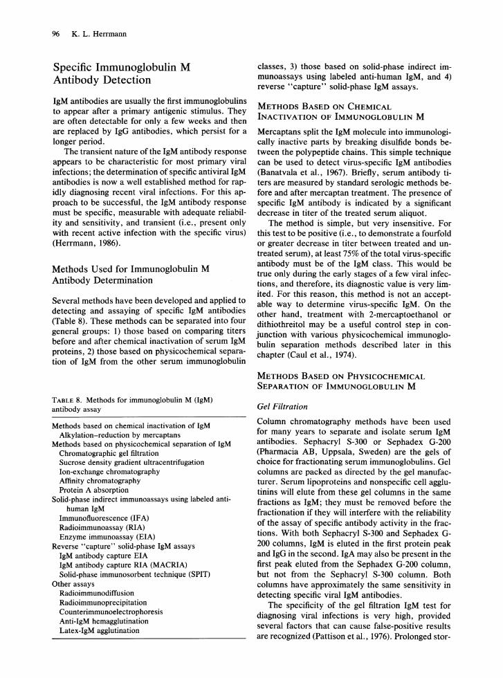
96 K. L. Herrmann
Specific Immunoglobulin M Antibody Detection
IgM antibodies are usually the first immunoglobulins to appear after a primary antigenic stimulus. They are often detectable for only a few weeks and then are replaced by IgG antibodies, which persist for a longer period.
The transient nature of the IgM antibody response appears to be characteristic for most primary viral infections; the determination of specific antiviral IgM antibodies is now a well established method for rapidly diagnosing recent viral infections. For this approach to be successful, the IgM antibody response must be specific, measurable with adequate reliability and sensitivity, and transient (i.e., present only with recent active infection with the specific virus) (Herrmann, 1986).
Methods Used for Immunoglobulin M Antibody Determination
Several methods have been developed and applied to detecting and assaying of specific IgM antibodies (Table 8). These methods can be separated into four general groups: 1) those based on comparing titers before and after chemical inactivation of serum IgM proteins, 2) those based on physicochemical separation of IgM from the other serum immunoglobulin
TABLE 8. Methods for immunoglobulin M (IgM) antibody assay
Methods based on chemical inactivation of IgM Alkylation-reduction by mercaptans
Methods based on physicochemical separation of IgM Chromatographic gel filtration Sucrose density gradient ultracentrifugation Ion-exchange chromatography Affinity chromatography Protein A absorption
Solid-phase indirect immunoassays using labeled anti-humanIgM
Immunofluorescence (IF A) Radioimmunoassay (RIA) Enzyme immunoassay (EIA)
Reverse "capture" solid-phase IgM assays IgM antibody capture EIA IgM antibody capture RIA (MACRIA) Solid-phase immunosorbent technique (SPIT)
Other assays Radioimmunodiffusion Radioimmunoprecipitation Counterimmunoelectrophoresis Anti-IgM hemagglutination Latex-IgM agglutination
classes, 3) those based on solid-phase indirect immunoassays using labeled anti-human IgM, and 4) reverse "capture" solid-phase IgM assays.
METHODS BASED ON CHEMICAL INACTIVATION OF IMMUNOGLOBULIN M
Mercaptans split the IgM molecule into immunologically inactive parts by breaking disulfide bonds between the polypeptide chains. This simple technique can be used to detect virus-specific IgM antibodies (Banatvala et al., 1967). Briefly, serum antibody titers are measured by standard serologic methods before and after mercaptan treatment. The presence of specific IgM antibody is indicated by a significant decrease in titer of the treated serum aliquot.
The method is simple, but very insensitive. For this test to be positive (i.e., to demonstrate a fourfold or greater decrease in titer between treated and untreated serum), at least 75% of the total virus-specific antibody must be of the IgM class. This would be true only during the early stages of a few viral infections, and therefore, its diagnostic value is very limited. For this reason, this method is not an acceptable way to determine virus-specific IgM. On the other hand, treatment with 2-mercaptoethanol or dithiothreitol may be a useful control step in conjunction with various physicochemical immunoglobulin separation methods described later in this chapter (Caul et al., 1974).
METHODS BASED ON PHYSICOCHEMICAL SEPARATION OF IMMUNOGLOBULIN M
Gel Filtration
Column chromatography methods have been used for many years to separate and isolate serum IgM antibodies. Sephacryl S-300 or Sephadex G-200 (pharmacia AB, Uppsala, Sweden) are the gels of choice for fractionating serum immunoglobulins. Gel columns are packed as directed by the gel manufacturer. Serum lipoproteins and nonspecific cell agglutinins will elute from these gel columns in the same fractions as IgM; they must be removed before the fractionation if they will interfere with the reliability of the assay of specific antibody activity in the fractions. With both Sephacryl S-300 and Sephadex G-200 columns, IgM is eluted in the first protein peak and IgG in the second. IgA may also be present in the first peak eluted from the Sephadex G-200 column, but not from the Sephacryl S-300 column. Both columns have approximately the same sensitivity in detecting specific viral IgM antibodies.
The specificity of the gel filtration IgM test for diagnosing viral infections is very high, provided several factors that can cause false-positive results are recognized (Pattison et aI., 1976). Prolonged stor-
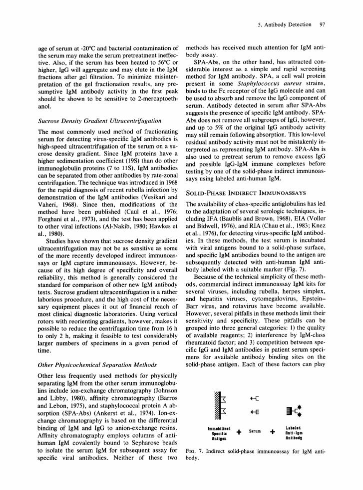
age of serum at -20°C and bacterial contamination of the serum may make the serum pretreatment ineffective. Also, if the serum has been heated to 56°C or higher, IgG will aggregate and may elute in the IgM fractions after gel filtration. To minimize misinterpretation of the gel fractionation results, any presumptive IgM antibody activity in the first peak should be shown to be sensitive to 2-mercaptoethanol.
Sucrose Density Gradient Ultracentrifugation
The most commonly used method of fractionating serum for detecting virus-specific IgM antibodies is high-speed ultracentrifugation of the serum on a sucrose density gradient. Since IgM proteins have a higher sedimentation coefficient (19S) than do other immunoglobulin proteins (7 to lIS), IgM antibodies can be separated from other antibodies by rate-zonal centrifugation. The technique was introduced in 1968 for the rapid diagnosis of recent rubella infection by demonstration of the IgM antibodies (Vesikari and Vaheri, 1968). Since then, modifications of the method have been published (Caul et al., 1976; Forghani et al., 1973), and the test has been applied to other viral infections (AI-Nakib, 1980; Hawkes et al., 1980).
Studies have shown that sucrose density gradient ultracentrifugation may not be as sensitive as some of the more recently developed indirect immunoassays or IgM capture immunoassays. However, because of its high degree of specificity and overall reliability, this method is generally considered the standard for comparison of other new IgM antibody tests. Sucrose gradient ultracentrifugation is a rather laborious procedure, and the high cost of the necessary equipment places it out of financial reach of most clinical diagnostic laboratories. Using vertical rotors with reorienting gradients, however, makes it possible to reduce the centrifugation time from 16 h to only 2 h, making it feasible to test considerably larger numbers of specimens in a given period of time.
Other Physicochemical Separation Methods
Other less frequently used methods for physically separating IgM from the other serum immunoglobulins include ion-exchange chromatography (Johnson and Libby, 1980), affinity chromatography (Barros and Lebon, 1975), and staphylococcal protein A absorption (SPA-Abs) (Ankerst et al., 1974). Ion-exchange chromatography is based on the differential binding of IgM and IgG to anion-exchange resins. Affinity chromatography employs columns of antihuman IgM covalently bound to Sepharose beads to isolate the serum IgM for subsequent assay for specific viral antibodies. Neither of these two
5. Antibody Detection 97
methods has received much attention for IgM antibody assay.
SPA-Abs, on the other hand, has attracted considerable interest as a simple and rapid screening method for IgM antibody. SPA, a cell wall protein present in some Staphylococcus aureus strains, binds to the Fc receptor of the IgG molecule and can be used to absorb and remove the IgG component of serum. Antibody detected in serum after SPA-Abs suggests the presence of specific IgM antibody. SPAAbs does not remove all subgroups of IgG, however, and up to 5% of the original IgG antibody activity may still remain following absorption. This low-level residual antibody activity must not be mistakenly interpreted as representing IgM antibody. SPA-Abs is also used to pretreat serum to remove excess IgG and possible IgG-IgM immune complexes before testing by one of the solid-phase indirect immunoassays using labeled anti-human IgM.
SOLID-PHASE INDIRECT IMMUNOASSAYS
The availability of class-specific antiglobulins has led to the adaptation of several serologic techniques, including IFA (Baublis and Brown, 1968), EIA (Voller and Bidwell, 1976), and RIA (Chau et al., 1983; Knez et al., 1976), for detecting virus-specific IgM antibodies. In these methods, the test serum is incubated with viral antigens bound to a solid-phase surface, and specific IgM antibodies bound to the antigen are subsequently detected with anti-human IgM antibody labeled with a suitable marker (Fig. 7).
Because of the technical simplicity of these methods, commercial indirect immunoassay IgM kits for several viruses, including rubella, herpes simplex, and hepatitis viruses, cytomegalovirus, EpsteinBarr virus, and rotavirus have become available. However, several pitfalls in these methods limit their sensitivity and specificity. These pitfalls can be grouped into three general categories: 1) the quality of available reagents; 2) interference by IgM-class rheumatoid factor; and 3) competition between specific IgG and IgM antibodies in patient serum specimens for available antibody binding sites on the solid-phase antigen. Each of these factors can play
~ +-C
+-E ~ ImIllDbilllld + SIMlIII +
Llbilid Splcilic a.tHglII bUgl. a.tibadg
FIG. 7. Indirect solid-phase immunoassay for IgM antibody.
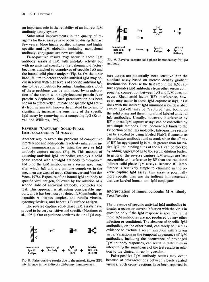
98 K. L. Herrmann
an important role in the reliability of an indirect IgM antibody assay system.
Substantial improvements in the quality of reagents for these assays have occurred during the past few years. More highly purified antigens and highly specific anti-IgM globulin, including monoclonal antibody, conjugates are now available.
False-positive results may occur in these IgM antibody assays if IgM with anti-IgG activity but with no antiviral specificity (i.e., rheumatoid factor) becomes attached to complexes of specific IgG and the bound solid-phase antigen (Fig. 8). On the other hand, failure to detect specific antiviral IgM may occur in serum with high levels of specific antiviral IgG due to the competition for antigen binding sites. Both of these problems can be minimized by preadsorption of the serum with staphylococcal protein A or protein A-Sepharose. Such preadsorption has been shown to effectively eliminate nonspecific IgM activity from serum with known rheumatoid factor and to significantly increase the sensitivity of the specific IgM assay by removing most competing IgG (Kronvall and Williams, 1969).
REVERSE "CAPTURE" SOLID-PHASE IMMUNOGLOBULIN M ASSAYS
Another way to avoid the problems of competitive interference and nonspecific reactivity inherent in indirect immunoassays is by using the reverse IgM antibody capture method (Fig. 9). This method of detecting antiviral IgM antibodies employs a solid phase coated with anti-IgM antibody to "capture" and bind the IgM . antibodies in a serum specimen, after which IgG and any immune complexes in the specimen are washed away (Duermeyer and Van der Veen, 1978). Exposure of the bound IgM antibody to specific viral antigen, followed by the addition of a second, labeled anti-viral antibody, completes the test. This approach is attracting considerable support, and it has been used to detect IgM antibodies to hepatitis A, herpes simplex, and rubella viruses, cytomegalovirus, and hepatitis B surface antigen.
The reverse capture solid-phase IgM assays have proved to be very sensitive and specific (Mortimer et al., 1981). Our experience confirms that the IgM cap-
ImmDbilind Spoclflc IgG Splcific + RllllbDd, Rllligill
Igm + Rllti-lgG +
(RF)
Labelld RIlII-lgm Rllllbadg
FIG. 8. False-positive results due to rheumatoid factor (RF) interference in the indirect solid-phase immunoassay.
Immabllind Rllli-Igm + Rlltiglll
3-+
:r-
SiMlm + Splcific Rlltiglll
Llblllid + Splciflc Rnllbadg
FIG. 9. Reverse capture solid-phase immunoassay for IgM antibody.
ture assays are potentially more sensitive than the standard assay based on sucrose density gradient fractionation. Because the first step in the IgM capture separates IgM antibodies from other serum components, competition between IgG and IgM does not occur. Rheumatoid factor (RF) interference, however, may occur in these IgM capture assays, as it does with the indirect IgM immunoassays described earlier. IgM-RF may be "captured" and bound on the solid phase and then in tum bind labeled antiviral IgG antibodies. Usually, however, interference by RF in these IgM capture assays can be controlled by two simple methods. First, because RF binds to the Fc portion ofthe IgG molecule, false-positive results can be avoided by using labeled F(ab')2 fragments as the indicator antibody; and second, since the affinity of RF for aggregated Ig is much greater than for native IgG, the binding sites of the RF can be blocked by adding aggregated Ig to the serum dilution buffer. In general, the reverse capture IgM assays are less susceptible to interference by RF than are traditional indirect solid-phase IgM assays. Because RF interference is relatively simple to eliminate in the reverse capture IgM assay, this assay is potentially more specific than are the indirect immunoassays that use labeled human anti-IgM conjugates.
Interpretation of Immunoglobulin M Antibody Test Results
The presence of specific antiviral IgM antibodies indicates a recent or current infection with the virus in question only if the IgM response is specific (i.e., if these IgM antibodies are not produced by any other infection or condition). The absence of specific IgM antibodies, on the other hand, can rarely be used as evidence to exclude a recent infection with a given virus. Variations in the temporal appearance of IgM antibodies, including the occurrence of prolonged IgM antibody responses, can result in difficulties in interpreting the significance of the test results in relation to the clinical illness in question.
False-positive IgM antibody results may occur because of cross-reactions between closely related viruses. Such cross-reactions have been reported in
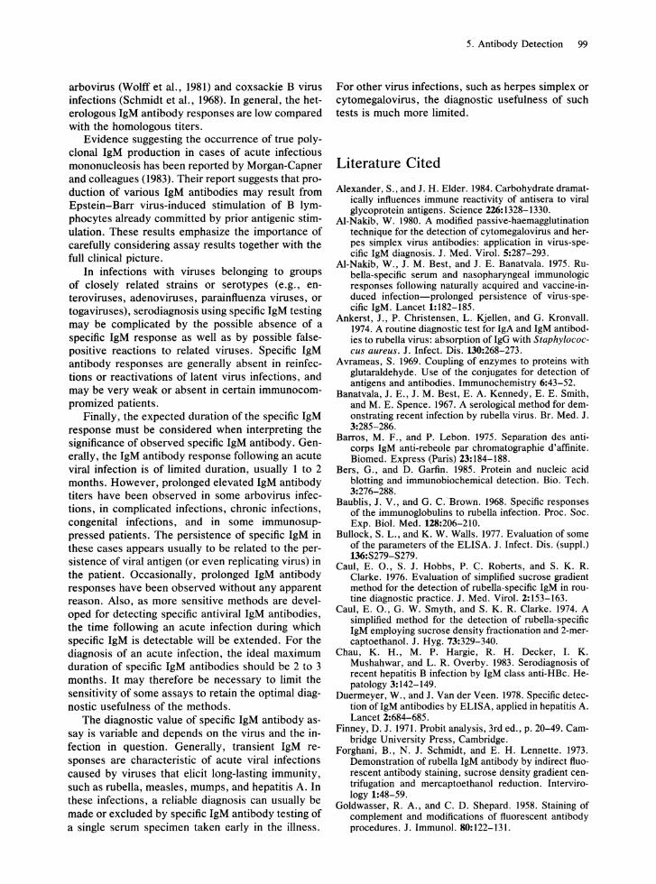
arbovirus (Wolff et al., 1981) and coxsackie B virus infections (Schmidt et al., 1968). In general, the heterologous IgM antibody responses are low compared with the homologous titers.
Evidence suggesting the occurrence of true polyclonal IgM production in cases of acute infectious mononucleosis has been reported by Morgan-Capner and colleagues (1983). Their report suggests that production of various IgM antibodies may result from Epstein-Barr virus-induced stimulation of B lymphocytes already committed by prior antigenic stimulation. These results emphasize the importance of carefully considering assay results together with the full clinical picture.
In infections with viruses belonging to groups of closely related strains or serotypes (e.g., enteroviruses, adenoviruses, parainfluenza viruses, or togaviruses), serodiagnosis using specific IgM testing may be complicated by the possible absence of a specific IgM response as well as by possible falsepositive reactions to related viruses. Specific IgM antibody responses are generally absent in reinfections or reactivations of latent virus infections, and may be very weak or absent in certain immunocompromized patients.
Finally, the expected duration of the specific IgM response must be considered when interpreting the significance of observed specific IgM antibody. Generally, the IgM antibody response following an acute viral infection is of limited duration, usually 1 to 2 months. However, prolonged elevated IgM antibody titers have been observed in some arbovirus infections, in complicated infections, chronic infections, congenital infections, and in some immunosuppressed patients. The persistence of specific IgM in these cases appears usually to be related to the persistence of viral antigen (or even replicating virus) in the patient. Occasionally, prolonged IgM antibody responses have been observed without any apparent reason. Also, as more sensitive methods are developed for detecting specific antiviral IgM antibodies, the time following an acute infection during which specific IgM is detectable will be extended. For the diagnosis of an acute infection, the ideal maximum duration of specific IgM antibodies should be 2 to 3 months. It may therefore be necessary to limit the sensitivity of some assays to retain the optimal diagnostic usefulness of the methods.
The diagnostic value of specific IgM antibody assay is variable and depends on the virus and the infection in question. Generally, transient IgM responses are characteristic of acute viral infections caused by viruses that elicit long-lasting immunity, such as rubella, measles, mumps, and hepatitis A. In these infections, a reliable diagnosis can usually be made or excluded by specific IgM antibody testing of a single serum specimen taken early in the illness.
5. Antibody Detection 99
For other virus infections, such as herpes simplex or cytomegalovirus, the diagnostic usefulness of such tests is much more limited.
Literature Cited
Alexander, S., andJ. H. Elder. 1984. Carbohydrate dramatically influences immune reactivity of antisera to viral glycoprotein antigens. Science 226:1328-1330.
Al-Nakib, W. 1980. A modified passive-haemagglutination technique for the detection of cytomegalovirus and herpes simplex virus antibodies: application in virus-specific IgM diagnosis. J. Med. Virol. 5:287-293.
Al-Nakib, W., J. M. Best, and J. E. Banatvala. 1975. Rubella-specific serum and nasopharyngeal immunologic responses following naturally acquired and vaccine-induced infection-prolonged persistence of virus-specific IgM. Lancet 1:182-185.
Ankerst, J., P. Christensen, L. Kjellen, and G. Kronvall. 1974. A routine diagnostic test for IgA and IgM antibodies to rubella virus: absorption of IgG with Staphylococcus aureus. J. Infect. Dis. 130:268-273.
Avrameas, S. 1969. Coupling of enzymes to proteins with glutaraldehyde. Use of the conjugates for detection of antigens and antibodies. Immunochemistry 6:43-52.
Banatvala, J. E., J. M. Best, E. A. Kennedy, E. E. Smith, and M. E. Spence. 1967. A serological method for demonstrating recent infection by rubella virus. Br. Med. J. 3:285-286.
Barros, M. F., and P. Lebon. 1975. Separation des anticorps IgM anti-rebeole par chromatographie d'affinite. Biomed. Express (Paris) 23:184-188.
Bers, G., and D. Gartin. 1985. Protein and nucleic acid blotting and immunobiochemical detection. Bio. Tech. 3:276-288.
Baublis, J. V., and G. C.~Brown. 1968. Specific responses of the immunoglobulins to rubella infection. Proc. Soc. Exp. BioI. Med. 128:206-210.
Bullock, S. L., and K. W. Walls. 1977. Evaluation of some of the parameters of the ELISA. J. Infect. Dis. (suppl.) 136:S279-S279.
Caul, E. 0., S. J. Hobbs, P. C. Roberts, and S. K. R. Clarke. 1976. Evaluation of simplified sucrose gradient method for the detection of rubella-specific IgM in routine diagnostic practice. J. Med. Virol. 2:153-163.
Caul, E. 0., G. W. Smyth, and S. K. R. Clarke. 1974. A simplified method for the detection of rubella-specific IgM employing sucrose density fractionation and 2-mercaptoethanol. J. Hyg. 73:329-340.
Chau, K. H., M. P. Hargie, R. H. Decker, I. K. Mushahwar, and L. R. Overby. 1983. Serodiagnosis of recent hepatitis B infection by IgM class anti-HBc. Hepatology 3:142-149.
Duermeyer, W., and J. Van der Veen. 1978. Specific detection of IgM antibodies by ELISA, applied in hepatitis A. Lancet 2:684-685.
Finney, D. J. 1971. Probit analysis, 3rd ed., p. 20-49. Cambridge University Press, Cambridge.
Forghani, B., N. J. Schmidt, and E. H. Lennette. 1973. Demonstration of rubella IgM antibody by indirect fluorescent antibody staining, sucrose density gradient centrifugation and mercaptoethanol reduction. Intervirology 1:48-59.
Goldwasser, R. A., and C. D. Shepard. 1958. Staining of complement and modifications of fluorescent antibody procedures. J. Immunol. 80:122-131.
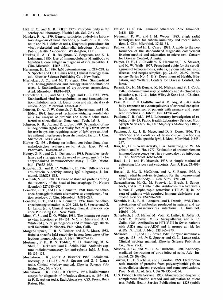
100 K. L. Herrmann
Hall, E. C., and M. B. Felker. 1970. Reproducibility in the serological laboratory. Health Lab. Sci. 7:63-68.
Hawkes, R. A. 1979. General principles underlying laboratory diagnosis of viral infections, p. 3-48. In E. H. Lennette and N. J. Schmidt (ed.), Diagnostic procedures for viral, rickettsial and chlamydial infections. American Public Health Association, Washington, D.C.
Hawkes, R. A., C. R. Boughton, V. Ferguson, and N. I. Lehmann. 1980. Use of immunoglobulin M antibody to hepatitis B core antigen in diagnosis of viral hepatitis. J. Clin. Microbiol. 11:581-583.
Herrmann, K. L. 1986. IgM determinations, p. 219-228. In S. Specter and G. J. Lancz (ed.), Clinical virology manual. Elsevier Science Publishing Co., New York.
Hierholzer, J. C., and M. T. Suggs. 1969. Standardized viral hemagglutination and hemagglutination-inhibition tests. I. Standardization of erythrocyte suspensions. Appl. Microbiol. 18:816-823.
Hierholzer, J. C., and M. T. Suggs, and E. C. Hall. 1969. Standardized viral hemagglutination and hemagglutination-inhibition tests. II. Description and statistical evaluation. Appl. Microbiol. 18:824-833.
Johnson, D. A., J. W. Gautsch, J. R. Sportsman, and J. H. Elder. 1984. Improved technique utilizing non-fat dry milk for analysis of proteins and nucleic acids transferred to nitrocellulose. Gene Anal. Tech. 1:3-8.
Johnson, R. B., Jr., and R. Libby. 1980. Separation ofimmunoglobulin (lgM) essentially free of IgG from serum for use in systems requiring assay of IgM-type antibodies without interference from rheumatoid factor. J. Clin. Microbiol. 12:451-454.
Kiirber, G. 1931. Beitrag zur kollektiven behandlung pharmakologischer reihenversuche. Arch. Exp. Pathol. Pharmakol. 162:480-483.
Kenny, G. E., and C. L. Dunsmoor. 1983. Principles, problems, and strategies in the use of antigenic mixtures for enzyme-linked immunosorbent assay. J. Clin. MicrobioI. 17:655-665.
Knonvall, G., and R. C. Williams, Jr. 1%9. Differences in anti-protein A activity among IgG subgroups. J. Immunol. 103:828-833.
Laemmli, V. K. 1970. Cleavage of standard proteins during the assembly of the head of bacteriaphage T4. Nature (London) 227:680-685.
Lennette, E. T., and D. A. Lennette. 1978. Immune adherence hemagglutination: alternative to complement-fumtion serology. J. Clin. Microbiol. 7:282-285.
Lennette, E. T., and D. A. Lennette. 1986. Immune adherence hemagglutination, p. 209-218. In S. Specter and G. L. Lancz (ed.), Clinical virology manual. Elsevier Science Publishing Co., New York.
Mims, C. S., and D. O. White. 1984. The immune response to viral infection, p. 87-131. In C. S. Mims and D. O. White (ed.), Viral pathogenesis and immunology. Blackwell Scientific Publishers, Palo Alto, Calif.
Morgan-Capner, P., R. S. Tedder, and J. E. Mace. 1983. Rubella-specific IgM reactivity in sera from cases of infectious mononucleosis. J. Hyg. 90:407-413.
Mortimer, P. P., R. S. Tedder, M. H. Hambling, M. S. Shaft, F. Burkhardt, and U. Schilt. 1981. Antibody capture radioimmunoassay for anti-rubella IgM. J. Hyg. 86:139-153.
Mushahwar, I. K., and T. A. Brawner. 1986. Radioimmunoassay, p. 111-131. In S. Specter and G. J. Lancz (ed.), Clinical virology manual. Elsevier Science Publishing Co., New York.
Mushahwar, I. K., and L. R. Overby. 1983. Radioimmune assays for diagnosis of infectious diseases, p. 167-194. In F. S. Ashkar (ed.), Radiobioassays. CRC Press, Boca Raton, Fla.
Nelson, D. S. 1963. Immune adherence. Adv. Immunol. 3:131-180.
Neumann, P. W., and J. M. Weber. 1983. Single radial hemolysis test for rubella immunity and recent infection. J. Clin. Microbiol. 17:28-34.
Palmer, D. F., and H. L. Casey. 1981. A guide to the performance of the standardized diagnostic complement fixation method and adaptation to micro test. Centers for Disease Control, Atlanta.
Palmer, D. F., J. J. Cavallaro, K. Herrmann, J. A. Stewart, and K. W. Walls. 1977. Procedural guide for the serodiagnosis oftoxoplasmosis, rubella, cytomegalic inclusion disease, and herpes simplex, pp. 24-56, 90-99. Immunology Series No.5. U.S. Department of Health, Education, and Welfare, Centers for Disease Control, Atlanta.
Parratt, D., H. McKenzie, K. H. Nielsen, and S. J. Cobb. 1982. Radioimmunoassay of antibody and its clinical applications, p. 19-31. John Wiley & Sons Ltd., Chichester, England.
Pass, R. F., P. D. Griffiths, and A. M. August. 1983. Antibody response to cytomegalovirus after renal transplantation: comparison of patients with primary and recurrent infections. J. Infect. Dis. 147:40-46.
Pattison, J. R. (ed.). 1982. Laboratory investigation of rubella, p. 19-25. Public Health Laboratory Service, Monograph Series No. 16, Her Majesty's Stationery Office, London.
Pattison, J. R., J. E. Mace, and D. S. Dane. 1976. The detection and avoidance of false-positive reactions in tests for rubella-specific IgM. J. Med. Microbiol. 9:355-357.
Rao, N., D. T. Waruszewski, J. A. Armstrong, R. W. Atchison, and M. Ho. 1977. Evaluation of anticomplement immunofluorescence test in cytomegalovirus infection. J. Clin. Microbiol. 6:633-638.
Reed, L. J., and H. Muench. 1938. A simple method of estimating fifty per cent endpoints. Am. J. Hyg. 27:493-497.
Russell, S. M., D. McCahon, and A. S. Beare. 1975. A single radial hemolysis technique for the measurement of influenza antibody. J. Gen. Virol. 27:1-10.
Samgadharan, M. G., M. Popovic, L. Bruch, J. Schupbach, and R. C. Gallo. 1984. Antibodies reactive with a human T lymphotropic retrovirus (HTLV-III) in the sera of patients with acquired immune deficiency syndrome. Science 224:506-508.
Schmidt, N. J., E. H. Lennette, and J. Dennis. 1968. Characterization of antibodies produced in natural and experimental coxsackievirus infections. J. Immunol. 100:99-106.
Schupbach, J., O. Haller, M. Vogt, R. Luthy, H. Joller, O. Oelz, M. Popovic, M. G. Samgadharan, and R. C. Gallo. 1985. Antibodies to HTLV-III in Swiss patients with AIDS and pre-AIDS and in groups at risk for AIDS. N. Engl. J. Med. 312:265-270.
Shekarchi, I. C., and J. L. Sever. 1986. Enzyme immunoassay, p. 133-146. In S. Specter and G. L. Lancz (ed.), Clinical virology manual. Elsevier Science Publishing Co., New York.
Sissons, J. G., and M. B. A. Oldstone. 1980. Antibodymediated destruction of virus infected cells. Adv. Immunol. 29:209-260.
Towbin, H., T. Staehelin, andJ. Gordon. 1979. Electrophoretic transfer of proteins from polyacrylamide gels to nitrocellulose sheets: procedure and some applications. Proc. Natl. Acad. Sci. USA 76:4350-4354.
U.S. Public Health Service. 1965. Standardized diagnostic complement fixation method and adaptation to micro test. Public Health Service Publication no. 1228 (public

health monograph no. 74). U.S. Government Printing Office, Washington, D.C.
Vesikari, T., and A. Vaheri. 1968. Rubella: a method for rapid diagnosis of a recent infection by demonstration of the IgM antibodies. Br. Med. J. 1:221-223.
Voller, A., and D. E. Bidwell. 1975. A simple method for detecting antibodies to rubella. Br. J. Exp. Pathol. 56:338-339.
Voller, A., and D. E. Bidwell. 1976. Enzyme immunoassays for antibodies in measles, cytomegalovirus infections and after rubella vaccination. Br. J. Exp. Pathol. 57:243-247.
5. Antibody Detection 101
Wilson, M. B., and P. K. Nakane. 1978. Recent developments in the periodate method of conjugating horseradish peroxidase, p. 215-224. In W. Knass, K. Holubar and G. Wick (ed.), Immunofluorescence and related staining techniques. Elsevier/North Holland, Amsterdam.
Wolff, K. L., D. J. Muth, B. W. Hudson, and D. W. Trent. 1981. Evaluation of the solid-phase radioimmunoassay for diagnosis of St. Louis encephalitis infection in humans. J. Clin. Microbiol. 14:135-140.
Wood, R. J., and T. M. Durham. 1980. Reproducibility of serological titers. J. Clin. Microbiol. 11:541-545.
Related Documents
![Springer link[1]](https://static.cupdf.com/doc/110x72/5589ccf2d8b42a472e8b45b0/springer-link1.jpg)