Animal welfare, etológia és tartástechnológia Animal welfare, ethology and housing systems Volume 9 Issue 3 Különszám/Special Issue Gödöllő 2013

Welcome message from author
This document is posted to help you gain knowledge. Please leave a comment to let me know what you think about it! Share it to your friends and learn new things together.
Transcript

Animal welfare, etológia és tartástechnológia
Animal welfare, ethology and housing systems
Volume 9 Issue 3
Különszám/Special Issue
Gödöllő
2013
RFFCH Gödöllő, 2013
401
INFLUENCE OF ELECTROMAGNETIC RADIATION ON SELECTED ORGANS IN RATS Viera Almášiová1, Katarína Holovská1, Viera Cigánková1, Enikö Račeková2
1Department of Anatomy, Histology and Physiology, University of Veterinary Medicine and Pharmacy, Kosice, Slovak Republic 2Institute of Neurobiology, Slovak Academy of Sciences, Kosice, Slovak Republic
Abstract The immediate whole body electromagnetic radiation was used to investigate testicular and kidney structure of the Wistar rats. Sexually mature (48 days old) rats were subjected to pulsed electromagnetic fields at frequency of 2.45 GHz and mean power density 2.8 mW/cm2 daily applications of 3 h per 3 weeks. Histological structure of the testicular and kidney parenchyma was evaluated in 3 hours after the last irradiation. The light microscopy revealed diffuse degenerative changes in both examined organs in experimental animals. The testes contained irregular seminiferous tubules, the seminiferous epithelium showed the signs of cellular sloughing, and sex cells were often degenerative or even necrotizing. The kidney parenchyma manifested degenerative changes within all nephron components as well as collecting tubules. The necrotizations were extremely intensive mainly within the medullary region. The interstitium showed the signs of inflammation. These findings confirmed an adverse effect of EMR, and the evidence that the testes and kidneys are amongst the most susceptible organs to the EMR. Key words: electromagnetic radiation, testes, kidney, rats Introduction Nowadays, human population is unavoidably influenced to the constant electromagnetic fields produced from variety of different sources. Frequencies between 30 kHz and 300 GHz are commonly used for telecommunication, including broadcast radio and television, and involve the radio frequency band which can be classified to as non-ionizing radiation in the microwave range. Mobile telephone systems exactly operate at radiofrequency electromagnetic waves, RF-EMW 900MHz and 1800MHz. Such the levels are far below the high frequency EMW of X-rays and gamma rays ionizing radiation. Although the low level of energy of non-ionizing radiation cannot break the covalent bonds in biological molecules, the human body due its electrical properties such the permittivity and conductivity is able to receive and induce electrical fields and currents inside the tissues (Sysoev et al., 2013). Thermal and non-thermal effects are the main mediators of EMW interaction with biological systems. The tissue damage in this case is caused due to tissue´s inability to eliminate the excessive heat. Non-thermal or direct effect is not completely understood, and comprises a wide range of various metabolic pathways. It is often associated with the plasma membrane injury, cellular signal transduction effects, nervous system excitability perturbation, neuroendocrine and immune system injury (Bhat, 2013, Zecca et al., 2006). However the testis and kidneys depend chiefly on surface admittance rather than blood flowage for temperature regulation, which performs an interesting aspect of thermal effect of RF-EMW, the mode of action of RF-EMW will be probably a combination of the thermal and nontermal effects. However many works showed that RF-EMR from a mobile phones, Wi-Fi, microwaves or other devices affected negatively male fertility (Lukac et al., 2011, Hales et al., 2005),or general health (Braune et al., 2002), a number of studies in contrast did not note any abnormalities (Dasdag et al.,2003; Chung et al., 2005). This study investigated the possible effect of immediate whole body irradiation by pulsed EMF on testis and kidney structure of rats. Materials and methods The research material consisted of 40 male albino rats strain Wistar. At the begining of the experiment, animals were randomly divided into two groups – the control and experimental (20 rats
RFFCH Gödöllő, 2013
402
in each). Animals were kept in ordinary cages under controlled temperature of 21±1ºC, and had ad libitum access to food and water. The lighting was turned off or on under a 12 h cycle. Experimental rats were irradiated by a pulse-wave EMF of 2.45 GHz, at mean power density 2.8 mW/cm2 in a purpose-designed chamber (Orendac et al., 2005), in 3 h daily applications per 3 weeks. Uniformity of the EMF was analysed with a spectral analyser to determine the optimal placement of animals. 3 hours after the last irradiation the animals from experimental and control groups were anesthetized by i.p. injection of xylazine+ketamine, and subsequently perfused with 4% paraformaldehyde solution in phosphate buffer (0.1 M, pH 7.3). The tissue excisions of size 1 mm3 were fixed by immersion in 3% glutaraldehyde and postfixed in 1% osmium tetraoxide (both in 0.15 M phosphate buffer, pH 7.3). After dehydration in acetone they were transferred to propylene oxide and embedded in Durcupan ACM. Semi-thin sections of specimen were cut using an ultramicrotome LKB Nova, stained with toluidine blue and examined under a light microscope Zeiss Axio Lab A1, and documented with camera Axio Cam ERc 5. The use and care of animals were approved by the Animal Care Committee of the Institute of Neurobiology, Slovak Academy of Sciences.
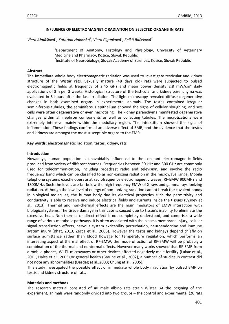
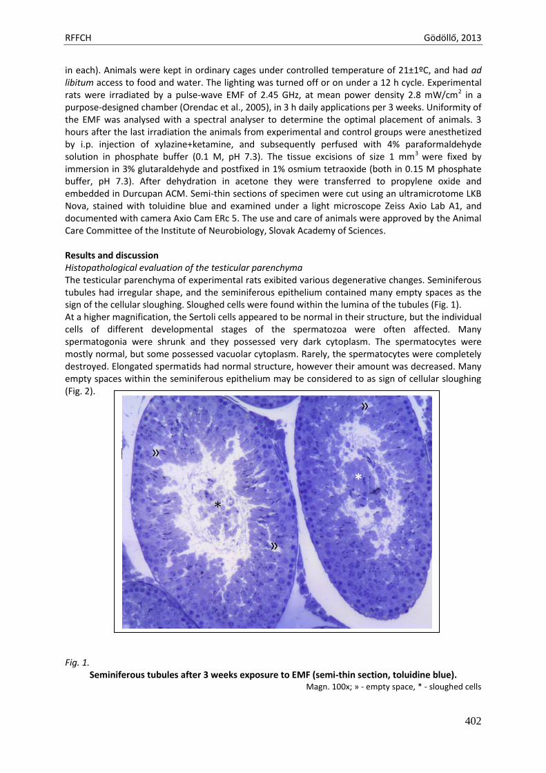
Results and discussion Histopathological evaluation of the testicular parenchyma The testicular parenchyma of experimental rats exibited various degenerative changes. Seminiferous tubules had irregular shape, and the seminiferous epithelium contained many empty spaces as the sign of the cellular sloughing. Sloughed cells were found within the lumina of the tubules (Fig. 1). At a higher magnification, the Sertoli cells appeared to be normal in their structure, but the individual cells of different developmental stages of the spermatozoa were often affected. Many spermatogonia were shrunk and they possessed very dark cytoplasm. The spermatocytes were mostly normal, but some possessed vacuolar cytoplasm. Rarely, the spermatocytes were completely destroyed. Elongated spermatids had normal structure, however their amount was decreased. Many empty spaces within the seminiferous epithelium may be considered to as sign of cellular sloughing (Fig. 2). Fig. 1.
Seminiferous tubules after 3 weeks exposure to EMF (semi-thin section, toluidine blue). Magn. 100x; » - empty space, * - sloughed cells
RFFCH Gödöllő, 2013
403
Fig. 2.
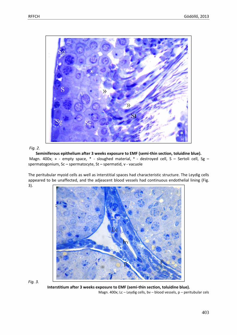
Seminiferous epithelium after 3 weeks exposure to EMF (semi-thin section, toluidine blue). Magn. 400x; » - empty space, * - sloughed material, ᵜ - destroyed cell, S – Sertoli cell, Sg – spermatogonium, Sc – spermatocyte, St – spermatid, v - vacuole The peritubular myoid cells as well as interstitial spaces had characteristic structure. The Leydig cells appeared to be unaffected, and the adjeacent blood vessels had continuous endothelial lining (Fig. 3). Fig. 3.
Interstitium after 3 weeks exposure to EMF (semi-thin section, toluidine blue). Magn. 400x; Lc – Leydig cells, bv – blood vessels, p – peritubular cels
RFFCH Gödöllő, 2013
404
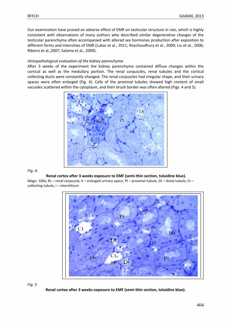
Our examination have proved an adverse effect of EMR on testicular structure in rats, which is highly consistent with observations of many authors who described similar degenerative changes of the testicular parenchyma often accompanied with altered sex hormones production after exposition to different forms and intensities of EMR (Lukac et al., 2011; Roychoudhury et al., 2009; Liu et al., 2006; Ribeiro et al.,2007; Salama et al., 2009). Histopathological evaluation of the kidney parenchyma After 3 weeks of the experiment the kidney parenchyma contained diffuse changes within the cortical as well as the medullary portion. The renal corpuscles, renal tubules and the cortical collecting ducts were constantly changed. The renal corpuscles had irregular shape, and their urinary spaces were often enlarged (Fig. 4). Cells of the proximal tubules showed high content of small vacuoles scattered within the cytoplasm, and their brush border was often altered (Figs. 4 and 5). Fig. 4.
Renal cortex after 3 weeks exposure to EMF (semi-thin section, toluidine blue). Magn. 100x; Rc – renal corpuscle, ¤ – enlarged urinary space, Pt – proximal tubule, Dt – distal tubule, Ct – collecting tubule, i – interstitium
Fig. 5
Renal cortex after 3 weeks exposure to EMF (semi-thin section, toluidine blue).
RFFCH Gödöllő, 2013
405
Magn. 100x; Pt – proximal tubule, Dt – distal tubule, Ct – collecting tubule, i – interstitium, bb – brush border, + - necrotizing cell
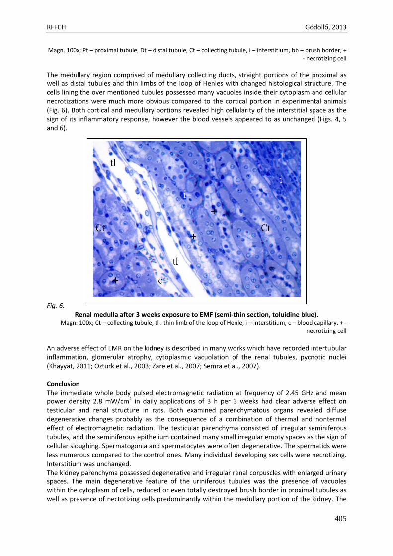
The medullary region comprised of medullary collecting ducts, straight portions of the proximal as well as distal tubules and thin limbs of the loop of Henles with changed histological structure. The cells lining the over mentioned tubules possessed many vacuoles inside their cytoplasm and cellular necrotizations were much more obvious compared to the cortical portion in experimental animals (Fig. 6). Both cortical and medullary portions revealed high cellularity of the interstitial space as the sign of its inflammatory response, however the blood vessels appeared to as unchanged (Figs. 4, 5 and 6). Fig. 6.
Renal medulla after 3 weeks exposure to EMF (semi-thin section, toluidine blue). Magn. 100x; Ct – collecting tubule, tl . thin limb of the loop of Henle, i – interstitium, c – blood capillary, + -
necrotizing cell
An adverse effect of EMR on the kidney is described in many works which have recorded intertubular inflammation, glomerular atrophy, cytoplasmic vacuolation of the renal tubules, pycnotic nuclei (Khayyat, 2011; Ozturk et al., 2003; Zare et al., 2007; Semra et al., 2007). Conclusion The immediate whole body pulsed electromagnetic radiation at frequency of 2.45 GHz and mean power density 2.8 mW/cm2 in daily applications of 3 h per 3 weeks had clear adverse effect on testicular and renal structure in rats. Both examined parenchymatous organs revealed diffuse degenerative changes probably as the consequence of a combination of thermal and nontermal effect of electromagnetic radiation. The testicular parenchyma consisted of irregular seminiferous tubules, and the seminiferous epithelium contained many small irregular empty spaces as the sign of cellular sloughing. Spermatogonia and spermatocytes were often degenerative. The spermatids were less numerous compared to the control ones. Many individual developing sex cells were necrotizing. Interstitium was unchanged. The kidney parenchyma possessed degenerative and irregular renal corpuscles with enlarged urinary spaces. The main degenerative feature of the uriniferous tubules was the presence of vacuoles within the cytoplasm of cells, reduced or even totally destroyed brush border in proximal tubules as well as presence of nectotizing cells predominantly within the medullary portion of the kidney. The
RFFCH Gödöllő, 2013
406
collecting tubules within the cortex and medulla contained cells with vacuolar cytoplasm. The necrotization within the collecting tubules was evident especially within the medullary region. The interstitium contained rich infiltrations with blood formed elements as the sign of inflammation. Acknowledgements This study was supported by VEGA Projects 1/0037/12 References Bhat MA, 2013. Effects of Electromagnetic Waves Emitted by Mobile Phones on Male Fertility. Comp
Engineering and Intelligent Systems, 4(3) Braune S, Riedel A, Schulte-Monting J, 2002. Influence of a radiofrequency electromagnetic field on
cardiovascular and hormonal parameters of the autonomic nervous system in healthy individuals. Radiation Res. 158: 352-356
Dasdag S, Zulkuf AM, Aksen F, 2003. Whole body exposure of rats to microwavws emitted from a cell phone does not affect the testes. Bioelectromagnetics (24): 182-188
Hales DB, Allen JA, Shankara T, Janus P, Buck S, Diemer T, 2005. Mitochondrial function in Leydig cell steroidogenesis. Ann NY Acad. Sci 1061: 120-134
Chung MK, Lee SJ, Kim YB, Park SC, Shin DH, Kim SH, Kim JC, 2005. Evaluation of spermatogenesis and fertility in F1 male rats after in utero and neonatal exposure to extremely low frequency electromagnetic fields. Asian J Androl. 7 (2): 189-194
Khayyat Latifa Ishaq, 2011. The histopathological effects of an electromagnetic field on the kidney and testis of mice. Eurasia J Biosci, 5: 103-109
Liu GW, Gong PS, Zhao HG, Wang ZC, Gong SL, Cai L, 2006. Effect of low level radiation on the death of male germ cells. Radiat.Res., 165:379-389
Lukac N, Massanyi P, Roychoudhury S, Capcarova M, Tvrda E, Knazicka Z, Kolesarova A, Danko J, 2011. J Envir Sci Health A Tox Hazard Subst Environ Eng. 46(12): 1417-1423
Orendac M, Fenik A, Mojzis M, Orendacova J, 2005. Biological effects of electromagnetic radiation on living systems with respect to the brain. Psychiatrie (2): 83-85
Ozturk A, Baltaci AK, Mogulkoc R, Oztekin E, 2003. Zinc prevention of electromagnetically induced damage to rat testicle and kidney tissues. Biol Trace Elem res. 96 (1-3): 247-254
Ribeiro EP, Rhoden EL, Horn MM, Rhoden C, Lima LP, Toniolo, 2007. Effects of subchronic exposure to radio frequency from a conventional cellular telephone on testicular function in adult rats. J Urol.177(1): 395-399
Roychoudhury S, Jedlicka J, Parkanyi V, Rafay J, Ondruska L, Massanyi P, Bulla J, 2009 Semra E, 2007. The effect of electromagnetic field (50Hz, 6 mT) on rat liver and kidney. Journal of Adnan
Menderes Medical Faculty 8(1): 5-11. Sysoev VN, Lukyanov GN, Serov IN, 2013. Electromagnetic Radiation Influence on Human Health.
http://www.aires.spb.ru/docs/eng/36-1-eng.pdf Zare S, Alivandi S, Ebadi AG, 2007. Histological studies of the low frequency electromagnetic fields effect on
liver, testes and kidney in quinea pig. World Applied Sciences Journal 2 (5): 509-511 Zecca L, Mantegazza C, Margonato V, Cerretelli P, Caniatti M, Piva F, 2006. Biological effects of prolonged
exposure to ELF electromagnetic fields in rats. Bioelectromagnetics 19(1): 57-66
Animal welfare, etológia és tartástechnológia
Animal welfare, ethology and housing systems
Volume 9 Issue 3
Különszám/Special Issue
Gödöllő
2013
RFFCH Gödöllő, 2013
407
CYTOCHROME P450 1A INDUCTION IN CHICKEN OVARIAN FOLLICLES EXPOSED TO 2,3,7,8-TETRACHLORODIBENZO-P-DIOXIN
Antos P.*, Sechman A.
University of Agriculture in Krakow, Department of Animal Physiology & Endocrinology, Poland *[email protected]
Cytochrome P450 monooxygenases (CYPs) comprise a large family of proteins that are involved in catalyzing the oxidation of wide variety of xenobiotics, including dioxins and dioxin-like compounds. Chickens have two CYP1A isoforms (CYP1A4 and CYP1A5) which are orthologous to mammalian CYP1A1 and CYP1A2, respectively. It is well-known that the chicken liver expresses both CYP1As, and dioxins induce CYP1A4 and A5 activities in this organ. Recently, we have found that chicken ovarian follicles express CYP1A4 and CYP1A5 mRNA. The aim of the present experiment was to measure in vitro CYP1A activity in control and 2,3,7,8-tetrachlorodibenzo-p-dioxin (TCDD) exposed ovarian follicles of the laying hen.
The experiment was performed on Hy-Line Brown hens at the age of 25 weeks. Birds were
fed ad libitum and kept in individual cages at a neutral temperature (18-20C), and under a photoperiodic regime of 14L:10D. Hens were decapitated 2 h after ovulation. Small (1-4 mm) and large (4-8 mm) white follicles and 3 the largest preovulatory follicles (20-36 mm; F3-F1; F3<F2<F1) were isolated from the ovary. In respect to the preovulatory follicles, the granulosa layer was separated from the theca one by Gilbert’s method. The white follicles and fragments of granulosa
and theca layers were incubated for 24 h at 39C in 1 ml Eagle’s medium supplemented with TCDD (Dr. Ehrenstorfer GmbH, Germany) at concentrations of 0 (control) or 10 nM, ovine LH (oLH; 10 ng/ml) and combination of oLH (10 ng/ml) with TCDD (10 nM). After incubation, tissues were
collected and kept at -80C till the measurement of the CYP1A enzyme activity. CYP1A activity was determined by fluorometric MROD assay using 7-methoxyresorufin as a substrate. The methoxyresorufin metabolite, resorufin, was measured using the excitation wavelength of 530 nm and the emission wavelength of 590 nm. Protein concentration was measured using fluorescamine at the excitation wavelength of 400 nm and the emission wavelength of 460 nm. The measurements were done in triplicates using 96-well plates with BioTek FLx-800TBI microplate reader. MROD activity was expressed as nmol resorufin/mg protein/min. The statistical analysis of the results was performed by ANOVA for repeated measures followed by Tukey test. Significance of differences was considered at the level of P<0.05.
The MROD activity was detected in all investigated ovarian follicles. The basal MROD activity ranged from 0.18 in the granulosa layer of the F3 follicle to 2.5 nmol resorufin/mg protein/min in the theca layer of the F1 preovulatory one (P<0.01). In comparison to control group, TCDD or oLH significantly (P<0.05-0.01) increased the MROD activity in all investigated follicles and tissues. The highest MROD activity following TCDD exposure was found in the granulosa layer of F3 preovulatory follicle (14.0 nmol resorufin/mg protein/min; P<0.01) while following oLH treatment in the granulosa layer of the F2 follicle (11.2 nmol resorufin/mg protein/min; P<0.01). Incubation of these follicles in medium supplemented with both oLH and TCDD revealed that TCDD did not affect oLH stimulated MROD activity.
In conclusion, the basal and TCDD inducible CYP1A activity in ovarian follicles suggest that the chicken ovary is involved in the process of xenobiotic detoxication. Supported by grant N N303 561 339 (2010-2013) & BM-4224/2013.
Animal welfare, etológia és tartástechnológia
Animal welfare, ethology and housing systems
Volume 9 Issue 3
Különszám/Special Issue
Gödöllő
2013
RFFCH Gödöllő, 2013
408
Mercury concentrations in the digestive tract of muskrat from southern Poland
Łukasz J. Binkowski, Sylwia Marek, Joanna Banaś, Tomasz Łaciak, Włodzimierz Wojtaś, Bartłomiej Zyśk, Marek Guzik, Lucjan Schimscheiner and Robert Stawarz
Institute of Biology, Pedagogical University of Cracow, PolandPodbrzezie 3, 31-054 Cracow, Poland Corresponding author: Łukasz J. Binkowski ([email protected])
Mercury is a xenobiotic metal which occurs in the environment in its most toxic form (methylmercury) mostly in sediments. The transfer level from the environment to living organism depends on many factors and is different in various environments. The aim of our study was to inquire the concentrations of total mercury in chosen parts of the digestive tract of muskrat Ondatra zibetrhicus L. from Zator fishponds area in southern Poland. Animals were shot in April 2012, weighed, skinned and frozen (-18ᵒC). Age was determined on the basis of molar wear. During the section in the laboratory of the Institute of Biology (Pedagogical University of Cracow) animals were sexed (5 females and 6 males) and samples of liver, stomach, large intestine, cecum and their content were taken. Concentrations of mercury were measured with CV-AA spectrometer (NIC MA-2; limit of quantification 0.2 ng) in the wet weight of samples. The differences in mercury concentrations between sexes were tested with T test. All analyses were performed with MS Excel 2010 PL and Statistica 10 EN. All individuals were 24 ± 3 months old. The statistical differences in mercury concentrations between sexes were not statistically significant so the results of both groups were combined. The highest mean mercury concentrations in tissues was noted in caecum (0.0221 µg/g), the lowest one in liver (0.0105 µg/g). The concentrations in the content of the digestive tract fitted into the range from 0.0034 (caecum content) to 0.0096 µg/g (large intestine content). These results show that mercury is available in the studied ecosystem. In comparison of the found concentrations in liver to the literature data they can be evaluated as comparable to ones found in other mammals in Poland and Slovakia region.

Animal welfare, etológia és tartástechnológia
Animal welfare, ethology and housing systems
Volume 9 Issue 3
Különszám/Special Issue
Gödöllő
2013
RFFCH Gödöllő, 2013
409
THE EFFECT OF BEE POLLEN ON MACROSCOPIC STRUCTURE OF FEMORA IN ADULT FEMALE RATS AFTER AN EXPERIMENTAL ADDITION IN DIET
Ivana BOBOŇOVÁ*1, Monika MARTINIAKOVÁ1, Hana CHOVANCOVÁ1, Radoslav OMELKA2, Róbert TOMAN3, Zuzana HÁJKOVÁ3, Robert STAWARZ4
1Constantine the Philosopher University, Faculty of Natural Sciences, Department of Zoology and Antropology, Nabrezie Mladeze 91, 949 76 Nitra, Slovakia, phone number: +421 37 64 08 720 2Constantine the Philosopher University, Faculty of Natural Sciences, Department of Botany and Genetics, Nabrezie Mladeze 91, 949 76 Nitra, Slovakia 3Slovak University of Agriculture, Faculty of Agrobiology and Food Resources, Department of Veterinary Disciplines, Tr. A. Hlinku 2, 949 76 Nitra, Slovakia 4Pedagogical University, Department of Vertebrate Zoology and Human Biology, Podbrzezie 3, 310 54 Krakow, Poland *Corresponding author: [email protected]
ABSTRACT
Bee pollen is considered a super food because it contains proteins and is rich in vitamins, minerals and phytochemicals. According to these benefits is bee pollen often used as a dietary additive, but its role on growth characteristics and bone metabolism is still poorly understood. Therefore the objective of this study was to determine the effect of diet supplementation with bee pollen on selected growth characteristics (body weight, femoral weight and femoral length) in adult female rats. One-month-old female Wistar rats were randomly divided into four groups of 5 animals each. In the control group (1), rats were fed a commercial diet throughout the experiment (90 days). Rats of three other experimental groups received standard diets with a different content of bee pollen: group 2 – 0.2%, group 3 – 0.5% and group 4 – 0.75% of bee pollen for 90 days. The statistical analysis of obtained data showed a statistically significant decrease of femoral weight in rats from experimental group 4 as compared to control one (1) and also relevant differences were found between rats from the experimental groups 2 and 4 (P<0.05). The results produced by the current study allow better understanding of the role of bee pollen on growth and bone metabolism in rats. Keywords: Bee pollen. Body weight. Femoral weight. Femoral length. Rats. INTRODUCTION
Bee pollen is composed of flower pollen mixed with nectar and bee secretions (Silva et al., 2006). Bee pollen is one of the widely used natural supplements. It contains many essential nutritional elements important for growth and development of animals and humans (Bell et al., 1983; Orzaez Villanueva et al., 2002; Haščík et al., 2011; Capcárová et al., 2012; Petruška et al., 2012). Bees use pollen as their nutritional source of proteins (25-30%), carbohydrates (30-55%), lipids, including fatty acids and sterols (1-20%), vitamins and minerals. Futhermore, bee pollen is rich in carotenoids, flavonoids, phytosterols, polyphenols and other beneficial compounds (Baltrušaityté et al., 2007; Moreira et al., 2008). This natural product is recognized to be a valuable apitherapeutic product with potential for medical and nutritional applications (Almeida-Muradian et al., 2005). Bee pollen is promoted as a health food with a wide range of nutritional and therapeutic properties (Yamaguchi et al., 2006), triggering beneficial effects to human health and the prevention of prostate problems (Shoskes, 2002), allergy desensitization (Mizrahi and Lensky, 1997), arteriosclerosis (Wojcicki et al., 1986) and
RFFCH Gödöllő, 2013
410
tumors (Zhang et al., 1995). Its important physiological functions have already been widely praised. It has been reported that bee pollen accelerates mitotic rate, promotes tissue repair, enhances greater toxic elimination and reduces excessive cholesterol levels (Morais et al., 2011). Its radical scavenging activity has already been reported (Baltrušaityté et al., 2007). Moreover Yamaguchi et al. (2004) demonstrated that the water-solubilized extract of bee pollen (Cistus ladaniferus) has an anabolic effect on several bone components in rats. The extract of bee pollen has a stimulatory effect on bone formation and an inhibitory effect on bone resorption in vitro (Yamaguchi et al., 2004; Hamamoto et al., 2006) and also stimulates bone caltification (Yamaguchi et al., 2004).
Growth and development of animals and humans is affected by numerous factors, including nutritional regime, genetic factors, sex, age, management conditions and production system. Recent years have witnessed an increasing interest in the use of various feed additives and dietary supplements believed to improve growth characteristics of animals. Therefore the aim of this study was to determine the effect of bee pollen as alternative feed additive on selected growth characteristics (body weight, femoral weight and femoral length) in adult female rats. MATERIAL AND METHODS
Our study was carried out on twenty one-month-old female Wistar rats. The animals were housed individually in plastic containers (Techniplast, Italy) under the same laboratory conditions of temperature (20-24°C) and relative humidity (55±10%) with acces to food (feed mixture M3, Bonagro, Czek Republic) and drinking water ad libitum. All experiments were provided in accordance with accepted standards of animal care in accredited laboratory (SK PC 50004) of the Slovak University of Agriculture in Nitra.
At the age of four weeks the young rats were divided into 4 groups, of 5 animals each. The control group (1) was fed with feed mixture without bee pollen additive. Experimental group 2 was fed with the bee pollen addition in concentration of 0.2%, group 3 with addition of bee pollen in concentration of 0.5% and group 4 with addition of bee pollen in concentration of 0.75% for 90 days. All procedures were approved by the Animal Experimental Committee of the Slovak Republic.
At the end of the experiment, all animals were killed, weighed and their femora were used for macroscopic analysis. Femora were weighed by analytical scales and their length was measured by a sliding instrument. Values for macroscopic analysis were expressed as mean ± standard deviation. Comparisons between experimental and control groups were assessed by the one-way analysis of variance (ANOVA) and Post Hoc Tukey’s test. The significance level was accepted at p<0.05. RESULTS AND DISCUSSION
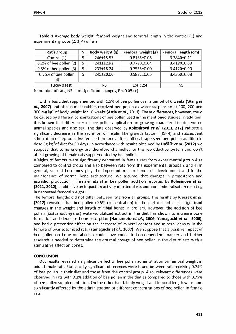
Our results demonstrate no significant effect of bee pollen application on body weight and femoral length in adult female rats. Statistically significant differences were found only for femoral weight in rats from experimental group 4 in comparison with those from the control one (1) and also relevant differences were observed between rats from the experimental groups 2 and 4 (Table 1).
In general, bee pollen contains a wide spectrum of amino acids, vitamins, hormones, and
minerals, as well as enzymes and co-enzymes necessary for good digestion and growth. Our study revealed a non-significant effect of bee pollen administration (in concentrations of 0.2%, 0.5% and 0.75%) on body weight in adult female rats. In the contrary Haro et al. (2000) reported that male rats fed with multifloral bee pollen (10g.kg-1 of diet for 10 days) had increased body weight. Significantly increased body weight was also observed in chicken fed
RFFCH Gödöllő, 2013
411
Table 1 Average body weight, femoral weight and femoral length in the control (1) and experimental groups (2, 3, 4) of rats.
Rat’s group N Body weight (g) Femoral weight (g) Femoral length (cm)
Control (1) 5 246±15.57 0.8185±0.05 3.3840±0.11
0.2% of bee pollen (2) 5 241±12.92 0.7780±0.04 3.4180±0.03
0.5% of bee pollen (3) 5 237±18.24 0.7535±0.09 3.4120±0.09
0.75% of bee pollen (4)
5 245±20.00 0.5832±0.05 3.4360±0.08
Tukey’s test NS 1:4+; 2:4+ NS
N: number of rats, NS: non-significant changes, P < 0.05 (+)
with a basic diet supplemented with 1.5% of bee pollen over a period of 6 weeks (Wang et al., 2007) and also in male rabbits received bee pollen as water suspension at 100, 200 and 300 mg.kg-1 of body weight for 10 weeks (Attia et al., 2011). These differences, however, could be caused by different concentrations of bee pollen used in the mentioned studies. In addition, it is known that differences of bee pollen application on growing characteristics depend on animal species and also sex. The data observed by Kolesárová et al. (2011, 212) indicate a significant decrease in the secretion of insulin like growth factor I (IGF-I) and subsequent stimulation of reproductive female hormones after unifloral rape seed bee pollen addition in dose 5g.kg-1of diet for 90 days. In accordance with results obtained by Haščík et al. (2012) we suppose that some energy are therefore channelled to the reproductive system and don’t affect growing of female rats supplemented by bee pollen. Weights of femora were significantly decreased in female rats from experimental group 4 as compared to control group and also between rats from the experimental groups 2 and 4. In general, steroid hormones play the important role in bone cell development and in the maintenance of normal bone architecture. We assume, that changes in progesteron and estradiol production in female rats after bee pollen addition reported by Kolesárová et al. (2011, 2012), could have an impact on activity of osteoblasts and bone mineralisation resulting in decreased femoral weight. The femoral lengths did not differ between rats from all groups. The results by Kleczek et al. (2012) revealed that bee pollen (0.5% concentration) in the diet did not cause significant changes in the weight and length of tibial bones in broilers. However, the addition of bee pollen (Cistus ladaniferus) water-solubilized extract in the diet has shown to increase bone formation and decrease bone resorption (Hamamoto et al., 2006; Yamaguchi et al., 2006), and had a preventive effect on the decrease of mineral content and mineral density in the femora of ovariectomized rats (Yamaguchi et al., 2007). We suppose that a positive impact of bee pollen on bone metabolism could have concentration-dependent manner and further research is needed to determine the optimal dosage of bee pollen in the diet of rats with a stimulative effect on bones. CONCLUSION
Out results revealed a significant effect of bee pollen administration on femoral weight in adult female rats. Statistically significant differences were found between rats receiving 0.75% of bee pollen in their diet and those from the control group. Also, relevant differences were observed in rats with 0.2% addition of bee pollen in the diet as compared to those with 0.75% of bee pollen supplementation. On the other hand, body weight and femoral length were non-significantly affected by the administration of different concentrations of bee pollen in female rats.
RFFCH Gödöllő, 2013
412
Acknowledgments: This study was supported by the grants KEGA 025UKF-4/2012 and VEGA 1/0790/11. REFERENCES ALMEIDA-MURADIAN, L.B., PAMPLONA, L.C., COIMBRA, S., BARTH, O.M. 2005. Chemical composition
and botanical evaluation of dried bee pollen pellets. Journal of Food Composition and Analysis, 18, 105-111.
ATTIA, Y.A., ALL-HANOUN, A., BOVERA, F. 2011. Effect of different levels of bee pollen on performance and blood profile of New Zealand White bucks and growth performance of their offspring during summer and winter months. Journal of Animal Physiology and Animal Nutrition, 95, 17-26.
BALTRUŠAITYTÉ, V., VENSKUTONIS, P.R., ČKSTARYTÉ, V. 2007. Radical scavenging activity of different floral origin honey and beebread phenolic extracts. Food Chemistry, 101, 502-514.
BELL, R.R., THRONBER, E.J., SEET, J.L., GROVES, M.T., HO, N.P., BELL, D.T. 1983. Composition and protein quality of honeybee-collected pollen of Eucalyptus marginata and Eucalyptus calophylla. Journal of Nutrition, 113, 2479-2484.
CAPCÁROVÁ, M., SLAMEČKA, J., ABBAS, K., KOLESÁROVÁ, A., KALAFOVÁ, A., VÁLENT, M., FILIPEJOVÁ, T., CHRASTINOVÁ, L., ONDRUŠKA, L., MASSANYI, P. 2012. Effect of diatery inclusion of Rhuscoriaria on internal milieu of rabbits. Journal of Animal Physiology and Animal Nutrition, 96, 459-465.
HAMAMOTO, R., ISHIYAMA, K., YAMAGUCHI, M. 2006. Inhibitory effects of bee pollen cistus ladaniferus extract on bone resorption in femoral tissues and osteoclast-like cell formation in bone marrow cellsin vitro. Journal of Health Science, 52, 268-275.
HARO, A., LOPEZ-ALLIAGA, I., LISBONA, F., BARRIONUEVO, M., ALFEREZ, M.J.M., CAMPOS, M.S. 2000. Beneficial effect of pollen nad/or propolis on the metabolism of iron, calcium, phosphorus, and magnesium in rats with nutritional ferropenic anemia. Journal of Agricultural and Food Chemistry, 48, 5715-5722.
HAŠČÍK, P., ELIMAN, I.E., BOBKO, M., KAČÁNIOVÁ, M., POCHOP, J., GARLIK, J., KROČKO, M., ČUBOŇ, J., VAVRIŠÍNOVÁ, K., ARPÁŠOVÁ, H., CAPCÁROVÁ, M., BENCZOVÁ, M. 2011. Oxidative stability of chicken meat after pollen extract application in their diet. Journal of Microbiology, Biotechnology and Food Sciences, 1, 70-82.
HAŠČÍK, P., ELIMAN, I.E., GARLIK, J., KAČÁNIOVÁ, M., ČUBOŇ, J., BOBKO, M., ABDULLA, H. 2012. Impact of bee pollen as feed suplements on the body weight of broiler Ross 308. African Journal of Biotechnology, 11, 15596-15599.
KOLESÁROVÁ, A., BAKOVÁ, Z., CAPCÁROVÁ, M., GARLIK, J., JURACEK, M., SIMKO, M., TOMAN, R., SIROTKIN, A.V. 2012. Consumption of bee pollen affects rat ovarian functions. Journal of Animal Physiology and Animal Nutrition, 9, 1-7.
KOLESÁROVÁ, A., CAPCÁROVÁ, M., BAKOVÁ, Z., GÁLIK, B., JURÁČEK, M., ŠIMKO, M., SIROTKIN, A. 2011. The effect of bee pollen on secretion activity, markers of proliferation and apoptosis of porcine ovarian granulosa cells in vitro. Journal of Environmental Science and Health, Part B. Pesticides, Food Contaminants, and Agricultural Wastes, 46, 207-212.
MIZRAHI, A., LENSKY, Y. 1997. Bee Products: Properties, Aplications and Apitherapy. Springer-Verlag: New York, USA.
MORAIS, M., MOREIRA, L., FEÁS, X., ESTEVINHO, L.M. 2011. Honeybee-collected pollen from five Portuguese Natural Parks: Palynological origin, phenolic content, antioxidant properties and antimicrobial activity. Food and Chemical Toxicology, 39, 1096-1101.
MOREIRA, L., DIAS, G., PEREIRA, E. 2008. Antioxidant and antimicrobial effects of phenolic compounds extracts of Northeast Portugam honey. Food and Chemical Toxicology, 46, 3774-3779.
ORZAEZ VILLANUEVA, M.T., DIAZ MARQUINA, A., BRAVO SERRANO, R., BLAZQUEZ ABELIAN, G. 2002. The importance of bee-collected pollen in the diet: a study of its composition. International Journal of Food Sciences Nutrition, 53, 217-224.
PETRUŠKA, P., TUŠIMOVÁ, E., KALAFOVÁ, A., HAŠČÍK, P., KOLESÁROVÁ, A., CAPCÁROVÁ, M. 2012. Effect of propolis in chicken diet on selected parameters of mineral profile. Journal of Microbiology, Biotechnology and Food Sciences, 1, 593-600.
SHOSKES, D.A. 2002. Phytotherapy in chronic prostatitis. Urology, 60, 35-37.
RFFCH Gödöllő, 2013
413
SILVA, T.M.S., CAMARA, C.A., LINS, A.C.S., BARBOSAFILHO, J.M., SILVA, E.M., FREITAS, B.M., SANTOS, F.A.R. 2006. Chemical composition and free radical scavenging activity of pollen loads from stingless bee Melipona subnitida Ducke. Journal of Food Composition and Analysis, 19, 507-511.
WANG, J., LI, S., WANG, Q., XIN, B., WANG, H. 2007. Trophic effect of bee pollen on small intestine in broiler chickens. Journal of Medicinal Food, 10, 276-280.
WOJCICKI, J., SAMACHOWIEC, L., BARTLOMOWICZ, B., HINEK, A., JAWORSKA, M., GAWRONSKA-SZKLARZ, B. 1986. Effect of pollen extract on the development of experimental atherosclerosis in rabbits. Atherosclerosis, 62, 39-45.
YAMAGUCHI, M., HAMAMOTO, R., UCHIYAMA, S., ISHIYAMA, K., HASHIMOTO, K. 2006. Anabolic effects of bee pollen Cistus ladaniferus extract on bone components in the femoral diaphyseal and metaphyseal tissues of rats in vitro and in vivo. Journal of Health Science, 52, 43-49.
YAMAGUCHI, M., IGARASHI, A., UCHIYAMA, S., MORITA, S., SUGAWARA, K., SUMIDA, T. 2004. Prolonged intake of juice (Citrus unshiu) reinforced with β-cryptoxanthin has an affect on circulating bone biochemical markers in normal individuals. Journal of Health Science, 50, 619-624.
YAMAGUCHI, M., UCHIYAMA, S., NAKAGAWA, T. 2007. Preventive effects of bee pollen cistus ladaniferus extract on bone loss in ovariectomized rats in vivo. Journal of Health Science, 53, 571-575.
ZHANG, X., HABIB, F.K., ROSS, M., BURGER, U., LEWENSTEIN, A., ROSE, K., JATON,J. 1995. Isolation and characterization of a cyclic hydroxamic acid from a pollen extract, which inhibits cancerous cell growth in vitro. Journal of Medicinal Chemistry, 38, 735-738.
RFFCH Gödöllő, 2013
414
Animal welfare, etológia és tartástechnológia
Animal welfare, ethology and housing systems
Volume 9 Issue 3
Különszám/Special Issue
Gödöllő
2013
RFFCH Gödöllő, 2013
414
HEMATOLOGICAL ALTERATIONS IN COMMON CARP (CYPRINUS CARPIO L.) EXPOSED TO MANKOZEB
Bojarski B., Lutnicka H., Ludwikowska A.
University of Agriculture in Cracow, Faculty of Animal Science, Department of Poultry and Fur Animal Breeding and Animal Hygiene, Poland
ABSTRACT In this study the influence of the fungicide mankozeb on hematological parameters of common carp was investigated. The study was conducted on fish weighing 60 (± 10) g in aquaria under controlled environmental conditions. Fish were exposed to the fungicide for 14 days at concentration of 1 mg L-1. Next, aminals were transported to clean water for 30 days (recovery period). It was determined that mankozeb caused a decrease in the values of hematocrit (HCT) and hemoglobin content (Hb) after 14 days of exposure. Increase in the mean corpuscular volume (MCV) and in white blood cell (WBC) number after the same time was observed. Increase in red blood cell (RBC) number was observed after 24 hours of exposure. The values of mean corpuscular hemoglobin (MCH) and mean corpuscular hemoglobin concentration (MCHC) were decreased after recovery period. Evaluation of hematological parameters in fish is useful in detection of environmental stressors such as fungicides. This work has been financially supported by DS 3210/KHDZFiZ Key words: fungicides, mankozeb, hematological parameters

Animal welfare, etológia és tartástechnológia
Animal welfare, ethology and housing systems
Volume 9 Issue 3
Különszám/Special Issue
Gödöllő
2013
RFFCH Gödöllő, 2013
415
COBALT-INDUCED ACTIVATION OF HEAT SHOCK PROTEIN 70 AND ANTIOXIDANT STATUS IN OVARIAN FRAGMENTS OF RATS
Marcela Capcarová1, Adriana Kolesarova1, Branislav Galik2, Milan Simko2, Miroslav Juracek2, Robert Toman3, Alexander V. Sirtokin4
1Department of Animal Physiology, Faculty of Biotechnology and Food Sciences, 2Department of Animal Nutrition, Faculty of Agrobiology and Food Resources, 3Department of Veterinary Disciplines, Faculty of Agrobiology and Food Resources, Slovak University of Agriculture in Nitra, Tr. A. Hlinku 2, 949 76 Nitra, Slovak Republic 4Department of Genetics and Reproduction, Animal Production Research Centre Nitra, Hlohovecka 2, 949 01 Nitra, Slovak Republic
Abstract The aim of this study was to determine the expression of heat shock protein 70 (Hsp70), activity of superoxide dismutase (SOD) and total antioxidant status (TAS) of rat ovarian fragments exposed to cobalt sulphate (Co) in vitro. Fragments were incubated with Co administrations as follows: group E1 (0.09 mg.ml-1), group E2 (0.17 mg.ml-1), group E3 (0.33 mg.ml-1), group E4 (0.5 mg.ml-1), group E5 (1.0 mg.ml-1) and the control group without any additions for 18 h. Co administration increase stress reaction by accumulation of Hsp70 what resulted in increasing activity of SOD. TAS of ovary fragments increased with higher doses of Co whereas low doses had no effect. Trace elements can adversely affect animal female reproductive system and its functions, through either direct or indirect effects on oxidative stress induction
Key words: cobalt, ovary, rats, antioxidants, HSP70
Introduction Cobalt (Co) has important role in many processes, including reproduction (Ashmead, 1993), it is an essential element, but at high concentrations is toxic (Kubrak et al, 2011). However, Co can be also acutely toxic in larger doses, cytotoxic and induce apoptosis and at higher concentrations necrosis with inflammatory response. Cobalt metal and salts are also genotoxic, mainly cause oxidative DNA damage by reactive oxygen species, perhaps combined with inhibition of DNA repair (Simonses et al, 2012). Chronic overexposure to Co may result in neurotoxic effects and exposure of pregnant and lactating rats resulted in the development of oxidative stress and the impairment of defence systems (Garoui et al, 2013). Exposure of rats to Co during late pregnancy and early postnatal period affected antioxidant enzyme activities and lipid peroxidation (Garoui et al, 2011). Co exposure of animal organism usually causes the activation of defence systems against reactive oxygen species (ROS) (Kubrak et al, 2012). In addition to its well-known function as an integral part of cobalamin (vitamin B₁₂), Co has recently been shown to be a mimetic of hypoxia and a stimulator of the production of ROS (Kubrak et al, 2011). In recent years, there has been growing interest in the roles of ROS in female reproduction. ROS are key signals in the initiation of apoptosis in antral follicles and granulosa cells of antral follicles by diverse stimuli, such as exposure to exogenous toxicants, and that antioxidants protect against these stimuli (Devine et al, 2012). When ROS are overproduced, oxidative stress may develop in the body (Jones, 2008). Superoxide dismutase (SOD) serves as front-line antioxidant defence (Scandalios, 2005), reacts with superoxide anion radicals to form oxygen and H2O2 (Ho et al, 1998). Total antioxidant status (TAS) represents the level of cumulative antioxidant reserve of the body and enables evaluation of the average antioxidant potential. Thus, the overall antioxidant status will give more relevant biological information compared to that obtained by the measurement of individual components (Millet et al, 1993). Heat shock proteins (HSPs) belong to a large and diverse group of unrelated proteins known as chaperones that assist in correct non-covalent assembly and/or disassembly of other polypeptide-containing structure (Ellis, 1997). A variety of stressful situations including environmental stimuli
RFFCH Gödöllő, 2013
416
(heavy metals) induce a marked increase in HSP synthesis, known as the stress response (Jaattela, 1999; Tsan and Gao, 2004). The aim of present study was to determinate dose-dependent changes in activity of SOD, TAS and expression of Hsp70 in ovarian fragments of rats exposed to Co in vitro.
Material and Methods Preparation, culture and processing of ovary Rats (Wister rats, Slovak University of Agriculture in Nitra, Slovak Republic) in age 150 days were kept under standard conditions at Slovak University of Agriculture in Nitra. Isolated ovaries were washed in a sterile physiological solution. Ovaries were cut by razor blade into fragments (totally 72 pieces) approximately 2 mm in size. Fragments of ovaries were washed in sterile DMEM/F12 1:1 medium (BioWhittakerTM, Verviers, Belgium) and incubated for 24 h in culture plates (NuncTM, Roskilde, Denmark, 1 ml/well) in the same medium with 10% fetal calf serum (BioWhittakerTM, Verviers, Belgium), 1% antibiotic-antimycotic solution (Sigma, St. Louis, Mo, USA), with or without cobalt sulphate CoSO4.7H2O (Co) addition in various doses (Table 1). Culture media from plate wells were aspirated and stored at –20 ºC for further assay. Cells intended for SOD activity analyse and Western immunoblotting were lyzed in ice-cold lysis buffer (1% Triton X-100, 0.5% Igepal NP-40, 5mM EDTA, 20µg/mlphenylmethylsuphonyl fluoride, 10 µg/ml aprotonin, 10 µg/ml leupeptin, 5 µg/ml pepstatin, 10mM sodium orthovanadate in phosphate-buffered saline, pH 7.5, all from Sigma, 50µg/well) (Sirotkin and Bauer, 2011).
Table 1. Cobalt concentrations used in the study
Group CoSO4.7H2O
mg.ml-1 Medium
ml Dilution
rate
Concentration of
CoSO4.7H2O (mg.ml-1)
Control 0 1 0:1 0
E1 0.09 0.91 1:10 0.09
E2 0.17 0.83 1:5 0.17
E3 0.33 0.67 1:2 0.33
E4 0.5 0.5 1:1 0.5
E5 1 0 1:0 1.0
SOD and TAS analysis The activity of SOD and TAS of ovarian fragments of rats was assayed by spectrophotometer Genesys 10 (using antioxidant RANDOX kits (Randox Labs., Crumlin, UK) according to the manufacturer’s instructions.
Western blotting The separation of HSP70 performed using SDS-PAGE and its subsequent visualization by Western immunoblotting using mouse monoclonal antibody against HSP70 and housekeeping protein GAPDH (1:250 dilution; all from Santa Cruz, Santa Cruz, CA, USA), secondary HRP-conjugated anti-mouse IG antibodies (Sevac. Prague, Czech Republic), ECL detection reagents and ECL Hyper-film (Amersham International) was performed as described previously (Sirotkin and Makarevich 1999; Sirotkin and Bauer, 2011). The primary antisera against HSP70 and GAPDH were specific for mouse, rat, human, porcine and bovine cells. Incubation medium without cells, or samples processed in the absence of primary antibody, were used as negative controls. The molecular weights of fractions were evaluated using a molecular weight calibration set (18, 24, 45 and 67 kDa; ICN Biomedicals, Inc., Irvine, CA, USA). Band intensity was evaluated by densitometry analysis (not shown here).
RFFCH Gödöllő, 2013
417
Statistical analysis Each experimental group was represented by four culture wells of cultured ovary fragments. Assays of substances in incubation medium were performed in duplicate. The data presented are means of values obtained in three separate experiments performed on separate days using separate pools of ovaries from 10 – 12 animals. Significant differences between the control and experimental groups were evaluated by one-way ANOVA test using statistical software Sigma Plot 11.0 (Jandel, Corte Madera, USA). The data are expressed as means±SD. Differences were compared for statistical significance at the level P<0.05.
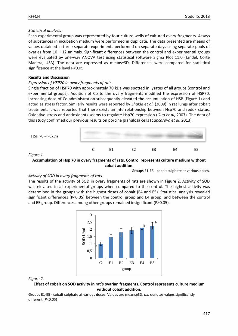
Results and Discussion Expression of HSP70 in ovary fragments of rats Single fraction of HSP70 with approximately 70 kDa was spotted in lysates of all groups (control and experimental groups). Addition of Co to the ovary fragments modified the expression of HSP70. Increasing dose of Co administration subsequently elevated the accumulation of HSP (Figure 1) and acted as stress factor. Similarly results were reported by Shukla et al. (2009) in rat lungs after cobalt treatment. It was reported that there exists an interrelationship between Hsp70 and redox status. Oxidative stress and antioxidants seems to regulate Hsp70 expression (Guo et al, 2007). The data of this study confirmed our previous results on porcine granulosa cells (Capcarova et al, 2013).
C E1 E2 E3 E4 E5 Figure 1.
Accumulation of Hsp 70 in ovary fragments of rats. Control represents culture medium without cobalt addition.
Groups E1-E5 - cobalt sulphate at various doses.
Activity of SOD in ovary fragments of rats The results of the activity of SOD in ovary fragments of rats are shown in Figure 2. Activity of SOD was elevated in all experimental groups when compared to the control. The highest activity was determined in the groups with the highest doses of cobalt (E4 and E5). Statistical analysis revealed significant differences (P<0.05) between the control group and E4 group, and between the control and E5 group. Differences among other groups remained insignificant (P>0.05).
Figure 2.
Effect of cobalt on SOD activity in rat’s ovarian fragments. Control represents culture medium without cobalt addition.
Groups E1-E5 - cobalt sulphate at various doses. Values are means±SD. a,b denotes values significantly different (P<0.05)
0
0,5
1
1,5
2
2,5
3
C E1 E2 E3 E4 E5
SO
D U
/ml
group
HSP 70 – 70kDa
a
b b
RFFCH Gödöllő, 2013
418
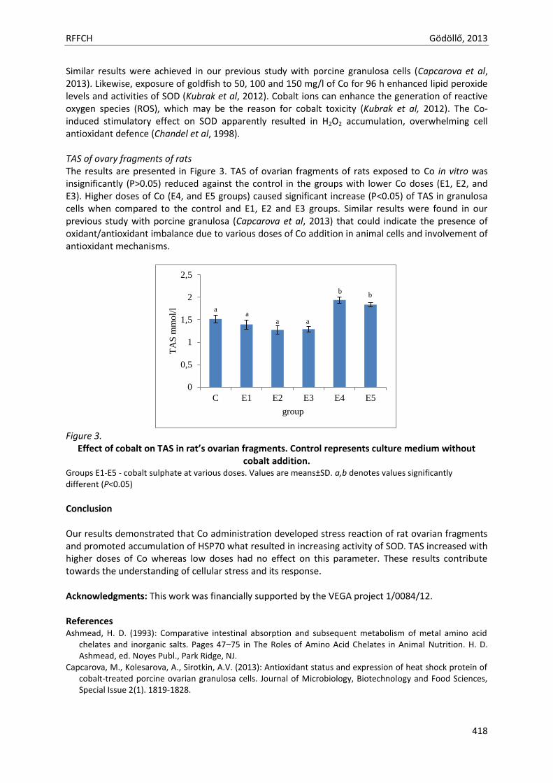
Similar results were achieved in our previous study with porcine granulosa cells (Capcarova et al, 2013). Likewise, exposure of goldfish to 50, 100 and 150 mg/l of Co for 96 h enhanced lipid peroxide levels and activities of SOD (Kubrak et al, 2012). Cobalt ions can enhance the generation of reactive oxygen species (ROS), which may be the reason for cobalt toxicity (Kubrak et al, 2012). The Co-induced stimulatory effect on SOD apparently resulted in H2O2 accumulation, overwhelming cell antioxidant defence (Chandel et al, 1998). TAS of ovary fragments of rats The results are presented in Figure 3. TAS of ovarian fragments of rats exposed to Co in vitro was insignificantly (P>0.05) reduced against the control in the groups with lower Co doses (E1, E2, and E3). Higher doses of Co (E4, and E5 groups) caused significant increase (P<0.05) of TAS in granulosa cells when compared to the control and E1, E2 and E3 groups. Similar results were found in our previous study with porcine granulosa (Capcarova et al, 2013) that could indicate the presence of oxidant/antioxidant imbalance due to various doses of Co addition in animal cells and involvement of antioxidant mechanisms.
Figure 3.
Effect of cobalt on TAS in rat’s ovarian fragments. Control represents culture medium without cobalt addition.
Groups E1-E5 - cobalt sulphate at various doses. Values are means±SD. a,b denotes values significantly different (P<0.05)
Conclusion Our results demonstrated that Co administration developed stress reaction of rat ovarian fragments and promoted accumulation of HSP70 what resulted in increasing activity of SOD. TAS increased with higher doses of Co whereas low doses had no effect on this parameter. These results contribute towards the understanding of cellular stress and its response.
Acknowledgments: This work was financially supported by the VEGA project 1/0084/12. References Ashmead, H. D. (1993): Comparative intestinal absorption and subsequent metabolism of metal amino acid
chelates and inorganic salts. Pages 47–75 in The Roles of Amino Acid Chelates in Animal Nutrition. H. D. Ashmead, ed. Noyes Publ., Park Ridge, NJ.
Capcarova, M., Kolesarova, A., Sirotkin, A.V. (2013): Antioxidant status and expression of heat shock protein of cobalt-treated porcine ovarian granulosa cells. Journal of Microbiology, Biotechnology and Food Sciences, Special Issue 2(1). 1819-1828.
0
0,5
1
1,5
2
2,5
C E1 E2 E3 E4 E5
TA
S m
mo
l/l
group
a a
a a
b
a a
b
a a
RFFCH Gödöllő, 2013
419
Chandel, N.S., Maltepe, E., Goldwasser, E., Mathieu, C.E., Simon, M.C., Schumaker, P.T. (1998): Mitochondrial reactive oxygen species trigger hypoxia-induced transcription. Proceedings of the National Academy of Sciences, 95. 11715-11720.
Devine, P.J., Perreault, S.D., Luderer, U., (2012): Roles of reactive oxygen species and antioxidants in ovarian toxicity. Biology of Reproduction, 86. 1-10.
Ellis, R.J. (1997): Do molecular chaperones have to be proteins? Biochemical and Biophysical Research Communications, 238. 687-692.
Garoui el M., Fetouri, H., Ayadi Makni F., Boudawara, T., Zeghal, N. (2011): Cobalt chloride induces hepatotoxicity in adult rats and their suckling pups. Experiemntal and Toxicologic Pathology, 63. 9-15.
Garoui, E., Amara, I.B., Driss, D., Elwej, A., Chaabouni, S.E., Boudawara, T., Zeghal, N. (2013): Effects of Cobalt on Membrane ATPases, Oxidant, and Antioxidant Values in the Cerebrum and Cerebellum of Suckling Rats. Biological Trace Elements Research, in press.
Guo, S., Wharton, W., Moseley, P., Shi, H. (2007): Heat shock protein 70 regulates cellular redox status by modulating glutathione-related enzyme activities. Cell Stress and Chaperones, 12. 245-254.
Ho Y-S, Cargano M, Cao J, Bronson RT, Heimler I, Hutz RJ, 1998. Reduced fertility in female mice lacking copper-zinc superoxide dismutases. Journal of Biological Chemistry, 273. 7765-7769.
Jones DP, 2008. Radical-free biology of oxidative stress. American Journal of Physiology: Cell Physiology, 295. C849-C868.
Jaattela, M. (1999): Heat shock proteins as cellular lifeguards. Annual Medicine, 31. 261-271. Kubrak, O.I., Husak, V.V., Rovenko, B.M., Storey, J.M., Storey, K.B., Lushchak, V.I. (2011): Cobalt-induced
oxidative stress in brain, liver and kidney of goldfish Carassius auratus. Chemosphere, 85. 983-989. Kubrak, O.I., Rovenko, B.M., Husak, V.V., Vasylkiv, O.Y., Storey, K.B., Storey, J.M., Lushchak, V.I. (2012): Goldfish
exposure to cobalt enhances hemoglobin level and triggers tissue-specific elevation of antioxidant defenses in gills, heart and spleen. Comparative Biochemistry and Physiology C, 155. 325-332.
Miller, N., Rice-Evans, C., Davies, M.J. (1993): A novel method for measuring antioxidant capacity and its application to monitoring the antioxidant status in premature neonates. Clinical Science, 84. 407-412.
Scandalios JG, 2005. Oxidative stress: molecular perception and transduction of signals triggering antioxidant gene defences. Brazilian Journal of Medical and Biological Research, 38. 995-1014.
Shukla, D., Saxena, S., Jayamurthy, P., Sairam, M., Singh, M., Jain, S.K., Bansal, A., Ilavazagham, G. (2009): Hypoxic preconditioning with cobalt attenuates hypobaric hypoxia-induced oxidative damage in rat lungs. High Altitude Medicine and Biology, 10. 57-69.
Simonsen, L.O., Harbak, H., Bennekou, P. (2012): Cobalt metabolism and toxicology – a brief update. The Science of Total Environment, 432. 210-215.
Sirotkin, A.V., Makarevich, A.V. (1999): GH regulates secretory activity and apoptosis in cultured bovine granulosa cells through the activation of the cAMP/protein dinase A system. Journal of Endocrinology, 163. 317–327.
Sirotkin, A.V., Bauer, M. (2011): Heat shock proteins in porcine ovary: synthesis, accumulation and regulation by stress and hormones. Cell Stress and Chaperons, 16. 379-387.
Tsan, M.F., Gao, B. (2004): Cytokine function of heat shock proteins. American Journal of Physiology – ZPI, 243 p. ISBN 80-85 120-37-2.

Animal welfare, etológia és tartástechnológia
Animal welfare, ethology and housing systems
Volume 9 Issue 3
Különszám/Special Issue
Gödöllő
2013
RFFCH Gödöllő, 2013
420
OSTEOTOXIC EFFECT OF SIMULTANEOUS ADMINISTRATION TO CADMIUM AND DIAZINON ON BONE IN ADULT MALE RATS
Hana CHOVANCOVÁ1, Monika MARTINIAKOVÁ1, Ivana BOBOŇOVÁ1, Radoslav OMELKA2, Mária ADAMKOVIČOVÁ2, Robert STAWARZ3, Róbert TOMAN4
1Department of Zoology and Anthropology, Constantine the Philosopher University, 949 74 Nitra, Slovakia 2Department of Botany and Genetics, Constantine the Philosopher University, 949 74 Nitra, Slovakia 3Institute of Biology, Krakow Pedagogical University, Krakow 31 054, Poland 4Department of Veterinary Sciences, Slovak University of Agriculture, 949 76 Nitra, Slovakia
Abstract Bone is a metabolically active tissue which can be influenced by various toxicants presented in the environment. The study was aimed to investigate the osteotoxic effect of simultaneous peroral administration to heavy metal cadmium (Cd) and nonselective organophosphorous insecticide diazinon (DZN) on bone in adult male rats. A total of twenty 1-month-old male Wistar rats were randomized into two experimental groups. In the first group (A), young males were dosed with combination of 30 mg CdCl2/L and 40 mg DZN/L in drinking water, for 90 days. Ten 1-month-old males without toxicant administration served as a control group (B). After treatment period, detailed histological analysis of compact bone was performed in each group. We found that rats from the group A displayed different microstructure in the middle part of the substantia compacta where primary vascular radial bone tissue appeared (due to radial extension of vascular canals from the endosteal surfaces). In some cases, vascular expansion was so enormous that canals were also present near the periost. On the other hand, they occurred only near the endosteal surfaces in rats from the group B. In Cd-DZN-exposed rats, a smaller number of primary and secondary osteons was also identified signalizing reduced bone mechanical properties. Our results suggest an adaptive response of bone to Cd-DZN-induced toxicity in rats in order to prevent osteonecrosis. Key words: Bone. Osteotoxicology. Cadmium. Diazinon. Rats. Introduction Cadmium (Cd) is a toxic metal which still attracts the attention of researchers and the public because its level in food products often exceeds the maximum allowable limits (Toman et al., 2011). The diet is the major source (~ 99%) of Cd exposure in the general non-smoking population (Järup and Akesson, 2009). Concentrations of Cd were determined in various organs of experimental animals (Massányi et al., 2001; Kolesárová et al., 2008). In respect to bone, which is one of the important organs for Cd toxicity (WHO, 1992), exposure to Cd has been linked to bone loss, low bone mass, and osteoporosis and even to an increased incidence of bone fractures (Wilson et al., 1996; Wang et al., 2003). The results obtained by Brzóska and Moniuszko-Jakoniuk (2005) have shown that chronic, even low-level exposure to Cd disturbs bone metabolism during skeletal development and maturity by affecting bone turnover most probably through a direct influence on bone formation and resorption, and indirectly via disorders in Ca metabolism. Diazinon (DZN) is a contact organophosphate pesticide which is extensively used in agriculture (Salehi et al., 2009). Like other organophosphates (OPs), the main toxic action of DZN is inhibition of acetylcholinesterase activity (AChE) which results in accumulation of acetylcholine
RFFCH Gödöllő, 2013
421
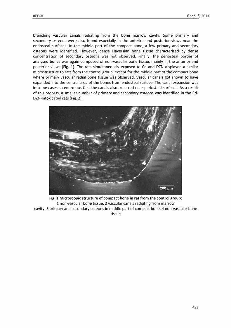
(ACh) and associated neurotoxicity (Oruc and Usta, 2007). According to Garg et al. (2004), a potential target of pesticide toxicity is the skeletal system. Marked impairment in the development of the backbone in ducklings due to OPs toxicity has been observed in the study by Ludle et al. (1979). Higher amounts of DZN caused additional defects in quail and chicken including folding of the spinal chord, shortening of the neck (Wyttenbach and Hwang, 1984), fusing and twisting of vertebrae, abnormal development of ribs and breastbone (Meneely and Wyttenbach, 1989), curled claws, reduced growth of leg and wing bones (Cho and Lee, 1990), and reduced bone calcification (Cho and Lee, 1991). In addition, OPs cause a significant reduction in bone mass and density in individuals following chronic low-level intoxication (Compston et al., 1999). Results by Lari et al. (2011) indicate that DZN exposure is associated with decrease in trabecular and cortical bone density and might be one of the causes for worldwide increasing prevalence of osteoporosis. The aim of current study was to investigate the osteotoxic effect of peroral Cd-DZN co-administration on bone in adult male rats. Materials and methods Our experiment was conducted on twenty 1-month-old male Wistar rats obtained from the accredited experimental laboratory (number SK PC 50004) of the Slovak University of Agriculture in Nitra. Clinically healthy rats were randomly divided into two groups, of 10 animals each. In the first group (A), young males were dosed with a daily intake of 30 mg CdCl2/L in combination with 40 mg DZN/L in drinking water for 90 days. The second group without Cd and DZN supplementation served as a control (group B). The xenobiotics used in our experiment were chosen on the basis of their possible occurrence in the human and animal food (Toman et al., 2011). Indeed, correlation coefficients found between Cd and DZN in men (0.70) and women (0.69) indicate high probability of exposure to both compounds (Toman et al., 2012). The doses of Cd and DZN were high enough to reach a toxicity level but also safe enough to prevent animal mortality. All procedures were approved by the Animal Experimental Committee of the Slovak Republic. At the end of the experiment, all animals were killed and their right femora were used for microscopic analysis. Each right femur was sectioned at the midshaft of its diaphysis. The obtained segments were placed in HistoChoice fixative (Amresco, USA). Specimens were then dehydrated in ascending grades of ethanol and embedded in epoxy resin Biodur (Günter von Hagens, Heidelberg, Germany) according to Martiniaková et al. (2007). Transverse thin sections (70-80 μm) were prepared with a sawing microtome (Leitz 1600, Leica, Wetzlar, Germany) and affixed to glass slides by Eukitt (Merck, Darmstadt, Germany) as previously described (Martiniaková et al., 2008). The qualitative histological characteristics of the compact bone were determined according to the internationally accepted classification systems of Enlow and Brown (1956) and Ricqlés et al. (1991), who classified bone into three main categories: primary vascular tissue, non-vascular tissue and Haversian bone tissue. Various patterns of vascularization can occur in primary vascular bone: longitudinal, radial, reticular, plexiform, laminar, lepidosteoid, acellular, fibriform and protohaversian. There are three subcategories indentified in Haversian bone tissue: irregular, endosteal and dense. Results Femoral diaphyses of rats from the control group had the following microstructure in common. The internal layer surrounding the medullary cavity (i.e. endosteal border) was formed by non-vascular bone tissue in all views of the thin sections. The bone tissue contained cellular lamellae and osteocytes. Primary and/or secondary osteons were not present. Additionally, there were also identified some areas of primary vascular radial bone tissue in anterior, posterior and lateral views. This type of bone tissue was created by branching or non-
RFFCH Gödöllő, 2013
422
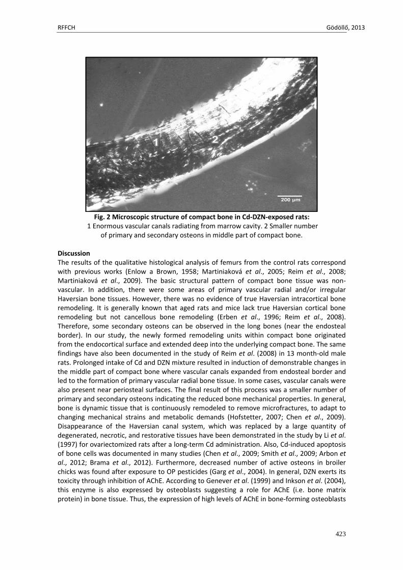
branching vascular canals radiating from the bone marrow cavity. Some primary and secondary osteons were also found especially in the anterior and posterior views near the endosteal surfaces. In the middle part of the compact bone, a few primary and secondary osteons were identified. However, dense Haversian bone tissue characterized by dense concentration of secondary osteons was not observed. Finally, the periosteal border of analysed bones was again composed of non-vascular bone tissue, mainly in the anterior and posterior views (Fig. 1). The rats simultaneously exposed to Cd and DZN displayed a similar microstructure to rats from the control group, except for the middle part of the compact bone where primary vascular radial bone tissue was observed. Vascular canals got shown to have expanded into the central area of the bones from endosteal surface. The canal expansion was in some cases so enormous that the canals also occurred near periosteal surfaces. As a result of this process, a smaller number of primary and secondary osteons was identified in the Cd-DZN-intoxicated rats (Fig. 2).
Fig. 1 Microscopic structure of compact bone in rat from the control group:
1 non-vascular bone tissue. 2 vascular canals radiating from marrow cavity. 3 primary and secondary osteons in middle part of compact bone. 4 non-vascular bone
tissue
RFFCH Gödöllő, 2013
423
Fig. 2 Microscopic structure of compact bone in Cd-DZN-exposed rats:
1 Enormous vascular canals radiating from marrow cavity. 2 Smaller number of primary and secondary osteons in middle part of compact bone.
Discussion The results of the qualitative histological analysis of femurs from the control rats correspond with previous works (Enlow a Brown, 1958; Martiniaková et al., 2005; Reim et al., 2008; Martiniaková et al., 2009). The basic structural pattern of compact bone tissue was non-vascular. In addition, there were some areas of primary vascular radial and/or irregular Haversian bone tissues. However, there was no evidence of true Haversian intracortical bone remodeling. It is generally known that aged rats and mice lack true Haversian cortical bone remodeling but not cancellous bone remodeling (Erben et al., 1996; Reim et al., 2008). Therefore, some secondary osteons can be observed in the long bones (near the endosteal border). In our study, the newly formed remodeling units within compact bone originated from the endocortical surface and extended deep into the underlying compact bone. The same findings have also been documented in the study of Reim et al. (2008) in 13 month-old male rats. Prolonged intake of Cd and DZN mixture resulted in induction of demonstrable changes in the middle part of compact bone where vascular canals expanded from endosteal border and led to the formation of primary vascular radial bone tissue. In some cases, vascular canals were also present near periosteal surfaces. The final result of this process was a smaller number of primary and secondary osteons indicating the reduced bone mechanical properties. In general, bone is dynamic tissue that is continuously remodeled to remove microfractures, to adapt to changing mechanical strains and metabolic demands (Hofstetter, 2007; Chen et al., 2009). Disappearance of the Haversian canal system, which was replaced by a large quantity of degenerated, necrotic, and restorative tissues have been demonstrated in the study by Li et al. (1997) for ovariectomized rats after a long-term Cd administration. Also, Cd-induced apoptosis of bone cells was documented in many studies (Chen et al., 2009; Smith et al., 2009; Arbon et al., 2012; Brama et al., 2012). Furthermore, decreased number of active osteons in broiler chicks was found after exposure to OP pesticides (Garg et al., 2004). In general, DZN exerts its toxicity through inhibition of AChE. According to Genever et al. (1999) and Inkson et al. (2004), this enzyme is also expressed by osteoblasts suggesting a role for AChE (i.e. bone matrix protein) in bone tissue. Thus, the expression of high levels of AChE in bone-forming osteoblasts
RFFCH Gödöllő, 2013
424
and their progenitors supports a toxic effect of AChE inhibitors (including DZN) on these cells (Genever et al., 1999; Grisaru et al., 1999; Inkson et al., 2004; Hoogduijn et al., 2006). On the basis of all mentioned findings we propose that the formation of primary vascular bone tissue, mainly in the central area of the femur, could be explained as an adaptive response of bone to Cd-DZN toxicity, in order to protect the tissue against death of cells and subsequent necrosis. Conclusions Simultaneous peroral exposure to Cd and DZN had the osteotoxic effect on femurs in adult male rats. Co-administration to Cd and DZN affected mainly the middle part of rats’ bones where primary vascular radial bone tissue was identified as a result of adaptive response to xenobiotic-induced osteonecrosis. On the other hand, the vascular canal expansion into central area of substantia compacta led to a smaller number of primary and secondary osteons signalizing weakened mechanical properties of the bones. Acknowledgments: This study was supported by the grants KEGA 035UKF-4/2013, VEGA 1/0790/11 and UGA VII/28/2013 References ARBON, K. S., C. M. CHRISTENSEN, W. A. HARVEY a S. J. HEGGLAND, 2012. Cadmium exposure activates
the ERK signaling pathway leading to altered osteoblast gene expression and apoptotic death in Saos-2 cells. In: Food Chem. Toxicol. Vol. 50, pp. 198-205.
BRAMA, M., L. POLITI, P. SANTINI, S. MIGLIACCIO a R. SCANDURRA, 2012. Cadmium-induced apoptosis and necrosis in human osteoblasts: role of caspases and mitogen-activated protein kinases pathways. In: J. Endocrinol. Invest. Vol. 35, pp. 198-208.
BRZÓSKA, M. M. a J. MONIUSZKO-JAKONIUK, 2005. Disorders in bone metabolism of female rats chronically exposed to cadmium. In: Toxicol. Appl. Pharmacol. Vol. 202, pp. 68–83.
CHEN, X., G. ZHU, S. GU, T. JIN a C. SHAO, 2009. Effects of cadmium on osteoblasts and osteoclasts in vitro. In: Environ. Toxicol. Pharmacol. Vol. 28, pp. 232–236.
CHO, J. a C. LEE, 1990. Effects of diazinon on the anatomical and embryological changes in the developing chick embryo. In: Res. Rep. RDA(V). Vol. 32, pp. 35-47.
CHO, J. a C. LEE, 1991. Studies on diazinon induced inhibition of skeletal mineralization in chick embryo. In: Res. Rep. RDA(V). Vol. 33, pp. 41-60.
COMPSTON, J. E., S. VEDI, A. B. STEPHEN, S. BORD, A. R. LYONS, S. J. HODGES a B. E. SCAMMELL, 1999. Reduced bone formation after exposure to organophosphates. In: Lancet. Vol. 354, pp. 1791–1792.
ENLOW, D. H. a S. O. BROWN, 1956. A comparative histological study of fossil and recent bone tissues. Part I. In: Texas J. Sci. Vol. 8, pp. 405-412.
ENLOW, D. H. a S. O. BROWN, 1958. A comparative histological study of fossil and recent bone tissues. Part III. In: Texas J. Sci. Vol. 10, pp. 187-230.
ERBEN, R. G., 1996. Trabecular and endocortical bone surfaces in the rat: modeling or remodeling? In: Anat. Rec. Vol. 246, pp. 39–46.
GARG, U. K., A. K. PAL, G. J. JHA a S. B. JADHAO, 2004. Pathophysiological effects of chronic toxicity with synthetic pyrethroid, organophosphate and chlorinated pesticides on bone health of broiler chicks. In: Toxicologic. Pathol. Vol. 32, pp. 364–369.
GENEVER, P. G., M. A. BIRCH, E. BROWN a T. M. SKERRY, 1999. Osteoblast-derived acetylcholinesterase: a novel mediator of cell-matrix interactions in bone? In: Bone. Vol. 24, pp. 297–304.
GRISARU, D., E. LEV-LEHMAN, M. SCHAPIRA, E. CHAIKIN, J. B. LESSING, A. ELDOR, F. ECKSTEIN a H. SOREQ, 1999. Human osteogenesis involves differentiation-dependent increases in the morphogenically active 39 alternative splicing variant of acetylcholinesterase. In: Mol. Cell Biol. Vol. 19, pp. 788-795.
HOFSTETTER, W., 2007. Bone remodeling. In: Eur. Cell. Mater. Vol. 14, p. 31. HOOGDUIJN, M. J., Z. RAKONCZAY a P. G. GENEVER, 2006. The effects of anticholinergic insecticides on
human mesenchymal stem cells. In: Toxicol. Sci. Vol. 94, pp. 342–350.
RFFCH Gödöllő, 2013
425
INKSON, C. A., A. C. BRABBS, T. S. GREWAL, T. M. SKERRY a P. G. GENEVER, 2004. Characterization of acetylcholinesterase expression and secretion during osteoblast differentiation. In: Bone. Vol. 35, pp. 819–827.
JÄRUP, L. a A. ÁKESSON, 2009. Current status of cadmium as an environmental health problem. In: Toxicol. Appl. Pharmacol. Vol. 238, pp. 201–208.
KOLESÁROVÁ, A., J. SLAMEČKA, R. JURČÍK, F. TATARUCH, N. LUKÁČ, J. KOVÁČIK, M. CAPCAROVÁ, M. VALENT a P. MASSÁNYI. 2008. Environmental levels of cadmium, lead and mercury in brown hares and their relation to blood metabolic parameters. In: J. Envir. Sci. Health A, Tox. Hazard. Subst. Environ. Eng. Vol. 43, pp. 646-650.
LARI, R., M. H. ELAHI a P. LARI, 2011. Diazinon exposure reduces trabecular and cortical bone mineral density. In: „Promoting Health Through Sustainable Chemical and Drug Safety Initiatives“: 10th Scientific Congress of the Asia Pacific Association of Medical Toxicology. Penang, Malaysia: Universiti Sains Malaysia, s. P85.
LI, J. P., T. AKIBA a F. MARUMO, 1997. Long-term, low-dose, cadmium-induced nephropathy with renal osteopathy in ovariectomized rats. In: J. Toxicol. Sci. Vol. 22, pp. 185-198.
LUDLE, J. L., M. P. MEHRLE, L. M. FOSTER a T. EARLKAISER, 1979. Bone development in black ducks as affected by dietarytoxophene. In: Pestic. Biochem. Physiol. Vol. 10, pp. 168–173.
MARTINIAKOVÁ, M., B. GROSSKOPF, M. VONDRÁKOVÁ, R. OMELKA a M. FABIŠ, 2005. Observation of the microstructure of rat cortical bone tissue. In: Scripta Med. Vol. 78, pp. 45-50.
MARTINIAKOVÁ, M., B. GROSSKOPF, R. OMELKA, K. DAMMERS, M. VONDRÁKOVÁ a M. BAUEROVÁ, 2007. Histological study of compact bone tissue in some mammals: a method for species determination. In: Int. J. Osteoarch. Vol. 17, pp. 82-90.
MARTINIAKOVÁ, M., R. OMELKA, B. GROSSKOPF, A. V. SIROTKIN a P. CHRENEK, 2008. Sex-related variation in compact bone microstructure of the femoral diaphysis in juvenile rabbits. In: Acta Vet. Scand. Vol. 50, p. 15.
MARTINIAKOVÁ, M., R. OMELKA, B. GROSSKOPF, Z. MOKOŠOVÁ a R. TOMAN, 2009. Histological analysis of compact bone tissue in adult laboratory rats. In: Slovak J. Anim. Sci. Vol. 42, pp. 56-59.
MASSÁNYI, P., P. NAĎ, R. TOMAN a J. KOVÁČIK, 2001. Concentration of cadmium, lead, nickel, copper and zinc in various muscles of sheep. In: Die Bodenkultur. Vol. 52, pp. 255-258. ISSN 0006-5471.
MENEELY, G. A. a C. R. WYTTENBACH, 1989. Effects of the organophosphate insecticides diazinon and parathion on bobwhite quail embryos: Skeletal defects and acetylcholinesterase embryos: Skeletal defects and acetylcholinesterase activity. In: J. Exp. Zool. Vol. 252, pp. 60-70.
ORUC, Ö. E. a D. USTA, 2007. Evaluation of oxidative stress responses and neurotoxicity potential of diazinon in different tissues of Cyprinus carpio. In: Environ. Toxicol. Pharmacol. Vol. 23, pp. 48–55.
REIM, N. S., B. BREIG, K. STAHR, J. EBERLE, A. HOEFLICH, E. WOLF a R. G. ERBEN, 2008. Cortical bone loss in androgen-deficient aged male rats is mainly caused by increased endocortical bone remodeling. In: J. Bone Miner. Res. Vol. 23, pp. 694-704.
RICQLÉS, A. J. de, F. J. MEUNIER, J. CASTANET a H. FRANCILLON–VIEILLOT, 1991. Comparative microstructure of bone. In: B. K. Hall, (ed.): Bone 3, Bone Matrix and Bone Specific Products. Boca Raton: CRC Press, s. 1-78. ISBN 0-8493-8823-6.
SALEHI, M., M. JAFARI, M. S. MOQADAM, M. SALIMIAN, A. R. ASGHARI, M. NATEGHI, M. ABASNEJAD a M. HAGGHOLAMALI, 2009. The effect of diazinon on rat brain antioxidant system. In: Toxicol. Lett. Vol. 189S, p. 123S.
SMITH, S. S., J. R. REYES, K. S. ARBON, W. A. HARVEY, L. M. HUNT a S. J. HEGGLAND, 2009. Cadmium-induced decrease in RUNX2 mRNA expression and recovery by the antioxidant N-acetylcysteine (NAC) in the human osteoblast-like cell line, Saos-2. In: Toxicol. In Vitro. Vol. 23, pp. 60–66.
TOMAN, R., M. ADAMKOVIČOVÁ, S. HLUCHÝ, M. CABAJ a J. GOLIAN, 2011. Quantitative analysis of the rat testes after an acute cadmium and diazinon administration. In: Animal Sci. Biotech. Vol. 44, pp. 188-191.
TOMAN, R., S. HLUCHÝ, J. GOLIAN, M. CABAJ a M. ADAMKOVIČOVÁ, 2012. Diazinon and cadmium neurotoxicity in rats after an experimental administration. In: Scientific Papers: Animal Science and Biotechnologies. Vol. 45, pp. 137-141.
WANG H., G. ZHU, Y. SHI, S. WENG, T. JIN, Q. KONG a G. F. NORDBERG, 2003. Influence of environmental cadmium exposure on forearm bone density. In: J. Bone Miner. Res. Vol. 18, pp. 553–560.
WHO: Environmental Health Criteria 134, Cadmium. Geneva: IPCS; 1992.

RFFCH Gödöllő, 2013
426
WILSON, A. K., E. A. CERNY, B. D. SMITH, A. WAGH a M. H. BHATTACHARYYA, 1996. Effects of cadmium on osteoclast formation and activity in vitro. In: Toxicol. Appl. Pharmacol. Vol. 140, pp. 451–460.
WYTTENBACH, C. R. a J. D. HWANG, 1984. Relationship between insecticide-induced short and wry neck and cervical defects visible histologically shortly after treatment of chick embryos. In: J. Exp. Zool. Vol. 229, pp. 437-446.

Animal welfare, etológia és tartástechnológia
Animal welfare, ethology and housing systems
Volume 9 Issue 3
Különszám/Special Issue
Gödöllő
2013
RFFCH Gödöllő, 2013
427
USE OF SISTER CHROMATID EXCHANGES AND COMET ASSAY IN GENETIC RISK ASSESMENT
Monika DRÁŽOVSKÁ, Katarína ŠIVIKOVÁ, Ján DIANOVSKÝ, Beáta HOLEČKOVÁ, Martina GALDÍKOVÁ
Department of Biology and Genetics, University of Veterinary Medicine and Pharmacy in Košice Corresponding author: [email protected]
ABSRACT
Sister chromatid exchanges (SCEs) and comet assay (SCGE) are considered as sensitive bioindicators for detection of genotoxic activity of chemicals agents. Both methods, SCEs and SCGE were used for evaluation of genotoxic/cytotoxic effects after treatment of cultured bovine peripheral lymphocytes with neonicotinoid insecticide Calypso 480 SC (active ingredient: thiacloprid). Frequency of SCEs, proliferation indices (PI) and % DNA in tail were evaluated. For 24h, a weak statistical significance increase of SCEs was observed at the concentration of 240 and 480 µg.ml-1 in donor 2. Reduction of PI was obtained only at the highest concentration. For 48h statistical significant elevation in the SCE frequency was found at concentrations ranging from 120 to 480 µg.ml-1 in each donor. Reduction of the PI was recorded at these concentrations too, but most significantly at highest dose (480 µg.ml-1) in both donors. A statistical significance in the increase of DNA strand breaks was seen at the concentration ranged from 60 to 480 µg.ml-1 for 1h with the same compound by comet assay. Keywords: sister chromatid exchanges, comet assay, genotoxicity, cytotoxicity, thiacloprid INTRODUCTION SCEs belong to the most frequently employed cytogenetic biomarkers. Despite the lack of specificity in the detection of mutagenic activity of chemical agents they remain very attractive in short term assays (Tucker and Preston, 1996) in vivo and also in vitro (Tucker et al., 1993). Most studies that involve SCEs analysis have been carried out in humans and rodents (Arruga et al., 1992). As the basic component of the feed of ruminants is of plant origin, they represent the first consumers exposed to environmental mutagens. Thus biomonitoring studies using farm animals could be very useful and sensitive indicator to evaluate the genotoxic effect chemical substances with predictive value for human health risks (Parada and Jaszczak, 1993).
SCEs represent the cytological manifestation of interchanges between DNA replication products at apparently homologous loci (Tucker et al., 1993). The mechanism of SCE formation is still not known, though they appear to be a consequence of errors of DNA replication, possibly at the replication fork itself (Painter, 1980). There is no direct association between SCE induction and an adverse health outcome. Thus, the analysis of SCEs has limited value in risk assessment; however, they are except as a biomarker of exposure (Tucker and Preston, 1996).
The comet assay or single cell gel electrophoresis (SCGE) is a simple and frequently used method for measuring DNA strand breaks in eukaryotic cells. The assay attracts adherents except its simplicity also by its sensitivity, versatility, speed and economy. This assay has become one of standard methods for assessing DNA damage, with applications in genotoxicity testing, ecogenotoxicology, human biomonitoring as well as fundamental research in DNA damage and repair (Collins, 2004). At present, two basic modifications of the comet assay are used – alkaline SCGE, and neutral SCGE. Both methods were developed by
RFFCH Gödöllő, 2013
428
Östling and Johanson (1984). Alkaline SCGE is applied for detection single strand breaks; neutral SCGE is applied for detection double strand breaks (Dvořák and Matejovičová, 2008).
The principle of alkaline SCGE is that cells embedded in agarose on a microscope slide are lysed with detergent and high salt to form nucleoids containing supercoiled loops of DNA. Loops containing a break lose their supercoiling and become free to extend toward the anode therefore they escape from the nucleoid comet head to form a tail. Finally, electrophoresis at high pH result in structures resembling comets, which can be observed by fluorescence microscopy (Collins, 2004; Horváthová et al., 2004).
In this study, SCE analysis and SCGE were used for assessing of genotoxic/cytotoxic effects of commercial neonicotinoid insecticide Calypso 480 SC (active ingredient: thiacloprid) in bovine peripheral lymphocyte. As additional endpoints, the proliferation indices (PI) were evaluated to detect cytotoxic activity of the insecticide. Neonicotinoids represent a new class of synthetic insecticides that are widely used to control sucking insects. They act as agonist at the insect nicotinic acetylcholine receptor (Tomizawa and Casida, 2003). Genotoxic effects are considered as most serious of the possible potential side effects of agricultural agents (Anwar, 1997). The genotoxic effect of the thiacloprid insecticide was reported by Kocaman et al. (in press) in human peripheral blood lymphocytes using chromosome aberrations, sister chromatid exchanges and micronuclei. In another study Comet assay was applied to detect the genotoxic and cytotoxic effects of neonicotinoid insecticides in human peripheral lymphocytes in vitro Calderón-Segura et al. (in press). MATERIAL AND METHODS
The experiments were performed with peripheral blood obtained from two healthy bull donors (Slovak spotted cattle, 6 months old). Calypso 480 SC (with active component thiacloprid) was used as toxic substance. The agent was dissolved in purified water and applied into culture flasks at concentration of 30, 60, 120, 240 and 480 µg.ml-1. For the SCEs analysis, mitomycin C (MMC, Sigma, St. Louis, MO, USA; 0.4 µg.ml-1) and for the comet assay, hydrogen peroxide (H2O2, Mikrochem, SR; 300 µM) were used as positive control agents. Sister chromatid exchanges test Whole blood (0.5 ml) was cultivated for 72 h at 38°C in 5 ml of RPMI 1640 medium supplemented with L-glutamine and HEPES (Sigma, St. Louis, MO, USA), 15% foetal calf serum, antibiotics (streptomycin 250 µg.ml-1 and penicillin 250 U.ml-1) and phytohaemagglutinine (PHA, Welcome, Dartford, England; 180 µg.ml-1).
Chromosome preparations were obtained by the standard cytogenetic method. Bromodeoxyuridine (BrdU, Sigma, St. Louis, MO, USA; 8 µg.ml-1) was supplemented to all groups 24 h after the initial divisions. Experimental lymphocyte cultures were exposed to the insecticide Calypso 480 SC for the last 24 h and 48 h. 2 h before harvest, colchicine (Merck, Darmstadt, Germany; 5 µg.ml-1) was added to control and also to experimental cultures. The slides were prepared by an air-dried method and stained with the fluorescence plus Giemsa (FPG) technique to differentiate sister chromatids and cell cycles. Comet assay Whole blood was diluted with PBS buffer (1:1) and subsequently suspension was layered at separation medium (Histopaque). After centrifugation at 1000 rpm for 15 min according to Singh and Stephens (1997) lymphocyte were harvested and suspended with PBS buffer in total volume of 1 ml.
Bovine peripheral lymphocytes (0,3 ml) were incubated with insecticide Calypso 480 SC at each concentration in 4,7 ml of 1640 RPMI medium supplemented with L-glutamine and
RFFCH Gödöllő, 2013
429
HEPES (Sigma) including 15% fetal calf serum, 2% phytohemagglutinin (PHA, Welcome, Dartford, England; 180 µg.ml-1) and antibiotics (streptomycin 250 µg.ml-1 and penicillin 250 U.ml-1) at 37°C for 1 h according to Chen et al. (2008). After treatments, the cells were centrifuged at 1800 rpm for 3 min and then were mixed with low melting point agarose for the alkaline comet assay. Each concentration was represented by three microscope slides or gels. Plain glass microscope slides were pre-coated by dipping in a solution of 1% normal melting point agarose (NMP, Serva, Germany) in distilled water and drying at room temperature. This pre-coating ensures adhesion of agarose gels that has been used subsequently. 100 µl 1% NMP diluted in PBS buffer and 85 µl cells suspension mixed in 0.75 % low melting point agarose by Serva were placed on microscope slide. Each layer was left to set at 4°C and cover-slips were removed. Microscope slides were placed into lysis solution (2.5 M NaCl, 0.1 M Na2EDTA, 10 mM Tris, plus 1% Triton X-100) for 1 h at 4°C. Slides were then placed in horizontal gel electrophoresis tank and immersed in 0.3 M NaOH, 1 mM Na2EDTA for 40 min, before electrophoresis at 25 V (300 mA) for 30 min at 4°C. After electrophoresis, slides were neutralized by washing three times for 5 min with 0.4 M Tris-HCl, pH 7.5. Finely slides were stained with 20 µl ethidium bromide (5 µg/ml) and covered with cover-slip. Comets were analyzed by NIKON Labophot 2A fluorescence microscope, equipped with single band pass filter Texas Red.
For SCEs frequencies, fifty differentially stained well-spread metaphases (Figure 1a) per donor and at the different concentrations were examined and 100 metaphases were analyzed for determination of M1, M2 and M3 mitotic divisions and subsequent calculation of the proliferation index according to Lamberti et al. (1983).
A total of 100 comets on each slide were classified visually into five categories (Figure 1b, 1c); based on the DNA fluorescence intensity in proportion comet tail and head, and an overall score for each gel of between 0 (all undamaged) and 400 (all maximally damaged) was calculated. Statistical analysis of results was performed using simple analysis of variance (ANOVA), and then the Student's t test was applied to evaluate SCE occurrence and DNA breaks between treated and untreated groups. Chi square test (χ2) was applied for estimation of the cell cycle delay too. Figure 1
a – metaphases in M2 mitotic division; b, c – different categories of comets RESULTS and DISCUSSION The results obtained from the SCE studies and lymphocyte proliferation kinetics after exposure to thiacloprid-based insecticide for 24 and 48 h are summarized in Figure 2 and 3.
For 24h no significant elevation in the SCE frequency was obtained in donor 1. A weak statistical significance in the increase of SCEs was observed in donor 2 after exposure to the insecticide at the concentration of 240 and 480 µg.ml-1. The highest concentration also reflected in a reduction of the PI (p < 0.05, ANOVA, Student's t test, and χ2 test, respectively, Fig. 2). In the experiments for 48h a statistical significance in the increase of frequency SCEs was found after the treatment with the neonicotinoid insecticide Calypso 480 SC at
a b c
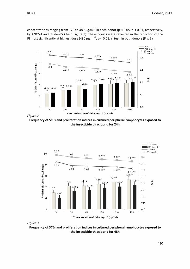
RFFCH Gödöllő, 2013
430
concentrations ranging from 120 to 480 µg.ml-1 in each donor (p < 0.05, p < 0.01, respectively, by ANOVA and Student's t test, Figure 3). These results were reflected in the reduction of the PI most significantly at highest dose (480 µg.ml-1, p < 0.01, χ2 test) in both donors (Fig. 3) Figure 2
Frequency of SCEs and proliferation indices in cultured peripheral lymphocytes exposed to the insecticide thiacloprid for 24h
Figure 3
Frequency of SCEs and proliferation indices in cultured peripheral lymphocytes exposed to the insecticide thiacloprid for 48h
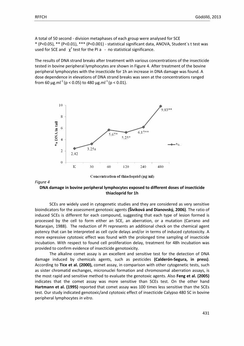
RFFCH Gödöllő, 2013
431
A total of 50 second - division metaphases of each group were analysed for SCE * (P<0.05), ** (P<0.01), *** (P<0.001) - statistical significant data, ANOVA, Student`s t test was used for SCE and χ2 test for the PI a - no statistical significance. The results of DNA strand breaks after treatment with various concentrations of the insecticide tested in bovine peripheral lymphocytes are shown in Figure 4. After treatment of the bovine peripheral lymphocytes with the insecticide for 1h an increase in DNA damage was found. A dose dependence in elevations of DNA strand breaks was seen at the concentrations ranged from 60 µg.ml-1 (p < 0.05) to 480 µg.ml-1 (p < 0.01).
Figure 4
DNA damage in bovine peripheral lymphocytes exposed to different doses of insecticide thiacloprid for 1h
SCEs are widely used in cytogenetic studies and they are considered as very sensitive
bioindicators for the assessment genotoxic agents (Šiviková and Dianovský, 2006). The ratio of induced SCEs is different for each compound, suggesting that each type of lesion formed is processed by the cell to form either an SCE, an aberration, or a mutation (Carrano and Natarajan, 1988). The reduction of PI represents an additional check on the chemical agent potency that can be interpreted as cell cycle delays and/or in terms of induced cytotoxicity. A more expressive cytotoxic effect was found with the prolonged time sampling of insecticide incubation. With respect to found cell proliferation delay, treatment for 48h incubation was provided to confirm evidence of insecticide genotoxicity.
The alkaline comet assay is an excellent and sensitive test for the detection of DNA damage induced by chemicals agents, such as pesticides (Calderón-Segura, in press). According to Tice et al. (2000), comet assay, in comparison with other cytogenetic tests, such as sister chromatid exchanges, micronuclei formation and chromosomal aberration assays, is the most rapid and sensitive method to evaluate the genotoxic agents. Also Feng et al. (2005) indicates that the comet assay was more sensitive than SCEs test. On the other hand Hartmann et al. (1995) reported that comet assay was 100 times less sensitive than the SCEs test. Our study indicated genotoxic/and cytotoxic effect of insecticide Calypso 480 SC in bovine peripheral lymphocytes in vitro.
RFFCH Gödöllő, 2013
432
Aknowledgements This work was supported by the Ministry of Education, Science, Research and Sport of the Slovak Republic through contract no. VEGA1/0125/11 and VEGA 1/0117/13. REFERENCES ANWAR, W.A. 1997. Biomarkers of human exposure to pesticides. Environmental Health Perspectives,
105 (4), 801-806. ARRUGA, M.V., CATALÁN, J., MORENO, C. 1992. The effect of chloramphenicol on sister chromatid
exchange (SCE) in bovine fibroblasts. Research in Veterinary Science, 52, 256-259. COLLINS, A.R., 2004. The Comet Assay for DNA Damage and Repair. Molecular Biotechnology, 26 (3),
249-261. CALDERÓN-SEGURA, M.E., GÓMEZ-ARROYO, S., VILLALOBOS-PIETRINI, R., MARTÍNEZ-VALENZUELA, C.,
CARBAJAL-LÓPEZ, Y., CALDERÓN-ESQUERRO, M.C., CORTÉS-ESLAVA, J., GARCÍA-MARTINÉZ, R., FLORES-RAMÍREZ, D., RODRÍGUEZ- ROMERO, M.I., MÉNDEZ-PÉREZ, P., BAÑUELOS-RUÍZ, E. 2012. Evaluation of Genotoxic and Cytotoxic Effects in Human Peripheral Blood Lymphocytes Exposed In Vitro to Neonicotinoid Insecticides News. Journal of Toxicology, in press doi:10.1155/2012/612647
CARRANO, A.V., NATARAJAN, A.T. 1988. Considerations for population monitoring using cytogenetic technics. Mutation Research, 204, 379–406.
DVOŘÁK, M., MATEJOVIČOVÁ, M. 2008. Principy a využití kometové analýzy při detekci poškození DNA. Chemické listy, 102, 977-983.
GARCIA-REPETTO, R., MARTÍNEZ, M., REPETTO, M. 1997. Biodisposition study of the organophosphorus pesticide, methyl-parathion, Bull. Environmental Contamination and Toxicology, 59, 901-908.
HARTMANN, A., HERKOMMER, K., GLUCK, M., SPEIT, G. 1995. DNA-damaging effect of cyclophosphamide on human blood cells in vivo and in vitro studied with the single.cell gel test (comet assay). Environmental and Molecular Mutagenesis, 25, 180-187.
HORVÁTHOVÁ, E., DUŠINSKÁ, M., SHAPOSHNIKOV, S., COLLINS, A.R. 2004. DNA damage and repair measured in different genomic regions using the comet assay with fluorescent in situ hybridization. Mutagenesis, 19 (4), 269-276.
CHEN, C.S., HSEU, Y.C., LIANG, S.H., KUO, J-Y., CHEN, S.C. 2008. Assessment of genotoxicity of methyl-tert-butyl ether, benzene, toluene, ethylbenzene, and xylene to human lymphocytes using comet assay. Journal of Hazardeous Materials, 153, 351-356.
KOCAMAN A. Y., RENCΫZOĞULLARI E., TOPAKTAŞ M. 2012. In Vitro Investigation of the Genotoxic and Cytotoxic Effects of Thiacloprid in Cultured Human Peripheral Blood Lymphocytes. Environmental Toxicology, in press, DOI 10.1002/tox.21790.
LAMBERTI, L., PONZETTO, B.P., ARDITO, G. 1983. Cell kinetics and sister chromatid exchange frequency in human lymphocytes. Mutation Research, 319, 193-199.
ÖSTLING, O., JOHANSON, K.J., 1984. Microelectrophoretic study of radiation-induced DNA damages in individual mammalian cells. Biochemical and Biophysical Research Communications, 123, 291-298.
PARADA, R., JASZCZAK, K. 1993. A cytogenetic study of cows from a highly industrial or an agricultural region. Mutation Research, 300, 259-263.
SINGH, N.P., STEPHENS, R.E. 1997. Microgel electrophoresis: sensitivity, mechanisms, and DNA electrostretching. Mutation Research, 383, 167-175.
ŠIVIKOVÁ, K., DIANOVSKÝ, J. 2006. Cytogenetic effect of technical glyphosate on cultivated bovine peripheral lymphocytes. International Journal of Hygiene and Environmental Health, 209, 15-20.
TICE, R.R., AGURELL, E., ANDERSON, D., BURLINSON, B., HARTMANN, A., KOBAYASHI, H., MIYAMAE, Y., ROJAS, E., RYU, J.-C., SSAKI, Y.F. 2000. Single Cell Gel/Comet Assay: Guidelines for In Vivo Genetic Toxicology Testing. Environmental and Molecular Mutagenesis, 35, 206-221.
TOMIZAWA, M., CASIDA, J.E. 2003. Selective Toxicity of Neonicotinoids Attributable to Specificity of Insect and Mammalian Nicotinic Receptors. Annual Review of Entomology, 48, 339-364.
TUCKER, J.D., AULETTA, A., CIMINO, M.C., DEARFIELD, K.L., JACOBSON-KRAM, D., TICE, R.R., CARRANO, A.V. 1993. Sister-chromatid exchange: Second report of the Gene-Tox program. Mutation Research, 297, 101-180.
TUCKER, J.D., PRESTON, R.J., 1996. Chromosome aberrations, micronuclei, aneuploidy, sister chromatid exchanges, and cancer risk assessment. Mutation Research, 365, 147-159.

Animal welfare, etológia és tartástechnológia
Animal welfare, ethology and housing systems
Volume 9 Issue 3
Különszám/Special Issue
Gödöllő
2013
RFFCH Gödöllő, 2013
433
Ultrastructural changes in kidneys of newly hatched chicks exposed to cadmium during embryogenesis
Dżugan M.1, Lutnicka H.2, Lis M.2, Droba M.1, Niedziółka J.2
1Department of Chemistry and Food Toxicology, University of Rzeszów, Ćwiklińskiej 2, 35-601 Rzeszów, Poland, e-mail: [email protected]
2Department of Poultry and Fur Animals Breeding and Animal Hygiene, University of Agriculture in Kraków, Al. Mickiewicza 24/28, 30-059 Kraków, Poland
Cadmium, an important environmental toxic agent has the kidney as its most important
target organ. It is concentrated mainly in the renal cortex. In humans, excessive renal accumulation of cadmium causes well defined morphological and ultrastructural, pathological changes in the proximal tubules. In this study, the effect of cadmium (Cd) injected in ovo on ultrastructural alterations of kidney of chicken embryos were studied using transmission electron microscopy. Cadmium (3 and 6 µg per egg) was injected, on the 4th day of incubation, to broilers eggs (n=50 per group) with live embryos whereas control group (n=50) was injected with saline solution. After hatching, kidneys were taken and fixed in glutaraldehyde solution for electron microscopic observations.
Ultrastructural changes in kidney of chicks exposed to cadmium were observed in all
organelle and cytoplasm of the epithelial cells in renal tubules. Moreover strong adverse effect with increasing cadmium dose was observed. The most advanced ultrastructural changes were observed in mitochondria. Part of them was completely destroyed. Many of them were swollen. Their external and internal membranes were locally destroyed. Mitochondrial inner structure was partially damaged. The most characteristic pathological change in mitochondria was dilation of the space between mitochondrial crests and these places had lower electron density in relation to other matrix. The membranes of other organelle were locally destroyed. The rough endoplasmic reticulum (RER) was observed rarely as well as its ribosomes. The strong process of RER fragmentation was observed. The ends of the cisterns of Golgi apparatus were dilated. The structure of brush border of the epithelial cells was slightly and locally destroyed. Some lysosomes were observed. The nuclear membranes of the epithelial cells of the renal tubules were locally damaged. The lysis of cytoplasm of some cells was seen.
It was established that cadmium was toxic for kidney and caused structural damage.
Cadmium injected in ovo resulted mainly in severe damage of mitochondria, but other organelle and cytoplasm were damaged too. The pathological alterations were depended on Cd dose. Key words: cadmium, chicken embryos, kidney, pathological alterations, electron microscopy
Animal welfare, etológia és tartástechnológia
Animal welfare, ethology and housing systems
Volume 9 Issue 3
Különszám/Special Issue
Gödöllő
2013
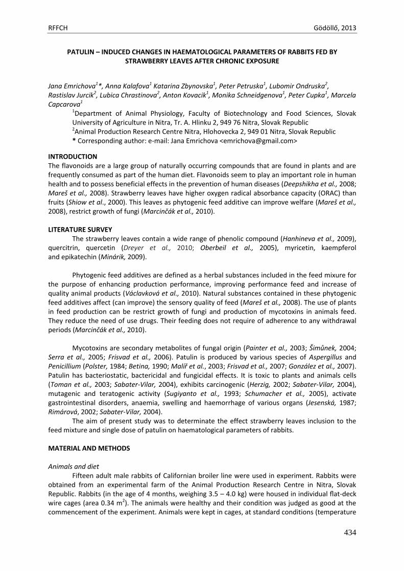
RFFCH Gödöllő, 2013
434
PATULIN – INDUCED CHANGES IN HAEMATOLOGICAL PARAMETERS OF RABBITS FED BY STRAWBERRY LEAVES AFTER CHRONIC EXPOSURE
Jana Emrichova1*, Anna Kalafova1 Katarina Zbynovska1, Peter Petruska1, Lubomir Ondruska2, Rastislav Jurcik2, Lubica Chrastinova2, Anton Kovacik1, Monika Schneidgenova1, Peter Cupka1, Marcela Capcarova1
1Department of Animal Physiology, Faculty of Biotechnology and Food Sciences, Slovak University of Agriculture in Nitra, Tr. A. Hlinku 2, 949 76 Nitra, Slovak Republic 2Animal Production Research Centre Nitra, Hlohovecka 2, 949 01 Nitra, Slovak Republic * Corresponding author: e-mail: Jana Emrichova <[email protected]>
INTRODUCTION The flavonoids are a large group of naturally occurring compounds that are found in plants and are frequently consumed as part of the human diet. Flavonoids seem to play an important role in human health and to possess beneficial effects in the prevention of human diseases (Deepshikha et al., 2008; Mareš et al., 2008). Strawberry leaves have higher oxygen radical absorbance capacity (ORAC) than fruits (Shiow et al., 2000). This leaves as phytogenic feed additive can improve welfare (Mareš et al., 2008), restrict growth of fungi (Marcinčák et al., 2010). LITERATURE SURVEY
The strawberry leaves contain a wide range of phenolic compound (Hanhineva et al., 2009), quercitrin, quercetin (Dreyer et al., 2010; Oberbeil et al., 2005), myricetin, kaempferol and epikatechin (Minárik, 2009).
Phytogenic feed additives are defined as a herbal substances included in the feed mixure for
the purpose of enhancing production performance, improving performance feed and increase of quality animal products (Václavková et al., 2010). Natural substances contained in these phytogenic feed additives affect (can improve) the sensory quality of feed (Mareš et al., 2008). The use of plants in feed production can be restrict growth of fungi and production of mycotoxins in animals feed. They reduce the need of use drugs. Their feeding does not require of adherence to any withdrawal periods (Marcinčák et al., 2010).
Mycotoxins are secondary metabolites of fungal origin (Painter et al., 2003; Šimůnek, 2004;
Serra et al., 2005; Frisvad et al., 2006). Patulin is produced by various species of Aspergillus and Penicillium (Polster, 1984; Betina, 1990; Malíř et al., 2003; Frisvad et al., 2007; González et al., 2007). Patulin has bacteriostatic, bactericidal and fungicidal effects. It is toxic to plants and animals cells (Toman et al., 2003; Sabater-Vilar, 2004), exhibits carcinogenic (Herzig, 2002; Sabater-Vilar, 2004), mutagenic and teratogenic activity (Sugiyanto et al., 1993; Schumacher et al., 2005), activate gastrointestinal disorders, anaemia, swelling and haemorrhage of various organs (Jesenská, 1987; Rimárová, 2002; Sabater-Vilar, 2004).
The aim of present study was to determinate the effect strawberry leaves inclusion to the feed mixture and single dose of patulin on haematological parameters of rabbits.
MATERIAL AND METHODS Animals and diet
Fifteen adult male rabbits of Californian broiler line were used in experiment. Rabbits were obtained from an experimental farm of the Animal Production Research Centre in Nitra, Slovak Republic. Rabbits (in the age of 4 months, weighing 3.5 – 4.0 kg) were housed in individual flat-deck wire cages (area 0.34 m2). The animals were healthy and their condition was judged as good at the commencement of the experiment. Animals were kept in cages, at standard conditions (temperature
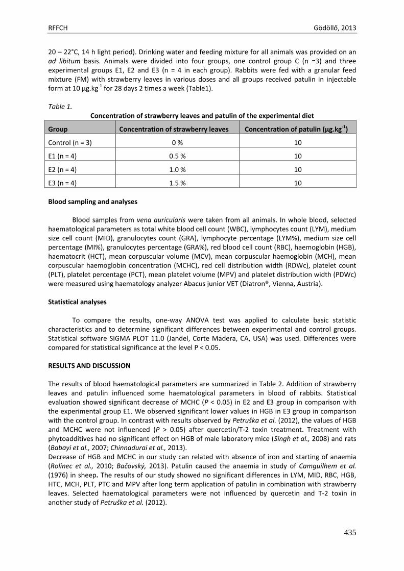
RFFCH Gödöllő, 2013
435
20 – 22°C, 14 h light period). Drinking water and feeding mixture for all animals was provided on an ad libitum basis. Animals were divided into four groups, one control group C (n =3) and three experimental groups E1, E2 and E3 (n = 4 in each group). Rabbits were fed with a granular feed mixture (FM) with strawberry leaves in various doses and all groups received patulin in injectable form at 10 µg.kg-1 for 28 days 2 times a week (Table1). Table 1.
Concentration of strawberry leaves and patulin of the experimental diet
Group Concentration of strawberry leaves Concentration of patulin (µg.kg-1)
Control (n = 3) 0 % 10
E1 (n = 4) 0.5 % 10
E2 (n = 4) 1.0 % 10
E3 (n = 4) 1.5 % 10
Blood sampling and analyses
Blood samples from vena auricularis were taken from all animals. In whole blood, selected
haematological parameters as total white blood cell count (WBC), lymphocytes count (LYM), medium size cell count (MID), granulocytes count (GRA), lymphocyte percentage (LYM%), medium size cell percentage (MI%), granulocytes percentage (GRA%), red blood cell count (RBC), haemoglobin (HGB), haematocrit (HCT), mean corpuscular volume (MCV), mean corpuscular haemoglobin (MCH), mean corpuscular haemoglobin concentration (MCHC), red cell distribution width (RDWc), platelet count (PLT), platelet percentage (PCT), mean platelet volume (MPV) and platelet distribution width (PDWc) were measured using haematology analyzer Abacus junior VET (Diatron®, Vienna, Austria). Statistical analyses
To compare the results, one-way ANOVA test was applied to calculate basic statistic
characteristics and to determine significant differences between experimental and control groups. Statistical software SIGMA PLOT 11.0 (Jandel, Corte Madera, CA, USA) was used. Differences were compared for statistical significance at the level P < 0.05. RESULTS AND DISCUSSION The results of blood haematological parameters are summarized in Table 2. Addition of strawberry leaves and patulin influenced some haematological parameters in blood of rabbits. Statistical evaluation showed significant decrease of MCHC (P < 0.05) in E2 and E3 group in comparison with the experimental group E1. We observed significant lower values in HGB in E3 group in comparison with the control group. In contrast with results observed by Petruška et al. (2012), the values of HGB and MCHC were not influenced (P > 0.05) after quercetin/T-2 toxin treatment. Treatment with phytoadditives had no significant effect on HGB of male laboratory mice (Singh et al., 2008) and rats (Babayi et al., 2007; Chinnadurai et al., 2013). Decrease of HGB and MCHC in our study can related with absence of iron and starting of anaemia (Rolinec et al., 2010; Bačovský, 2013). Patulin caused the anaemia in study of Camguilhem et al. (1976) in sheep. The results of our study showed no significant differences in LYM, MID, RBC, HGB, HTC, MCH, PLT, PTC and MPV after long term application of patulin in combination with strawberry leaves. Selected haematological parameters were not influenced by quercetin and T-2 toxin in another study of Petruška et al. (2012).
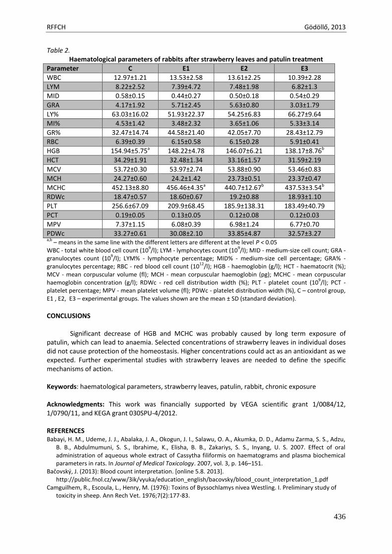
RFFCH Gödöllő, 2013
436
Table 2. Haematological parameters of rabbits after strawberry leaves and patulin treatment
Parameter C E1 E2 E3
WBC 12.97±1.21 13.53±2.58 13.61±2.25 10.39±2.28
LYM 8.22±2.52 7.39±4.72 7.48±1.98 6.82±1.3
MID 0.58±0.15 0.44±0.27 0.50±0.18 0.54±0.29
GRA 4.17±1.92 5.71±2.45 5.63±0.80 3.03±1.79
LY% 63.03±16.02 51.93±22.37 54.25±6.83 66.27±9.64
MI% 4.53±1.42 3.48±2.32 3.65±1.06 5.33±3.14
GR% 32.47±14.74 44.58±21.40 42.05±7.70 28.43±12.79
RBC 6.39±0.39 6.15±0.58 6.15±0.28 5.91±0.41
HGB 154.94±5.75a 148.22±4.78 146.07±6.21 138.17±8.76b
HCT 34.29±1.91 32.48±1.34 33.16±1.57 31.59±2.19
MCV 53.72±0.30 53.97±2.74 53.88±0.90 53.46±0.83
MCH 24.27±0.60 24.2±1.42 23.73±0.51 23.37±0.47
MCHC 452.13±8.80 456.46±4.35a 440.7±12.67b 437.53±3.54b
RDWc 18.47±0.57 18.60±0.67 19.2±0.88 18.93±1.10
PLT 256.6±67.09 209.9±68.45 185.9±138.31 183.49±40.79
PCT 0.19±0.05 0.13±0.05 0.12±0.08 0.12±0.03
MPV 7.37±1.15 6.08±0.39 6.98±1.24 6.77±0.70
PDWc 33.27±0.61 30.08±2.10 33.85±4.87 32.57±3.27 a,b
– means in the same line with the different letters are different at the level P < 0.05 WBC - total white blood cell count (10
9/l); LYM - lymphocytes count (10
9/l); MID - medium-size cell count; GRA -
granulocytes count (109/l); LYM% - lymphocyte percentage; MID% - medium-size cell percentage; GRA% -
granulocytes percentage; RBC - red blood cell count (1012
/l); HGB - haemoglobin (g/l); HCT - haematocrit (%); MCV - mean corpuscular volume (fl); MCH - mean corpuscular haemoglobin (pg); MCHC - mean corpuscular haemoglobin concentration (g/l); RDWc - red cell distribution width (%); PLT - platelet count (10
9/l); PCT -
platelet percentage; MPV - mean platelet volume (fl); PDWc - platelet distribution width (%), C – control group, E1 , E2, E3 – experimental groups. The values shown are the mean ± SD (standard deviation).
CONCLUSIONS
Significant decrease of HGB and MCHC was probably caused by long term exposure of patulin, which can lead to anaemia. Selected concentrations of strawberry leaves in individual doses did not cause protection of the homeostasis. Higher concentrations could act as an antioxidant as we expected. Further experimental studies with strawberry leaves are needed to define the specific mechanisms of action. Keywords: haematological parameters, strawberry leaves, patulin, rabbit, chronic exposure Acknowledgments: This work was financially supported by VEGA scientific grant 1/0084/12, 1/0790/11, and KEGA grant 030SPU-4/2012. REFERENCES Babayi, H. M., Udeme, J. J., Abalaka, J. A., Okogun, J. I., Salawu, O. A., Akumka, D. D., Adamu Zarma, S. S., Adzu,
B. B., Abdulmumuni, S. S., Ibrahime, K., Elisha, B. B., Zakariys, S. S., Inyang, U. S. 2007. Effect of oral administration of aqueous whole extract of Cassytha filiformis on haematograms and plasma biochemical parameters in rats. In Journal of Medical Toxicology. 2007, vol. 3, p. 146–151.
Bačovský, J. (2013): Blood count interpretation. [online 5.8. 2013]. http://public.fnol.cz/www/3ik/vyuka/education_english/bacovsky/blood_count_interpretation_1.pdf
Camguilhem, R., Escoula, L., Henry, M. (1976): Toxins of Byssochlamys nivea Westling. I. Preliminary study of toxicity in sheep. Ann Rech Vet. 1976;7(2):177-83.
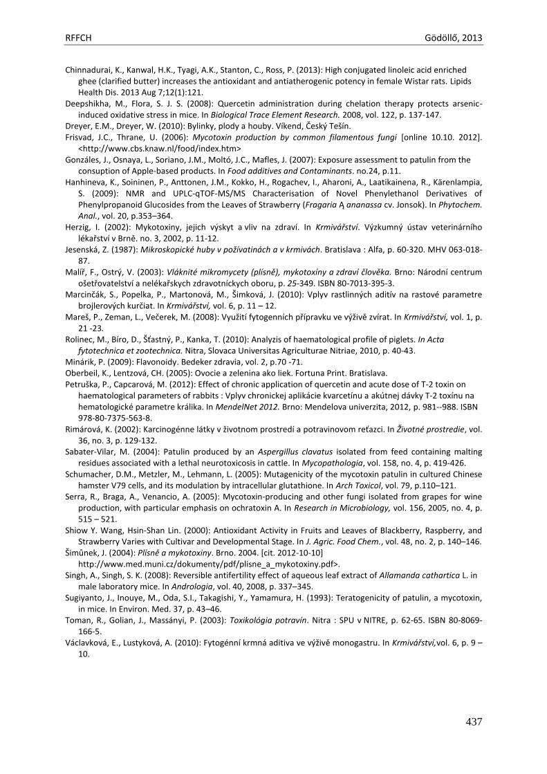
RFFCH Gödöllő, 2013
437
Chinnadurai, K., Kanwal, H.K., Tyagi, A.K., Stanton, C., Ross, P. (2013): High conjugated linoleic acid enriched ghee (clarified butter) increases the antioxidant and antiatherogenic potency in female Wistar rats. Lipids Health Dis. 2013 Aug 7;12(1):121.
Deepshikha, M., Flora, S. J. S. (2008): Quercetin administration during chelation therapy protects arsenic-induced oxidative stress in mice. In Biological Trace Element Research. 2008, vol. 122, p. 137-147.
Dreyer, E.M., Dreyer, W. (2010): Bylinky, plody a houby. Víkend, Český Tešín. Frisvad, J.C., Thrane, U. (2006): Mycotoxin production by common filamentous fungi [online 10.10. 2012].
<http://www.cbs.knaw.nl/food/index.htm> Gonzáles, J., Osnaya, L., Soriano, J.M., Moltó, J.C., Mafles, J. (2007): Exposure assessment to patulin from the
consuption of Apple-based products. In Food additives and Contaminants. no.24, p.11. Hanhineva, K., Soininen, P., Anttonen, J.M., Kokko, H., Rogachev, I., Aharoni, A., Laatikainena, R., Kärenlampia,
S. (2009): NMR and UPLC-qTOF-MS/MS Characterisation of Novel Phenylethanol Derivatives of Phenylpropanoid Glucosides from the Leaves of Strawberry (Fragaria Ą ananassa cv. Jonsok). In Phytochem. Anal., vol. 20, p.353–364.
Herzig, I. (2002): Mykotoxiny, jejich výskyt a vliv na zdraví. In Krmivářství. Výzkumný ústav veterinárního lékařství v Brně. no. 3, 2002, p. 11-12.
Jesenská, Z. (1987): Mikroskopické huby v požívatinách a v krmivách. Bratislava : Alfa, p. 60-320. MHV 063-018-87.
Malíř, F., Ostrý, V. (2003): Vláknité mikromycety (plísně), mykotoxíny a zdraví člověka. Brno: Národní centrum ošetřovatelství a nelékařskych zdravotníckych oboru, p. 25-349. ISBN 80-7013-395-3.
Marcinčák, S., Popelka, P., Martonová, M., Šimková, J. (2010): Vplyv rastlinných aditív na rastové parametre brojlerových kurčiat. In Krmivářství, vol. 6, p. 11 – 12.
Mareš, P., Zeman, L., Večerek, M. (2008): Využití fytogenních přípravku ve výživě zvírat. In Krmivářství, vol. 1, p. 21 -23.
Rolinec, M., Bíro, D., Šťastný, P., Kanka, T. (2010): Analyzis of haematological profile of piglets. In Acta fytotechnica et zootechnica. Nitra, Slovaca Universitas Agriculturae Nitriae, 2010, p. 40-43.
Minárik, P. (2009): Flavonoidy. Bedeker zdravia, vol. 2, p.70 -71. Oberbeil, K., Lentzová, CH. (2005): Ovocie a zelenina ako liek. Fortuna Print. Bratislava. Petruška, P., Capcarová, M. (2012): Effect of chronic application of quercetin and acute dose of T-2 toxin on
haematological parameters of rabbits : Vplyv chronickej aplikácie kvarcetínu a akútnej dávky T-2 toxínu na hematologické parametre králika. In MendelNet 2012. Brno: Mendelova univerzita, 2012, p. 981--988. ISBN 978-80-7375-563-8.
Rimárová, K. (2002): Karcinogénne látky v životnom prostredí a potravinovom reťazci. In Životné prostredie, vol. 36, no. 3, p. 129-132.
Sabater-Vilar, M. (2004): Patulin produced by an Aspergillus clavatus isolated from feed containing malting residues associated with a lethal neurotoxicosis in cattle. In Mycopathologia, vol. 158, no. 4, p. 419-426.
Schumacher, D.M., Metzler, M., Lehmann, L. (2005): Mutagenicity of the mycotoxin patulin in cultured Chinese hamster V79 cells, and its modulation by intracellular glutathione. In Arch Toxicol, vol. 79, p.110–121.
Serra, R., Braga, A., Venancio, A. (2005): Mycotoxin-producing and other fungi isolated from grapes for wine production, with particular emphasis on ochratoxin A. In Research in Microbiology, vol. 156, 2005, no. 4, p. 515 – 521.
Shiow Y. Wang, Hsin-Shan Lin. (2000): Antioxidant Activity in Fruits and Leaves of Blackberry, Raspberry, and Strawberry Varies with Cultivar and Developmental Stage. In J. Agric. Food Chem., vol. 48, no. 2, p. 140–146.
Šimůnek, J. (2004): Plísně a mykotoxiny. Brno. 2004. [cit. 2012-10-10] http://www.med.muni.cz/dokumenty/pdf/plisne_a_mykotoxiny.pdf>.
Singh, A., Singh, S. K. (2008): Reversible antifertility effect of aqueous leaf extract of Allamanda cathartica L. in male laboratory mice. In Andrologia, vol. 40, 2008, p. 337–345.
Sugiyanto, J., Inouye, M., Oda, S.I., Takagishi, Y., Yamamura, H. (1993): Teratogenicity of patulin, a mycotoxin, in mice. In Environ. Med. 37, p. 43–46.
Toman, R., Golian, J., Massányi, P. (2003): Toxikológia potravín. Nitra : SPU v NITRE, p. 62-65. ISBN 80-8069-166-5.
Václavková, E., Lustyková, A. (2010): Fytogénní krmná aditiva ve výživě monogastru. In Krmivářství,vol. 6, p. 9 – 10.

Animal welfare, etológia és tartástechnológia
Animal welfare, ethology and housing systems
Volume 9 Issue 3
Különszám/Special Issue
Gödöllő
2013
RFFCH Gödöllő, 2013
438
SURVEY OF THE MYCOTOXIN CONTAMINATION OF FEED COMMODITIES IN DIFFERENT REGIONS OF HUNGARY
Márta Erdélyi*,Zsolt Ancsin, Andrea Bócsai, Krisztián Balogh, Miklós Mézes
Szent István University, Department of Nutrition, H-2103 Gödöllő, Hungary *[email protected]
Introduction Feed supply is important for animal production and any factor that affects the security of the
feed supply is a significant constraint to production. Feed spoilage by moulds may result in heating, reduced palatability and the loss of nutritive value (Christensen, 1974). In addition, the affected commodity may become contaminated with mycotoxins. The biological reactions following ingestion of one or a combination of mycotoxins vary from acute, overt disease with high morbidity and death to chronic, insidious disorders with reduced animal productivity. Fortunately, mycotoxin contamination levels in animal feedstuffs are usually not high enough to cause an overt disease, but may result in economical loss due to changes in growth, production and immunosuppression (Richard, 2007).
It has been estimated that some 30% of the world’s grain crops is affected annually by fungal invasion and mycotoxin contamination, and with global warming, the threat from fungal invasion of crops is likely to increase (Garrett et al., 2006). The accumulation of mycotoxins, both before and after harvest, largely reflects climatic conditions. Fusarium toxins (e.g. DON, ZEN, T-2 and HT-2 toxin) are produced in cereal grains in high moisture conditions around harvest (Munkvold and Desjardins, 1997), whereas pre-harvest aflatoxin contamination of maize (Payne, 1998) is associated with high temperatures, insect damage and prolonged drought conditions. During the last decade, aflatoxin producing Aspegillus moulds were found even in the field of different regions in Hungary (Dobolyi et al., 2011). Moreover, because Aspergillus can tolerate lower water activity than Fusarium, it is more likely to contaminate commodities both pre- and post-harvest, whereas Fusarium is more likely to be found as a pre-harvest contaminant (Abramson, 1998). The quantity of mycotoxins changes only moderately during fermentation (e.g. silage production); thus, if a batch of maize was contaminated with mycotoxins already at the time of being loaded into the silo, the quantity of mycotoxins will moderately decrease as a result of partial detoxification that occurs during the ensiling process (Bodra and Morgavi, 2009; Richard et al., 2007). Feed manufacturing processes do not substantially decrease the mycotoxin content of feedstuffs, and the efficiency of different chemical and/or heat treatments is questionable (Tóth et al., 2012).
Among aflatoxins, the occurrence of AFB1 or AFG1 is the highest in feed plants. Those are extremely toxic mycotoxins, LD50 in farm animals varies between 0.3 to 20 µg/kg b.w, depending on species, age and sex. Those toxins have mainly hepatotoxic effects, but also inhibit protein synthesis, therefore decrease immune response, as well. In human, International Agency for Research on Cancer (1993) has defined AFB1, as human 1A carcinogen, and also its hydroxylated metabolites, such as AFM1 in milk or AFB2 in egg are potential human carcinogen.
Fusarium moulds produce trichothecene mycotoxins (e.g. DON, T-2 and HT-2 toxin) which have protein synthesis inhibitory activity, and zearalenone which has estrogenic properties, therefore this latter one is genotoxic (Steyn, 1995).
Materials and methods
Feed samples – cereal grains, extracted oil seeds (e.g. soybean meal and sunflower meal) and forages (corn, alfalfa and grass silage) were taken from farms located at different regions of Hungary, and the plants cultivated in the same region in the years 2012 and 2013, except imported soybean meal.
RFFCH Gödöllő, 2013
439
Analyses of mycotoxins determined in the present study were carried out with HPLC method after immunoaffinity cleanup using relevant AOAC methods for AFB1 and zearalenone, according to Czerwiecki and Wilczyńska (2003) for DON, and Trebstein et al (2008) for T-2 and HT-2 toxin. Results and discussion Results of the survey are shown in Table 1. It has been revealed that proportion of positive samples was different in the different regions. It was the highest in the Southern part of the country, possibly because of the higher average temperature and the lowest rainfall during the summer season..
Table 1.
Mycotoxin contamination of feedingstuffs from different regions of Hungary (mean; minimum-maximum values)
Region
number of
positive samples
positive samples
(%) AFB1 µg/kg
DON mg/kg
ZEN mg/kg
T-2 toxin mg/kg
HT-2 toxin mg/kg
Northern 4 31,2 5,93
(1,38-16,86) 0,57
(0,12-1,16) n.d. n.d. n.d.
North Eastern
18 46,8 9,77
(1,71-7,38) 2,23
(0,23-12,90) 0,03
(0,01-0,05) 0,20
(0,17-0,24) 0,36
(0,30-0,40)
South Eastern
16 98,3 5,87
(1,10-19,60) 0,20
(0,06-0,39) n.d.
0,56 (0,11-1,46)
0,86 (0,21-2,55)
Central 6 64,7 7,55
(1,30-13,80) 0,16
(0,05-0,26) n.d. n.d. n.d.
Middle Western
18 51,5 1,63
(0,30-3,02) 0,31
(0,07-0,69) 0,13
(0,02-0,24) 0,33
(0,05-0,51) 0,40
(0,08-0,58)
South Western
16 94,8 2,81
(1,19-4,42) 0,40
(0,35-0,49) 0,06
(0,02-0,08) 0,61
(0,13-0,87) 0,99
(0,37-1,48)
Western 4 41,9 n.d. n.d. 0,02
(0,01-0,03) n.d. n.d.
The results showed that the mean mycotoxin contamination of feedingstuffs in each investigated region was below the maximum permitted level for AFB1 (20 µg/kg; Regulation No. 574/2011/EU) and proposals for the other analysed mycotoxins (Recommendation No. 576/2006/EC). Otherwise mean values were found to be higher in the South Eastern and South Western regions for T-2 and HT-2 toxins, for which we do not have official limit value proposal in the EU, but generally accepted the threshold value of 0.5 mg/kg for all farm animals, as it was proposed by Eriksen and Pettersson (2004). However, the results has also revealed that AFB1 has occurred even in crops cultivated in different regions of Hungary due to the climate changes in the last decade. This was found mainly in maize silage, in which the actual value was higher than the EU maximum permitted level. T-2 and HT-2 toxin levels were also higher in some cases than the proposed threshold value, mainly in grains cultivated in Southern Hungary. Acknowledgement The research was supported by the project TÁMOP-4.2.1.B-11/2/KMR-2011-0003 „Improvement of the level of education and research at the Szent István University“
RFFCH Gödöllő, 2013
440
References Abramson, D., 1998. Mycotoxin formation and environmental factors. In: Sinha, K.K., Bhatnagar, D. (Eds.),
Mycotoxins in Agriculture and Food Safety. Marcel Dekker, Inc, New York, pp. 255–277. AOAC Official Method 990.33 Aflatoxins in corn and peanutbutter. Association of Official Analytical Chemists, Arlington. AOAC Official method 985.18. Zearalenone in corn. Association of Official Analytical Chemists, Arlington Bodra, H., Morgavi, D.P. 2009. Silage could be a way to detoxify maize contaminated by mycotoxins.
Proceedings. ISM Conference 2009, Tulln, p. 106. Christensen, C.M., 1974. Storage of cereal grains and products. 2
nd ed. Am. Assoc. Cereal Chem., St. Paul,
Minnesota Czerwiecki, L., Wilczyńska, G. 2003. Determination of deoxynivalenol in cereals by HPLC-UV. Mycotox. Res.
19:31-34. Dobolyi, Cs., Sebők, F., Varga, J., Kocsubé, S., Szigeti, Gy., Baranyi, N., Szécsi, Á., Lustyik, Gy., Micsinai, A., Tóth,
B., Varga, M., Kriszt, B., Kukolya, J. 2011. Occurrence of aflatoxin-producing Aspergillus flavus strains in maize kernels in Hungary [In Hungarian]. Növényvédelem, 47, 125–133.
Eriksen, G.S., Pettersson H. 2004. Toxicological evaluation of trichothecenes in animal feed. Anim. Feed Sci. Technol. 114: 205–239.
Garrett, K.A., Dendy, S.P., Frank, E.E., Rouse, M.N., Travers, S.E., 2006. Climate change effects on plant disease: genome to ecosystems. Annu. Rev. Phytopathol. 44: 489–509.
International Agency for Research on Cancer (IARC) 1993. Some naturally occurring substances: food items and constituents, heterocyclic amines and mycotoxins. IARC Monographs on Evaluation of Carcinogenic Risk to Humans, Lyon, France, pp. 56.
Munkvold, G.P., Desjardins, A.E., 1997. Fumonisins in maize: can we reduce the occurrence? Plant Dis. 81: 556–565.
Payne, G.A. 1998. Process of contamination by aflatoxin-producing fungi and their impact on crops. In: Sinha, K.K., Bhatnagar, D. (Eds.), Mycotoxins in Agriculture and Food Safety. Marcel Dekker, Inc, New York, pp. 279–306.
Richard, J.L., 2007. Some major mycotoxins and their mycotoxicoses; An overview. Int. J. Food Microbiol. 119, 3–10.
Richard, E., Heutte, N., Sage, L., Pottier, D., Bouchart, V., Lebailly, P., Garon, D. 2007. Toxigenic fungi and mycotoxins in mature corn silage. Food Chem. Toxicol., 45: 2420–2425.
Steyn, P.S. 1995. Mycotoxins, general view, chemistry and structure. Toxicol. Lett. 82/83: 843-851. Tóth K. Balogh K., Bócsai A., Mézes M. 2012. Reduction of the mycotoxin contamination of forage plants during
cultivation, storage and processing. Acta Aliment. 41: 465–474. Trebstein A, Seefelder W, Lauber U, Humpf HU. 2008. Determination of T-2 and HT-2 toxins in cereals including
oats after immunoaffinity cleanup by liquid chromatography and fluorescence detection. J Agric Food Chem. 56:4968-4975.
Animal welfare, etológia és tartástechnológia
Animal welfare, ethology and housing systems
Volume 9 Issue 3
Különszám/Special Issue
Gödöllő
2013
RFFCH Gödöllő, 2013
441
SYSTEMIC TOXICITY OF ACRYLAMIDE IN SWISS MICE
FORMICKI G., KOPAŃSKA M., KRASKA K., GREŃ A., KAPUSTA E., MUCHACKA R., SZAROMA W., GOC Z., SEMLA M.
Department of Animal Physiology and Toxicology, Cracow Pedagogical University, ul. Podbrzezie 3, 31-054 Kraków, email: [email protected]
We studied systemic influence of acrylamide toxicity on the antioxidative system of SWISS mice. The mice were injected with two doses of acrylamide 20 and 40 and 80 mg/kg b.w. The injections were made once a week for 8 weeks. Mice were decapitated after 12, 24, 48 hours and in one week periods between 1 st and 8 th week of the experiment. Next blood samples, brain hemispheres, cerebellum, brainstem, eye lenses, liver, kidneys, testes were taken. In blood serum the concentrations of glucose, cholesterol and triglyceride were measured. In the rest of samples the concentration of reduced glutathione, and activity of superoxide dismutase, glutathione peroxidase and catalase were estimated. In neural system activity of acetylcholinesterase was estimated additionally. Systemic influence of acrylamide was generally related to significant depletion of reduced glutathione content in tested organs and significant changes in the concentration of glucose, cholesterol and triglyceride in blood serum. The decreases of reduced glutathione content were accompanied by affected activity of antioxidative enzymes. In the studied brain structures clear reduction of acetylcholinesterase activity occurred. Our studies indicated that acrylamide toxicity is related to disturbances in redox balance and disturbances in basic blood parameters. The inhibition of acetylcholinesterase participates in neurotoxic properties of acrylamide. Keywords: acrylamide, antioxidants, glucose, triglycerides, cholesterol, acetylcholinesterase, mice organs

Animal welfare, etológia és tartástechnológia
Animal welfare, ethology and housing systems
Volume 9 Issue 3
Különszám/Special Issue
Gödöllő
2013
RFFCH 2013, Gödöllő
442
EVALUATION OF CHROMOSOMAL DAMAGE INDUCED BY THIACLOPRID-BASED INSECTICIDE IN BOVINE PERIPHERAL
LYMPHOCYTES IN VITRO
Martina GALDÍKOVÁ, Katarína ŠIVIKOVÁ, Ján DIANOVSKÝ, Beáta HOLEČKOVÁ, Monika DRÁŽOVSKÁ
University of Veterinary Medicine and Pharmacy, Department of Biology and Genetics, Košice, Slovak Republic; Tel: +421 915 984 775; Fax: 055/6711674; Email: [email protected]
Abstract Thiacloprid-based insecticide was evaluated for its ability to induce chromosome aberrations in cultured bovine peripheral lymphocytes. The cultures were treated with the insecticides at the concentrations ranged from 30 to 480 µg.ml-1 for the last 24 and 48 h of incubation. Dose dependence in the increase of CAs was observed after exposure to thiacloprid formulation ranged from 120 to 480 µg. ml-1. The highest concentration of the insecticide also reflected in reduction of mitotic index in donor 2. For detection of structural and numerical aberrations, three whole chromosome painting probes (WCPs) were used in our experiments. We observed numerical aberrations, but without statistical significance. Introduction Thiacloprid, a neurotoxic insecticide, belongs to the new and commercially very successful family of the neonicotinoids; relatively new class of synthetic organic insecticide, which are now widely used to control piercing and sucking insect pests around the world (Pandey et al., 2009). Thiacloprid is an acute contact and stomach poison, with systemic properties. Neonicotinoid insecticides act as agonists on the insect nicotinic acetylcholine receptors (nAChRs), which plays an important role, in synaptic transmission, in the central nervous system (Muccio et al., 2006). Because of widespread application of thiacloprid, the assessment of its possible cytotoxic, genotoxic and/or carcinogenic potential effects is very important. Despite massive use, only manufactory’s reports about its mutagenic potential are available. It has been described not to be mutagenic in in vitro bacterial mutation and in vivo cytogenetic mammalian assays (Pesticide residues in food, 2006). We report here the cytogenetic effect of the thiacloprid-based insecticide (Calypso 480 SC) on the induction of chromosome aberrations (CA) in bovine peripheral lymphocytes in vitro. Chromosome aberrations are generally considered as biological endpoints to determine the level of genetic damage. Our interest was to detect also the frequency of stable aberrations, which do not result in the loss of chromosome material and it is assumed to be heritable. For this purpose, fluorescence in situ hybridization technique (FISH) was applied. In addition, changes in expression of bovine GSTA2 and GSTM3 after exposure to insecticide were investigated using real-time PCR method. Glutathione S-transferase (GST) family is considered as one of the most important detoxification enzymes groups (Isgor et al., 2010). Keywords: thiaclopri-based insecticide, bovine peripheral lymphocytes, fluorescence in situ hybridization Materials and Methods Chemicals The thiacloprid-based insecticide, (trade name Calypso 480 SC, with active agent N-{3-[(6-Chloro-3-pyridinyl)methyl]-1,3-thiazolan-2-yliden}cyanamide (Bayer AG, Germany), was solved in water and used in the experiments. Mitomycin C (MMC, Sigma, St. Louis, MO, USA, 0.4µmol) and ethyl methanesulfonate (EMS, Sigma, St. Louis, MO, USA) at a concentration of 250 µg.ml-1 were used as positive control agents.
RFFCH 2013, Gödöllő
443
Cell cultivation 0,5 ml of heparinised whole blood of 2 healthy young bulls (Slovak spotted cattle, 6-8 months old) was added to 5 ml of chromosome medium RPMI 1640 supplemented with L- glutamine, 15 µmol/L HEPES (Sigma, St. Louis, MO, USA), 15% foetal calf serum (BOFES, Sigma, Chemical Co. St. Louis, MO, USA), antibiotics (penicillin 250 U/ml and streptomycin 250 µg. ml-1 and phytohaemagglutinin (PHA, 180 µg. ml-1, Welcome, Dartford, England). Lymphocyte cultures were incubated at 37oC for 72 h Lymphocyte cultures were treated with 30, 60, 120, 240 and 480 µg. ml-1of thiacloprid insecticide for the last 24 and 48 h of the incubation. One and half hour before the end of cultivation, colchicine (Merck, Darmstadt, Germany) was added at the final concentration of 5 µg/ml. For the standard cytogenetic analysis the slides were stained with Giemsa solution. One hundred well-spread metaphases were analysed for the CA including chromatid, isochromatid breaks (CB, IB) and chromatid, isochromatid exchanges (CE, IE). Gaps (G) were examined separately. The mitotic index (MI) was calculated as the metaphase ratio of the total number of 3000 cells. Fluorescence in situ hybridization Orange-red labelled whole chromosome painting probes (WCPs), specific for the bovine chromosome 1 and 7, and green labelled WCP, specific for the bovine chromosomes 5 (Kubičková et al., 2002) were used for hybridization, simultaneously. The painting probe in hybridization mixture (50% formamide in 2xSSC, 10% dextran sulphate, salmon sperm DNA, competitor DNA) was denatured at 72˚C for 10 min and reannealed at 37˚C for 90 min. The denaturation of slides was performed in 70% formamide in 2xSSC (pH 7.0) at 72˚C for 2 min and followed by a dehydration procedure (70, 80, 90% ethanol, -20˚C and 96% ethanol, RT) for 2 min. After overnight hybridization at 37˚C, the slides were washed in 2xSSC (3-5 min, room temperature), in 0, 4 xSSC with 0, 3% Igepal at 72˚C for 2 min and again in 2xSSC at room temperature from 5 sec to 2 min and TNT (Tris-NaCl-Tween 20 buffer) (2 x 5 min, RT). The slides were counterstained in DAPI/Antifade (4´, 6´-diamidino-2-phenylindole, Q-BIOgene, UK). One thousand of well spread metaphases were analysed for each and all experimental cultures and controls. Aberrations were scored according to the PAINT nomenclature (Tucker et al., 1995). A fluorescent microscope NIKON Labophot 2A/2, equipped with a single (Texas Red and Fluorescein isothiocyanate - FITC) and dual band pass filters (FITC/Texas Red; DAPI/FITC) was used for visualisation of painting probes. Statistical analysis was performed using the χ2 test for the estimation of CAs and MI in both experimental conditions. Real time PCR RNA was extracted using the AurumTM Total RNA Mini Kit (BioRad, USA). cDNA was prepared by iScriptTM cDNA Synthesis Kit (BioRad, USA). Real time PCR was performed using CFX96 Touch™ Real-Time PCR Detection System (BioRad). Relative expression values were calculated according to comparative threshold cycle method. Results The frequency of chromosomal aberrations induced by thiacloprid-based insecticide in bovine lymphocyte cultures can be seen in Tables 1 and 2. When compared with controls a weak higher percentage of chromosomal breaks were found after treatment with the fungicide for 24 h (Table 1). A dose dependence of CAs was found after treatment with the insecticide at concentrations ranging from 120 to 480 µg.ml-1 (p<0.05 or p<0.01). A slightly decrease in mitotic indices was seen in each donor, with statistical significance at the highest concentration of the insecticide in donor 2 (p<0.05). In the CA assay for 48 h, no statistically significant increases in the induction of chromosome aberrations were obtained (Table 2). After the exposure to the
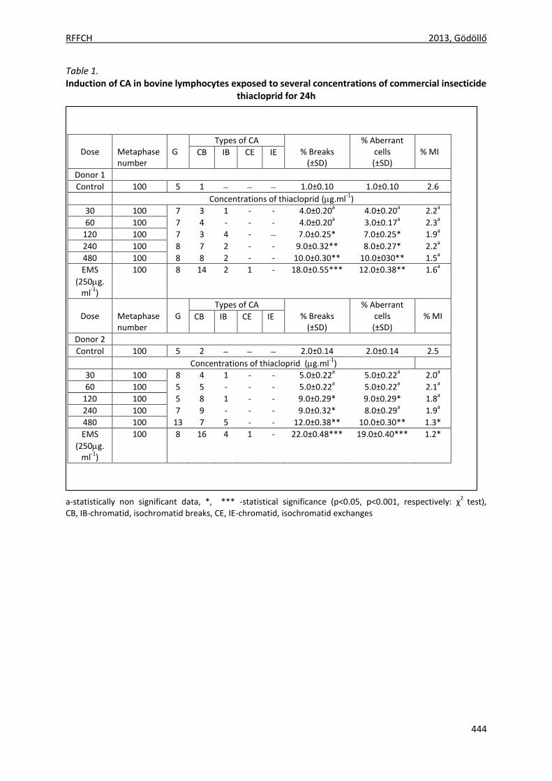
RFFCH 2013, Gödöllő
444
Table 1. Induction of CA in bovine lymphocytes exposed to several concentrations of commercial insecticide
thiacloprid for 24h
a-statistically non significant data, *, *** -statistical significance (p<0.05, p<0.001, respectively: χ2
test), CB, IB-chromatid, isochromatid breaks, CE, IE-chromatid, isochromatid exchanges
Dose
Metaphase number
G
Types of CA % Breaks
(±SD)
% Aberrant cells (±SD)
% MI CB IB CE IE
Donor 1
Control 100 5 1 1.0±0.10 1.0±0.10 2.6
Concentrations of thiacloprid (g.ml-1
)
30 100 7 3 1 - - 4.0±0.20a 4.0±0.20
a 2.2
a
60 100 7 4 - - - 4.0±0.20a 3.0±0.17
a 2.3
a
120 100 7 3 4 - 7.0±0.25* 7.0±0.25* 1.9a
240 100 8 7 2 - - 9.0±0.32** 8.0±0.27* 2.2a
480 100 8 8 2 - - 10.0±0.30** 10.0±030** 1.5a
EMS
(250g.ml
-1)
100 8 14 2 1 - 18.0±0.55*** 12.0±0.38** 1.6a
Dose
Metaphase number
G
Types of CA % Breaks
(±SD)
% Aberrant cells (±SD)
% MI CB IB CE IE
Donor 2
Control 100 5 2 2.0±0.14 2.0±0.14 2.5
Concentrations of thiacloprid (g.ml-1
)
30 100 8 4 1 - - 5.0±0.22a 5.0±0.22
a 2.0
a
60 100 5 5 - - - 5.0±0.22a 5.0±0.22
a 2.1
a
120 100 5 8 1 - - 9.0±0.29* 9.0±0.29* 1.8a
240 100 7 9 - - - 9.0±0.32* 8.0±0.29a 1.9
a
480 100 13 7 5 - - 12.0±0.38** 10.0±0.30** 1.3*
EMS
(250g.ml
-1)
100 8 16 4 1 - 22.0±0.48*** 19.0±0.40*** 1.2*
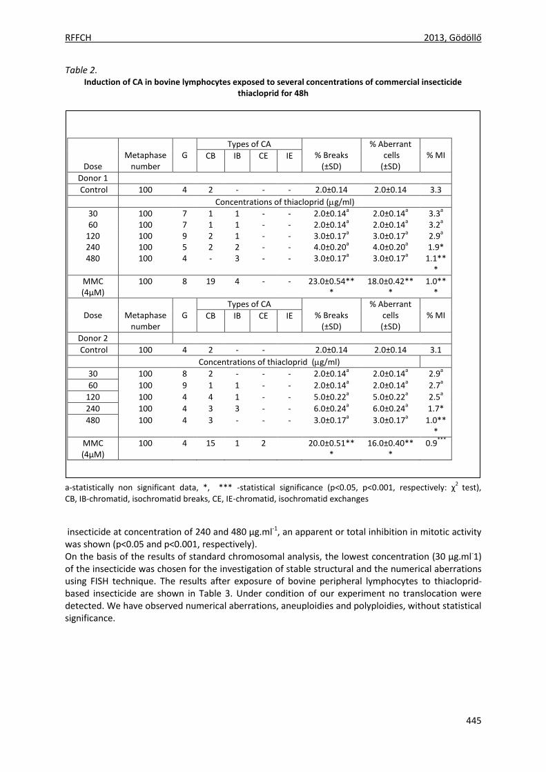
RFFCH 2013, Gödöllő
445
Table 2. Induction of CA in bovine lymphocytes exposed to several concentrations of commercial insecticide
thiacloprid for 48h
a-statistically non significant data, *, *** -statistical significance (p<0.05, p<0.001, respectively: χ2
test), CB, IB-chromatid, isochromatid breaks, CE, IE-chromatid, isochromatid exchanges
insecticide at concentration of 240 and 480 µg.ml-1, an apparent or total inhibition in mitotic activity was shown (p<0.05 and p<0.001, respectively). On the basis of the results of standard chromosomal analysis, the lowest concentration (30 µg.ml-1) of the insecticide was chosen for the investigation of stable structural and the numerical aberrations using FISH technique. The results after exposure of bovine peripheral lymphocytes to thiacloprid-based insecticide are shown in Table 3. Under condition of our experiment no translocation were detected. We have observed numerical aberrations, aneuploidies and polyploidies, without statistical significance.
Dose
Metaphase
number
G
Types of CA % Breaks
(±SD)
% Aberrant cells (±SD)
% MI CB IB CE IE
Donor 1
Control 100 4 2 - - - 2.0±0.14 2.0±0.14 3.3
Concentrations of thiacloprid (g/ml)
30 100 7 1 1 - - 2.0±0.14a 2.0±0.14
a 3.3
a
60 100 7 1 1 - - 2.0±0.14a 2.0±0.14
a 3.2
a
120 100 9 2 1 - - 3.0±0.17a 3.0±0.17
a 2.9
a
240 100 5 2 2 - - 4.0±0.20a 4.0±0.20
a 1.9*
480 100 4 - 3 - - 3.0±0.17a 3.0±0.17
a 1.1**
*
MMC (4µM)
100 8 19 4 - - 23.0±0.54***
18.0±0.42***
1.0***
Dose
Metaphase
number
G
Types of CA % Breaks
(±SD)
% Aberrant cells (±SD)
% MI CB IB CE IE
Donor 2
Control 100 4 2 - - 2.0±0.14 2.0±0.14 3.1
Concentrations of thiacloprid (g/ml)
30 100 8 2 - - - 2.0±0.14a 2.0±0.14
a 2.9
a
60 100 9 1 1 - - 2.0±0.14a 2.0±0.14
a 2.7
a
120 100 4 4 1 - - 5.0±0.22a 5.0±0.22
a 2.5
a
240 100 4 3 3 - - 6.0±0.24a 6.0±0.24
a 1.7*
480 100 4 3 - - - 3.0±0.17a 3.0±0.17
a 1.0**
*
MMC (4µM)
100 4 15 1 2 20.0±0.51***
16.0±0.40***
0.9***
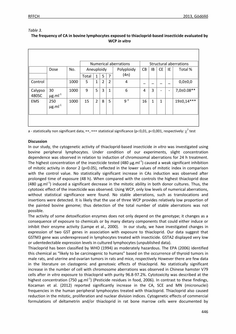
RFFCH 2013, Gödöllő
446
Table 3. The frequency of CA in bovine lymphocytes exposed to thiacloprid-based insecticide evaluated by
WCP in vitro
a - statistically non significant data, , statistical significance (p0,01, p0,001, respectively: 2 test
Discussion In our study, the cytogenetic activity of thiacloprid-based insecticide in vitro was investigated using bovine peripheral lymphocytes. Under condition of our experiments, slight concentration dependence was observed in relation to induction of chromosomal aberrations for 24 h treatment. The highest concentration of the insecticide tested (480 µg.ml-1) caused a weak significant inhibition of mitotic activity in donor 2 (p<0.05), reflected in the lower values of mitotic index in comparison with the control value. No statistically significant increase in CAs induction was observed after prolonged time of exposure (48 h). When compared with the controls the highest thiacloprid dose (480 μg.ml-1) induced a significant decrease in the mitotic ability in both donor cultures. Thus, the cytotoxic effect of the insecticide was observed. Using WCP, only low levels of numerical aberrations, without statistical significance were found. No stable aberrations, such as translocations and insertions were detected. It is likely that the use of three WCP provides relatively low proportion of the painted bovine genome; thus detection of the total number of stable aberrations was not possible. The activity of some detoxification enzymes does not only depend on the genotype; it changes as a consequence of exposure to chemicals or by many dietary components that could either induce or inhibit their enzyme activity (Lampe et al., 2000). In our study, we have investigated changes in expression of two GST genes in association with exposure to thiacloprid. Our data suggest that GSTM3 gene was underexpressed in lymphocytes treated with insecticide. GSTA2 displayed very low or udentedectable expression levels in cultured lymphocytes (unpublished data). Thiacloprid has been classified by WHO (1994) as moderately hazardous. The EPA (2006) identified this chemical as “likely to be carcinogenic to humans” based on the occurrence of thyroid tumors in male rats, and uterine and ovarian tumors in rats and mice, respectively However there are few data in the literature on clastogenic and genotoxic effects of thiacloprid. No statistically significant increase in the number of cell with chromosome aberrations was observed in Chinese hamster V79 cells after in vitro exposure to thiacloprid with purity 96.8-97.2%. Cytotoxicity was described at the highest concentration (750 μg.ml-1) (Pesticide residues in food, 2006). In contrast to these findings, Kocaman et al. (2012) reported significantly increase in the CA, SCE and MN (micronuclei) frequencies in the human peripheral lymphocytes treated with thiacloprid. Thiacloprid also caused reduction in the mitotic, proliferation and nuclear division indices. Cytogenetic effects of commercial formulations of deltametrin and/or thiacloprid in rat bone marrow cells were documented by
Numerical aberrations Structural aberrations
Dose No. Aneuploidy Polyploidy (4n)
CB IB CE IE Total %
Total 1 5 7
Control 1000 5 1 2 2 4 _ _ _ _ 0,0±0,0
Calypso 480SC
30 μg.ml-1
1000 9 5 3 1 6 4 3 - - 7,0±0.08**
EMS 250 µg.ml-1
1000 15 2 8 5 9 a 16 1 1 19±0,14***
RFFCH 2013, Gödöllő
447
Sekeroglu et al. (2011). They found significantly increase in the CA and MN frequencies. Other authors (Calderón-Segura et al., 2012) have investigated neonicotinoid insecticides (thiacloprid, clothianidin and imidacloprid) in vitro in human peripheral lymphocytes using comet assay. They have demonstrated that commercial formulations, Jade, Gaucho, Calypso and Poncho directly induce DNA damage in a concentration-dependent manner. In conclusion, our results indicated ability of thiacloprid formulation to induce clastogenic/genotoxic and/or cytotoxic effects in bovine peripheral lymphocytes. Acknowledgements This work was supported by the Ministry of Education and Science of the Slovak Republic through contract no. VEGA1/0125/11 and VEGA 1/0117/13 References CALDERÓN-SEGURA, M.E., GÓMEZ-ARROYO, S., VILLALOBOS-PIETRINI, R., MARTÍNEZ-VALENZUELA, C., CARBAJAL-LÓPEZ,
Y., CALDERÓN-EZQUERRO, MDEL, C., CORTÉS-ESLAVA, J., GARCÍA-MARTÍNEZ, R., FLORES-RAMÍREZ, D., RODRÍGUEZ-ROMERO, M. I, MÉNDEZ-PÉREZ, P., BAÑUELOS-RUÍZ, E. 2012. Evaluation of genotoxic and cytotoxic effects in human peripheral blood lymphocytes exposed in vitro to neonicotinoid insecticides news. Journal of Toxicology, 2012,1-11.
ISGOR, B.S., CORUH, N., ISCAN, M. 2010. Soluble Glutathione S-Transferases in Bovine Liver: Existence of GST T2. Journal of Biological Sciences, 667-675.
KOCAMAN, A. Y., RENCÜZOĞULLARI, E., TOPAKTAŞ, M. 2012. In vitro investigation of the genotoxic and cytotoxic effects of thiacloprid in cultured human peripheral blood lymphocytes. Environmental Toxicology, 1-11.
KUBÍčKOVÁ, S., ČERNOHORSKÁ, H., MUSILOVÁ, P., RUBEŠ J. 2002. The use of laser microdissection for the preparation of chromosome-specific painting probes in farm animals. Chromosome Research,10(7),571-577.
LAMPE, J.W., CHEN, C., LI, S., PRUNTY, J., GRATE, M.T., MEEHAN, D., BARALE, K.V., DIGHTMAN, D.A., FENG, Z., POTTER, J.D. 2000. Modulation of human glutathione S-transferases by botanically defined vegetable diets. Cancer Epidemiology Biomarkers & Prevention,9,787–793.
MUCCIO, A.D., FIDENTE, P., BARBINI, D.A., DOMMARCO, R., SECCIA, S., MORRICA, P. 2006. Application of solid-phase extraction and liquid chromatography–mass spectrometry to the determination of neonicotinoid pesticide residues in fruit and vegetables. Journal of Chromatography A,1108,1–6.
PANDEY, G., DORRIAN, S.J., RUSSELL, R.J., OAKESHOTT, J.G. 2009. Biotransformation of the neonicotinoid insecticides imidacloprid and thiamethoxam by Pseudomonas sp. 1G. Biochemical and Biophysical Research Commununications, 380(3),710-714.
SEKEROĞLU, V., SEKEROĞLU, Z. A., KEFELIOĞLU, H. 2011. Cytogenetic effects of commercial formulations of deltamethrin and/or thiacloprid on Wistar rat bone marrow cells. Environmental Toxicology, 1-8.
TUCKER, J.D., MORGAN, W.F., AWA, A.A., BAUCHINGER, M., BLAKEY, D., CORNFORTH, M.N., LITTLEFIELD, L.G., NATARAJAN, A.T., SHASSERRE, C. 1995. A proposed system for scoring structural aberrations detected by chromosome painting. Cytogenetics and Cell Genetics, 68, 211-221.
[USEPA Office of Pesticide Programs, Health Effects Division, Science Information Management Branch: "Chemicals Evaluated for Carcinogenic Potential" (April 2006)] http://www.fluoridealert.org/pesticides/pesticides.cancer.potential.2006.pdf World Health Organization, WHO,2004. The WHO recommended classification of pesticides and guidelines to classification. 2004 World Health Organization.
Animal welfare, etológia és tartástechnológia
Animal welfare, ethology and housing systems
Volume 9 Issue 3
Különszám/Special Issue
Gödöllő
2013
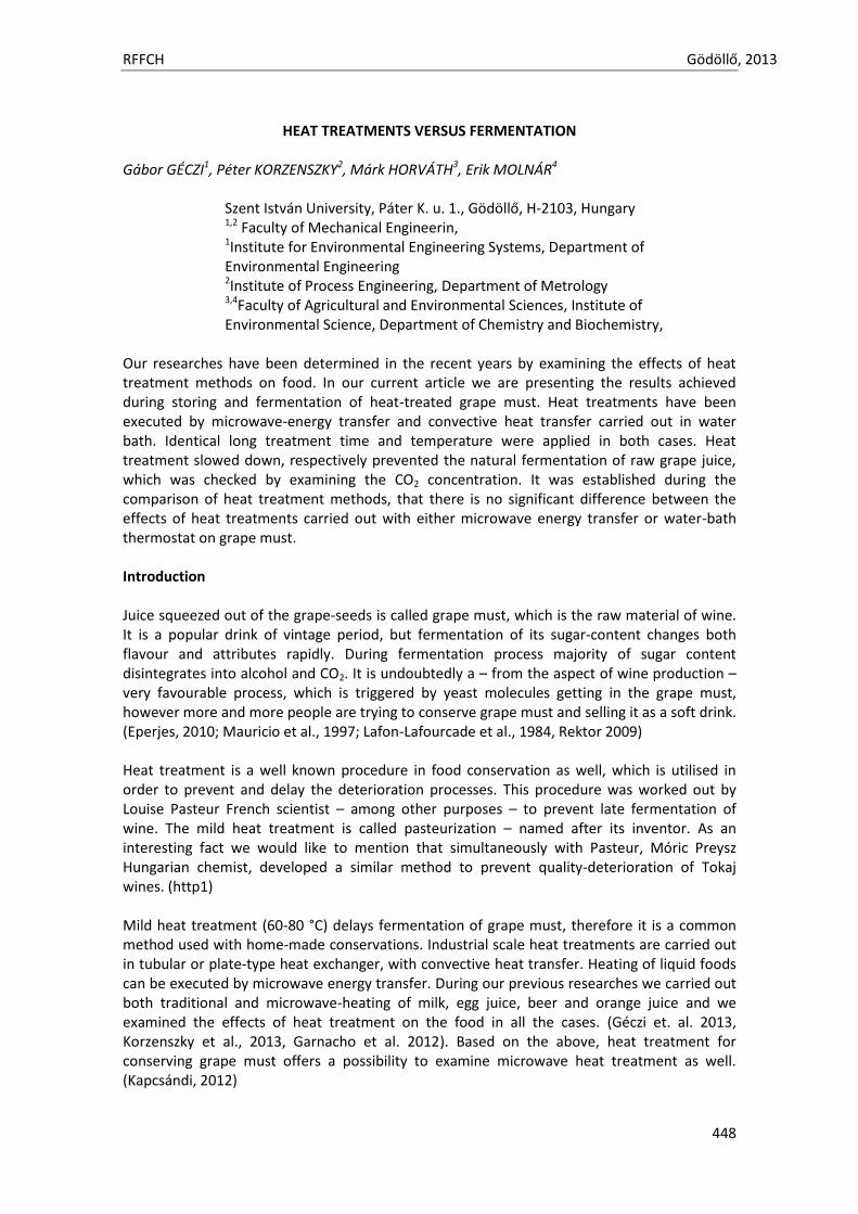
RFFCH Gödöllő, 2013
448
HEAT TREATMENTS VERSUS FERMENTATION Gábor GÉCZI1, Péter KORZENSZKY2, Márk HORVÁTH3, Erik MOLNÁR4
Szent István University, Páter K. u. 1., Gödöllő, H-2103, Hungary 1,2 Faculty of Mechanical Engineerin, 1Institute for Environmental Engineering Systems, Department of Environmental Engineering 2Institute of Process Engineering, Department of Metrology 3,4Faculty of Agricultural and Environmental Sciences, Institute of Environmental Science, Department of Chemistry and Biochemistry,
Our researches have been determined in the recent years by examining the effects of heat treatment methods on food. In our current article we are presenting the results achieved during storing and fermentation of heat-treated grape must. Heat treatments have been executed by microwave-energy transfer and convective heat transfer carried out in water bath. Identical long treatment time and temperature were applied in both cases. Heat treatment slowed down, respectively prevented the natural fermentation of raw grape juice, which was checked by examining the CO2 concentration. It was established during the comparison of heat treatment methods, that there is no significant difference between the effects of heat treatments carried out with either microwave energy transfer or water-bath thermostat on grape must. Introduction Juice squeezed out of the grape-seeds is called grape must, which is the raw material of wine. It is a popular drink of vintage period, but fermentation of its sugar-content changes both flavour and attributes rapidly. During fermentation process majority of sugar content disintegrates into alcohol and CO2. It is undoubtedly a – from the aspect of wine production – very favourable process, which is triggered by yeast molecules getting in the grape must, however more and more people are trying to conserve grape must and selling it as a soft drink. (Eperjes, 2010; Mauricio et al., 1997; Lafon-Lafourcade et al., 1984, Rektor 2009) Heat treatment is a well known procedure in food conservation as well, which is utilised in order to prevent and delay the deterioration processes. This procedure was worked out by Louise Pasteur French scientist – among other purposes – to prevent late fermentation of wine. The mild heat treatment is called pasteurization – named after its inventor. As an interesting fact we would like to mention that simultaneously with Pasteur, Móric Preysz Hungarian chemist, developed a similar method to prevent quality-deterioration of Tokaj wines. (http1) Mild heat treatment (60-80 °C) delays fermentation of grape must, therefore it is a common method used with home-made conservations. Industrial scale heat treatments are carried out in tubular or plate-type heat exchanger, with convective heat transfer. Heating of liquid foods can be executed by microwave energy transfer. During our previous researches we carried out both traditional and microwave-heating of milk, egg juice, beer and orange juice and we examined the effects of heat treatment on the food in all the cases. (Géczi et. al. 2013, Korzenszky et al., 2013, Garnacho et al. 2012). Based on the above, heat treatment for conserving grape must offers a possibility to examine microwave heat treatment as well. (Kapcsándi, 2012)

RFFCH Gödöllő, 2013
449
Material and methods Comparison-examinations were executed on the experimental instrument set up in the laboratory of Szent István University, Faculty of Mechanical Engineering. Analysing and checking the grape must examples took place at Szent István University, Department of Chemistry and Biochemistry. Examples heat treated with microwave (MH), respectively water bath thermostat (TH) were compared with each other and untreated (WH) example as well. Grape must used for the experiments at the end of July 2013 was prepared from Italian Vittoria white grape and Black Magic red grape. In august was used Bianca white grape and Nero red grape from Hungary. Properties of grape must is summarised in Table 1. Table 1
Attributes of grape must used in the experiments.
Vittoria white grape (Italy)
Black Magic red grape (Italy)
Nero red grape (Hungary)
Bianca white grape (Hungary)
Dry matter content [g/100ml]
13.55±0.03 16.63±0.05 23.31±0.12 23.74±0.08
Density [g/ml] 1.0402±0.011 1.0327±0.013 1.083±0.02 1.079 pH 2.08±0.1 2.56±0.08 3.52±0.11 3.26 Conductivity [μS/cm] 1477±11 1497±17 2350±15 1620 Degrees Brix [°Bx]* 13 16 22 23
*determined by OG-101/A type manual refractometer At the Faculty of Mechanical Engineering of Szent István University a group of researchers dealing with food treatment created an experimental instrument capable of continuous service microwave heating and heat treatment. They managed to create a measuring circuit capable of comparison-examinations, which allowed heating up liquid foods by usage of microwave energy transfer and convective heat transfer, by upgrading this instrument in the recent years. The „spirit” of the comparison-circuit is a glass spiral, in which the food to be treated is circulated – grape must in our case – by a STENNER 85M5 type pump (Stenner Pump Company, Jacksonville, FL, U.S.A.) which has continuously adjustable flow rate. The glass spiral can be placed into either the Whirlpool AT 314WH type (Whirlpool Corporation, U.S.A.) household microwave device or T-PHYWE (Lauda DR. R. Wobser GmbH, Lauda-Königshofen, Germany) water-bath thermostat for executing the heating procedure. In case of the microwave gadget we applied continuous 900 Watt output power, whereas with the water-bath thermostat a water-bath of appropriate temperature (70-95°C) for the desired target temperature was used. During the heat treatment of continuous flowing, the liquid foods circulating in the glass spiral – depending on the length of the glass spiral and the volume flow of the pump – can be heated to the desired temperature.
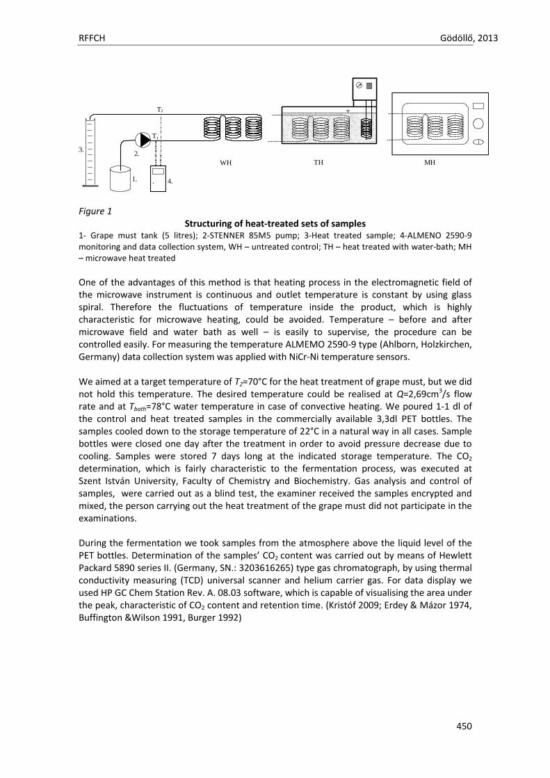
RFFCH Gödöllő, 2013
450
Figure 1 Structuring of heat-treated sets of samples
1- Grape must tank (5 litres); 2-STENNER 85M5 pump; 3-Heat treated sample; 4-ALMENO 2590-9 monitoring and data collection system, WH – untreated control; TH – heat treated with water-bath; MH – microwave heat treated
One of the advantages of this method is that heating process in the electromagnetic field of the microwave instrument is continuous and outlet temperature is constant by using glass spiral. Therefore the fluctuations of temperature inside the product, which is highly characteristic for microwave heating, could be avoided. Temperature – before and after microwave field and water bath as well – is easily to supervise, the procedure can be controlled easily. For measuring the temperature ALMEMO 2590-9 type (Ahlborn, Holzkirchen, Germany) data collection system was applied with NiCr-Ni temperature sensors. We aimed at a target temperature of T2=70°C for the heat treatment of grape must, but we did not hold this temperature. The desired temperature could be realised at Q=2,69cm3/s flow rate and at Tbath=78°C water temperature in case of convective heating. We poured 1-1 dl of the control and heat treated samples in the commercially available 3,3dl PET bottles. The samples cooled down to the storage temperature of 22°C in a natural way in all cases. Sample bottles were closed one day after the treatment in order to avoid pressure decrease due to cooling. Samples were stored 7 days long at the indicated storage temperature. The CO2
determination, which is fairly characteristic to the fermentation process, was executed at Szent István University, Faculty of Chemistry and Biochemistry. Gas analysis and control of samples, were carried out as a blind test, the examiner received the samples encrypted and mixed, the person carrying out the heat treatment of the grape must did not participate in the examinations. During the fermentation we took samples from the atmosphere above the liquid level of the PET bottles. Determination of the samples’ CO2 content was carried out by means of Hewlett Packard 5890 series II. (Germany, SN.: 3203616265) type gas chromatograph, by using thermal conductivity measuring (TCD) universal scanner and helium carrier gas. For data display we used HP GC Chem Station Rev. A. 08.03 software, which is capable of visualising the area under the peak, characteristic of CO2 content and retention time. (Kristóf 2009; Erdey & Mázor 1974, Buffington &Wilson 1991, Burger 1992)
T2
T1
1.
2.3.
. 4..
MHTHWH
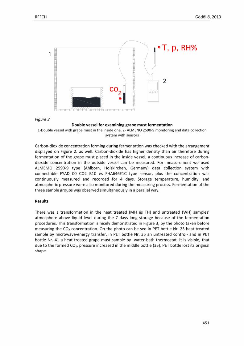
RFFCH Gödöllő, 2013
451
Figure 2
Double vessel for examining grape must fermentation 1-Double vessel with grape must in the inside one, 2- ALMENO 2590-9 monitoring and data collection
system with sensors
Carbon-dioxide concentration forming during fermentation was checked with the arrangement displayed on Figure 2. as well. Carbon-dioxide has higher density than air therefore during fermentation of the grape must placed in the inside vessel, a continuous increase of carbon-dioxide concentration in the outside vessel can be measured. For measurement we used ALMEMO 2590-9 type (Ahlborn, Holzkirchen, Germany) data collection system with connectable FYAD 00 CO2 B10 és FHA646E1C type sensor, plus the concentration was continuously measured and recorded for 4 days. Storage temperature, humidity, and atmospheric pressure were also monitored during the measuring process. Fermentation of the three sample groups was observed simultaneously in a parallel way. Results There was a transformation in the heat treated (MH és TH) and untreated (WH) samples’ atmosphere above liquid level during the 7 days long storage because of the fermentation procedures. This transformation is nicely demonstrated in Figure 3, by the photo taken before measuring the CO2 concentration. On the photo can be see in PET bottle Nr. 23 heat treated sample by microwave-energy transfer, in PET bottle Nr. 35 an untreated control- and in PET bottle Nr. 41 a heat treated grape must sample by water-bath thermostat. It is visible, that due to the formed CO2, pressure increased in the middle bottle (35), PET bottle lost its original shape.
2co
T, p, RH%1
2
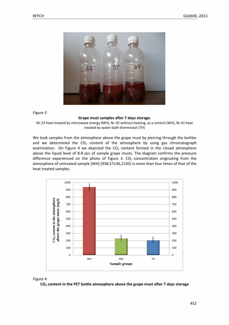
RFFCH Gödöllő, 2013
452
Figure 3
Grape must samples after 7 days storage. Nr.23 heat-treated by microwave energy (MH), Nr.35 without heating, as a control (WH), Nr.41 heat
treated by water-bath thermostat (TH)
We took samples from the atmosphere above the grape must by piercing through the bottles and we determined the CO2 content of the atmosphere by using gas chromatograph examination. On Figure 4 we depicted the CO2 content formed in the closed atmosphere above the liquid level of 8-8 pcs of sample grape musts. The diagram confirms the pressure difference experienced on the photo of Figure 3. CO2 concentration originating from the atmosphere of untreated sample (WH) (938,57±36,21SD) is more than four times of that of the heat treated samples.
Figure 4
CO2 content in the PET bottle atmosphere above the grape must after 7 days storage
0
100
200
300
400
500
600
700
800
900
1000
0
100
200
300
400
500
600
700
800
900
1000
WH MH TH
CO
2-c
on
ten
t in
th
e a
tmo
sph
ere
ab
ov
e th
e g
rap
e m
ust
, [m
g/l
]
Sample groups
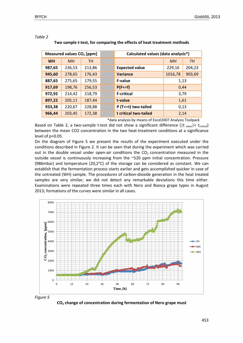
RFFCH Gödöllő, 2013
453
Table 2 Two sample t-test, for comparing the effects of heat treatment methods
Measured values CO2 [ppm] Calculated values (data analysis*)
WH MH TH MH TH
987,65 236,53 213,86 Expected value 229,16 204,23
945,60 278,65 176,43 Variance 1016,78 903,69
887,65 275,65 179,55 F-value 1,13
917,69 198,76 256,53 P(F<=f) 0,44
972,92 214,42 218,79 F-critical 3,79
897,22 205,11 187,44 t-value 1,61
933,38 220,67 228,88 P (T<=t) two-tailed 0,13
966,44 203,45 172,38 t critical two-tailed 2,14
*data analysis by means of Excel2007 Analysis Toolpack
Based on Table 2, a two-sample t-test did not show a significant difference (|t value|< tcritical) between the mean CO2 concentration in the two heat-treatment conditions at a significance level of p=0.05. On the diagram of Figure 5 we present the results of the experiment executed under the conditions described in Figure 2. It can be seen that during the experiment which was carried out in the double vessel under open-air conditions the CO2 concentration measured in the outside vessel is continuously increasing from the ~520 ppm initial concentration. Pressure (986mbar) and temperature (20,2°C) of the storage can be considered as constant. We can establish that the fermentation process starts earlier and gets accomplished quicker in case of the untreated (WH) sample. The procedures of carbon-dioxide generation in the heat treated samples are very similar; we did not detect any remarkable deviations this time either. Examinations were repeated three times each with Nero and Bianca grape types in August 2013; formations of the curves were similar in all cases.
Figure 5
CO2 change of concentration during fermentation of Nero grape must
0
1000
2000
3000
4000
5000
6000
7000
8000
0 12 24 36 48 60 72 84 96
CO
2co
nce
ntr
ati
on
, [p
pm
]
Time, [h]
TH
MH
WH
RFFCH Gödöllő, 2013
454
Conclusions Grape must proved to be an appropriate subject for examining microwave heat transfer. During vine-making technology grape must starts to ferment and its sugar content transforms into alcohol and carbon-dioxide. Fermentation of grape must can be tracked by measuring the latter component. Fermentation process might be delayed, slowed down by using heat treatment methods. (A known method for home-made grape must conservation.) Just as during our previous examinations, we were searching for an answer whether microwave heat treating can be matched with any further advantages or disadvantages apart from the known thermal-effect. Fermentation of the grape must heat treated with microwave method was compared to fermentation of samples heat treated with convective method (water bath thermostat). We examined fermentation in PET bottle, closed conditions and in double glass vessel fermentation under natural conditions. It can be established that the effect of heat treatment is obviously detectable in both examinations; however we could not find any significant difference between the effects of heat treatments in either of the cases. Acknowledgments This work was financially supported by TÁMOP 4.2.1.B-11/2/KMR-2011-0003 Improvement of Research and Education Standard of Szent István University project and by KTIA-AIK-12 project. References Buffington R & Wilson K. M.(1991): Detectors for Gas Chromatography. HP Hewlett Packard, Awandale,
37-42 p. Burger K. (1992): Basics of quantitative analysis – chemical and instrumental analysis. (Hungarian
language) Semmelweis Publisher, Budapest 449-454 p. Imre Eperjes (2010): Wineyard technology – Wine making 1. (Hungarian language) Mezőgazda Publisher,
314p. L. Erdey & L. Mázor (1974): Analitical handbook. (Hungarian language) Technical Publisher, Budapest,
pp. 303-342. G Géczi, M Horváth, T Kaszab, G G Alemany (2013): No Major Differences Found between the Effects of
Microwave-Based and Conventional Heat Treatment Methods on Two Different Liquid Foods PLOS ONE 8:(1) pp. 1-12.
Gonzalo Garnacho, Tímea Kaszab, Márk Horváth, Gábor Géczi (2012): COMPARATIVE STUDY OF HEAT-TREATED ORANGE JUICE JOURNAL OF MICROBIOLOGY BIOTECHNOLOGY AND FOOD SCIENCES 2:(3) pp. 446-457.
V. Kapcsándi, E. Lakatos, A. J. Kovács, M. Neményi (2012): Effects of low power microwave radiation on the fermentation of Italian Riesling grape must. (Hungarian language) XXXIV. Óvár Scientific Day, Mosonmagyaróvár, NyME, pp. 62-67.
Péter Korzenszky, Péter Sembery, Gábor Géczi (2013): Microwave Milk Pasteurization without Food Safety Risk POTRAVINARSTVO 7:(1) pp. 45-48.
K. Kristóf (2009): Structure of the gas chromatograh, principles of measurements with gas chromatograh. (Hungarian language) Educational lecture 1-5 p.
S. Lafon-Lafourcade, C. Geneix and P. Ribéreau-Gayon (1984): Inhibition of Alcoholic Fermentation of Grape Must by Fatty Acids Produced by Yeasts and Their Elimination by Yeast Ghosts Appl. Environ. Microbiol. June 1984 vol. 47 no. 6 1246-1249
J.C. Mauricio, J. Moreno, L. Zea, J.M. Ortega, M. Medina (1997):The effects of grape must fermentation conditions on volatile alcohols and esters formed by Saccharomyces cerevisiae Journal of the Science of Food and Agriculture 75 (6) 155-160.
Attila Rektor (2009): Processing milk whey and grape must with complex membrane-technology methods (PhD) dissertation (Hungarian language) Budapest Corvinus University 119 p.
http1://www.ng.hu/Tudomany/2003/10/Skot_es_magyar_felfedezok_Pasteur_elott

Animal welfare, etológia és tartástechnológia
Animal welfare, ethology and housing systems
Volume 9 Issue 3
Különszám/Special Issue
Gödöllő
2013
RFFCH Gödöllő, 2013
455
THE CONCENTRATION OF MERCURY IN KIDNEY CANCEROUS AND CONTROL TISSUES OF HUMAN Marta GŁOGOWSKA1
1Department of Vertebrate Zoology and Human Biology, Pedagogical University of Cracow, Poland e-mail: [email protected]
ABSTRACT
The aims of this study was determinate the average accumulation of mercury in kidney healthy and cancerous tissues of human in relation age and sex.
Both inorganic and organic forms of mercury are highly toxic to humans; however, inorganic mercury is not as easily absorbed by the body. Inorganic mercury, such as mercury vapor, is toxic if inhaled in large concentrations. As the body tries to rid itself of these toxins, gaseous mercury is oxidized to divalent mercury, which accumulates in the kidneys and can cause kidney damage.
Tissues were taken during surgery and post-mortem from Military Hospital, PROSMED Health Center and Oncology Centre of Maria Skłodowska - Curie in Cracow. Fragments of tissues cancerous (tissues away from the tumor, tissues surrounding the tumor, tissues tumor) and tissues normal (control group) were taken from kidney and then frozen. Average mass of each sample hesitated from 0,5-1g. Samples were taken from 98 (n=98) cancerous patients and from 22 (n=22) healthy patients (control tissues). Mercury contents were detected using CVAAS methods. All results were expressed in ppm.
Mean content of mercury in men (20-50 years old) in control tissues is 0,015±0,00001 ppm. The average content of mercury in tissues away from the tumor is 0,12±0,091 ppm. The average content of mercury in tissues surrounding the tumor is 0,099±0,064 ppm. The average content of mercury in tumor tissues is 0,036±0,021 ppm. Mean content of mercury in men (50-90 years old) in control tissues is 0,023±0,009 ppm. The average content of mercury in tissues away from the tumor is 0,164±0,154 ppm. The average content of mercury in tissues surrounding the tumor is 0,106±0,094 ppm. The average content of mercury in tumor tissues is 0,035±0,031 ppm. Mean content of mercury in women (20-50 years old) in control tissues is 0,0005±0,0003 ppm. The average content of mercury in tissues away from the tumor is 0,174±0,005 ppm. The average content of mercury in tissues surrounding the tumor is 0,151±0,061 ppm. The average content of mercury in tumor tissues is 0,011±0,009 ppm. Mean content of mercury in women (50-90 years old) in control tissues is 0,021±0,013 ppm. The average content of mercury in tissues away from the tumor is 0,093±0,069 ppm. The average content of mercury in tissues surrounding the tumor is 0,086±0,073 ppm. The average content of mercury in tumor tissues is 0,068±0,063 ppm.
Mercury content is much higher in patients with tumors (both men and women) in comparison to healthy tissues of both sexes. In all groups, mercury concentration was the highest in tissues away from the kidney tumor, in comparison to mercury content in adjacent mucous membrane and tumor tissues of this organ. The results suggest that some types of cancerous tissues may accumulate more effectively toxic elements. Key words: kidney, mercury, CVAAS, cancerous tissues Introduction
Around 208,500 new cases of kidney cancer are diagnosed in the world each year, accounting for just under 2% of all cancers (Lindblad and Adami). The most recent estimates of incidence of kidney cancer suggest that there are 63,300 new cases annually in the EU25. In Europe, kidney cancer accounts for nearly 3% of all cancer cases (Ferlay et al., 2007). In the UK kidney cancer is the eighth most common cancer in men (5,377 new cases diagnosed in 2008), and the ninth most common cancer in women (3,380 new cases in 2008), giving a male: female ratio of over 3:2. The number of cases of kidney cancer in men in the UK has doubled from 7 per 100,000 to 14.8 per
RFFCH Gödöllő, 2013
456
100,000 between 1975-1977 and 2006-2008. In women the rates have more than doubled over the same period, rising from 3.2 to 7.5 per 100,000 (Cancer Research). The incidence of kidney cancer is also increasing in the United States. This is thought to be a real increase, not only due to changes in the way the disease is diagnosed (Lynch et al., 2007).
Historically medical practitioners would expect a person to present with what is known as the classic triad of symptoms (Cohen and McGovern, 2005). This triad of symptoms includes: hematuria which is when there is blood present in the urine. Flank pain, which is pain on the side of the body between the hip and ribs. An abdominal mass, similar to bloating but larger. It is now known that this classic triad of symptoms only occurs in 10-15% of cases, and can be indicative of the Renal cell carcinoma (RCC) in an advanced stage (Cohen and McGovern, 2005). Today RCC is fairly asymptomatic (meaning little to no symptoms) and is generally detected incidentally when a person is being examined for other ailments (Motzer et al., 1996). VHL is a rare disease that runs in some families. It is caused by changes in the VHL gene. An abnormal VHL gene increases the risk of kidney cancer. It also can cause cysts or tumors in the eyes, brain, and other parts of the body. Family members of those with this syndrome can have a test to check for the abnormal VHL gene. For people with the abnormal VHL gene, doctors may suggest ways to improve the detection of kidney cancer and other diseases before symptoms develop.
Both inorganic and organic forms of mercury are highly toxic to humans; however, inorganic mercury is not as easily absorbed by the body. Inorganic mercury, such as mercury vapor, is toxic if inhaled in large concentrations. Inhaled gaseous mercury is absorbed into the blood. Once in the circulatory system, it can pass through the blood-brain barrier and accumulate in the brain, damaging the central nervous system. As the body tries to rid itself of these toxins, gaseous mercury is oxidized to divalent mercury, which accumulates in the kidneys and can cause kidney damage (Honda et al., 2006). Most people are not exposed to inorganic mercury but rather absorb methylmercury through the consumption of fish and shellfish. Methylmercury is easily absorbed in the digestive tract, where it forms a complex with the amino acid cysteine. This new complex resembles a large neutral amino acid found in the body, methionine, and can more easily gain entry into cells. As with inorganic mercury, once in the bloodstream, methylmercury will accumulate in the brain and cause damage to the central nervous system. Methylmercury is naturally removed from the body over time. Eventually, this methylmercury-cysteine complex is transported to the liver where it is secreted into bile, after which enzymes break the complex down into its amino acid and methylmercury parts. Some of this methylmercury then comes in contact with the bacteria in the intestine and is broken down into inorganic mercury and carbon. The inorganic mercury is poorly absorbed in the digestive tract and 90 percent is excreted in the feces. The rest of the methylmercury that does not interact with bacteria is reabsorbed by the body and goes through the process again. It takes about 30 to 40 hours for methylmercury to be distributed to the tissues of the body (Clarkson and Magos, 2006).
Material and methods
Tissues were taken during surgery and post-mortem from Military Hospital, PROSMED Health Center and Oncology Centre of Maria Skłodowska - Curie in Cracow. Permission for research was given by Local Bioethical Commission. Fragments of tissues cancerous (tissues away from the tumor, tissues surrounding the tumor, tissues tumor) and tissues normal (control group) were taken from kidney and then frozen. Average mass of each sample hesitated from 0,5-1g. Samples were taken from 98 (n=98) cancerous patients and from 22 (n=22) healthy patients (control tissues).
Mercury contents were detected using CVAAS methods. All results were expressed in ppm. Hypothesis falsification were made by U Mann-Whitney test, and Kruskal-Wallis ANOVA. Results
Results were analyzed statistically by using Statistica 10 program. In statistical description the following statistics were used: the arithmetic mean and standard deviation. Normality of the distribution was examined by Shapiro-Wilk test. Because none of the normal distributions were noticed, Kruskal-Wallis test for independent trials was used. Statistical analysis was to use the U
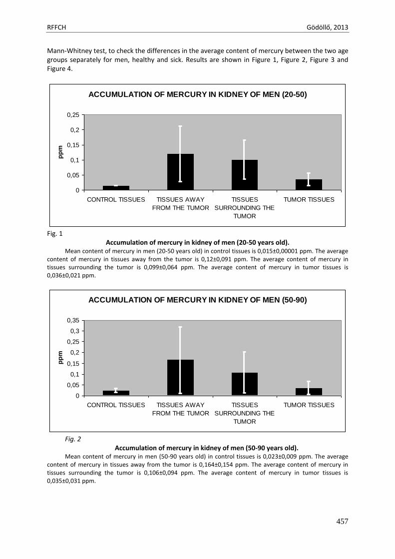
RFFCH Gödöllő, 2013
457
Mann-Whitney test, to check the differences in the average content of mercury between the two age groups separately for men, healthy and sick. Results are shown in Figure 1, Figure 2, Figure 3 and Figure 4.
ACCUMULATION OF MERCURY IN KIDNEY OF MEN (20-50)
0
0,05
0,1
0,15
0,2
0,25
CONTROL TISSUES TISSUES AWAY
FROM THE TUMOR
TISSUES
SURROUNDING THE
TUMOR
TUMOR TISSUES
pp
m
Fig. 1
Accumulation of mercury in kidney of men (20-50 years old). Mean content of mercury in men (20-50 years old) in control tissues is 0,015±0,00001 ppm. The average
content of mercury in tissues away from the tumor is 0,12±0,091 ppm. The average content of mercury in tissues surrounding the tumor is 0,099±0,064 ppm. The average content of mercury in tumor tissues is 0,036±0,021 ppm.
ACCUMULATION OF MERCURY IN KIDNEY OF MEN (50-90)
0
0,05
0,1
0,15
0,2
0,25
0,3
0,35
CONTROL TISSUES TISSUES AWAY
FROM THE TUMOR
TISSUES
SURROUNDING THE
TUMOR
TUMOR TISSUES
pp
m
Fig. 2
Accumulation of mercury in kidney of men (50-90 years old). Mean content of mercury in men (50-90 years old) in control tissues is 0,023±0,009 ppm. The average
content of mercury in tissues away from the tumor is 0,164±0,154 ppm. The average content of mercury in tissues surrounding the tumor is 0,106±0,094 ppm. The average content of mercury in tumor tissues is 0,035±0,031 ppm.
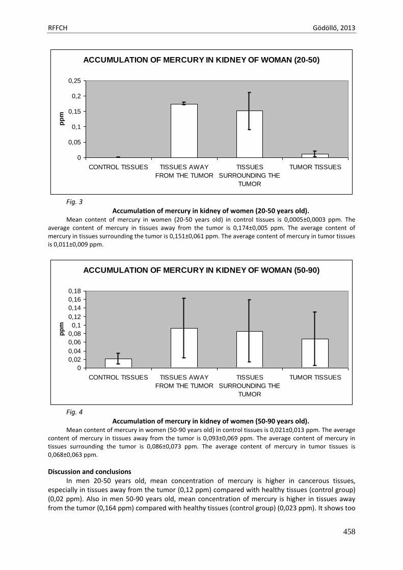
RFFCH Gödöllő, 2013
458
ACCUMULATION OF MERCURY IN KIDNEY OF WOMAN (20-50)
0
0,05
0,1
0,15
0,2
0,25
CONTROL TISSUES TISSUES AWAY
FROM THE TUMOR
TISSUES
SURROUNDING THE
TUMOR
TUMOR TISSUES
pp
m
Fig. 3
Accumulation of mercury in kidney of women (20-50 years old). Mean content of mercury in women (20-50 years old) in control tissues is 0,0005±0,0003 ppm. The
average content of mercury in tissues away from the tumor is 0,174±0,005 ppm. The average content of mercury in tissues surrounding the tumor is 0,151±0,061 ppm. The average content of mercury in tumor tissues is 0,011±0,009 ppm.
ACCUMULATION OF MERCURY IN KIDNEY OF WOMAN (50-90)
0
0,02
0,04
0,06
0,08
0,1
0,12
0,14
0,16
0,18
CONTROL TISSUES TISSUES AWAY
FROM THE TUMOR
TISSUES
SURROUNDING THE
TUMOR
TUMOR TISSUES
pp
m
Fig. 4
Accumulation of mercury in kidney of women (50-90 years old). Mean content of mercury in women (50-90 years old) in control tissues is 0,021±0,013 ppm. The average
content of mercury in tissues away from the tumor is 0,093±0,069 ppm. The average content of mercury in tissues surrounding the tumor is 0,086±0,073 ppm. The average content of mercury in tumor tissues is 0,068±0,063 ppm.
Discussion and conclusions In men 20-50 years old, mean concentration of mercury is higher in cancerous tissues,
especially in tissues away from the tumor (0,12 ppm) compared with healthy tissues (control group) (0,02 ppm). Also in men 50-90 years old, mean concentration of mercury is higher in tissues away from the tumor (0,164 ppm) compared with healthy tissues (control group) (0,023 ppm). It shows too
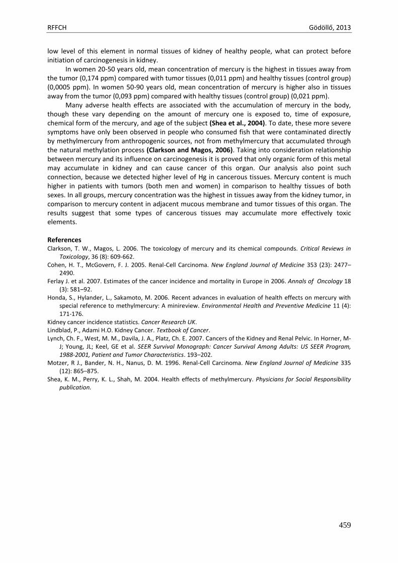
RFFCH Gödöllő, 2013
459
low level of this element in normal tissues of kidney of healthy people, what can protect before initiation of carcinogenesis in kidney.
In women 20-50 years old, mean concentration of mercury is the highest in tissues away from the tumor (0,174 ppm) compared with tumor tissues (0,011 ppm) and healthy tissues (control group) (0,0005 ppm). In women 50-90 years old, mean concentration of mercury is higher also in tissues away from the tumor (0,093 ppm) compared with healthy tissues (control group) (0,021 ppm).
Many adverse health effects are associated with the accumulation of mercury in the body, though these vary depending on the amount of mercury one is exposed to, time of exposure, chemical form of the mercury, and age of the subject (Shea et al., 2004). To date, these more severe symptoms have only been observed in people who consumed fish that were contaminated directly by methylmercury from anthropogenic sources, not from methylmercury that accumulated through the natural methylation process (Clarkson and Magos, 2006). Taking into consideration relationship between mercury and its influence on carcinogenesis it is proved that only organic form of this metal may accumulate in kidney and can cause cancer of this organ. Our analysis also point such connection, because we detected higher level of Hg in cancerous tissues. Mercury content is much higher in patients with tumors (both men and women) in comparison to healthy tissues of both sexes. In all groups, mercury concentration was the highest in tissues away from the kidney tumor, in comparison to mercury content in adjacent mucous membrane and tumor tissues of this organ. The results suggest that some types of cancerous tissues may accumulate more effectively toxic elements.
References Clarkson, T. W., Magos, L. 2006. The toxicology of mercury and its chemical compounds. Critical Reviews in
Toxicology, 36 (8): 609-662. Cohen, H. T., McGovern, F. J. 2005. Renal-Cell Carcinoma. New England Journal of Medicine 353 (23): 2477–
2490. Ferlay J. et al. 2007. Estimates of the cancer incidence and mortality in Europe in 2006. Annals of Oncology 18
(3): 581–92. Honda, S., Hylander, L., Sakamoto, M. 2006. Recent advances in evaluation of health effects on mercury with
special reference to methylmercury: A minireview. Environmental Health and Preventive Medicine 11 (4): 171-176.
Kidney cancer incidence statistics. Cancer Research UK. Lindblad, P., Adami H.O. Kidney Cancer. Textbook of Cancer. Lynch, Ch. F., West, M. M., Davila, J. A., Platz, Ch. E. 2007. Cancers of the Kidney and Renal Pelvic. In Horner, M-
J; Young, JL; Keel, GE et al. SEER Survival Monograph: Cancer Survival Among Adults: US SEER Program, 1988-2001, Patient and Tumor Characteristics. 193–202.
Motzer, R J., Bander, N. H., Nanus, D. M. 1996. Renal-Cell Carcinoma. New England Journal of Medicine 335 (12): 865–875.
Shea, K. M., Perry, K. L., Shah, M. 2004. Health effects of methylmercury. Physicians for Social Responsibility publication.

Animal welfare, etológia és tartástechnológia
Animal welfare, ethology and housing systems
Volume 9 Issue 3
Különszám/Special Issue
Gödöllő
2013
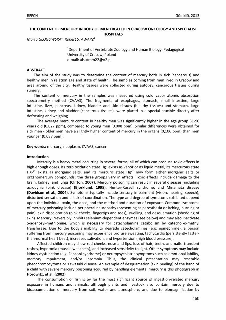
RFFCH Gödöllő, 2013
460
THE CONTENT OF MERCURY IN BODY OF MEN TREATED IN CRACOW ONCOLOGY AND SPECIALIST HOSPITALS
Marta GŁOGOWSKA1, Robert STAWARZ1
1Department of Vertebrate Zoology and Human Biology, Pedagogical University of Cracow, Poland e-mail: [email protected]
ABSTRACT
The aim of the study was to determine the content of mercury both in sick (cancerous) and healthy men in relation age and state of health. The samples coming from men lived in Cracow and area around of the city. Healthy tissues were collected during autopsy, cancerous tissues during surgery.
The content of mercury in the samples was measured using cold vapor atomic absorption spectrometry method (CVAAS). The fragments of esophagus, stomach, small intestine, large intestine, liver, pancreas, kidney, bladder and skin tissues (healthy tissues) and stomach, large intestine, kidney and bladder (cancerous tissues), were placed in a special crucible directly after defrosting and weighing.
The average mercury content in healthy men was significantly higher in the age group 51-90 years old (0,027 ppm), compared to young men (0,008 ppm). Similar differences were obtained for sick men - older men have a slightly higher content of mercury in the organs (0,106 ppm) than men younger (0,088 ppm). Key words: mercury, neoplasm, CVAAS, cancer Introduction
Mercury is a heavy metal occurring in several forms, all of which can produce toxic effects in high enough doses. Its zero oxidation state Hg0 exists as vapor or as liquid metal, its mercurous state Hg2
2+ exists as inorganic salts, and its mercuric state Hg2+ may form either inorganic salts or organomercury compounds; the three groups vary in effects. Toxic effects include damage to the brain, kidney, and lungs (Clifton, 2007). Mercury poisoning can result in several diseases, including acrodynia (pink disease) (Bjorklund, 1995), Hunter-Russell syndrome, and Minamata disease (Davidson et al., 2004). Symptoms typically include sensory impairment (vision, hearing, speech), disturbed sensation and a lack of coordination. The type and degree of symptoms exhibited depend upon the individual toxin, the dose, and the method and duration of exposure. Common symptoms of mercury poisoning include peripheral neuropathy (presenting as paresthesia or itching, burning or pain), skin discoloration (pink cheeks, fingertips and toes), swelling, and desquamation (shedding of skin). Mercury irreversibly inhibits selenium-dependent enzymes (see below) and may also inactivate S-adenosyl-methionine, which is necessary for catecholamine catabolism by catechol-o-methyl transferase. Due to the body's inability to degrade catecholamines (e.g. epinephrine), a person suffering from mercury poisoning may experience profuse sweating, tachycardia (persistently faster-than-normal heart beat), increased salivation, and hypertension (high blood pressure).
Affected children may show red cheeks, nose and lips, loss of hair, teeth, and nails, transient rashes, hypotonia (muscle weakness), and increased sensitivity to light. Other symptoms may include kidney dysfunction (e.g. Fanconi syndrome) or neuropsychiatric symptoms such as emotional lability, memory impairment, and/or insomnia. Thus, the clinical presentation may resemble pheochromocytoma or Kawasaki disease. An example of desquamation (skin peeling) of the hand of a child with severe mercury poisoning acquired by handling elemental mercury is this photograph in Horowitz, et al. (2002).
The consumption of fish is by far the most significant source of ingestion-related mercury exposure in humans and animals, although plants and livestock also contain mercury due to bioaccumulation of mercury from soil, water and atmosphere, and due to biomagnification by
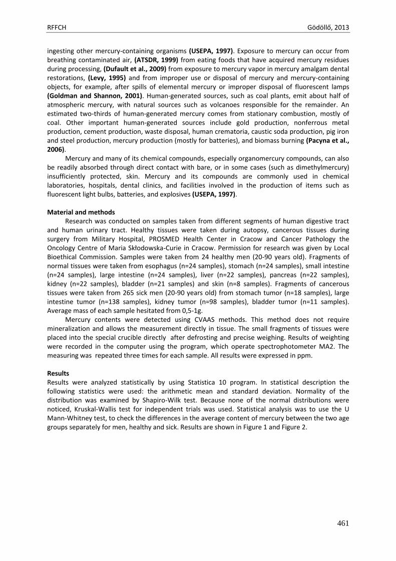
RFFCH Gödöllő, 2013
461
ingesting other mercury-containing organisms (USEPA, 1997). Exposure to mercury can occur from breathing contaminated air, (ATSDR, 1999) from eating foods that have acquired mercury residues during processing, (Dufault et al., 2009) from exposure to mercury vapor in mercury amalgam dental restorations, (Levy, 1995) and from improper use or disposal of mercury and mercury-containing objects, for example, after spills of elemental mercury or improper disposal of fluorescent lamps (Goldman and Shannon, 2001). Human-generated sources, such as coal plants, emit about half of atmospheric mercury, with natural sources such as volcanoes responsible for the remainder. An estimated two-thirds of human-generated mercury comes from stationary combustion, mostly of coal. Other important human-generated sources include gold production, nonferrous metal production, cement production, waste disposal, human crematoria, caustic soda production, pig iron and steel production, mercury production (mostly for batteries), and biomass burning (Pacyna et al., 2006).
Mercury and many of its chemical compounds, especially organomercury compounds, can also be readily absorbed through direct contact with bare, or in some cases (such as dimethylmercury) insufficiently protected, skin. Mercury and its compounds are commonly used in chemical laboratories, hospitals, dental clinics, and facilities involved in the production of items such as fluorescent light bulbs, batteries, and explosives (USEPA, 1997).
Material and methods
Research was conducted on samples taken from different segments of human digestive tract and human urinary tract. Healthy tissues were taken during autopsy, cancerous tissues during surgery from Military Hospital, PROSMED Health Center in Cracow and Cancer Pathology the Oncology Centre of Maria Skłodowska-Curie in Cracow. Permission for research was given by Local Bioethical Commission. Samples were taken from 24 healthy men (20-90 years old). Fragments of normal tissues were taken from esophagus (n=24 samples), stomach (n=24 samples), small intestine (n=24 samples), large intestine (n=24 samples), liver (n=22 samples), pancreas (n=22 samples), kidney (n=22 samples), bladder (n=21 samples) and skin (n=8 samples). Fragments of cancerous tissues were taken from 265 sick men (20-90 years old) from stomach tumor (n=18 samples), large intestine tumor (n=138 samples), kidney tumor (n=98 samples), bladder tumor (n=11 samples). Average mass of each sample hesitated from 0,5-1g.
Mercury contents were detected using CVAAS methods. This method does not require mineralization and allows the measurement directly in tissue. The small fragments of tissues were placed into the special crucible directly after defrosting and precise weighing. Results of weighting were recorded in the computer using the program, which operate spectrophotometer MA2. The measuring was repeated three times for each sample. All results were expressed in ppm. Results Results were analyzed statistically by using Statistica 10 program. In statistical description the following statistics were used: the arithmetic mean and standard deviation. Normality of the distribution was examined by Shapiro-Wilk test. Because none of the normal distributions were noticed, Kruskal-Wallis test for independent trials was used. Statistical analysis was to use the U Mann-Whitney test, to check the differences in the average content of mercury between the two age groups separately for men, healthy and sick. Results are shown in Figure 1 and Figure 2.
RFFCH Gödöllő, 2013
462
0
0,005
0,01
0,015
0,02
0,025
0,03
ppm
YOUNG MEN (20-50 years old) OLDER MEN (51-90 years old)
ACCUMULATION OF MERCURY IN HEALTHY MEN
Fig. 1
Accumulation of mercury in healthy men organs
0
0,02
0,04
0,06
0,08
0,1
0,12
ppm
YOUNG MEN (20-50 years old) OLDER MEN (51-90 years old)
ACCUMULATION OF MERCURY IN SICK MEN
Fig. 2
Accumulation of mercury in sick men organs
Mean content of mercury for tissues young (<50) healthy men is 0,008 ppm. The average content of mercury for tissues older (>50) healthy men is 0,027 ppm. The average content of mercury for tissues young (<50) sick men is 0,088 ppm. The average content of mercury for tissues older (>50) sick men is 0,106 ppm. Statistically significant differences were found between healthy and sick men tissues. In the case of healthy tissues, mercury concentrations was higher in older men.
Discussion and conclusions
In our study we determined presence of Hg in cancerous and normal (healthy) tissues of similar men’s organs (bladder, kidney, esophagus, stomach, small intestine, large intestine, liver, pancreas and skin). Compounds of mercury tend to be much more toxic than the elemental form, and organic compounds of mercury are often extremely toxic and have been implicated in causing brain and liver damage. The most dangerous mercury compound, dimethylmercury, is so toxic that even a few microliters spilled on the skin, or even a latex glove, can cause death. Mercuric chloride may cause
RFFCH Gödöllő, 2013
463
cancer as it has caused increases in several types of tumors in rats and mice, while methyl mercury has caused kidney tumors in male rats. The EPA has classified mercuric chloride and methyl mercury as possible human carcinogens.
Metals – such as mercury - are involved in different physical and chemical processes, which are common in cancer development. There are different effects caused by metals in the process of carcinogenesis, such as: damage in DNA, formation of DNA cross-links, the interference of metals with DNA-repair enzymes. Some metals affect the transduction signal by initiation of gene expression disturbances and intracellular communication. Moreover metals may cause of negative effects of the immune system function and disturbances of cellular homeostasis. It is crucial to realize that mercury is especially dangerous because it can be found in things and places which are surrounding us for example tuna has a capacity to accumulate high quantities of mercury in muscles.
It can be stated unequivocally much higher mercury content in cancerous tissues of sick men (0,106 ppm and 0,088 ppm), compared to healthy tissues (control group) (0.027 ppm and 0,008 ppm). Older people have a greater tendency to accumulate heavy metals in their tissues, perhaps because the metals are not the ability to direct carcinogenesis, may interact with other carcinogenic factors, such as metals can inhibit the DNA repair process, increasing the likelihood of mutation and, as a thus - cancer (Hartwig, 1998, Hu et al., 2004a, 2004b). The nucleic acids are capable of intermolecular interaction with different chemical compounds and metal ions. References ATSDR Mercury ToxFAQ. 1999. ToxFAQs: Mercury. Agency for Toxic Substances and Disease Registry. Retrieved
2007-07-25. Bjørklund, G. 1995. Mercury and Acrodynia. Journal of Orthomolecular Medicine 10, 145–146. Clifton, J.C. 2007. Mercury exposure and public health. Pediatr Clin North Am 54 (2): 237–69. Davidson, P.W., Myers, G.J., Weiss, B. 2004. Mercury exposure and child development outcomes. Pediatrics
113 (4 Suppl): 1023–9. Dufault, R., LeBlanc, B., Schnoll, R. et al. 2009. Mercury from chlor-alkali plants: measured concentrations in
food product sugar. Environ Health 8 (1): 2. Goldman, L.R., Shannon, M.W. Technical report: mercury in the environment: implications for pediatricians.
Pediatrics 108 (1): 197–205. Hartwig, A. 1998. Carcinogenicity of metal compounds: possible role of DNA repair inhibition. Toxicology
Letters 102-103, 235-239. Horowitz, Y., Greenberg, D., Ling, G., Lifshitz, M. 2002. Acrodynia: a case report of two siblings. Arch Dis Child
86 (6): 453. Hu, W., Feng, Z., Tang, M.S. 2004. Chromium(VI) enhances (+/-)-anti-7beta,8alpha-dihydroxy-9alpha,10alpha-
epoxy-7,8,9,10 tetrahydrobenzo[a]pyrene-induced cytotoxicity and mutagenicity in mammalian cells through its inhibitory effect on nucleotide excision repair. Biochemistry 43, 14282-14289.
Hu, W., Feng, Z., Tang, M.S. 2004. Nickel (II) enhances benzo[a]pyrene diol epoxide-induced mutagenesis through inhibition of nucleotide excision repair in human cells: a possible mechanism for nickel (II)-induced carcinogenesis. Carcinogenesis 25, 455-462.
Levy, M. 1995. Dental Amalgam: toxicological evaluation and health risk assessment. J Cdn Dent Assoc 61: 667–8, 671–4.
Pacyna, E.G., Pacyna, J.M., Steenhuisen, F., Wilson, S. 2006. Global anthropogenic mercury emission inventory for 2000. Atmos Environ 40 (22): 4048–63.
United States Environmental Protection Agency. 1997. Mercury Study Report to Congress 3. Washington, D.C.: United States Environmental Protection Agency.
United States Environmental Protection Agency. 1997. Mercury Study Report to Congress 4. Washington, D.C.: United States Environmental Protection Agency.
Animal welfare, etológia és tartástechnológia
Animal welfare, ethology and housing systems
Volume 9 Issue 3
Különszám/Special Issue
Gödöllő
2013
RFFCH Gödöllő, 2013
464
Influence of smoking on selected metals and antioxidants in breast milk
GOC Z., KILIAN K., STAWARZ R., FORMICKI G., SZAROMA W., GREŃ A., KAPUSTA E. Institute of Biology, Pedagogical University of Cracow, Podbrzezie 3, 31-045 Cracow, Poland
Abstract
The aim of the study was comparison of selected breast milk parameters taken from smoking and non-smoking mothers, being in different age. Concentrations of calcium, mercury, and glutathione (GSH) were measured in milk samples. Also activities of superoxide dismutase (Cu/Zn-SOD), were fixed. Breast milk samples were collected in the morning hours from women were between 20 and 37 years old, living in Cracow in Lesser Poland. All samples were divided in accordance with women’ age and smoking addict. According to their age status, 90 mothers were classified into three groups of age: 20-25, 26-31 and 32-37 years. Samples were taken from the selected women between 4th and 14th day postpartum (transitional milk). Results indicate that mercury was present in all analyzed milk samples and its concentrations were higher in milk samples taken from smoking women than taken from non-smoking in all age groups. Mercury concentration in milk samples of smoking women was lower than in milk samples taken from elder women. This fact suggests that smoking can affect mechanisms of detoxification and mercury excretion into breast milk. Mean calcium level was the highest in milk samples taken from smoking mothers in age of 32-37 years old and the lowest level was observed in milk from youngest women. Smoking habit did not have influence on calcium concentration in milk.
Differences between superoxide dismutase (Cu/Zn-SOD) activity and glutathione (GSH)
level in milk samples taken from smoking and samples taken from non-smoking women were statistically insignificant in all age groups. This fact suggests that antioxidant properties of milk are always stable and independent of women’ age and cigarette smoking.

Animal welfare, etológia és tartástechnológia
Animal welfare, ethology and housing systems
Volume 9 Issue 3
Különszám/Special Issue
Gödöllő
2013
RFFCH Gödöllő, 2013
465
Investigation of the Paramecium Toxicity Test for Detecting of Heavy Metals in an Aquatic Environment
Magdalena Greczek-Stachura, Patrycja Zagata
Institute of Biology, Pedagogical University 30-089 Kraków, Podchorążych 2
Heavy metals are ubiquitous elements in the natural environment and also but their presence in the environment is associated with contamination by industrial waste. Contamination of aquatic ecosystems with heavy metals has increased, worldwide. The heavy metals can enter the aquatic food chain through direct consumption of water or through consumption of organism living in the contaminated water. Microorganisms and specially ciliates are the key components for the decomposition of organic materials. They are an important link in the aquatic food-chain and they are involved in water purification. Many studies conducted on heavy metals polluted waters have revealed changes in the dynamics of protist communities. The ciliate species present in the polluted sites could become useful bioindicator for the detection of environmental disturbances and assessment of the trophic state.
Paramecium bursaria is a unicellular organisms, which is widely distributed in aquatic
environment. Among ciliates Paramecium species, only Paramecium bursaria maintains several hundred symbiotic algae in their cytoplasm. The algae can supply the host with a photosynthetic product, maltose enable P. bursaria to survive under starvation condition.
The present study of Paramecium bursaria and its interaction with cadmium, chromium,
nickel, copper, zinc may be useful for bioremediation of heavy metals contaminated environment.

Animal welfare, etológia és tartástechnológia
Animal welfare, ethology and housing systems
Volume 9 Issue 3
Különszám/Special Issue
Gödöllő
2013
RFFCH Gödöllő, 2013
466
Effect of resveratrol on antioxidants in diabetic mice
Greń Agnieszka*, Formicki Grzegorz*, Tvrdá Eva**, Goc Zofia*, Kňažická Zuzana**, Lukáčová Jana**, Lukáč Norbert**, Massányi Peter**, Kapusta Edyta*
* Pedagogical University of Cracow , Department of Animal Physiology and Toxicology, Institute of Biology, ul. Podbrzezie 3, Kraków, Poland,
**Slovak University of Agriculture, Department of Animal Physiology, Tr. Andreja Hlinku 2, Nitra, Slovak Republic
*Corresponding author: [email protected]
Abstract Diabetes mellitus (DM) is characterized by chronic hyperglycemia, resulting from defects in insulin secretion, action, or both, leading to disturbance in carbohydrate, lipid and protein metabolism. This state induces immediate oxidant stress and free radicals which trigger oxidative stress-linked diabetic complications. Resveratrol (RSV) (3,5,4’-trihydroxystilbene) is a natural polyphenol present in various foodstuffs, mainly in grapes where it is synthesized in response to environmental stress conditions. Many studies have demonstrated that this molecule exhibits a wide range of biological and pharmacological activities both in vivo and in vitro. A series of studies showed that resveratrol has anti-oxidant properties, anti-inflammatory properties, and anticancer activity.
The present study was to investigate the effect of resveratrol on alloxan-induced diabetic mice, through analysis of basic biochemical parameters, enzymic as well as non-enzymic activities – superoxide dismutase (SOD), catalase (CAT) and reduced glutathione (GSH).
The experiments were carried out male mice, average body weight 25 – 26g, bred in the constant light conditions LD 12:12 and fed with standard diet with unlimited access to water. Male mice were divided into four groups (n = 6): normal control, diabetic control, normal rats treated with resveratrol, and diabetic rats treated with resveratrol. Alloxan was administered as a single dose (75 mgKg −1 body weight) to induce diabetes in mice. A dose of resveratrol (Sigma Aldrich, St. Louis, MO, USA) (20 mgKg −1 body weight/for 14 days) were administrated orally, to the alloxan-induced diabetic mice. Thirty minutes after the last injection animals were anaesthetized and decapitated. The blood samples were collected from the carotid artery. All blood samples were processed immediately and frozen at minus 70 degrees centigrade until assay.
In animals, the control group average of enzyme activity was respectively: SOD=21,61 U/mg protein; CAT=50,13 U/mg protein and level of GSH=1,93 µM/ml blood. The levels of antioxidants were significantly decreased in blood serum in alloxan-induced diabetic mice. A significant (p<0.05) reduction in SOD=14,27 U/mg protein; CAT=38,34 U/mg protein and level of GSH=1,14 µM/ml blood are observed in diabetic group as compared to control mice. After resveratrol administration, there were increases in superoxide dismutase (p<0.001), catalase (p<0.001) activities and reduced glutathione (p<0.01) concentration compared to diabetes mice.
Treating the diabetic mice with doses of resveratrol restored the changes in the above parameters analyzed. The present study, showed that resveratrol exerted antioxidant effects, consequently alleviate damage caused by alloxan-induced diabetes Keywords: resveratrol, oxidative stress, diabetes, mice

Animal welfare, etológia és tartástechnológia
Animal welfare, ethology and housing systems
Volume 9 Issue 3
Különszám/Special Issue
Gödöllő
2013
RFFCH Gödöllő, 2013
467
EFFECTS OF DIETARY FUSARIUM MYCOTOXINS ON ACTIVITY OF ANTIOXIDANT ENZYMES IN CHICKENS
Ľubomíra GREŠÁKOVÁ , Klaudia ČOBANOVÁ, Štefan FAIX
Institute of Animal Physiology, Slovak Academy of Sciences, Šoltesovej 4-6, 040 01, Košice, Slovak Republic
Abstract The effect of medium dietary levels of Fusarium mycotoxins deoxynivalenol (DON) with or without zearalenone (ZEA) on the activity of glutathione peroxidase (GPx) in blood and tissues,thioredoxin reductase (TrxR) in liver tissue, superoxide dismutase (SOD) in erythrocytes, the activity of γ-glutamyltransferase (GGT) in plasma and concentrations of malondialdehyde (MDA)in liver and kidney was investigated on Ross 308 hybrid broiler chickens of both sexes. After hatching, all chickens were fed an identical control diet for two weeks. Then chickens of Group 1 fed a diet contaminated with DON (3 mg DON/kg), while Group 2 was fed a contaminated diet containing DON and ZEA, each at a level of 3.4 mg/kg. All contaminated diets were prepared in the laboratory using maize cultivated with Fusarium graminearum. Intake of contaminated diet with the combination of mycotoxins DON and ZEA resulted in significantly increased levels of MDA in liver and kidney tissues, while the activity of TrxR in liver and GPx in both tissues did not affected. On the other hand, feeding of diet contaminated with 3 mg DON/kg and only background ZEA level (0.15 mg/kg) reduced activity of GPx in liver only and increased level of MDA in liver and kidney tissue. The activity of blood GPx and SOD in erythrocytes did not affected by dietary mycotoxins. The activity of GGT in plasma was significantly elevated in all chickens fed contaminated diets. Presented results demonstrate that diet contaminated with DON and ZEA at medium dietary levels is already able to induce oxidative stress in broilers. Introduction Mycotoxins as secondary metabolites of several moulds differ in their chemical structure, toxic properties and biological effects. In the mild climatic zone, maize is usually infected by members of the genus Fusarium, especially by the species Fusarium graminearum. The most important mycotoxins produced by these moulds are DON, its acetylated derivatives, ZEA and nivalenol (Binder et al., 2007). Deoxynivalenol (DON) is a member of trichothecene mycotoxin group. The mode of toxic action of DON is inhibition of protein synthesis, thus affecting rapidly dividing cells, such as those of the gastrointestinal tract and the immune system (Sergent et al., 2006). ZEA is a naturally occurring non-steroidal mycotoxin with oestrogenic properties, which is known to induce functional and morphological alteration in reproductive organs due to the activation of estrogen receptors. In addition, ZEA has been shown to be hepatotoxic, haemotoxic, immunotoxic and genotoxic (Zinedine et al., 2007). Adverse effects of mycotoxins on cells are also associated with the increased production of free radicals and reactive oxygen species resulting in oxidative damage of target tissues (Dvorska et al., 2007). The aim of our studies was to investigate the effects of diets contaminated with DON and ZEA in complete feed on parameters of tissue oxidative stress and antioxidant status in fattening chickens. Material and methods Experimental design, diets and analysis One-day-old chickens of Ross 308 hybrid broilers of both sexes were randomly divided into dietary treatment groups and all were fed an identical uncontaminated starter diet for 2 weeks. After this time, control broilers continued to be fed the uncontaminated diet and chickens from experimental Group 1 being fed a diet contaminated with DON (3 mg DON/kg) and only background ZEA level (0.15 mg/kg). Group 2 was given a diet contaminated with DON and ZEA, each at a level of 3.4 mg/kg. Rearing of the chickens was done with a lighting regimen of 23 h light to 1 h dark and lasted for four
RFFCH Gödöllő, 2013
468
weeks. The initial room temperature of 32–33°C was reduced weekly by 18°C to a final temperature of 28°C. All birds had free access to water and feed. To provide stable dietary contents of mycotoxins during the entire experimental period, the chickens were fed only one type of diet (HYD-01). The final diets were obtained by mixing the basal diet (BD, the part of complete diet before addition of 40% portion of control or contaminated maize) with control or contaminated maize batches. Contaminated batches of maize were obtained by their cultivation with Fusarium graminearum for four weeks at the Slovak Agriculture University in Nitra (Labuda et al. 2003). The contents of deoxynivalenol (DON), zearalenone (ZEA), total aflatoxins, and ochratoxin A in BD and experimental diets are shown in Table 1. Table 1
Final concentrations of mycotoxins in control and experimental diets Dietary treatment DON ZEA AFLA
total OTA
Control group 0.1 0.005 0.0 0.0004 Group 1
(DON contaminated diet) 3.0 0.15 0.0 0.0004
Group 2 (DON+ZEA contaminated diet)
3.4 3.4 0.0 0.0005
DON = deoxynivalenol; ZEA = zearalenone; AFLA total = total aflatoxins; OTA = ochratoxin A
At the end of the 4-week experiment, eight randomly chosen chickens from each treatment were euthanised and tissue samples from liver and kidney were collected. Samples of blood were collected by intracardial punction into heparinised tubes. Mucosa from the duodenum was obtained by scraping. The blood, plasma and tissue samples were stored at –65 °C until analysis. Mycotoxin concentrations in feed were detected using the commercial competitive enzyme-linked immunosorbent assay-based Veratox 5/5 kit (Neogen, Lansing, MI., USA). The activity of blood glutathione peroxidase (GPx) was determined using the method of Paglia and Valentine (1967) with the Ransel kit (Randox, UK). Malondialdehyde concentrations (MDA) in tissue were measured by modified fluorometric methods as described by Jo and Ahn (1998). Haemoglobin (Hb) content of blood and superoxide dismutase (SOD) activity (Arthur and Boyne, 1985) in erythrocytes was analysed using kits from Randox, UK. The protein concentrations in the tissues examined were measured by the spectrophotometric method of Bradford (1976). Spectrophotometric determination of thioredoxin reductase (TrxR) activity was done with a Thioredoxin Reductase Assay Kit by the method of Holmgren and Bjornstedt (1995). The γ-glutamyltransferase (GGT) activity was measured using the kit from Randox, UK, by the method of Szasz (1969). Statistical analysis Statistical analysis was done by one-way analysis of variance (ANOVA) with the post hoc Tukey multiple comparison test using GraphPad Software (USA). The results are given as means ± SEM. Result and discussion Two weeks of feeding contaminated diets with mycotoxins resulted in increased levels of MDA in
liver and kidney tissue in both experimental groups. A similar response was found in the MDA
concentration in kidney of all broilers fed contaminated diets. Intake of contaminated diet with DON
(3 mg DON/kg) led to significantly decreased GPx activity in liver tissue only, while no change of TrxR
activity in this tissue as well as the activity of GPx in kidney did not affected in any experimental
groups (Table 2).
RFFCH Gödöllő, 2013
469
Table 2 Concentration of malondialdehyde (MDA), activity of glutathione peroxidase (GPx) and
thioredoxin reductase (TrxR) in tissues of broilers fed diet contaminated with deoxynivalenol (DON, 3mg/kg) and combination DON with zearalenone (ZEA), both 3.4 mg/kg).
Parameter Control DON DON+ZEA
Liver MDA (nmol·g -1 protein) 195.2±32.4
a 382.6±44.8
b 397.3±54.7
b
GPx (U·g -1 protein) 13.8±1.7 a 7.8±1.3
b 10.4±1.4
a
TrxR (U·g -1 protein) 18.4±1.9 28.01±5.0 24.4±3.6 Kidney MDA (nmol·g -1 protein) 133.2±8.4
a 214.4±15.4
b 232.0±18.3
b
GPx (U·g -1 protein) 15.4±3.5 16.7±2.7 17.7±3.1 a, b: Different letters in superscript within a row mean significant difference (P<0.05).
Our results demonstrate increased production of MDA with concomitant significant reduction of GPx activity in liver tissue of broilers fed on diets contaminated with DON. It has been reported that lower GPx activity in tissues is closely connected with an increase in MDA concentration (Balogh et al. 2004). DON contaminated feed has been reported as a factor increasing TBARS formation in rat and mice liver (Rizzo et al. 1994). It was found that dietary inclusion of another trichothecene mycotoxin T-2 toxin also increased lipid peroxidation in the livers of chickens, ducks and geese (Mezes et al.,1999). The kidney tissue activities of GPx did not show any differences between dietary treatments. This could be in part ascribed to a considerably smaller MDA response in kidney tissue to contaminated diets than that in the liver of chickens. Currently it is not clear if mycotoxins stimulate lipid peroxidation directly by enhancing free radicals production, or if the increased tissue susceptibility to lipid peroxidation is a result of a compromised antioxidant system (Surai 2006). It has been shown that DNA damage and apoptosis rather than plasma membrane damage and necrosis may be responsible for toxicity of DON (Minervini et al., 2004). In the liver, the gluthathione and thioredoxin systems are major players in the regulation of cell redox status (Gromer et al., 2004). The decreased activity of GPx in liver tissue together with greater MDA production indicate that combinations of DON and ZEA in concentrations 3.4 mg/kg feed are capable of inducing significant oxidative stress in chickens. Intake of contaminated diet with the combination of mycotoxins DON and ZEA, both being 3.4 mg.kg -1 resulted in significantly increased GGT activity in plasma of chickens while the activity of blood GPx and SOD in erythrocytes did not elevated (Table 3). Table 3
Activity of glutathione peroxidase (GPx) in blood, superoxid dismutase (SOD) in red blood cells (RBC) and activity of γ-glutamyltransferase (GGT) in plasma of broilers fed diet
contaminated with deoxynivalenol (DON, 3mg/kg) and combination DON with zearalenone (ZEA, both 3.4 mg/kg).
Parameter Control DON DON+ZEA
GPx in blood (U.g -1
Hb) 132.3±12.3 132.9±10.5 134.5±11.2
SOD in erythrocytes (U.g -1
Hb) 1295±105 1566±113 1639±47
GGT in plasma (U.L -1
) 40.7±2.8 a 64.9±2.5
b 63.4±6.0
b
a, b: Different letters in superscript within a row mean significant difference (P<0.05).
The adverse effects of mycotoxins DON and ZEA on the livers of our broilers are demonstrated by findings of increased plasma activities of γ-glutamyltransferase (GGT). This enzyme is known to be involved in the transfer of amino acids across the cellular membrane and in glutathione metabolism. Serum GGT activity is widely used as a marker of liver dysfunction. Our results as well as previous studies show that serum GGT activity may be an early marker of oxidative stress (Lim et al., 2004).
RFFCH Gödöllő, 2013
470
Conclusion In conclusion, the results of this experiment demonstrate the adverse effects of Fusarium mycotoxins on the antioxidative status in chickens for fattening. It is shown that diets contaminated with DON with or without ZEA at medium levels of about 3 mg/kg feed are already able to induce oxidative stress in liver and kidney tissue. Acknowledgement This study was supported by the Slovak Research and Development Agency, Slovak Republic, Grants No. LPP-0213-06. References Arthur JR, Boyne R. 1985. Superoxide dismutase and glutathione peroxidase activities in neutrophils from selenium deficient and copper deficient cattle. Life Sci 36: 1569-1575. Binder, E., Tan, L., Chin, L., Handl, J. and Richard, J. (2007): Worldwide occurrence of mycotoxins in
commodities feeds and feed ingredients. Anim. Feed Sci. Technol. 137, 265–282. Bořutová, R., Faix, Š., Plachá, I., Grešáková, Ľ., Čobanová, K. and Leng, Ľ. (2008): Effects of deoxynivalenol and
zearalenone on oxidative stress and blood phagocytic activity in broilers. Arch. Anim. Nutr. 62, 303-312. Bradford M. 1976. A rapid and sensitive method for quantification of microgram quantities of protein utilizing
the principle of protein-dye binding. Anal Biochem 72: 248-254. Dvorska JE, Pappas AC, Karadas F, Speake BK, Surai PF. 2007. Protective effect of modified glucomannans and
organic selenium against antioxidant depletion in the chicken liver due to T-2 toxin-contaminated feed consumption. Comp Biochem Physiol C Toxicol Pharmacol 145: 582-7.
Gromer S, Urig S, Becker K. 2004. The thioredoxin system - from science to clinic. Med Res Rev 24: 40-89. Holmgren A, Bjornstedt M. 1995. Thioredoxin and thioredoxin reductase. Meth Enzymol 252: 199-208. Jo C, Ahn DU. 1998. Fluorimetric analysis of 2-thiobarbituric acid reactive substances in turkey. Poult Sci 77:
457-480. Labuda R, Tancinova D, Hudec K. 2003. Identification and enumeration of Fusarium species in poultry feed
mixtures from Slovakia. Ann Agric Environ Med 10: 61-6. Lim JS, Yang JH, Chung BY, Kam S, Jacobs Jr. DR, Lee DH. 2004. Is serum γ-glutamyltransferase inversely
associated with serum antioxidants as a marker of oxidative stress? Free Radical Bio Med 37: 1018-1023. Minervini F, Fornelli F, Flynn KM. 2004. Toxicity and apoptosis induced by the mycotoxins nivalenol,
deoxynivalenol and fumonisin B1 in a human erythroleukemia cell line. Toxicol In vitro 18: 21-28. Paglia DE, Valentine WN. 1967. Studies on quantitative and qualitative charactrezation of erythrocyte
glutathione peroxidase. J Lab Clin Med 70: 158-169. Sergent, T., Parys, M., Garson, S., Pussemier, L., Schneider, Y.J., Larondelle, Y., 2006. Deoxynivalenol transport
across human intestinal Caco-2 cells and its effects on cellular metabolism at realistic intestinal concentration. Toxicol. Lett. 164, 167–176.
Surai PF. 2006. Selenium and mycotoxins. In: Surai PF, editor. Selenium nutrition and health. Nothingham University Press. p 317-347.
Szasz G. 1969. A kinetic photometric method for serum γ-glutamyltranspeptidase. Clin Chem 15: 124-136. Zinedine, A., Soriano, JM., Molto, JC. and Manes, J. (2007): Review on the toxicity, occurrence, detoxification,
regulations and intake of zearalenone: An oestrogenic mycotoxin. Food Chem. Toxicol.45, 1-18.
Animal welfare, etológia és tartástechnológia
Animal welfare, ethology and housing systems
Volume 9 Issue 3
Különszám/Special Issue
Gödöllő
2013
RFFCH Gödöllő, 2013
471
POSSIBLE EFECT OF AMYGDALIN IN COMBINATION WITH DEOXYNIVALENOL ON SECRETION ACTIVITY OF PORCINE OVARIAN GRANULOSA CELLS IN VITRO
Marek Halenár*1, Marína Medveďová1, Nora Maruniaková1, Adriana Kolesárová1
1Department of Animal Physiology, Faculty of Biotechnology and Food Sciences, Slovak University of Agriculture in Nitra, Tr. A. Hlinku 2, 949 76 Nitra, Slovak Republic *Corresponding author: [email protected]
ABSTRACT Amygdalin is a controversial anti-tumor natural product found in plants of the Rosaceae
family that has been used as an alternative cancer drug for many years. One of the most widely distributed trichothecene contaminating food and animal feed is deoxynivalenol (DON) produced by Fusarium species. Deoxynivalenol has adverse effects on humans, animals, and crops that result in illnesses. The aim of the present in vitro study was to investigated the effect of natural compound amygdalin at the selected doses (1, 10, 100, 1000, 10 000 µg/ml) in combination with deoxynivalenol (1000 ng/ml) on secretion of steroid hormones (progesterone and estradiol) by ovarian granulosa cells from non-cyclic gilts. The release of progesterone and estradiol by granulosa cells was significantly (P≤0.05) increased in experimental group with the highest dose of amygdalin (10 000 µg/ml) in combination with deoxynivalenol (1000 ng/ml) compared to control group without administration of the natural compounds. On the other hand no significant differences in secretion of both steroid hormones were observed. Results from our study suggest possible effects of amygdalin in combination with mycotoxin-deoxynivalenol on secretion activity of porcine ovarian granulosa cells and possible involvement in the steroidogenesis.
Keywords: amygdalin, deoxynivalenol, estradiol, progesterone, health granulosa cells
INTRODUCTION Amygdalin is one of many nitrilosides, which are natural cyanide-containing substances
abundant in the seeds of apricots, almond, peaches, apples, and other rosaceous plants (Chang et al., 2006). Its oldest known use was by the ancient Egyptians as a poison for executing capital punishment: “penalty of the peach”. Amygdalin (D-mandelonitrile-β-D-gentiobioside, Fig. 1), C20H27NO11, is composed of two molecules of glucose, one of benzaldehyde, which induces an analgesic action, and one of hydrocyanic acid, which is an anti-neoplastic compound (Chang et al., 2006).
It has been used as a traditional drug because of its wide range of medicinal benefits, including curing or preventing cancer, relieving fever, suppressing cough, and quenching thirst. In the late 1970s and early 1980s, amygdalin was reported to selectively kill cancer cells at the tumor site without systemic toxicity and to effectively relieve pain in cancer patients (Zhou et al., 2012). However, the Food and Drug Administration (FDA) has not approved amygdalin as a cancer treatment owing to insufficient clinical evidence of its efficacy and potential toxicity. Despite the failure of clinical tests to demonstrate the anticancer effects of amygdalin in the U.S.A. and in Europe, amygdalin continues to be manufactured and administered as an anticancer therapy in northern Europe and Mexico (Chang et al.,2006; Kwon et al., 2010).
Figure 1
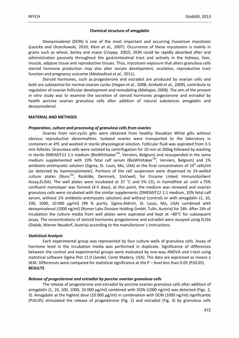
RFFCH Gödöllő, 2013
472
Chemical structure of amygdalin Deoxynivalenol (DON) is one of the most important and occurring Fussarium mycotoxin
(Łazicka and Orzechowski, 2010; Klem et al., 2007). Occurrence of these mycotoxins is mainly in grains such as wheat, barley and maize (Creppy, 2002). DON could be rapidly absorbed after oral administration passively throughout the gastrointestinal tract and actively in the kidneys, liver, muscle, adipose tissue and reproductive tissues. Thus, mycotoxin exposure that alters granulosa cells steroid hormone production may also alter oocyte development, ovulation, reproductive tract function and pregnancy outcome (Medveďová et al., 2011).
Steroid hormones, such as progesterone and estradiol are produced by ovarian cells and both are substantial for normal ovarian cycles (Hagan et al., 2008; Arnhold et al., 2009), contribute to regulation of ovarian follicular development and remodeling (Mahajan, 2008). The aim of the present in vitro study was to examine the secretion of steroid hormones progesterone and estradiol by health porcine ovarian granulosa cells after addition of natural substances amygdalin and deoxynivalenol. MATERIAL AND METHODS Preparation, culture and processing of granulosa cells from ovaries
Ovaries from non-cyclic gilts were obtained from healthy Slovakian White gilts without obvious reproductive abnormalities. Isolated ovaries were transported to the laboratory in containers at 4ºC and washed in sterile physiological solution. Follicular fluid was aspirated from 3-5 mm follicles. Granulosa cells were isolated by centrifugation for 10 min at 200xg followed by washing in sterile DMEM/F12 1:1 medium (BioWhittakerTM, Verviers, Belgium) and resuspended in the same medium supplemented with 10% fetal calf serum (BioWhittakerTM, Verviers, Belgium) and 1% antibiotic-antimycotic solution (Sigma, St. Louis, Mo, USA) at the final concentration of 106 cells/ml (as detected by haemocytometer). Portions of the cell suspension were dispensed to 24-welled culture plates (NuncTM, Roskilde, Denmark, 1ml/well; for Enzyme Linked ImmunoSorbent Assay,ELISA). The well plates were incubated at 37 °C and 5% CO2 in humidified air until a 75% confluent monolayer was formed (4-5 days), at this point, the medium was renewed and ovarian granulosa cells were incubated with the similar supplements (DMEM/F12 1:1 medium, 10% fetal calf serum, without 1% antibiotic-antimycotic solution) and without (control) or with amygdalin (1, 10, 100, 1000, 10 000 µg/ml) (99 % purity, Sigma-Aldrich, St. Louis, Mo, USA) combined with deoxynivalenol (1000 ng/ml) (Romer Labs Division Holding GmbH, Tulln, Austria) for 24h. After 24h of incubation the culture media from well plates were aspirated and kept at –80°C for subsequent assay. The concentrations of steroid hormones progesterone and estradiol were assayed using ELISA (Dialab, Wiener Neudorf, Austria) according to the manufacturer´s instructions. Statistical Analysis
Each experimental group was represented by four culture wells of granulosa cells. Assay of hormone level in the incubation media was performed in duplicate. Significance of differences between the control and experimental groups were evaluated by one-way ANOVA and t-test using statistical software Sigma Plot 11.0 (Jandel, Corte Madera, USA). The data are expressed as means ± SEM. Differences were compared for statistical significance at the P – level less than 0.05 (P≤0.05). RESULTS Release of progesterone and estradiol by porcine ovarian granulosa cells
The release of progesterone and estradiol by porcine ovarian granulosa cells after addition of amygdalin (1, 10, 100, 1000, 10 000 µg/ml) combined with DON (1000 ng/ml) was detected (Figs. 2, 3). Amygdalin at the highest dose (10 000 µg/ml) in combination with DON (1000 ng/ml) significantly (P≤0.05) stimulated the release of progesterone (Fig. 2) and estradiol (Fig. 3) by granulosa cells
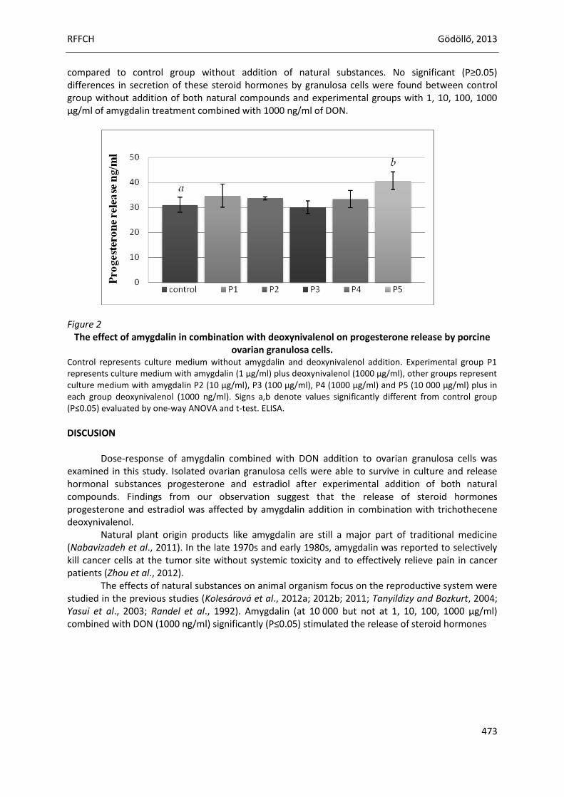
RFFCH Gödöllő, 2013
473
compared to control group without addition of natural substances. No significant (P≥0.05) differences in secretion of these steroid hormones by granulosa cells were found between control group without addition of both natural compounds and experimental groups with 1, 10, 100, 1000 µg/ml of amygdalin treatment combined with 1000 ng/ml of DON.
Figure 2
The effect of amygdalin in combination with deoxynivalenol on progesterone release by porcine ovarian granulosa cells.
Control represents culture medium without amygdalin and deoxynivalenol addition. Experimental group P1 represents culture medium with amygdalin (1 µg/ml) plus deoxynivalenol (1000 µg/ml), other groups represent culture medium with amygdalin P2 (10 µg/ml), P3 (100 µg/ml), P4 (1000 µg/ml) and P5 (10 000 µg/ml) plus in each group deoxynivalenol (1000 ng/ml). Signs a,b denote values significantly different from control group (P≤0.05) evaluated by one-way ANOVA and t-test. ELISA.
DISCUSION
Dose-response of amygdalin combined with DON addition to ovarian granulosa cells was examined in this study. Isolated ovarian granulosa cells were able to survive in culture and release hormonal substances progesterone and estradiol after experimental addition of both natural compounds. Findings from our observation suggest that the release of steroid hormones progesterone and estradiol was affected by amygdalin addition in combination with trichothecene deoxynivalenol.
Natural plant origin products like amygdalin are still a major part of traditional medicine (Nabavizadeh et al., 2011). In the late 1970s and early 1980s, amygdalin was reported to selectively kill cancer cells at the tumor site without systemic toxicity and to effectively relieve pain in cancer patients (Zhou et al., 2012).
The effects of natural substances on animal organism focus on the reproductive system were studied in the previous studies (Kolesárová et al., 2012a; 2012b; 2011; Tanyildizy and Bozkurt, 2004; Yasui et al., 2003; Randel et al., 1992). Amygdalin (at 10 000 but not at 1, 10, 100, 1000 µg/ml) combined with DON (1000 ng/ml) significantly (P≤0.05) stimulated the release of steroid hormones
RFFCH Gödöllő, 2013
474
Figure 3
The effect of amygdalin in combination with deoxynivalenol on estradiol release by porcine ovarian granulosa cells.
Control represents culture medium without amygdalin and deoxynivalenol addition. Experimental group E1 represents culture medium with amygdalin (1 µg/ml) plus deoxynivalenol (1000 ng/ml), other groups represent culture medium with amygdalin E2 (10 µg/ml), E3 (100 µg/ml), E4 (1000 µg/ml) and E5 (10 000 µg/ml) plus in each group deoxynivalenol (1000 ng/ml). Signs a,b denote values significantly different from control group (P≤0.05) evaluated by one-way ANOVA and t-test. ELISA.
progesterone and estradiol by porcine ovarian granulosa cells. On the other hand, our recent in vitro investigation showed that the release of steroid hormone progesterone by health granulosa cells from cyclic and non-cyclic porcine ovaries was not affected by the amygdalin addition at the doses 1, 10, 100, 1000 and 10 000 µg/ml (Halenár et al., 2013). Previous studies on amygdalin have focused on its purification, toxicity related to the release of cyanide, anti-tumor mechanism, and identification of its metabolites in plasma or herbs, and its pharmacological effect on cancers (Rauws et al., 1982). Possible effect of amygdalin on the male reproductive system was observed in previous study (Tanyildizi and Bozkurt, 2004). There are many studies which suggest dose-dependent impact of different mycotoxins on the secretion activity of porcine (Medveďová et al., 2011, Maruniaková et al., 2013, Ranzenigo et al., 2008) and rat ovarian cells (Kolesárová et al., 2011). The effect of deoxynivalenol on the steroid secretion by porcine ovarian granulosa cells was examined by Medveďová et al. (2011). Progesterone release by porcine ovarian granulosa cells was stimulated by DON treatment at the dose 1000 ng/ml but not at 10 and 100 ng/ml. Previous data also indicated dose-depended effects of DON on ovarian granulosa cells (Ranzenigo et al., 2008). On the other hand, the release of progesterone by porcine ovarian granulosa cells was significantly (P<0.05) inhibited after administration of trichothecene T-2 toxin at the doses 10, 100 and 1000 ng/ml (Maruniaková et al., 2013). The release of progesterone by porcine ovarian granulosa cells after exposure to toxic concentrations of DON, resveratrol, and their combination was studied by Kolesárová et al. (2012). These in vitro results suggest that reproductive toxicity of animals induced by a mycotoxin – deoxynivalenol can be inhibited by a protective natural substance - resveratrol.
The results of our in vitro experiments indicate that amygdalin combined with DON could modify secretion of steroid hormones by granulosa cells and potentially regulate process of steroidogenesis in porcine ovaries. CONCLUSION
This in vitro study was focused on assessment the interaction between natural anti-cancer substance amygdalin and mycotoxin deoxynivalenol. In conclusion, there are still a few studies that suggest possible positive or negative effects of natural substances depending on their toxicity. The
RFFCH Gödöllő, 2013
475
results suggest possible effects of amygdalin in combination with mycotoxin-deoxynivalenol on secretion activity of porcine ovarian granulosa cells and possible involvement in the steroidogenesis.
Acknowledgments: This work was financially supported by the Ministry of Education, Science, Research and Sport of the Slovak Republic projects no. 1/0790/11 and 1/0022/13 and European Community under project no 26220220180: Building Research Centre „AgroBioTech". REFERENCES Arnhold, I.J., Lofrano-Porto, A., Latronico, A.C. (2009): Inactivating mutations of luteinizing hormone beta-
subunit or luteinizing hormone receptor cause oligo-amenorrhea and infertility in women. In Hormone Research, vol. 71, 2009, no. 2, p. 75-82.
Creppy, E. E. (2002): Update of survey, regulation and toxic effects of mycotoxins in Europe. In Toxicology Letters, vol. 127, 2002, no. 1-3, p. 19-28.
Hagan, C.R., Faivre, E.J., Lange, C.A. (2008): Scaffolding actions of membrane-associated progesterone receptors. In Steroids, vol. 74, 2008, p. 568-572.
Halenár, M., Maruniaková, N., Medveďová, M., Kolesárová, A. (2013): The effect of amygdalin on porcine ovarian granulosa cells in vitro. In Journal of Microbiology, Biotechnology and Food Sciences, vol. 2, 2013, p. 14.
Chang, H.K., Shin, M.S., Yang, H.Y., Lee, J.W., Kim, Y.S., Lee, M.H., Kim, J., Kim, K.H.,Kim, C.J. (2006): Amygdalin Induces Apoptosis through Regulation of Bax and Bcl-2 Expressions in Human DU145 and LNCaP Prostate Cancer Cells. In Biological and Pharmaceutical Bulletin, vol. 29, 2006, no. 8, p. 1597-1602.
Klem, K., Váňová, M., Hajšlová, J., Lancová, K., Sehnalová, M. (2007): A neural network model for prediction of deoxynivalenol content in wheat grain based on weather data and preceding crop. In Plant and Soil Environment, vol. 53, 2007, no. 10, p. 421-429.
Kolesárová, A., Capcarová, M., Baková, Z., Gálik, B., Juracek, M., Simko, M., Sirotkin, A.V. (2011): The effect of bee pollen on secretion activity, markers of proliferation and apoptosis of porcine ovarian granulosa cells in vitro. In Journal of Environmental Science and Health, Part B, vol. 46, 2011, no. 3, p. 207-212.
Kolesárová, A., Capcarová, M., Maruniaková, N., Lukáč, N., Ciereszko, R.E., Sirotkin, A.V. (2012a): Resveratrol inhibits reproductive toxicity induced by deoxynivalenol. In Journal of Environmental Science and Health, Part A, vol. 47,
2012a, p. 1329-1334. Kolesárová, A., Baková, Z., Capcarová, M., Gálik, B., Juracek, M., Simko, M., Toman, R., Sirotkin, A.V. (2012b):
Consumption of bee pollen affects rat ovarian functions. In Journal of Animal Physiology and Animal Nutrition, (Berl).2012b Nov 9.doi: 10.1111/jpn.12013. [Epub ahead of print]
Kwon, H.J., Lee, J.H., Hong, S.P. (2010): Improvement of the extraction efficiency of D-amygdalin from Armeniacae Semen powder through inactivating emulsion and suppressing the epimerization of D-amygdalin. In Archives of Pharmacal Research, vol. 33, 2010, p. 81-86.
Łazicka, K.,Orzechowski, S. (2010): The characteristics of the chosen mycotoxins and their toxic influence on the human and animal metabolism. In Natural Science, vol. 2, 2010, no. 6, p. 544-550.
Mahajan, D.K. (2008): Pig model to study Dynamics of steroid during ovarian follicular growth and maturation. In Sourcebook of Models for Biomedical Research, Totowa, New Jersey: Humana Press, 2008, p. 778.
Maruniaková, N., Kolesárová, A., Kádasi, A., Medveďová, M., Halenár, M., Sirotkin, A.V., Grossmann, R., Bulla, J. (2013): Release of progesterone and testosterone by ovarian granulosa cells after addition of T-2 toxin and its combination with growth factor IGF-I. In Journal of Microbiology, Biotechnology and Food Sciences, vol. 2, 2013, p. 1864-1874.
Medveďová, M., Kolesárová, A., Capcarová, M., Labuda, R., Sirotkin, A.V., Kováčik, J., Bulla, J. (2011): The effect of deoxynivalenol on the secretory activity, proliferation and apoptosis of porcine ovarian granulosa cells in vitro. In Journal of Environmental Science and Health Part B, vol. 46, 2011, no. 3, p. 213-219.
Nabavizadeh, F., Alizadeh, A.M., Sadroleslami, Z.,Adeli, S. (2011):Gastroprotective effects of amygdalin on experimental gastric ulcer: Role of NO and TNF-α.
In Journal of Medical Plants Research, vol. 5, 2011, no. 14, p. 3122-3127. Randel, R.D., Chase, C.C.Jr., Wyse, S.J. (1992): Effects of gossypol and cottonseed products on reproduction of
mammals. In Journal of Animal Science, vol. 70, 1992, p. 1620- 1638. Ranzenigo, G., Caloni, F., Cremonesi, F., Aad, P.Y., Spicer, L.J. (2008): Effects of Fusariummycotoxins on steroid
production by porcine granulosa cells. In AnimalReproduction Science, vol. 107, 2008, no. 1-2, p. 115-130.
RFFCH Gödöllő, 2013
476
Rauws, A.G., Gramberg, L.G., Olling, M. (1982): Determination of amygdalin and its major metabolite prunasin in plasma and urine by high pressure liquid chromatography. In Pharmacy World & Science, vol. 6, 1982, p. 172-175.
Tanyildizi, S., Bozkurt, T. (2004):In Vitro Effects of Linamarin, Amygdalin and Gossypol Acetic Acid on Hyaluronidase Activity, Sperm Motility and Morphological Abnormality in Bull Sperm. In Turkish Journal of Veterinary and Animal Sciences, vol. 28, 2004, p. 819-824.
Yasui, T., Matsuzaki, T., Ushigoe, K., Kuwahara, A., Maegawa, M., Furumoto, H., Aono, T., Irahara, M. (2003): Stimulatory effect of the herbal medicine Keishi-bukuryo-ganon a cytokine-induced neutrophil chemoattractant, in rat ovarian cell culture. In American Journal of Reproductive Immunology, vol. 50, 2003, p. 90- 97.
Zhou, C., Qian, L.,Ma, H., Yu, X., Zhang, Y., Qu, W., Zhang, X., Xia, W. (2012): Enhancement of amygdalin activated with β-D-glucosidase on HepG2 cells proliferation and apoptosis. In Carbohydrate Polymers, vol. 90, 2012, p. 516-523.
Animal welfare, etológia és tartástechnológia
Animal welfare, ethology and housing systems
Volume 9 Issue 3
Különszám/Special Issue
Gödöllő
2013
RFFCH Gödöllő, 2013
477
THE EFFECT OF BEE POLLEN AS SUPPLEMENT DIETARY FOR MEAT pH, COOLING AND FREEZING LOSES ON BROILER CHICKENS MEAT
Peter Haščík, Ibrahim Elimam*, Jozef Garlík, Marek Bobk, Juraj Čuboň
Address: Department of Evaluation and Processing of Animal Products, Faculty of Biotechnology and Food Sciences, Slovak University of Agriculture, Tr. A. Hlinku 2, 949 76 Nitra, Slovak Republic. *Corresponding author: [email protected]
ABSTRACT
The present study was aimed to achieve the effect of bee pollen as a supplement dietary on broiler chickens for the meat pH value, cooling and freezing loss. A total 180 chicks in one day old which were divided into 6 groups (n = 30). To experimental groups were added bee pollen in different doses [(500 mg/kg (E1); 1500 mg/kg (E2); 2500 mg/kg (E3); 3500 mg/kg (E4) and 4500 mg/kg (E5)] into feed mixture. However the breast muscles pH value was greater in the experimental groups compared to the control groups except E1, E2 after 45 minutes and after 2 hours, and there were found significant differences (P≤0.05) between E1and E2 and between E2 with E4 after 2 hours. Also in thigh muscles the experimental groups were greater except E1, E2 and E3 after 45 minutes and after 2 hours and there found significant (P≤0.05) between E1and E4, and E4 an E5. Otherwise the cooling and freezingloss was higher in experimental groups except E5 in freezing loss and there were found significant differences (P≤0.05) between control group and E1, E2, E5 after 24 hours, also between control group with E2 and between E3 with E5 after 48 hours and there was significant (P≤0.05) between control and E5 groups with E2, E4 groups in freezing loss. The bee pollen has a positive effect on meat pH value but has a negative effect on cooling and freezing loss.
Keywords: bee pollen, broiler, pH meat value, cooling and freezing loss INTRODUCTION
Broilers are chickens bred and raised specifically for meat production, and it's one of the most common and widespread domestic animals. In this regard, numerous references exist on increasing poultry meat yields and improving carcass quality. For this reason, many ingredients have been used in broiler diets, in recent years (Karaoğlu et al., 2005). Complete feed mixtures for broiler chickens are often enriched with various additives as vegetable oils, probiotic, prebiotic and enzyme preparations (Lee et al., 2003, 2004; Shalmany and Shivazad, 2006). It is reported that additional benefits can be gained by supplementing broiler diets, particularly using of bee pollen as feed additives. Bee pollen has been used for many years in both traditional medicine and supplementary nutrition, as well as in alternative diets, mainly due to its nutritional properties and health benefits (Serra et al., 1997; Isla et al., 2001; Almeida-Muradian et al., 2005). Pollen loads are formed from the pollen of one plant species, ie. monofloral pollen (Carrión et al., 2003), is shaped pollen grains from different plant species called multifloral pollen, respectively (Stanley and Linskens, 1974; Barth et al., 2009; Modro et al., 2009). Pollen loads or bee pollen is the basic food for the colony as a source of protein for them (Tüylü and Sorkun, 2004). The protein content of pollen is 25-30% carbohydrates, 30-55% fats, including fatty acids and sterols 1-20% and also contains significant amounts of vitamins and minerals. Composition of the pollen provides valuablenutrients such as free amino acids, minerals, polyfenolytic substances and oligo-elements (Serra et al., 1997; Villanueva et al., 2002; Bastos et al., 2004; Almeida-Muradian et al., 2005; Cocan et al., 2005; Hamamoto et al., 2006; Human and Nicolson, 2006; Yamaguchi et al., 2006) and therefore is also used in the human diet, which provides a sense of well-being, contributes to functional and well-balanced body has antioxidant properties (Moreira et al., 2008) and prevents free radicals (Hejinen et al., 2002;Villanueva et al., 2002; Bastos et al., 2004; Almeida-Muradian et al., 2005; Silva et al., 2006;
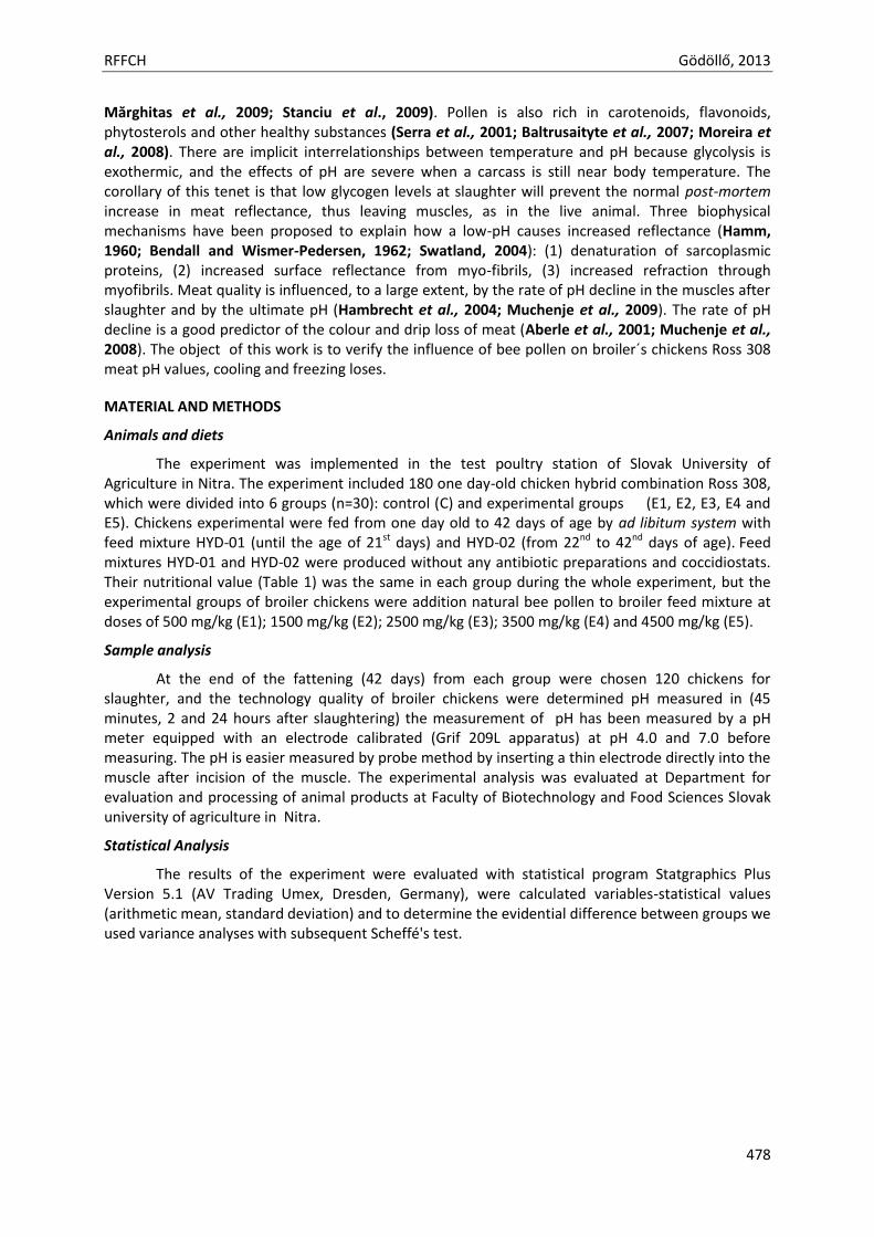
RFFCH Gödöllő, 2013
478
Mărghitas et al., 2009; Stanciu et al., 2009). Pollen is also rich in carotenoids, flavonoids, phytosterols and other healthy substances (Serra et al., 2001; Baltrusaityte et al., 2007; Moreira et al., 2008). There are implicit interrelationships between temperature and pH because glycolysis is exothermic, and the effects of pH are severe when a carcass is still near body temperature. The corollary of this tenet is that low glycogen levels at slaughter will prevent the normal post-mortem increase in meat reflectance, thus leaving muscles, as in the live animal. Three biophysical mechanisms have been proposed to explain how a low-pH causes increased reflectance (Hamm, 1960; Bendall and Wismer-Pedersen, 1962; Swatland, 2004): (1) denaturation of sarcoplasmic proteins, (2) increased surface reflectance from myo-fibrils, (3) increased refraction through myofibrils. Meat quality is influenced, to a large extent, by the rate of pH decline in the muscles after slaughter and by the ultimate pH (Hambrecht et al., 2004; Muchenje et al., 2009). The rate of pH decline is a good predictor of the colour and drip loss of meat (Aberle et al., 2001; Muchenje et al., 2008). The object of this work is to verify the influence of bee pollen on broiler´s chickens Ross 308 meat pH values, cooling and freezing loses.
MATERIAL AND METHODS
Animals and diets
The experiment was implemented in the test poultry station of Slovak University of Agriculture in Nitra. The experiment included 180 one day-old chicken hybrid combination Ross 308, which were divided into 6 groups (n=30): control (C) and experimental groups (E1, E2, E3, E4 and E5). Chickens experimental were fed from one day old to 42 days of age by ad libitum system with feed mixture HYD-01 (until the age of 21st days) and HYD-02 (from 22nd to 42nd days of age). Feed mixtures HYD-01 and HYD-02 were produced without any antibiotic preparations and coccidiostats. Their nutritional value (Table 1) was the same in each group during the whole experiment, but the experimental groups of broiler chickens were addition natural bee pollen to broiler feed mixture at doses of 500 mg/kg (E1); 1500 mg/kg (E2); 2500 mg/kg (E3); 3500 mg/kg (E4) and 4500 mg/kg (E5).
Sample analysis
At the end of the fattening (42 days) from each group were chosen 120 chickens for slaughter, and the technology quality of broiler chickens were determined pH measured in (45 minutes, 2 and 24 hours after slaughtering) the measurement of pH has been measured by a pH meter equipped with an electrode calibrated (Grif 209L apparatus) at pH 4.0 and 7.0 before measuring. The pH is easier measured by probe method by inserting a thin electrode directly into the muscle after incision of the muscle. The experimental analysis was evaluated at Department for evaluation and processing of animal products at Faculty of Biotechnology and Food Sciences Slovak university of agriculture in Nitra.
Statistical Analysis
The results of the experiment were evaluated with statistical program Statgraphics Plus Version 5.1 (AV Trading Umex, Dresden, Germany), were calculated variables-statistical values (arithmetic mean, standard deviation) and to determine the evidential difference between groups we used variance analyses with subsequent Scheffé's test.
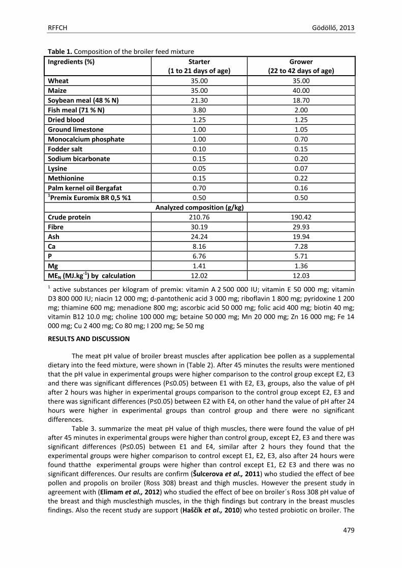
RFFCH Gödöllő, 2013
479
Table 1. Composition of the broiler feed mixture
Ingredients (%) Starter (1 to 21 days of age)
Grower (22 to 42 days of age)
Wheat 35.00 35.00
Maize 35.00 40.00
Soybean meal (48 % N) 21.30 18.70
Fish meal (71 % N) 3.80 2.00
Dried blood 1.25 1.25
Ground limestone 1.00 1.05
Monocalcium phosphate 1.00 0.70
Fodder salt 0.10 0.15
Sodium bicarbonate 0.15 0.20
Lysine 0.05 0.07
Methionine 0.15 0.22
Palm kernel oil Bergafat 0.70 0.16 1Premix Euromix BR 0,5 %1 0.50 0.50
Analyzed composition (g/kg)
Crude protein 210.76 190.42
Fibre 30.19 29.93
Ash 24.24 19.94
Ca 8.16 7.28
P 6.76 5.71
Mg 1.41 1.36
MEN (MJ.kg-1) by calculation 12.02 12.03
1 active substances per kilogram of premix: vitamin A 2 500 000 IU; vitamin E 50 000 mg; vitamin D3 800 000 IU; niacin 12 000 mg; d-pantothenic acid 3 000 mg; riboflavin 1 800 mg; pyridoxine 1 200 mg; thiamine 600 mg; menadione 800 mg; ascorbic acid 50 000 mg; folic acid 400 mg; biotin 40 mg; vitamin B12 10.0 mg; choline 100 000 mg; betaine 50 000 mg; Mn 20 000 mg; Zn 16 000 mg; Fe 14 000 mg; Cu 2 400 mg; Co 80 mg; I 200 mg; Se 50 mg
RESULTS AND DISCUSSION
The meat pH value of broiler breast muscles after application bee pollen as a supplemental dietary into the feed mixture, were shown in (Table 2). After 45 minutes the results were mentioned that the pH value in experimental groups were higher comparison to the control group except E2, E3 and there was significant differences (P≤0.05) between E1 with E2, E3, groups, also the value of pH after 2 hours was higher in experimental groups comparison to the control group except E2, E3 and there was significant differences (P≤0.05) between E2 with E4, on other hand the value of pH after 24 hours were higher in experimental groups than control group and there were no significant differences.
Table 3. summarize the meat pH value of thigh muscles, there were found the value of pH after 45 minutes in experimental groups were higher than control group, except E2, E3 and there was significant differences (P≤0.05) between E1 and E4, similar after 2 hours they found that the experimental groups were higher comparison to control except E1, E2, E3, also after 24 hours were found thatthe experimental groups were higher than control except E1, E2 E3 and there was no significant differences. Our results are confirm (Šulcerova et al., 2011) who studied the effect of bee pollen and propolis on broiler (Ross 308) breast and thigh muscles. However the present study in agreement with (Elimam et al., 2012) who studied the effect of bee on broiler´s Ross 308 pH value of the breast and thigh musclesthigh muscles, in the thigh findings but contrary in the breast muscles findings. Also the recent study are support (Haščík et al., 2010) who tested probiotic on broiler. The
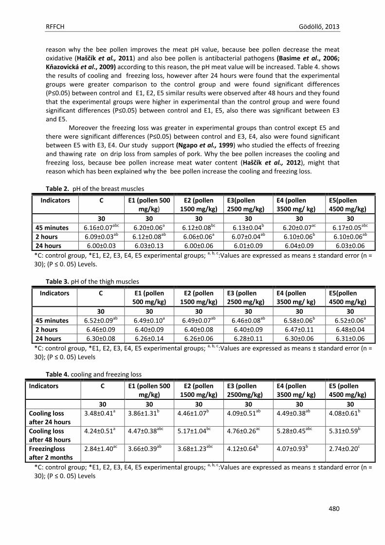
RFFCH Gödöllő, 2013
480
reason why the bee pollen improves the meat pH value, because bee pollen decrease the meat oxidative (Haščík et al., 2011) and also bee pollen is antibacterial pathogens (Basime et al., 2006; Kňazovická et al., 2009) according to this reason, the pH meat value will be increased. Table 4. shows the results of cooling and freezing loss, however after 24 hours were found that the experimental groups were greater comparison to the control group and were found significant differences (P≤0.05) between control and E1, E2, E5 similar results were observed after 48 hours and they found that the experimental groups were higher in experimental than the control group and were found significant differences (P≤0.05) between control and E1, E5, also there was significant between E3 and E5.
Moreover the freezing loss was greater in experimental groups than control except E5 and there were significant differences (P≤0.05) between control and E3, E4, also were found significant between E5 with E3, E4. Our study support (Ngapo et al., 1999) who studied the effects of freezing and thawing rate on drip loss from samples of pork. Why the bee pollen increases the cooling and freezing loss, because bee pollen increase meat water content (Haščík et al., 2012), might that reason which has been explained why the bee pollen increase the cooling and freezing loss. Table 2. pH of the breast muscles
Indicators C E1 (pollen 500 mg/kg)
E2 (pollen 1500 mg/kg)
E3(pollen 2500 mg/kg)
E4 (pollen 3500 mg/ kg)
E5(pollen 4500 mg/kg)
30 30 30 30 30 30
45 minutes 6.16±0.07abc 6.20±0.06a 6.12±0.08bc 6.13±0.04b 6.20±0.07ac 6.17±0.05abc
2 hours 6.09±0.03ab 6.12±0.08ab 6.06±0.06a 6.07±0.04ab 6.10±0.06b 6.10±0.06ab
24 hours 6.00±0.03 6.03±0.13 6.00±0.06 6.01±0.09 6.04±0.09 6.03±0.06
*C: control group, *E1, E2, E3, E4, E5 experimental groups; a, b, c:Values are expressed as means ± standard error (n = 30); (P ≤ 0. 05) Levels.
Table 3. pH of the thigh muscles
Indicators C E1 (pollen 500 mg/kg)
E2 (pollen 1500 mg/kg)
E3 (pollen 2500 mg/kg)
E4 (pollen 3500 mg/ kg)
E5(pollen 4500 mg/kg)
30 30 30 30 30 30
45 minutes 6.52±0.09ab 6.49±0.10a 6.49±0.07ab 6.46±0.08ab 6.58±0.06b 6.52±0.06a
2 hours 6.46±0.09 6.40±0.09 6.40±0.08 6.40±0.09 6.47±0.11 6.48±0.04
24 hours 6.30±0.08 6.26±0.14 6.26±0.06 6.28±0.11 6.30±0.06 6.31±0.06
*C: control group, *E1, E2, E3, E4, E5 experimental groups; a, b, c:Values are expressed as means ± standard error (n = 30); (P ≤ 0. 05) Levels
Table 4. cooling and freezing loss
Indicators C E1 (pollen 500 mg/kg)
E2 (pollen 1500 mg/kg)
E3 (pollen 2500mg/kg)
E4 (pollen 3500 mg/ kg)
E5 (pollen 4500 mg/kg)
30 30 30 30 30 30
Cooling loss after 24 hours
3.48±0.41a 3.86±1.31b 4.46±1.07b 4.09±0.51ab 4.49±0.38ab 4.08±0.61b
Cooling loss after 48 hours
4.24±0.51a 4.47±0.38abc 5.17±1.04bc 4.76±0.26ac 5.28±0.45abc 5.31±0.59b
Freezingloss after 2 months
2.84±1.40ac 3.66±0.39ab 3.68±1.23abc 4.12±0.64b 4.07±0.93b 2.74±0.20c
*C: control group; *E1, E2, E3, E4, E5 experimental groups; a, b, c:Values are expressed as means ± standard error (n = 30); (P ≤ 0. 05) Levels
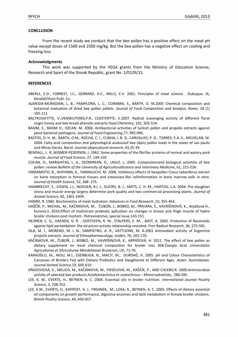
RFFCH Gödöllő, 2013
481
CONCLUSION
From the recent study we conduct that the bee pollen has a positive effect on the meat pH value except doses of 1500 and 2500 mg/kg. But the bee pollen has a negative effect on cooling and freezing loss.
Acknowledgments This work was supported by the VEGA grants from the Ministry of Education Science,
Research and Sport of the Slovak Republic, grant No. 1/0129/13. REFERENCES ABERLE, E.D., FORREST, J.C., GERRARD, D.E., MILLS, E.V. 2001. Principles of meat science . Dubuque. IA,
Kendall/Hunt Publ. Co. ALMEIDA-MURADIAN, L. B., PAMPLONA, L. C., COIMBRA, S., BARTH, O. M.2005 Chemical composition and
botanical evaluation of dried bee pollen pellets. Journal of Food Composition and Analysis, Rome, 18 (1) 105-111.
BALTRUSAITYTE, V.,VENSKUTONIS,P.R., CEKSTERYTE, V.2007. Radical scavenging activity of different floral origin honey and bee bread phenolic extracts.Food Chemistry, 101, 502-514.
BASIM, E., BASIM H., OZCAN. M. 2006. Antibacterial activities of turkish pollen and propolis extracts against plant bacterial pathogens. Journal of Food Engineering,77, 992-996.
BASTOS, D. H. M., BARTH, O.M., ROCHA, C. I., CUNHA, I. B. D., CARVALHO, P. D., TORRES, E.A. S., MICHELAN, M. 2004. Fatty acid composition and palynological analusisof bee (Apis) pollen loads in the states of sao paulo and Minas Gerais, Barzil. Journal ofapicultural research, 43,35-39.
BENDALL, J. R.,WISMER-PEDERSEN, J. 1962. Some properties of the fibrillar proteins of normal and watery pork muscle. Journal of Food Science, 27, 144-159.
COCAN, O., MARGHITAS, L. A., DEZMIREAN, D., LASLO, L. 2005. Compositionand biological activities of bee pollen: review.Bulletin of the University of AgriculturalScience and Veterinary Medicine, 61, 221-226.
HAMAMOTO, R., ISHIYAMA, K., YAMAGUCHI, M. 2006. Inhibitory effects of beepollen Cistus ladaniferus extract on bone resorption in femoral tissues and osteoclast-like cellmformation in bone marrow cells in vitro. Journal of Health Science, 52, 268- 275.
HAMBRECHT, E., EISSEN, J.J., NOOIJEN, R.I. J., DUCRO, B. J., SMITS, C. H. M., HARTOG, L.A. 2004. Pre-slaughter stress and muscle energy largery determine pork quality and two commercial processing plants. Journal of Animal Science, 82, 1401-1409.
HAMM, R. 1960. Biochemistry of meat hydration. Advances in Food Research, 10, 355-463. HAŠČÍK, P., MICHAL, M., KAČÁNIOVÁ, M., ČUBOŇ, J., BOBKO, M., PRÍVARA, Š., VAVRIŠINOVÁ, K., Arpášová H.,
Kunová.S. 2010.Effect of multistrain probiotic aplication on changes in breast and thigh muscle of hybro broiler chickens post mortem. Potravinárstvo, special issue,143-151.
HEJINEN, C. G., HAENEN, G. R. , OOSTVEEN, R. M., STALPERS, E. M. , BAST, A. 2002. Protective of flavonoids against lipid peroxidation: the structure activity relationship revisited. Free Radical Research, 36, 575-581.
ISLA, M. I., MORENO, M. I. N., SAMPIETRO, A. R., VATTUONE, M. A.2001 Antioxidant activity of Argentine propolis extracts. Journal of Ethnopharmacology, Leiden, 76, 165-170.
KAČÁNIOVÁ, M., ČUBOŇ, J., BOBKO, M., VAVRIŠINOVÁ, K., ARPÁŠOVÁ, H. 2011. The effect of bee pollen as dietary supplement on meat chemical composition for broiler ross 308.Časopis Acta Universitatis Agriculturae et Silviculturae Mendelianae Brunensis, LXI, 71-76.
KARAOĞLU, M., AKSU, M.İ., ESENBUGA, N., MACIT, M., DURDAĞ, H. 2005. pH and Colour Characteristics of Carcasses of Broilers Fed with Dietary Probiotics and Slaughtered at Different Ages. Asian- Australasian. Journal Animal Science,19, 605-610.
KŇAZOVICKÁ, V., MELICH, M., KAČÁNIOVÁ, M., FIKSELOVÁ, M., HAŠČÍK, P., AND CHLEBO.R. 2009.Antimicrobial activity of selected bee products.Actafytotechnica et zootechnica – Mimoriadnečíslo, 280-285.
LEE, K. W., EVERTS, H., BEYNEN, A. C. 2004. Essential oils in broiler nutrition. International Journal Poultry Science, 3, 738-752.
LEE, K.W., EVERTS, H., KAPPERT, H. J., FREHNER, M., LOSA, R., BEYNEN, A. C. 2003. Effects of dietary essential oil components on growth performance, digestive enzymes and lipid metabolism in female broiler chickens. British Poultry Science, 44, 450-457.
RFFCH Gödöllő, 2013
482
MĂRGHITAS, L.A., STANCIU, O.G., DEZMIREAN, D.S., BOBIS, O., POPESCU, O., BOGDANOV, S., CAMPOS, M.G. 2009. In vitro antioxidant capacity of honey bee collected pollen of selected floral origin harvested from Romania. Food Chemistry, 115, 878-883.
MOREIRA, L., DIAS, L. G., PEREIRA,J. A., ESTEVINHO, L. 2008. Antioxidant properties, total phenols and pollen analysis of propolis samples from Portugal.Food Chemistry and Toxicology, 46, 3482-3485.
MUCHENJE, V., DZAMA, K., CHIMONYO, M., STRYDOM, P. E., RAATS, J. G. 2009. Relationship between pre-slaughter stress responsiveness and beef quality in three cattle breeds. Meat Science, 81,653-657.
MUCHENJE, V., DZAMA, K., CHIMONYO, M., STRYDOM, P.E., HUGO, A., RAATS, J.G. 2008. Sensory evaluation and its relationship to quality attributes of beef from Nguni and Bonsmara steers raised on natural pasture. Animal, 2, 1700-1706.
NGAPOA, T.M., BABAREB,I.H.REYNOLDSB, J.,MAWSONB.R.F. 1999. Freezing and thawing rate effects on drip loss from samples of pork. Meat Science, 53, (3) 149-158.
SERRA BONVEHI, J., ESCOLA, JORDA, R. 1997. Nutrient composition and The microbiological quality of honey bee collected pollen in Spain.Journal of Agriculture and Food Chemistry, 45, 725-732.
SERRA BONVEHÍ, J., SOLIVA TORRENTÓ, M., CENTELLES LORENTE, E., 2001 Evaluation of polyphenolic and flavonoidcompounds in honeybee-collected pollen produced in Spain. Journal of Agricultural and Food Chemistry, Washington, 49,1848-1853.
SHALMANY, K.S., SHIVAZAD, M. 2006. The Effect of Diet Propolis Supplementation on Ross Broiler Chicks Performance. International Journal of Poultry Science, 5, 84-88.
SILVA, T.M.S., CAMARA, C. A., LINS, A.C.S., BARBOSA-FILHO, J. M., SILVA,E.M.S., FREITAS, B.M., SANTOS, F., DE A.R. 2006. Chemical composition and free radical scavenging activity of pollen loads from stingless bee Melipona subnitida duck. InJournal of Food Composition and Analysis, 19, 507-511.
STANCIU, O. G., MARGHITAS,L. A., DEZMIREAN, D.2009. Macro- and oligo-mineral elements from honeybee-collected pollen and beebread harvested from Transylvania (Romania). Bulletin UASVM Animal Science and Biotechnology, 66, 1-2.
ŠULCEROVÁ, H., MIHOK, M., JŮZL M. HAŠČÍK. P. 2011. Effect of addition of pollen and propolis to feeding mixture during the production of broiler chickens Ross 308 to the colour for thigh and breast muscles and pH determination. Časopis Acta Universitatis Agriculturae et Silviculturae Mendelianae Brunensis, 44, 359-366.
SWATLAND, H. J. 2004. Progress in understanding the paleness of meat with a low pH. South African Journal of Animal Science, 34,1-4.
TÜYLÜ, A. S., SORKUN, K. 2004. Organoleptic analysis of economically significant pollen samples collected in bursa region by Apis mellifera L. Mellifera, 4, 38-44.
VILLANUEVA, M. T. O., MARQUINA, A. D., SERRANO, R. B., ABELLÁN, G. B.2002. The importance of bee-collected pollen in the diet: a study of its composition. International Journal of Food Sciences and Nutrition, 53, 217-224.
YAMAGUCHI, M., HAMAMOTO, R., UCHIYAMA, S., ISHIYAMA, K., HASHIMOTO, K. 2006. Anabolic effects of bee pollen Cistus ladaniferus extract on bonecomponents in the femoral diaphyseal and metaphyseal tissues of rats in vitro and in vivo. Journal of Health Science, 52, 2006, 43-49.

Animal welfare, etológia és tartástechnológia
Animal welfare, ethology and housing systems
Volume 9 Issue 3
Különszám/Special Issue
Gödöllő
2013
RFFCH Gödöllő, 2013
483
THE EFFECT OF THE PROPOLIS EXTRACT ON BROILER HUBBARD JV INTERAL FAT Peter Haščík1, Jozef Garlík1, Ibrahim Omer Elimam Elamin1, Miroslava Kačániová2, Vladimíra Kňazovická1
1. Department of Animal Products Evaluation and Processing, Faculty of Biotechnology and Food Sciences, Slovak University of Agriculture in Nitra, Tr. A. Hlinku 2, 949 76 Nitra, Slovakia 2. Department of Microbiology, Faculty of Biotechnology and Food Sciences, Slovak University of Agriculture in Nitra, Tr. A. Hlinku 2, 949 76 Nitra, Slovakia
ABSTRACT The aim of the experiment was to evaluate the influence of feed mixtures with propolis extract on weight and fat creation in carcass body of chickens from Hubbard JV hybrid combination. Propolis extract was applied into the feed mixtures in amount of 150 mg kg-1 (1st experimental group), 450 mg kg-1 (2nd experimental group), 600 mg kg-1 (3rd experimental group) and 800 mg kg-1 (4th experimental group). Fattening was lasted for 42 days on deep litter (sawdust). Into the experiment, 500 pieces of 1-day chickens were included and then 5 groups of 100 pieces were created: control group – C (without propolis extract application) and 4 experimental groups – E1, E2, E3 and E4 (with a dose of propolis extract). Higher weight of carcass body (P ≥ 0.05) was found in E1 and E2 groups (1022.37 g - E1 and 1019.75 g - E2) compared with C group (1005.12 g) and lower weight of carcass body (P ≥ 0.05) was found in E3 and E4 group (927.37 and 916.75) compared with control group. Significant differences (P ≤ 0.05) were found between the groups E1 and E4, E2 and E4, E1 and E3 and between the groups E2 and E3. The lowest content of abdominal fat was recorded in control group and in E4 group (0.73%). In experimental groups, values of abdominal fat content were decreased by gradual increasing of propolis extract dose in Hubbard JV chicken nutrition (1.03% - E1, 0.99% - E2, 0.80% - E3 and 0.73% - E4). Gizzard fat from carcass body was value-balanced and ranged from 0.29% (E1) to 0.44% (E3). Heart fat was from 0.047% (P4) to 0.068% (P1).Internal fats were considered together from carcass body and the value ranged from 1.17% (P4) to 1.43% (P2). In control group, internal fat was 1.21%. Content of internal fat from carcass body as well as abdominal, gizzard and heart fat contents showed no significant differences (P ≥ 0.05) between the groups. We found that propolis extract positively influenced the carcass body weight only in E1 and E2 groups. In these groups, no distinctive impact on fat creation was detected. Propolis in the doses of 150 mg kg-1 and 450 mg kg-1 can be recommended to application in nutrition of Hubbard JV chickens. Higher concentration of propolis extract (600 mg kg-1 and 800 mg kg-1) may result in decrease of carcass body weight. Keywords: chickens Hubbard JV, propolis extract, fat INTRODUCTION Consumption of poultry meat has had a growing trend in Slovakia, it was approximately of 19.9 kg per head in 2007 (Nagy, 2009). Poultry is characteristic for the best conversion of nutrients into the meat and therefore production costs as well as prices of poultry products on the world market are relatively low compared with the other animal products. Effort is to breed more efficient hybrids, to improve the conversion of nutrients and to shorten the fattening period (Marcinčák et al., 2008). Primary importance of fat in human nutrition is presence of polyunsaturated fatty acids, which have an essential role in organism as precursors for variety of biologically active substances. Broiler chickens have fat content of 5-7%, of which approximately 30% are saturated fatty acids and 70 % are unsaturated fatty acids (Halaj and Golian, 2000). Poultry fat contains a higher amount of polyunsaturated fatty acids compared with the other carcass animals. It is caused by relatively high content of phospholipids in membrane structures of muscle cells (Bystrický and Dičáková, 1998). The complete feed mixtures are used for nutrition of broiler chickens. Over the last few years, feed mixtures have been enriched by probiotic preparations or by other available additions,
RFFCH Gödöllő, 2013
484
which are added into the feed mixtures as a possible substitution for excluded animal meals, antibiotic preparations and coccidiostats, what is in accordance with new legislation determined by laws and regulations of European Union and for the purpose of health state improvement, mortality reducing and the better utilization of feed nutrients as are described in papers by Fuller (1992), Nahashon et al. (1992), Kumprecht and Zobač (1998), Haščík et al. (2004, 2006, 2007) and Bobko et al.(2009). Integration of new component in animal nutrition has to retain technological, nutritional and sensorial properties of meat without their deterioration (Aleson-Carbonell et al., 2004; P´erez-Alvarez, 2006). As alternative substitutions in nutrition, bee products (pollen, propolis or their extracts) may be employed, because they can have positive influence on health state, economic use of feed and quality of product (Prytzyk et al., 2003; Wang et al., 2004; Haščík et al., 2005a, b, 2007; Angelovičová et al., 2006, 2008; Shalmany a Shivazad, 2006; Seven et al., 2008 and others.). Several plant supplements contain substances, which increase the gluttony and digestion (Barreto et al., 2008). Some researchers showed these substances may stimulate a natural immunity of poultry and may decrease the pathogen microorganisms’ activity (Cross, 2002; Dalloul et al., 2003; Kačániová et al., 2011). The contemporary basic requirements in poultry nutrition are to respect the poultry physiological demands on consumption of energy and nutrients, to keep a good animal health and a high yield (Suchý and Straková, 2004). Haščík et al. (2005a) stated that intensive selection and efficacy of feed mixtures used in chicken nutrition for the last decades shorten the fattening period to less than half, but intensive growth of chickens caused an increased fat deposition, mainly in abdominal cavity in form of abdominal fat. According to Simoneová (1999), large part of subcutaneous fat, fat placed in abdominal cavity and in intestinal region belong to the total body fat. Excessive accumulation of broiler abdominal fat is a result of imbalance between the energy intake and output, while amount of abdominal fat decreases with reduced energy consumption (Skřivan, 2000). Hood (1984) and Fischer et al. (1990) found that energy intake reduction in food mixture by 20-25% had an influence on substantial decrease and reduction of body fat. Lipid content in poultry meat depends on various factors as e. g. animal species, breed, gender, origin, and muscle anatomy (Benková et al., 2005). Fat is important in term of meat sensorial properties for consumer, because fat is a source of aromatic substances influencing mainly the taste (Pipek, 2000). Fatty compounds are considered as a taste medium, and only 1-2% fat from total animal weight is sufficient for this purpose (Winkelmayer et al., 2005). Meat of mammals and birds are different and birds’ meat, with certain exceptions, is culinary prepared and consumed with skin, subcutaneous connective tissue, together with blood vessels and nerves, which are in meat (Uhrín et al., 1993). All these facts influence substantially the taste properties of poultry meat. But certain negatives occur; it is primarily the increase of energy value, which is caused by subcutaneous fat connective tissue and by lipids of the skin. Fat in certain amount is an important part of meat. An excessive fat tissue formation is considered as an undesired consequence of modern broiler type selection for increased growth. Fat accumulated in broiler carcass bodies is a waste product for consumer; nutritional value of food increases unacceptably following with possible risk to human health and carcass yield is artificially increased (Mahmouda and Mihaly, 1998). According to Ochrimenko et al. (1997), chemical composition of broiler chickens fat causes an unpleasant smell and fatty acids placed in the fat negatively influenced the taste of broiler chickens. Also based on these factors, intestinal, heart, gizzard and abdominal fat represent certain economic losses, because of carcass yield decrease. Increased fat deposition in broiler bodies is not desirable for producer and for consumers, too (Cherian and Wolfe, 1996; Lichtenstein, 1999). Following the literature sources and new trends in poultry nutrition, the aim of the experiment was to verify the influence of propolis extract applied in various concentrations on carcass body weight and on proportion of internal fats in carcass body of Hubbard JV chickens. MATERIAL AND METHODOLOGY
Experiment was performed in poultry test station Zámostie Company. Tested animals were broiler chickens of Hubbard JV hybrid combination. Five hundreds of one-day old chickens were
RFFCH Gödöllő, 2013
485
included in the experiment and five groups of one hundred pieces per each were created as follows: control group – C (without propolis extract application) and four experimental groups – E1, E2, E3 and E4 (with certain dose of propolis extract). Chicken fattening lasted forty two days. Chickens were bred on deep litter (sawdust). Feed mixture was served using tubular feeders. Feed mixtures were mixed and prepared according to Gazette of Ministry of Agriculture and Rural Development (original name: Vestník MP SR) (2004) in Biofeed Company located in Kolárovo. Feed mixtures were analysed in term of basic nutrients and energy value at the Department of Animal Nutrition (Faculty of Agrobiology and Food Resources, Slovak University of Agriculture in Nitra). Feed mixture was manually served in periodic intervals each day and chickens were fed by the system ad libitum. All evaluated groups were fed by starter feed mixture HYD-01 (powder form) to 21st day of age and then, from 22nd day to 42nd day of age, all animals were fed by feed mixture HYD-02 (powder form). Feed mixtures HYD-01 and HYD-02 were produced without antibiotic preparations and coccidiostats. Nutritional value of the feed mixtures was uniform in each group during the experiment, but in experimental groups, propolis extract was added to both feed mixtures at a dose of 150 mg kg-1 (E1), 450 mg kg-1 (E2), 600 mg kg-1 (E3) and 800 mg kg-1 (E4). Propolis originated in Slovak Republic. Propolis extract was prepared from milled propolis and was mixed with 80% ethanol (Krell, 1996). Propolis solution was extracted in water bath at 80 °C for 1 hour under the reflux. Solution was centrifuged after extracting and cooling. Obtained supernatant was evaporated at 40-50 ° C using rotary vacuum evaporator and water bath. Then the residue was weighted. The evaporation of residues at dose of 15 g (E1), 45 g (E2), 60 g (E3) and 80 g (E4) were separately dissolved in 1000 cm3 of 80% ethanol and applied into the 100 kg of each feed mixture intended for evaluated group of Hubbard JV chickens. Water was available ad libitum by self-powered system using nipple drinkers with drip tray. At the end of fattening (42nd day), 60 pieces (30 male pieces and 30 female pieces) from each group were selected for carcass analysis, which was performed at Department of Animal Products Evaluation and Processing (Faculty of Biotechnology and Food Sciences, Slovak University of Agriculture in Nitra). We evaluated the carcass body weight and proportion of fat tissue from carcass body weight of Hubbard JV chickens (proportion of abdominal, gizzard, heart and total internal fat). Weights were measured on analytical scales of KERN 440-33N type with the accuracy of 0.01 g and calculated. Results of the experiment (arithmetic average, standard deviation) were statistically assessed using programme Statgraphics Plus version 5.1 (AV Trading, Umex, Dresden, Germany). Analysis of variance followed by Duncan test was used to determine significance of differences between the groups. RESULTS AND DISCUSSION Obtained values of carcass body weight, abdominal, gizzard, heart and total internal fat expressed as percentages from carcass body of Hubbard JV hybrid combination chickens are recorded in the tab 1.
Higher carcass body weight (P ≥ 0.05) was found in the 1st and 2nd experimental group (E1 – 1022.37 g, E2 – 1019.75 g) compared with the control group (1005.12 g). Lower carcass body weight (P ≥ 0.05) was detected in the 3rd and 4th experimental group (E3 - 927.37 g, E4 – 916.75 g) compared with control group. Significant difference (P ≤ 0.05) in carcass body weight was detected between the 1st and 4th experimental group, between the 2nd and 4th experimental group, between the 1st and 3rd experimental group and between the 2nd and 3rd experimental group. Carcass body weights detected in this experiment are higher than results of Seven et al. (2008) and are lower compared with results of Haščík et al. (2010, 2012), who used a probiotic preparation and pollen extracts in nutrition of Ross 308 chickens.
The lowest content of abdominal fat in carcass body was detected in control and 4th experimental group (0.73%). The values of abdominal fat in carcass body decreased in experimental groups by gradually increasing doses of propolis (E1 – 1.03%, E2 – 0.99%, E3 – 0.80%, E4 – 0.73%) without significant differences (P ≥ 0.05) between the groups. Angelovičová (1997) tested a reduced level of metabolisable energy in feed mixtures and found comparable results of abdominal fat in carcass body, in numbers below 1.00%. Results did not confirm a tendency of abdominal fat
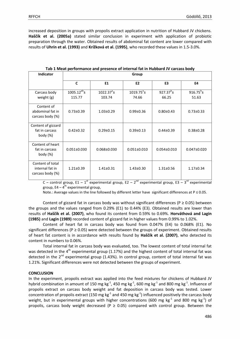
RFFCH Gödöllő, 2013
486
increased deposition in groups with propolis extract application in nutrition of Hubbard JV chickens. Haščík et al. (2005a) stated similar conclusion in experiment with application of probiotic preparation through the water. Obtained results of abdominal fat content are lower compared with results of Uhrín et al. (1993) and Krížková et al. (1995), who recorded these values in 1.5-3.0%.
Tab 1 Meat performance and presence of internal fat in Hubbard JV carcass body Indicator Group
C
E1
E2
E3
E4
Carcass body weight (g)
1005.12ab
± 115.77
1022.37a±
103.74 1019.75
a±
74.66 927.37
b±
66.25 916.75
b±
51.63
Content of abdominal fat in carcass body (%)
0.73±0.39 1.03±0.29 0.99±0.36 0.80±0.43 0.73±0.33
Content of gizzard fat in carcass
body (%) 0.42±0.32 0.29±0.15 0.39±0.13 0.44±0.39 0.38±0.28
Content of heart fat in carcass
body (%) 0.051±0.030 0.068±0.030 0.051±0.010 0.054±0.010 0.047±0.020
Content of total internal fat in
carcass body (%) 1.21±0.39 1.41±0.31 1.43±0.30 1.31±0.56 1.17±0.34
C – control group, E1 – 1st
experimental group, E2 – 2nd
experimental group, E3 – 3rd
experimental group, E4 – 4
th experimental group,
Note.: Average values in the line followed by different letter have significant differences at P ≤ 0.05.
Content of gizzard fat in carcass body was without significant differences (P ≥ 0.05) between
the groups and the values ranged from 0.29% (E1) to 0.44% (E3). Obtained results are lower than results of Haščík et al. (2007), who found its content from 0.59% to 0.69%. Horváthová and Lagin (1985) and Lagin (1989) recorded content of gizzard fat in higher values from 0.99% to 1.02%.
Content of heart fat in carcass body was found from 0.047% (E4) to 0.068% (E1). No significant differences (P ≥ 0.05) were detected between the groups of experiment. Obtained results of heart fat content is in accordance with results found by Haščík et al. (2007), who detected its content in numbers to 0.06%.
Total internal fat in carcass body was evaluated, too. The lowest content of total internal fat was detected in the 4th experimental group (1.17%) and the highest content of total internal fat was detected in the 2nd experimental group (1.43%). In control group, content of total internal fat was 1.21%. Significant differences were not detected between the groups of experiment. CONCLUSION In the experiment, propolis extract was applied into the feed mixtures for chickens of Hubbard JV hybrid combination in amount of 150 mg kg-1, 450 mg kg-1, 600 mg kg-1 and 800 mg kg-1. Influence of propolis extract on carcass body weight and fat deposition in carcass body was tested. Lower concentration of propolis extract (150 mg kg-1 and 450 mg kg-1) influenced positively the carcass body weight, but in experimental groups with higher concentrations (600 mg kg-1 and 800 mg kg-1) of propolis, carcass body weight decreased (P ≥ 0.05) compared with control group. Between the
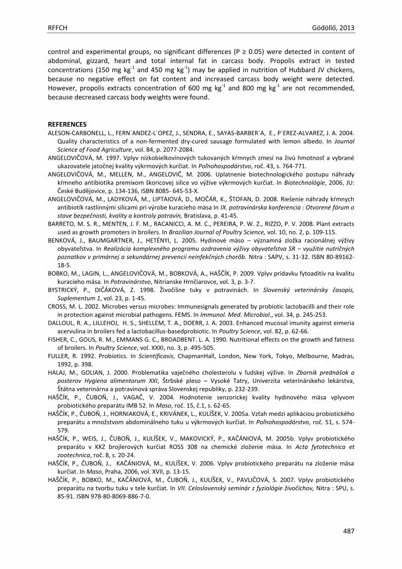
RFFCH Gödöllő, 2013
487
control and experimental groups, no significant differences (P ≥ 0.05) were detected in content of abdominal, gizzard, heart and total internal fat in carcass body. Propolis extract in tested concentrations (150 mg kg-1 and 450 mg kg-1) may be applied in nutrition of Hubbard JV chickens, because no negative effect on fat content and increased carcass body weight were detected. However, propolis extracts concentration of 600 mg kg-1 and 800 mg kg-1 are not recommended, because decreased carcass body weights were found. REFERENCES ALESON-CARBONELL, L., FERN´ANDEZ-L´OPEZ, J., SENDRA, E., SAYAS-BARBER´A, E., P´EREZ-ALVAREZ, J. A. 2004.
Quality characteristics of a non-fermented dry-cured sausage formulated with lemon albedo. In Journal Science of Food Agriculture, vol. 84, p. 2077-2084.
ANGELOVIČOVÁ, M. 1997. Vplyv nízkobielkovinových tukovaných kŕmnych zmesí na živú hmotnosť a vybrané ukazovatele jatočnej kvality výkrmových kurčiat. In Poľnohospodárstvo, roč. 43, s. 764-771.
ANGELOVIČOVÁ, M., MELLEN, M., ANGELOVIČ, M. 2006. Uplatnenie biotechnologického postupu náhrady kŕmneho antibiotika premixom škoricovej silice vo výžive výkrmových kurčiat. In Biotechnológie, 2006, JU: České Budĕjovice, p. 134-136, ISBN 8085- 645-53-X.
ANGELOVIČOVÁ, M., LADYKOVÁ, M., LIPTAIOVÁ, D., MOČÁR, K., ŠTOFAN, D. 2008. Riešenie náhrady kŕmnych antibiotík rastlinnými silicami pri výrobe kuracieho mäsa In IX. potravinárska konferencia : Otvorené fórum o stave bezpečnosti, kvality a kontroly potravín, Bratislava, p. 41-45.
BARRETO, M. S. R., MENTEN, J. F. M., RACANICCI, A. M. C., PEREIRA, P. W. Z., RIZZO, P. V. 2008. Plant extracts used as growth promoters in broilers. In Brazilian Journal of Poultry Science, vol. 10, no. 2, p. 109-115.
BENKOVÁ, J., BAUMGARTNER, J., HETÉNYI, L. 2005. Hydinové mäso – významná zložka racionálnej výživy obyvateľstva. In Realizácia komplexného programu ozdravenia výživy obyvateľstva SR – využitie nutričných poznatkov v primárnej a sekundárnej prevencii neinfekčných chorôb. Nitra : SAPV, s. 31-32. ISBN 80-89162-18-5.
BOBKO, M., LAGIN, L., ANGELOVIČOVÁ, M., BOBKOVÁ, A., HAŠČÍK, P. 2009. Vplyv prídavku fytoaditív na kvalitu kuracieho mäsa. In Potravinárstvo, Nitrianske Hrnčiarovce, vol. 3, p. 3-7.
BYSTRICKÝ, P., DIČÁKOVÁ, Z. 1998. Živočíšne tuky v potravinách. In Slovenský veterinársky časopis, Suplementum 1, vol. 23, p. 1-45.
CROSS, M. L. 2002. Microbes versus microbes: Immunesignals generated by probiotic lactobacilli and their role in protection against microbial pathogens. FEMS. In Immunol. Med. Microbiol., vol. 34, p. 245-253.
DALLOUL, R. A., LILLEHOJ, H. S., SHELLEM, T. A., DOERR, J. A. 2003. Enhanced mucosal imunity against eimeria acervulina in broilers fed a lactobacillus-basedprobiotic. In Poultry Science, vol. 82, p. 62-66.
FISHER, C., GOUS, R. M., EMMANS G. C., BROADBENT. L. A. 1990. Nutritional effects on the growth and fatness of broilers. In Poultry Science, vol. XXXI, no. 3, p. 495-505.
FULLER, R. 1992. Probiotics. In Scientificasis, ChapmanHall, London, New York, Tokyo, Melbourne, Madras, 1992, p. 398.
HALAJ, M., GOLIAN, J. 2000. Problematika vaječného cholesterolu v ľudskej výžive. In Zborník prednášok a posterov Hygiena alimentorum XXI, Štrbské pleso – Vysoké Tatry, Univerzita veterinárskeho lekárstva, Štátna veterinárna a potravinová správa Slovenskej republiky, p. 232-239.
HAŠČÍK, P., ČUBOŇ, J., VAGAČ, V. 2004. Hodnotenie senzorickej kvality hydinového mäsa vplyvom probiotického preparátu IMB 52. In Maso, roč. 15, č.1, s. 62-65.
HAŠČÍK, P., ČUBOŇ, J., HORNIAKOVÁ, E., KRIVÁNEK, L., KULÍŠEK, V. 2005a. Vzťah medzi aplikáciou probiotického preparátu a množstvom abdominálneho tuku u výkrmových kurčiat. In Poľnohospodárstvo, roč. 51, s. 574-579.
HAŠČÍK, P., WEIS, J., ČUBOŇ, J., KULÍŠEK, V., MAKOVICKÝ, P., KAČÁNIOVÁ, M. 2005b. Vplyv probiotického preparátu v KKZ brojlerových kurčiat ROSS 308 na chemické zloženie mäsa. In Acta fytotechnica et zootechnica, roč. 8, s. 20-24.
HAŠČÍK, P., ČUBOŇ, J., KAČÁNIOVÁ, M., KULÍŠEK, V. 2006. Vplyv probiotického preparátu na zloženie mäsa kurčiat. In Maso, Praha, 2006, vol. XVII, p. 13-15.
HAŠČÍK, P., BOBKO, M., KAČÁNIOVÁ, M., ČUBOŇ, J., KULÍŠEK, V., PAVLIČOVÁ, S. 2007. Vplyv probiotického preparátu na tvorbu tuku v tele kurčiat. In VII. Celoslovenský seminár z fyziológie živočíchov, Nitra : SPU, s. 85-91. ISBN 978-80-8069-886-7-0.
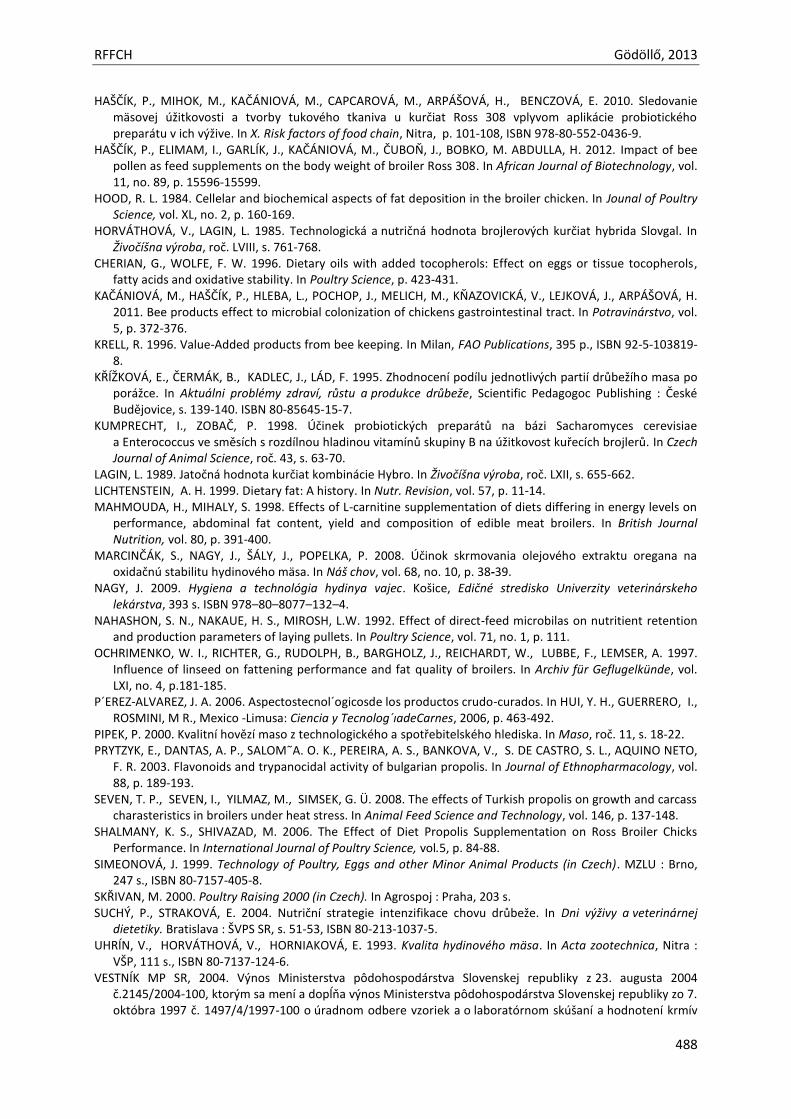
RFFCH Gödöllő, 2013
488
HAŠČÍK, P., MIHOK, M., KAČÁNIOVÁ, M., CAPCAROVÁ, M., ARPÁŠOVÁ, H., BENCZOVÁ, E. 2010. Sledovanie mäsovej úžitkovosti a tvorby tukového tkaniva u kurčiat Ross 308 vplyvom aplikácie probiotického preparátu v ich výžive. In X. Risk factors of food chain, Nitra, p. 101-108, ISBN 978-80-552-0436-9.
HAŠČÍK, P., ELIMAM, I., GARLÍK, J., KAČÁNIOVÁ, M., ČUBOŇ, J., BOBKO, M. ABDULLA, H. 2012. Impact of bee pollen as feed supplements on the body weight of broiler Ross 308. In African Journal of Biotechnology, vol. 11, no. 89, p. 15596-15599.
HOOD, R. L. 1984. Cellelar and biochemical aspects of fat deposition in the broiler chicken. In Jounal of Poultry Science, vol. XL, no. 2, p. 160-169.
HORVÁTHOVÁ, V., LAGIN, L. 1985. Technologická a nutričná hodnota brojlerových kurčiat hybrida Slovgal. In Živočíšna výroba, roč. LVIII, s. 761-768.
CHERIAN, G., WOLFE, F. W. 1996. Dietary oils with added tocopherols: Effect on eggs or tissue tocopherols, fatty acids and oxidative stability. In Poultry Science, p. 423-431.
KAČÁNIOVÁ, M., HAŠČÍK, P., HLEBA, L., POCHOP, J., MELICH, M., KŇAZOVICKÁ, V., LEJKOVÁ, J., ARPÁŠOVÁ, H. 2011. Bee products effect to microbial colonization of chickens gastrointestinal tract. In Potravinárstvo, vol. 5, p. 372-376.
KRELL, R. 1996. Value-Added products from bee keeping. In Milan, FAO Publications, 395 p., ISBN 92-5-103819-8.
KŘÍŽKOVÁ, E., ČERMÁK, B., KADLEC, J., LÁD, F. 1995. Zhodnocení podílu jednotlivých partií drůbežího masa po porážce. In Aktuálni problémy zdraví, růstu a produkce drůbeže, Scientific Pedagogoc Publishing : České Budějovice, s. 139-140. ISBN 80-85645-15-7.
KUMPRECHT, I., ZOBAČ, P. 1998. Účinek probiotických preparátů na bázi Sacharomyces cerevisiae a Enterococcus ve směsích s rozdílnou hladinou vitamínů skupiny B na úžitkovost kuřecích brojlerů. In Czech Journal of Animal Science, roč. 43, s. 63-70.
LAGIN, L. 1989. Jatočná hodnota kurčiat kombinácie Hybro. In Živočíšna výroba, roč. LXII, s. 655-662. LICHTENSTEIN, A. H. 1999. Dietary fat: A history. In Nutr. Revision, vol. 57, p. 11-14. MAHMOUDA, H., MIHALY, S. 1998. Effects of L-carnitine supplementation of diets differing in energy levels on
performance, abdominal fat content, yield and composition of edible meat broilers. In British Journal Nutrition, vol. 80, p. 391-400.
MARCINČÁK, S., NAGY, J., ŠÁLY, J., POPELKA, P. 2008. Účinok skrmovania olejového extraktu oregana na oxidačnú stabilitu hydinového mäsa. In Náš chov, vol. 68, no. 10, p. 38-39.
NAGY, J. 2009. Hygiena a technológia hydinya vajec. Košice, Edičné stredisko Univerzity veterinárskeho lekárstva, 393 s. ISBN 978–80–8077–132–4.
NAHASHON, S. N., NAKAUE, H. S., MIROSH, L.W. 1992. Effect of direct-feed microbilas on nutritient retention and production parameters of laying pullets. In Poultry Science, vol. 71, no. 1, p. 111.
OCHRIMENKO, W. I., RICHTER, G., RUDOLPH, B., BARGHOLZ, J., REICHARDT, W., LUBBE, F., LEMSER, A. 1997. Influence of linseed on fattening performance and fat quality of broilers. In Archiv für Geflugelkünde, vol. LXI, no. 4, p.181-185.
P´EREZ-ALVAREZ, J. A. 2006. Aspectostecnol´ogicosde los productos crudo-curados. In HUI, Y. H., GUERRERO, I., ROSMINI, M R., Mexico -Limusa: Ciencia y Tecnolog´ıadeCarnes, 2006, p. 463-492.
PIPEK, P. 2000. Kvalitní hovězí maso z technologického a spotřebitelského hlediska. In Maso, roč. 11, s. 18-22. PRYTZYK, E., DANTAS, A. P., SALOM˜A. O. K., PEREIRA, A. S., BANKOVA, V., S. DE CASTRO, S. L., AQUINO NETO,
F. R. 2003. Flavonoids and trypanocidal activity of bulgarian propolis. In Journal of Ethnopharmacology, vol. 88, p. 189-193.
SEVEN, T. P., SEVEN, I., YILMAZ, M., SIMSEK, G. Ü. 2008. The effects of Turkish propolis on growth and carcass charasteristics in broilers under heat stress. In Animal Feed Science and Technology, vol. 146, p. 137-148.
SHALMANY, K. S., SHIVAZAD, M. 2006. The Effect of Diet Propolis Supplementation on Ross Broiler Chicks Performance. In International Journal of Poultry Science, vol.5, p. 84-88.
SIMEONOVÁ, J. 1999. Technology of Poultry, Eggs and other Minor Animal Products (in Czech). MZLU : Brno, 247 s., ISBN 80-7157-405-8.
SKŘIVAN, M. 2000. Poultry Raising 2000 (in Czech). In Agrospoj : Praha, 203 s. SUCHÝ, P., STRAKOVÁ, E. 2004. Nutriční strategie intenzifikace chovu drůbeže. In Dni výživy a veterinárnej
dietetiky. Bratislava : ŠVPS SR, s. 51-53, ISBN 80-213-1037-5. UHRÍN, V., HORVÁTHOVÁ, V., HORNIAKOVÁ, E. 1993. Kvalita hydinového mäsa. In Acta zootechnica, Nitra :
VŠP, 111 s., ISBN 80-7137-124-6. VESTNÍK MP SR, 2004. Výnos Ministerstva pôdohospodárstva Slovenskej republiky z 23. augusta 2004
č.2145/2004-100, ktorým sa mení a dopĺňa výnos Ministerstva pôdohospodárstva Slovenskej republiky zo 7. októbra 1997 č. 1497/4/1997-100 o úradnom odbere vzoriek a o laboratórnom skúšaní a hodnotení krmív
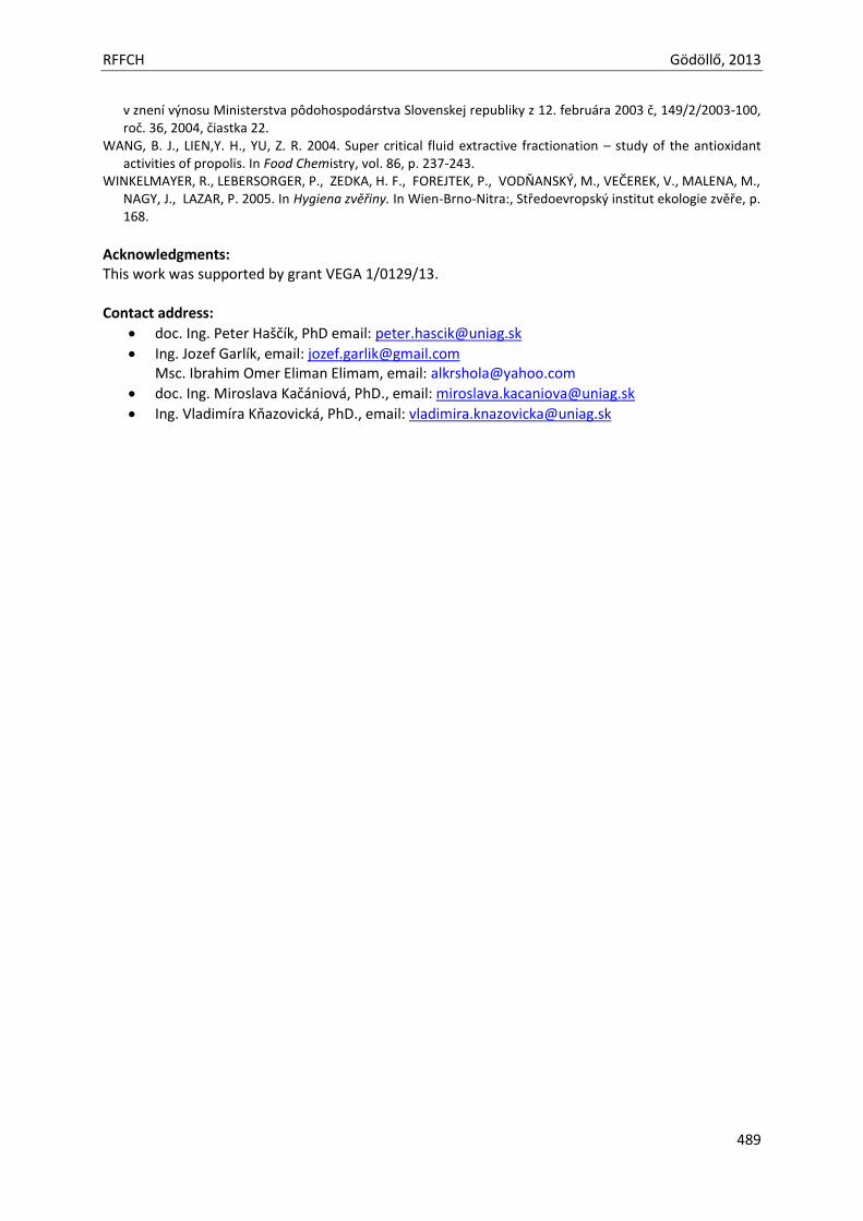
RFFCH Gödöllő, 2013
489
v znení výnosu Ministerstva pôdohospodárstva Slovenskej republiky z 12. februára 2003 č, 149/2/2003-100, roč. 36, 2004, čiastka 22.
WANG, B. J., LIEN,Y. H., YU, Z. R. 2004. Super critical fluid extractive fractionation – study of the antioxidant activities of propolis. In Food Chemistry, vol. 86, p. 237-243.
WINKELMAYER, R., LEBERSORGER, P., ZEDKA, H. F., FOREJTEK, P., VODŇANSKÝ, M., VEČEREK, V., MALENA, M., NAGY, J., LAZAR, P. 2005. In Hygiena zvěřiny. In Wien-Brno-Nitra:, Středoevropský institut ekologie zvěře, p. 168.
Acknowledgments: This work was supported by grant VEGA 1/0129/13. Contact address:
doc. Ing. Peter Haščík, PhD email: [email protected]
Ing. Jozef Garlík, email: [email protected] Msc. Ibrahim Omer Eliman Elimam, email: [email protected]
doc. Ing. Miroslava Kačániová, PhD., email: [email protected]
Ing. Vladimíra Kňazovická, PhD., email: [email protected]

Animal welfare, etológia és tartástechnológia
Animal welfare, ethology and housing systems
Volume 9 Issue 3
Különszám/Special Issue
Gödöllő
2013

RFFCH Gödöllő, 2013
490
XENOBIOTIC - INDUCED PEROXISOMES IN THE RABBIT LIVER HOLOVSKÁ K., ALMÁŠIOVÁ V., CIGÁNKOVÁ V.
Depatrment of Histology and Embryology, University of Veterinary Medicine and Pharmacy, Košice, Slovak Republic
ABSTRACT
The study investigated changes in ultrastructure of the liver in the rabbit (Oryctolagus cuniculus) which was administered bendiocarb for 3 days. Electron microscopy was used to observe changes in ultrastructure of hepatocytes. The changes were focal and the effect on the liver was not uniform. Some hepatocytes showed no obvious changes in their ultrastructure. In the changed hepatocytes, we observed dilatation of bile capillaries but no visible alterations in the intercellular contacts. Many hepatocytes showed considerable increase in the number of peroxisomes. We observed, that bendiocarb induced ultrastructural changes of hepatocytes but only to a moderate extent. INTRODUCTION
The current agricultural practice involves the use of many protective chemical substances which contribute to considerable environmental contamination. These environmental contaminants include also insect control preparations. Carbamate insecticides are widely used for agricultural and indoor purposes. They act as acetylcholinesterase (AChE) inhibitors which affect many organs, such as peripheral and central nervous system, muscles, liver, brain and heart. Bendiocarb is a toxic insecticide. In the body it undergoes hydrolysis to organic metabolites which can also affect the total toxicity of this insecticide. Bendiocarb is rapidly detoxified and excreted (Bendiocarb, 1982). Despite that many scientists observed various morphological changes in different organs. Immunosuppressive effects of bendiocarb were observed after its long-term administration (Mojžišová, 2004). It also induced morphological changes in lymphatic organs. Flešárová et al. (2007) performed morphometric analysis of the thymus exposed to bendiocarb and observed decreased number of cells and reduced volume of its medullar portion. An adverse effect of bendiocarb on the structure of lymph nodes was confirmed by Petrovová et al. (2010) and Špaleková et al. (2010). Almašiová et al. (2012) observed a subchronic influence of bendiocarbamate on histological structure of kidneys which exhibited changes of varying extent in both the cortex and medulla. Bendiocarb caused damage not only in renal corpuscles, but also in individual parts of renal tubules.
Carbamate insecticides are metabolised particularly in the liver due to various chemical reactions aimed at conversion of lipophilic compounds to hydrophilic ones. Because of that our study focused on the influence of bendiocarbamate on the structure and ultrastructure of liver in the common rabbit, the non-target species. MATERIAL AND METHODS Animals and diets
The experiment was carried out on 12 rabbits (Oryctolagus cuniculus), 54-day old, hybrid Hyla-27, mean weight 2.0±0.20 kg. The clinically healthy rabbits were kept in a well ventilated environment and received a standard diet Norm-type-0-10 (BIOFER, 7313/A) and water ad libitum. The animals were divided into two groups, six animals in each (control and day 3). Rabbits in experimental group were administrated bendiocarb (96 % Bendiocarb, Bayer) perorally at a dose of 5 mg/kg per day in capsule (Petrovová et al., 2010). The condition of the animals, their manipulation and use corresponded with the requirements of the ethical commission.
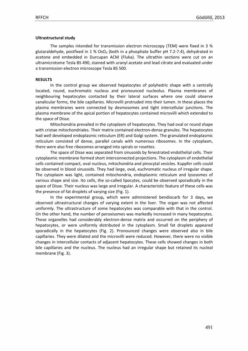
RFFCH Gödöllő, 2013
491
Ultrastructural study
The samples intended for transmission electron microscopy (TEM) were fixed in 3 % glutaraldehyde, postfixed in 1 % OsO4 (both in a phosphate buffer pH 7.2-7.4), dehydrated in acetone and embedded in Durcupan ACM (Fluka). The ultrathin sections were cut on an ultramicrotome Tesla BS 490, stained with uranyl acetate and lead citrate and evaluated under a transmission electron microscope Tesla BS 500. RESULTS
In the control group we observed hepatocytes of polyhedric shape with a centrally located, round, euchromatic nucleus and pronounced nucleolus. Plasma membranes of neighbouring hepatocytes contacted by their lateral surfaces where one could observe canalicular forms, the bile capillaries. Microvilli protruded into their lumen. In these places the plasma membranes were connected by desmosomes and tight intercellular junctions. The plasma membrane of the apical portion of hepatocytes contained microvilli which extended to the space of Disse.
Mitochondria prevailed in the cytoplasm of hepatocytes. They had oval or round shape with cristae mitochondriales. Their matrix contained electron-dense granules. The hepatocytes had well developed endoplasmic reticulum (ER) and Golgi system. The granulated endoplasmic reticulum consisted of dense, parallel canals with numerous ribosomes. In the cytoplasm, there were also free ribozomes arranged into spirals or rosettes.
The space of Disse was separated from sinusoids by fenestrated endothelial cells. Their cytoplasmic membrane formed short interconnected projections. The cytoplasm of endothelial cells contained compact, oval nucleus, mitochondria and pinocytal vesicles. Kuppfer cells could be observed in blood sinusoids. They had large, oval, euchromatic nucleus of irregular shape. The cytoplasm was light, contained mitochondria, endoplasmic reticulum and lysosomes of various shape and size. Ito cells, the so-called lipocytes, could be observed sporadically in the space of Disse. Their nucleus was large and irregular. A characteristic feature of these cells was the presence of fat droplets of varying size (Fig. 1).
In the experimental group, which were administered bendiocarb for 3 days, we observed ultrastructural changes of varying extent in the liver. The organ was not affected uniformly. The ultrastructure of some hepatocytes was comparable with that in the control. On the other hand, the number of peroxisomes was markedly increased in many hepatocytes. These organelles had considerably electron-dense matrix and occurred on the periphery of hepatocytes, or were uniformly distributed in the cytoplasm. Small fat droplets appeared sporadically in the hepatocytes (Fig. 2). Pronounced changes were observed also in bile capillaries. They were dilated and the microvilli were reduced. However, there were no visible changes in intercellular contacts of adjacent hepatocytes. These cells showed changes in both bile capillaries and the nucleus. The nucleus had an irregular shape but retained its nucleal membrane (Fig. 3).
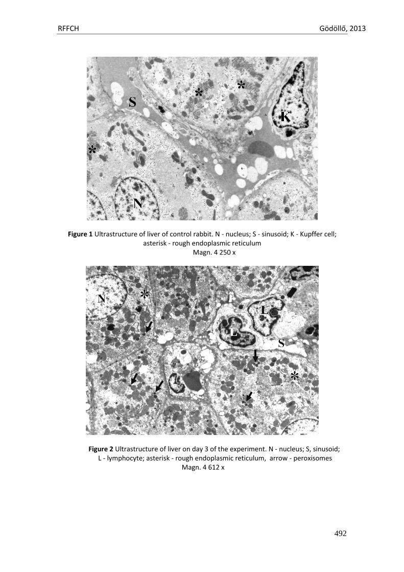
RFFCH Gödöllő, 2013
492
Figure 1 Ultrastructure of liver of control rabbit. N - nucleus; S - sinusoid; K - Kupffer cell; asterisk - rough endoplasmic reticulum
Magn. 4 250 x
Figure 2 Ultrastructure of liver on day 3 of the experiment. N - nucleus; S, sinusoid; L - lymphocyte; asterisk - rough endoplasmic reticulum, arrow - peroxisomes
Magn. 4 612 x
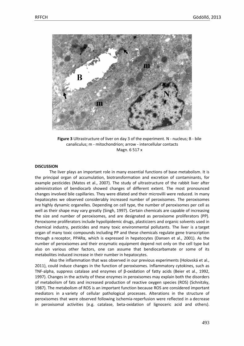
RFFCH Gödöllő, 2013
493
Figure 3 Ultrastructure of liver on day 3 of the experiment. N - nucleus; B - bile canaliculus; m - mitochondrion; arrow - intercellular contacts
Magn. 6 517 x DISCUSSION
The liver plays an important role in many essential functions of base metabolism. It is the principal organ of accumulation, biotransformation and excretion of contaminants, for example pesticides (Matos et al., 2007). The study of ultrastructure of the rabbit liver after administration of bendiocarb showed changes of different extent. The most pronounced changes involved bile capillaries. They were dilated and their microvilli were reduced. In many hepatocytes we observed considerably increased number of peroxisomes. The peroxisomes are highly dynamic organelles. Depending on cell type, the number of peroxisomes per cell as well as their shape may vary greatly (Singh, 1997). Certain chemicals are capable of increasing the size and number of peroxisomes, and are designated as peroxisome proliferators (PP). Peroxisome proliferators include hypolipidemic drugs, plasticizers and organic solvents used in chemical industry, pesticides and many toxic environmental pollutants. The liver is a target organ of many toxic compounds including PP and these chemicals regulate gene transcription through a receptor, PPARα, which is expressed in hepatocytes (Dansen et al., 2001). As the number of peroxisomes and their enzymatic equipment depend not only on the cell type but also on various other factors, one can assume that bendiocarbamate or some of its metabolites induced increase in their number in hepatocytes.
Also the inflammation that was observed in our previous experiments (Holovská et al., 2011), could induce changes in the function of peroxisomes. Inflammatory cytokines, such as TNF-alpha, suppress catalase and enzymes of β-oxidation of fatty acids (Beier et al., 1992, 1997). Changes in the activity of these enzymes in peroxisomes may explain both the disorders of metabolism of fats and increased production of reactive oxygen species (ROS) (Schnitzky, 1987). The metabolism of ROS is an important function because ROS are considered important mediators in a variety of cellular pathological processes. Alterations in the structure of peroxisomes that were observed following ischemia-reperfusion were reflected in a decrease in peroxisomal activities (e.g. catalase, beta-oxidation of lignoceric acid and others).
RFFCH Gödöllő, 2013
494
Investigations of the influence of bendiocarbamate on the activity of antioxidant enzymes in the rabbit showed no changes in the activity of catalase in experimental groups. On day three of the experiment, there were detected pronounced changes in the activity of glutathione peroxidase (GPH2O2) and increased content of TBARS, an important indicator of oxidative stress (Sobeková et al., 2009). CONCLUSION
Although bendiocarb does not accumulate in the body, it induced inflammatory changes in the liver of rabbits. They were observed under a light microscope, particularly on day 3 of the experiment. Inflammatory changes affected not only the portobiliary space but spread along the periphery of lobules. Electron microscopy confirmed changes in the ultrastructure of hepatocytes. Bendiocarb induced dilatation of bile capillaries and caused marked increase in the number of peroxisomes. The changes observed in our study were of moderate extent. Our observations allowed us to assume that due to its detoxication abilities the liver was capable of coping with the harmful effects of the investigated pesticide.
REFERENCES ALMÁŠIOVÁ V.- HOLOVSKÁ K.- TARABOVÁ L.- CIGÁNKOVÁ V. 2012. Sledovanie štrukturálnych zmien na
obličkách králikov po perorálnom podaní bendiokarbamátu. In Zborník referátov. 15. Košický morfologický deň. Cievny systém z hľadiska anatómie a histológie., p. 86-89.
BEIER K.- VOLKL A.- FAHIMI H. D. 1992. Suppression of peroxisomal lipid beta-oxidation enzymes by TNF-alpha. In FEBS Lett. 310, p. 273-276.
BEIER K.- VOLKL A.- FAHIMI H. D. 1997. TNF-alpha downregulates the peroxisome proliferarot activated receptor-alpha and mRNAs encoding peroxisomal proteins in rat liver. In FEBS Lett., 412, p. 385-387.
Bendiocarb, C. O. Pesticide residues in food: 1982 evaluations, file: //E: \Lymfatic\legath\carbamates\bendiocarb.htm
DANSEN T. B.- WIRTZ K. W. A. 2001. The peroxisome in Oxidative Stress. In Life, 51, p. 223-230. FLEŠÁROVÁ S.- LUKÁČ N.- DANKO J.- MASSANYI P.2007. Bendiocarbamate induced structural alterations
in rabbit thymus after experimental peroral administration. In Journal of Environmental Science and Health., vol. 42, no. 3, p. 29-334.
HOLOVSKÁ K.- ALMÁŠIOVÁ V.- TARABOVÁ L.- CIGÁNKOVÁ V. 2007. Effect of xenobiotics on the structure of rabbits liver. In Folia Veterinaria, 55, p. 69-72.
MATOS P.- FONTAINHAS-FERNANDES A.- PEIXOTO F.- CARROLA J.- ROCHA E.2007. Biochemical and histological hepatic changes of nile tilapia Oreochromis niloticus exposed to carbyl. In Pesticide Bioche. Physiol., 89, p. 73-80.
MOJŽIŠOVÁ J. 2004. The changes of the immunological and haematological parameters in rabbits after bendiocarbamate application In Proc. Agrochem. Morphogen., UVM, Košice, p. 37.
PETROVOVÁ E.- MAŽENSKÝ D.- VDOVIAKOVÁ K.- LUPTÁKOVÁ L. 2010. Changes in the structure of rabbit lymph nodes after bendiocarb administration. In Martinský morfologický zborník, p. 76-78.
SCHNITZKY M. 1987. Patterns of lipid changes in membranes of the aged brain. In Gerontology, 33, p. 149-154.
SCHRADER M.- FATIMI H. D. 2006. Peroxisomes and oxidative stress. In Biochimica Biophysica Acta, p. 1755-1766.
SINGH I. 1997. Biochemistry of peroxisomes in health and disease. In Mol. Cell. Biochem., 167, p. 1-29. SOBEKOVÁ A.- HOLOVSKÁ K.- LENÁRTOVÁ V.- FLEŠÁROVÁ S.- JAVORSKÝ P. 2009. The other toxic effect
of carbamate insecticides. In Acta Biologica Hungarica,60, p. 45-54. ŠPALEKOVÁ E.- MASSANYI P.- HOLOVSKÁ K.- ALMÁŠIOVÁ V.- PETROVOVÁ E.- DANKO J. 2010. Analýza
lymfatickej uzliny králika po experimentálnom podaní bendiokarbamátu. In X. Risk factor of food chain. Nitra: Slovak Agricultural University, p. 313-318.
Animal welfare, etológia és tartástechnológia
Animal welfare, ethology and housing systems
Volume 9 Issue 3
Különszám/Special Issue
Gödöllő
2013
RFFCH Gödöllő, 2013
495
Attempt to Reduce Salmonella Contamination in a Commercial Turkey Flock with Feed Additive Premixes
Janan J*.1, 2, Hutás I.2, Treuer Á.2, Csépányi B.2, Páli J.2
1Department of Animal Physiology and Health, Szent István University, Páter K. u. 1, H-2100 Gödöllő, Hungary 2Pharmatéka Bt., Cinkotai út 91/C., H-1148 Budapest, Hungary * [email protected]
Summary A water-soluble probiotic preparation with vitamin feed additive premix (Bactovit P) and a feed additive premix containing volatile oils (Turkey premix 10’), effective in vitro against Salmonella, were tested for reducing or preventing Salmonella contamination during the growing period of turkey production. Twenty thousand day-old turkey poults (sex ratio of 1: 1) being free from Salmonella were randomly assigned to a control (untreated) group and a treated group that received 1 kg Turkey Premix 10’ per ton ration for 42 days plus 0.15 g Bactovit P per kg BW in the drinking water on arrival and then at seven-day intervals. Composite faecal samples were taken from both groups per sex at weekly intervals and the population of Salmonella spp. enumerated using the Most Probable Number (MPN) method. The flock management data (number of animals, body weights, mortality and medications) were recorded by the farm. The feed additive premixes delayed initially the development of Salmonella infection of poults and reduced its level compared to the controls’ till week 4 to 5 in females and males, respectively. Thenceforward this effect discontinued. Of l4 faecal samples from the control and treated group each, 14 vs. 10 samples were Salmonella positive. The most frequent serovars was S. Bredney. The mortality rate was lower for the treated turkeys compared to the controls’. No appreciable improving or worsening was found between the control and experimental groups in the growth rate. Additional improvement of the products and their application seems reasonable to increase the persistency of their effect through the entire 6-week growing period. Introduction Salmonellosis is one of the most important food-born diseases and causes substantial medical and economical burdens worldwide. The main food sources of infections by Salmonella are poultry meat and eggs. Thus, a number of actions have been taken to reduce the prevalence of Salmonella serovars with public health significance in food-producing animals. Since 2003, all members of the European Union have to put into practice monitoring programs to control these pathogens (European Parliament and European Council, 2003). Salmonellae are carried within the gut of the birds and contaminate the environment by shedding from infected birds through the faeces, feather dust and eye secretions. In the past, antibiotics seemed an effective way of preventing Salmonella contamination. Antibiotic growth promoters have improved the performance and health status of poultry via their action on the intestinal micro-flora. However the ban on animal feed antibiotics in 2005 has led to a demand for alternatives to antibiotics. Possible non-therapeutic alternatives to antibiotics for poultry may be the ones which work via similar mechanisms, promoting growth while enhancing the efficacy of feed conversion Hughes P. and Heritage J. (2003). Such as are organic acids – exerting antimicrobial action through bowel pH depression; enzymes – improving ileac digestibility thereby indirectly overloading the mikroflora; probiotics – improving health and growth by altering intestinal microbial balance; prebiotics – selectively stimulating the growth or metabolic activity of a limited number of intestinal mikroflora; herbs & ether oils – antimicrobial agents by stimulating the endogenous digestive enzymes and immunostimulants – improving the immune system of the bird and enhancing the
RFFCH Gödöllő, 2013
496
resistance to disease (Reddy, 2004). The other method of Salmonella control is litter amendment with inorganic acidifiers. The purpose of this study was to evaluate the efficacy of two candidate feed additives for reducing Salmonella contamination in a turkey flock being free of Salmonella at the early age. Materials and Methods Feed additives tested Bactovit P (Pharmatéka Bt.): It is a water soluble probiotic (Lactic acid bacteria) preparation with vitamin feed additive premix. The application of the probiotic relives stress, enhances the immune system and inhibits the proliferation of harmful bacteria. It is used for preventive purposes. Dosage: 0.15 g per kg BW, dissolved in daily amount and given drinking water, once a week. Turkey Premix No.10 (Pharmatéka Bt.): It is a feed additive premix containing a mixture of volatile oils (Origanum vulgare, Thyme oil and Castanea sativa mill). Technological additive: Emulsion (E484), Carbovet. Dosage: mixed in the feed at a rate of 1 kg per ton feed (i.e. in a concentration of 0.1%) Experimental design The feeding trial was conducted in a partner farm of Pharmatéka Bt. in 2012. Twenty thousand day-old turkey poults (sex ratio = 1:1) were randomly assigned to a control (untreated) group and a treated group and housed separately in two henneries partitioned into two for isolating the sexes. The bedding material was fresh straw spread daily over the deep litter. Poults received the same turkey starter feed and drinking water ad libitum. The experimental group received 1 kg Turkey Premix No.10 per ton feed for 42 days plus 0.15 g Bactovit P per kg BW in the drinking water on arrival and then at seven-day intervals. Samplings and data collection Fresh faecal samples were collected across the entire hennery at weekly intervals, placed in 200 ml sterile plastic cups and covered. Two-two cups of composite faecal samples were prepared by treatment and sex, totalling 7 cups on each occasion. The faecal samples were examined for Salmonella prevalence in an accredited Laboratory. Flock management data (number of animals, body weights, mortality and medications) were recorded by the farm. Results and discussion Faecal samples from control poults were positive for Salmonella already at the of the first week, whilst the samples from treated poults became positive by the end of the first (females) to third week (males), but the level of infections was just above the minimum detectable level and it remained low by the end till week 4 to 5 in females and males, respectively, compared to the controls. However this between-group difference disappeared by the end of the sixth week. All the 14 faecal samples collected from the control group were positive for Salmonella spp.; while of 14 faecal samples from the treated group four (28.6%) were negative and ten (71.4%) were positive (Table 1). The most frequent serovars were S. Bredney, and in one or two samples S. Tenesse and S. Newport.
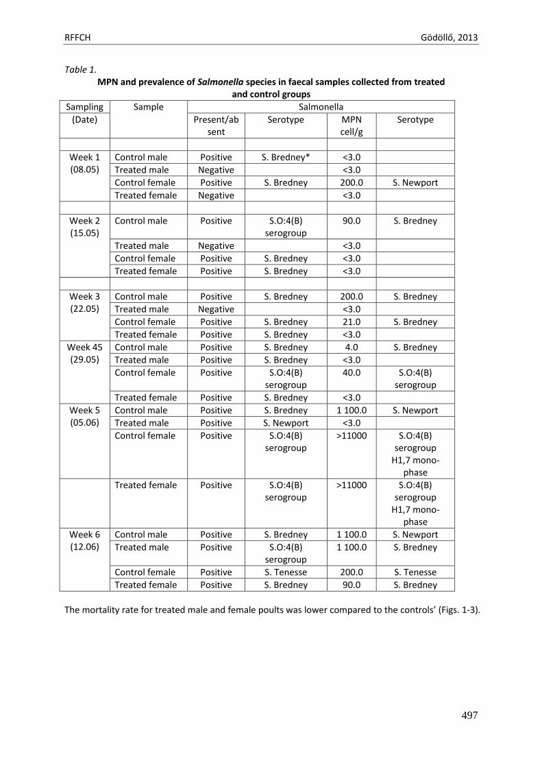
RFFCH Gödöllő, 2013
497
Table 1. MPN and prevalence of Salmonella species in faecal samples collected from treated
and control groups
Sampling Sample Salmonella
(Date) Present/absent
Serotype MPN cell/g
Serotype
Week 1 (08.05)
Control male Positive S. Bredney* <3.0
Treated male Negative <3.0
Control female Positive S. Bredney 200.0 S. Newport
Treated female Negative <3.0
Week 2 (15.05)
Control male Positive S.O:4(B) serogroup
90.0 S. Bredney
Treated male Negative <3.0
Control female Positive S. Bredney <3.0
Treated female Positive S. Bredney <3.0
Week 3 (22.05)
Control male Positive S. Bredney 200.0 S. Bredney
Treated male Negative <3.0
Control female Positive S. Bredney 21.0 S. Bredney
Treated female Positive S. Bredney <3.0
Week 45 (29.05)
Control male Positive S. Bredney 4.0 S. Bredney
Treated male Positive S. Bredney <3.0
Control female Positive S.O:4(B) serogroup
40.0 S.O:4(B) serogroup
Treated female Positive S. Bredney <3.0
Week 5 (05.06)
Control male Positive S. Bredney 1 100.0 S. Newport
Treated male Positive S. Newport <3.0
Control female Positive S.O:4(B) serogroup
>11000 S.O:4(B) serogroup
H1,7 mono-phase
Treated female Positive S.O:4(B) serogroup
>11000 S.O:4(B) serogroup
H1,7 mono-phase
Week 6 (12.06)
Control male Positive S. Bredney 1 100.0 S. Newport
Treated male Positive S.O:4(B) serogroup
1 100.0 S. Bredney
Control female Positive S. Tenesse 200.0 S. Tenesse
Treated female Positive S. Bredney 90.0 S. Bredney
The mortality rate for treated male and female poults was lower compared to the controls’ (Figs. 1-3).
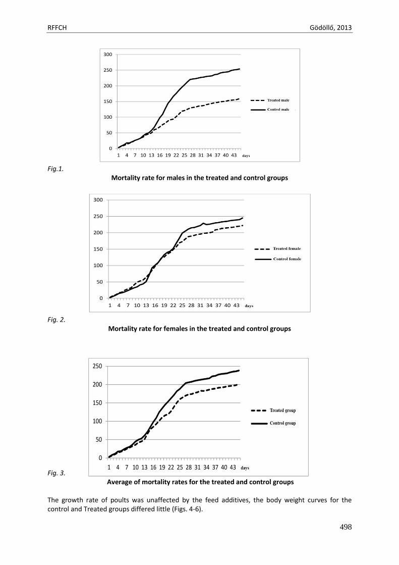
RFFCH Gödöllő, 2013
498
Fig.1.
Mortality rate for males in the treated and control groups
Fig. 2.
Mortality rate for females in the treated and control groups Fig. 3.
Average of mortality rates for the treated and control groups The growth rate of poults was unaffected by the feed additives, the body weight curves for the control and Treated groups differed little (Figs. 4-6).
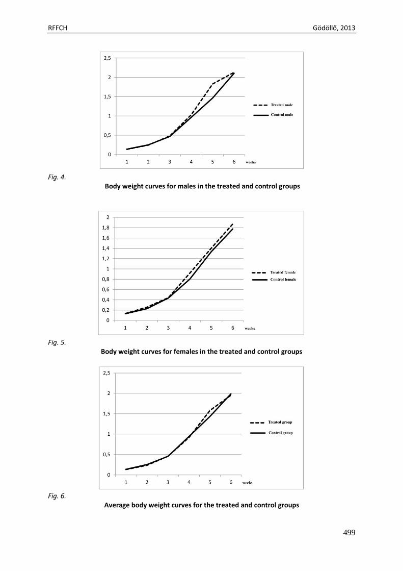
RFFCH Gödöllő, 2013
499
Fig. 4.
Body weight curves for males in the treated and control groups
Fig. 5.
Body weight curves for females in the treated and control groups
Fig. 6.
Average body weight curves for the treated and control groups
RFFCH Gödöllő, 2013
500
Conclusion Many candidate feed additives have been described in literature but there is still much uncertainty about their effect in vivo, about the persistency of their effect, and the potential interaction between different additives (Wageningen UR, 2012). This is true for our experiment presented here. In light of the results, the dietary inclusion of the two feed additive premixes appears a promising approach to reduce Salmonella contamination in turkey poults. However, additional improvement of the products and their application is desirable to increase the persistency of their effect through the entire growing period. References European Parliament and European Council (2003): Directive of the European Parliament and of the Council pf
17 November 2003 on the monitoring of zoonoses and zoonotic agents, amending Council Decision 90/424/EEC and repealing Council Directive 92/117/EEC.
Hughes P. and Heritage J. (2003): Antibiotic Growth-Promoters in Food Animals. http://www.fao.org/docrep/ARTICLE/AGRIPPA/555_EN.HTM
Reddy, V. R. (2004): The role of acidifiers in poultry nutrition. Avitech Technical Bulletin, July 2004. Wageningen University and Research Centre (2012): Law-emission animal feed bursting with energy.
http://www.wageningenur.nl/en/show/Lowemission-animal-feed-burs...
Acknowledgements This work was supported in part by Research Centre of Excellence-17586-4/2013/TUDPOL and GOP-1.1.1-09/1-2009-0097 grants
Animal welfare, etológia és tartástechnológia
Animal welfare, ethology and housing systems
Volume 9 Issue 3
Különszám/Special Issue
Gödöllő
2013
RFFCH Gödöllő, 2013
501
MICROFLORA OF RABBITS AFTER QUERCETIN AND T - 2 TOXIN APPLICATIONS
Miroslava Kačániová1*, Marcela Capcarová2, Jana Petrová1, Peter Haščík3, Lukáš Hleba1
1Department of Microbiology, Faculty of Biotechnology and Food Sciences, Slovak University of Agriculture, Nitra, Slovak Republic 2Deartment of animal physiology, Faculty of biotechnology and Food Sciences, Slovak University of Agriculture, Nitra, Slovak Republic 3Department of Animal Products Evaluation and Processing, Faculty of Biotechnology and Food Sciences, Slovak University of Agriculture, Nitra, Slovak Republic
*E-mail: [email protected]
Abstract The aim of this study was to compare the microbial species in gut microflora of rabbit in control group against experimental group were it was applied qertecin and T2 toxin. It is first study about caecum microflora study after qertecin and T2 toxin application. In this study for enumeration of bacteria were using classical method. Enterococci were counted on Slanetz-Bartley agar (Biolife, Italy) and incubated at 37 °C for 48-72 hours, lactobacilli were counted on MRS Lactobacillus agar (Biolife, Italy) and incubated at 37 °C for 48-72 hours and coliforms bacteria were counted on MacConkey agar (Biolife, Italy) and incubated at 37 °C for 24-48 hours. In our study the number of coliforms bacteria ranged between 4.23 to 5.50 log CFU.g-1, lactobacilli ranged between 3.20 to 4.54 log CFU.g-1 and enterococci ranged between 3.28 to 5.23 log CFU.g-1. Keywords: microflora, rabbits, quertecin, T2 toxin Introduction
Rabbits are monogastric herbivores, which are widely used for research purposes and husbandry. In rabbits, most food transformation events occur in the caecum, which is densely populated with bacteria and constitute an efficient continuous culture system. Rabbits help to maintain a relatively stable microbial population by caecotrophy. Notwithstanding this, relatively little attention has been paid to the microbial populations of the rabbit gut. Most studies have focused on the anaerobic microbiota of the caecum (Crociani et al. 1984; Forsythe and Parker, 1985) and intestinal athogens (Lelkes and Chang, 1987; Straw, 1988; Milon, 1996). These studies have revealed that the intestinal microbiota of rabbits consists predominantly of strictly anaerobic, Gram-negative bacteria belonging to the genus Bacteroides, while anaerobic sporulated Gram-positive bacteria are present in significantly lower numbers (Forsythe and Parker, 1985; Zomborszky-Kovacs et al. 2000). Facultative anaerobic bacteria isolated from the intestinal tract of rabbits belong to the Gram-positive genera Bacillus, Enterococcus and Staphylococcus, and Gram-negative Enterobacter and Escherichia (Forsythe and Parker, 1985; Canganella et al., 1992). In contrast with other mammals, lactobacilli are very rarely found (Yu and Tsen, 1993).
Probiotics along with other functional foods positively affect the health of the consumer (Horská, 2012). Flavonoids have high antioxidant activities as free radical scavengers and as inhibitors of enzymes generating reactive oxygen species (Hollman and Katan, 1999). Flavonoids may preserve β-cell function by reducing oxidative stress-induced tissue damage and therefore protect against the progression of insulin resistance to type 2 diabetes. In fact, quercetin, the main dietary flavonol, prevents and protects streptozotocin-induced oxidative stress and β-cell damage in rat pancreas (Coskun et al., 2005) and decreased blood glucose concentration in both alloxan- and streptozotocin-induced diabetic rats (Coskun et al., 2005; Romero et al., 2010). Moreover, the intake of some specific types of flavonoids, including quercetin, was inversely associated with a higher risk of incident type 2 diabetes (Knekt et al., 2002). Mycotoxins represent one of the most important categories of biologically produced natural toxins relative to human health and economic impact worldwide (Cetin and Bullerman, 2005; McKean et al., 2006). T-2 is a genotoxic and cytotoxic mycotoxin, which produce inhibition of protein synthesis by strong afinity for the 60S ribosomal unit
RFFCH Gödöllő, 2013
502
and inhibition of RNA and DNA synthesis. Moreover, it can induce mutation and apoptosis. All these effects were observed both in vivo and in vitro (Bouaziz et al., 2008; Chaudhari et al., 2009).
The purpose of this study was to compare the microbial species in gut microflora of rabbit in control group against experimental group were it was applied qertecin and T2 toxin. It is first study about caecum microflora study after qertecin and T2 toxin application. In this study for enumeration of bacteria were using classical method. Material and methods Animals and diet, blood collection and determination of the parameters Animals and diet
Adult female rabbits (n = 20) and male rabbits (n = 20) of meat line M91, maternal albinotic line (crossbreed Newzealand white, Buskat rabbit, French silver) and paternal acromalictic line (crossbreed Nitra’s rabbit, Californian rabbit, Big light silver) were used in experiment. Rabbits were healthy and their condition was judged as good at the commencement of the experiment. Water was available at any time from automatic drinking troughs. Groups of adult animals were balanced for age (150 days) and body weight (4 ± 0.5 kg) at the beginning of the experiment. Adult rabbits were fed diet of a 12.35 MJ.kg-1 of metabolizable diet (tab. 1) composed of a pelleted concentrate.
Table 1.
Chemical composition (g.kg-1) of the experimental diet
Component
Dry matter 926.26 Crude protein 192.06 Fat 36.08 Fibre 135.79 Non-nitrogen compounds 483.56 Ash 78.78 Organic matter 847.49 Calcium 9.73 Phosphorus 6.84 Magnesium 2.77 Sodium 1.81 Potassium 10.94 Metabolizable energy 12.35 MJ.kg-1
Animals were divided into two control groups (C1 and C2) and experimental groups (E1 – E6).
Experimental groups received quercetin (Sigma Aldrich, Saint Louis, USA) in injectable form (intramuscularly) at 10 µg.kg-1 in E1 and E2 group, 100 µg.kg-1 in E3 and E4 group and 1000 µg.kg-1 mg.kg-1 in E5 and E6 group without T-2 toxin for 90 days. T-2 toxin (Romer Labs Division Holding GmbH, Tulln, Austria) to C2, E2, E4 and E6 group at dose 0.08 mg per kg of body weight 72 hours before slaughter intramuscularly was applied.Control groups received injection water (Imuna Pharm a.s. Šarišské Michaľany, Slovak Republic).
In this animal study, institutional and national guidelines for the care and use of animals were followed, and all experimental procedures involving animals were approved by the State Veterinary and Food Institute of Slovak Republic, no. 3398/11-221/3.
RFFCH Gödöllő, 2013
503
Table 2. Application of quercetin and T-2 toxin in rabbit blood in vivo
Group Quercetin (µg.kg-1) T-2 toxin (mg.kg-1 of body weight, 72 hours before slaughter)
C1 0 0.00 C2 0 0.08 E1 10 0.00 E2 10 0.08 E3 100 0.00 E4 100 0.08 E5 1000 0.00 E6 1000 0.08
C – control group, E1 – E6 – experimental groups with various doses of quercetin alone or in combination with T-2 toxin Plate diluting method Determination of CFU counts: Plate diluting method was applied for quantitative CFU counts determination of respective groups of microorganisms in 1 g of substrate. Gelatinous nutritive substrate in Petri dishes was inoculated with 1 mL of chyme samples pour plate method in three replications. Homogenized samples of faecal chyme (chyme was taken to sterile Petri dishes) were prepared in advance by sequential diluting based on decimal dilution system application. Enterococci were counted on Slanetz-Bartley agar (Biolife, Italy) and incubated at 37 °C for 48-72 hours, lactobacilli were counted on MRS Lactobacillus agar (Biolife, Italy) and incubated at 37 °C for 48-72 hours and coliforms bacteria were counted on MacConkey agar (Biolife, Italy) and incubated at 37 °C for 24-48 hours. Isolated species, genera and groups of microorganisms and their fundamental identification were performed as per standard norms (Holt et al., 1994). Results and discussion
The digestive system of animals hosts several hundred species of bacteria. These bacteria (microbial flora or microflora) in the gastrointestinal tract form a complex ecosystem that plays an important role in maintaining the integrity of the host’s enterocyte, providing enzymes for metabolism of ingested of foods, modulation of metabolic processes for host needs (such as converting steroids and unconjugating bilirubin into more water soluble forms of urobilinogen), and protecting the host epithelial cells from colonization by toxin-producing or invasive intestinal pathogens. The intestinal microflora also provide certain nutrients and vitamins that are beneficial to the host. The relationship between the non-harmful microbiota and the host is often viewed as symbiotic; therefore, the organisms in the gastrointestinal tract are commensals under normal conditions. A complete understanding of the balance and composition of the gut microflora in the host animal species remains incomplete (Fortun-Lamothe and Boullier, 2007).
While lactobacilli, coliforms and streptococci constitute the main components of the intestinal microflora in the majority of farm animals, in the rabbit these bacteria can be found in traces only. In the first week of life their number is usually between 102 and 108, but subsequently they “disappear” and their count falls below 100. In the present experiment, the free nursing method (Group A) resulted in higher lactobacillus counts throughout and also higher coliform counts in the first four days of life. In Group C, a major rise was found in the coliform count by day 6, and on day 10 the coliform count was still higher than in the other two groups. This was presumably due to the slower establishment of the main microflora (i.e. the slower rate of colonisation of the caecum by Bacteroides). Although it could be established that the Lactobacillus and coliform counts were in inverse ratio to the Bacteroides count, a clear antagonism between the normal bacterial components of the microflora could still not be demonstrated (Kovacs et al., 2004).
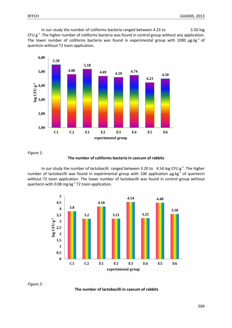
RFFCH Gödöllő, 2013
504
In our study the number of coliforms bacteria ranged between 4.23 to 5.50 log CFU.g-1. The higher number of coliforms bacteria was found in control group without any application. The lower number of coliforms bacteria was found in experimental group with 1000 µg.kg-1 of querticin without T2 toxin application.
Figure 1:
The number of coliforms bacteria in caecum of rabbits
In our study the number of lactobacilli ranged between 3.20 to 4.54 log CFU.g-1. The higher number of lactobacilli was found in experimental group with 100 application µg.kg-1 of quertecin without T2 toxin application. The lower number of lactobacilli was found in control group without quertecin with 0.08 mg.kg-1 T2 toxin application.
Figure 2: The number of lactobacilli in caecum of rabbits
5.50
4.80
5.18
4.69 4.59 4.74
4.23 4.50
1,00
2,00
3,00
4,00
5,00
6,00
C1 C2 E1 E2 E3 E4 E5 E6
log
CF
U.g
-1
experimental group
3.8
3.2
4.18
3.21
4.54
3.25
4.48
3.58
0
0,5
1
1,5
2
2,5
3
3,5
4
4,5
5
C1 C2 E1 E2 E3 E4 E5 E6
log
CF
U.g
-1
experimental group
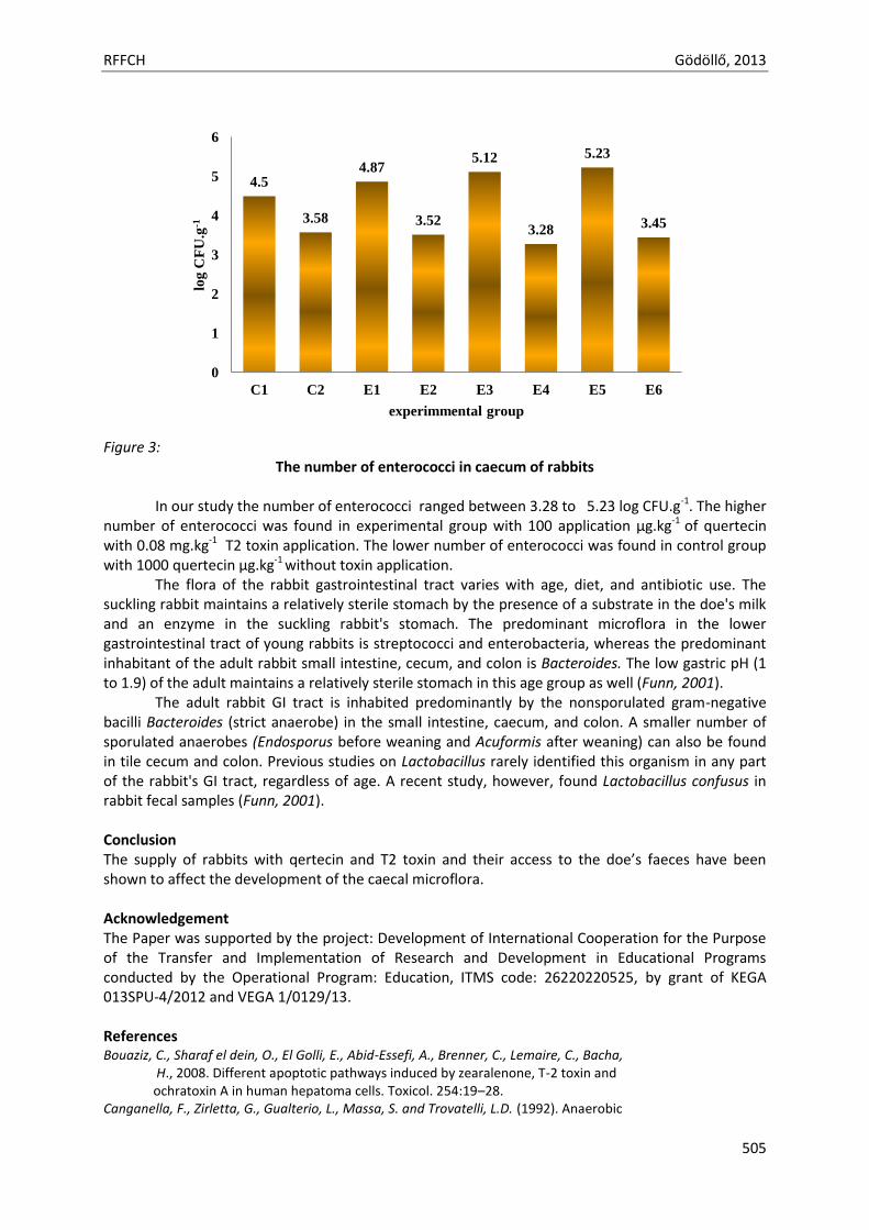
RFFCH Gödöllő, 2013
505
Figure 3:
The number of enterococci in caecum of rabbits
In our study the number of enterococci ranged between 3.28 to 5.23 log CFU.g-1. The higher number of enterococci was found in experimental group with 100 application µg.kg-1 of quertecin with 0.08 mg.kg-1 T2 toxin application. The lower number of enterococci was found in control group with 1000 quertecin µg.kg-1 without toxin application.
The flora of the rabbit gastrointestinal tract varies with age, diet, and antibiotic use. The suckling rabbit maintains a relatively sterile stomach by the presence of a substrate in the doe's milk and an enzyme in the suckling rabbit's stomach. The predominant microflora in the lower gastrointestinal tract of young rabbits is streptococci and enterobacteria, whereas the predominant inhabitant of the adult rabbit small intestine, cecum, and colon is Bacteroides. The low gastric pH (1 to 1.9) of the adult maintains a relatively sterile stomach in this age group as well (Funn, 2001).
The adult rabbit GI tract is inhabited predominantly by the nonsporulated gram-negative bacilli Bacteroides (strict anaerobe) in the small intestine, caecum, and colon. A smaller number of sporulated anaerobes (Endosporus before weaning and Acuformis after weaning) can also be found in tile cecum and colon. Previous studies on Lactobacillus rarely identified this organism in any part of the rabbit's GI tract, regardless of age. A recent study, however, found Lactobacillus confusus in rabbit fecal samples (Funn, 2001). Conclusion The supply of rabbits with qertecin and T2 toxin and their access to the doe’s faeces have been shown to affect the development of the caecal microflora. Acknowledgement The Paper was supported by the project: Development of International Cooperation for the Purpose of the Transfer and Implementation of Research and Development in Educational Programs conducted by the Operational Program: Education, ITMS code: 26220220525, by grant of KEGA 013SPU-4/2012 and VEGA 1/0129/13. References Bouaziz, C., Sharaf el dein, O., El Golli, E., Abid-Essefi, A., Brenner, C., Lemaire, C., Bacha, H., 2008. Different apoptotic pathways induced by zearalenone, T-2 toxin and ochratoxin A in human hepatoma cells. Toxicol. 254:19–28. Canganella, F., Zirletta, G., Gualterio, L., Massa, S. and Trovatelli, L.D. (1992). Anaerobic
4.5
3.58
4.87
3.52
5.12
3.28
5.23
3.45
0
1
2
3
4
5
6
C1 C2 E1 E2 E3 E4 E5 E6
log
CF
U.g
-1
experimmental group
RFFCH Gödöllő, 2013
506
facultative bacteria isolated from the gut of rabbits fed different diets. Zentralblatt fur Mikrobiologie 147:537–540. Cetin, Y., Bullerman, L.B., 2005. Cytotoxicity of Fusarium mycotoxins to mammalian cell cultures as determined by the MTT bioassay. Food Chem. Toxicol. 43:755–764. Chaudhari, M., Jayaraj, R., Bhaskar, A.S.B., Lakshmana Rao, P.V., 2009. Oxidative stress induction by T-2 toxin causes DNA damage and triggers apoptosis via caspase pathway in human cervical cancercells. Toxicol. 262:153–161. Coskun, O., Kanter, M., Korkmaz, A., Oter, S., (2005). Quercetin, a flavonoid antioxidant, prevents and protects streptozotocin-induced oxidative stress and beta-cell damage in rat pancreas. Pharmacol. Res. 51:117–123. Crociani, F., Biavati, B., Castagnoli, P., Matteuzzi, D. (1984). Anaerobic ureolytic bacteria from cecal content, soft faeces of rabbit. J. Appl. Bacter. 57:83–88. Forsythe, S.J., Parker, D.S. (1985). Nitrogen metabolism by the microbial flora of rabbit cecum. J. Appl. Bacter. 58:363–369. Fortun-Lamothe, F., Boullier, S. (2007). A review on the interactions between gut microflora and digestive mucosal immunity. Possible ways to improve the health of rabbits. Livestock Sci. 107:1–18. Holt, G. J., Krieg, N. R., Sneath, P. H. A., Staley, J.T., Williams, S.T. (1994). Bergey 's Manual of Determinative Bacteriology. Baltimore : Williams and Wilkins; 787. Horská, E. (2012). New Consumerism and Trends at the Food Market: within and Beyond Visegrad Borders. In Horská, E. Food Sciences & Business Studies. Nitra : SPU, 2012, 410 p. ISBN 978-80-552-0815-2. Knekt, P., Kumpulainen, J., Järvinen, R., Rissanen, H., Heliövaara, M., Reunanen, A., Hakulinen, T., Aromaa, A., (2002). Flavonoid intake and risk of chronic diseases. Am. J. Clin. Nutr. 76:560–568. Lelkes, L.. Chang, Ch.L. (1987) Microbial dysbiosis in rabbit mucoid enteropathy. Lab. Anim. Sci. 37, 757–764. Milon, A. (1996). Weaned rabbit colibacillosis: a model for study of enteropathogenic Escherichia coli (EPEC). In Proceedings of the 6th World Rabbit Congress, vol. 3. pp. 13–22. Toulouse, France. Romero,M., Jiménez, R., Hurtado,B ., Moreno, J.M., Rodríguez-Gómez, I., López-Sepúlveda, R., Zarzuelo, A., Pérez-Vizcaino, F., Tamargo, J., Vargas, F., Duarte, J. (2010). Lack of eneficial metabolic effects of quercetin in adult spontaneously hypertensive rats. Europ. J. Pharmacol. 627: 242–250. Straw, T.E. (1988). Bacteria of rabbit gut and their role in the health of the rabbit. J. Appl. Rabbit Res. 11:145–150. Yu, B., Tsen, H.Y. (1993) Lactobacillus cells in the rabbit digestive tract and the factors affecting their distribution. J. Appl. Bacter. 75:269–275. Zomborszky-Kovacs, M., Gyarmati, T., Parizs, T., Szendro, Z.S., Kametler, L., Toth, A. (2000) Some physiological properties of the digestive tract in traditionally reared and exclusively milk-fed young rabbits. In 7th World Rabbit Congress, Nutrition and digestive physiology short papers. Valencia, Spain.

Animal welfare, etológia és tartástechnológia
Animal welfare, ethology and housing systems
Volume 9 Issue 3
Különszám/Special Issue
Gödöllő
2013
RFFCH Gödöllő, 2013
507
THE EFFECT OF RESVERATROL ON PORCINE OVARIAN GRANULOSA CELL FUNCTIONS
Attila Kádasi*1, Adriana Kolesárová1, Nora Maruniaková1, Roland Grossmann3, Richard Alexa4, Aneta Štochmaľová4, Alexander V. Sirotkin2
1 Slovak University of Agriculture in Nitra, Faculty of Biotechnology and Food Sciences, Department of Animal Physiology, Department of Animal Physiology Tr. A. Hlinku2, 949 76 Nitra, Slovak Republic; 2 Institute for Genetics and Reproduction of Farm Animals, Animal Production Research Centre Nitra, Lužianky, Slovak Republic; 3Department of Functional Genomics and Bioregulation, Institute of Animal Science, Mariensee, 31535 Neustadt, Germany; 4Constantine the Philosopher University in Nitra, Faculty of Natural Sciences, Department of Zoology and Anthropology, *: [email protected]
Abstract
The aim of our study was to elucidate the role of resveratrol in control of ovarian functions. For this purpose, we have examined its effect (at the doses 0, 1, 10, 100 μg/ml) on proliferation, apoptosis and steroidogenesis in porcine ovarian granulosa cells in vitro. Accumulation of PCNA (marker of proliferation), BAX (marker of apoptosis) and the release of steroid hormones (progesterone and testosterone) were analysed by immunocytochemistry and RIA respectively. It was observed, that resveratrol addition decreased the percentage of proliferative (PCNA-positive) and increased the percentage of apoptotic (BAX-positive) cells at all used doses (1, 10 and 100 μg/ml). On the other hand decreased progesterone release (at all doses of resveratrol) and stimulated testosterone release (at 10 and 100 μg/ml but not at 1 μg/ml of resveratrol) by porcine granulosa cells was detected. Our results suggest a direct effect of resveratrol on proliferation, apoptosis and steroidogenesis in porcine ovaries. Taken together, these data suggest that resveratrol can affect reproductive (ovarian) functions of pigs.
Keywords: resveratrol, proliferation, apoptosis, progesterone, testosterone, porcine granulosa cells
Introduction
Phytoalexins are antimicrobial substances under natural conditions not found in plants. Their creation occurs in plants attacked by pathogens (Dixon and Paiva, 1995). Resveratrol (RSV) was chemically 3,4',5-trihydroxystilbene and is one of the natural phytoalexins (Hain et al., 1990). It is a naturally occurring phytoestrogen and antioxidant that is found in grapevines, but also in soft fruits and hazelnut (Frémont, 2000). RSV is well known for its phytoestrogenic, antioxidant properties, growth-inhibitory and apoptosis-inducing activities (Joe et al., 2002; Jiang et al., 2005; Baur and Sinclair, 2006). It is able to inhibit PI3K/Akt Protein Kinase/ mammalian target of rapamycin (PI3K/Akt/mTOR) pathway in various types of cells (Jiang et al., 2005; Shankar et al., 2007; Bai et al., 2010). RSV is favourable for biological processes in the body to protect against cardiovascular diseases. It also has anti-cancer properties because RSV inhibits proliferation and induces apoptosis in cells at different levels (Ferry-Dumazet et al, 2002, Haider et al, 2003).
Cell proliferation is the amount of cells in culture or in the body can be divided. The extent of DNA synthesis is marker for proliferation (Wyllie et al., 1998). Involving the protein to cell proliferation include PCNA (Tomanek and Chronowska, 2006). This protein is localized in the cell nucleus (Makarevich et al., 2000; Nahryzny and Lee, 2001) and located in granulosa cells of gilts (Sanislo et al. 2001).
Apoptosis is programmed death of cells. This process eliminates unnecessary and useless cells from the body (Wyllie et al., 1998). Apoptosis is supported by group of caspases, which include BAX
RFFCH Gödöllő, 2013
508
(Zwain and Amato, 2001). This protein is localized especially in mitochondria (Markström et al., 2002) and located in granulosa cells of gilts (Sanislo et al. 2001).
Progesterone (P4) is an ovarian steroid produced by ovarian granulosa cells (Kolesárová et al., 2010a; Kolesárová et al., 2010b; Medvedová et al., 2011) and corpus luteum (Gregoraszczuk, 1992; Gregoraszczuk, 1997) of pigs and contributes to regulation of ovarian follicular development and remodelling (Mahajan, 2008). It is a local paracrine or autocrine factor regulating luteal function (Gregoraszczuk, 1992; Gregoraszczuk, 1997). This progestin is essential for normal ovarian cycle of females (Hagan et al., 2009). Another hormone produced in ovary is testosterone (T) (Delort et al., 2009). T is steroid hormone as well as P4 are necessary as a precursor for the synthesis of estrogen (Mindnich et al., 2004).
The aim of our study was to research the effect of RSV treatment at doses 1, 10 and 100 µg/ml on accumulation of markers of proliferation (PCNA) and apoptosis (BAX) and secretory activity (steroid hormones of P4 and T) of porcine ovarian granulosa cells (GCs) in vitro.
Material and method
Preparation, culture and processing of granulosa cells from ovaries
Granulosa cells were collected from the ovaries of prepubertal Slovakian White gilts, after slaughter at a local abattoir. After aspiration and isolation of granulosa cells, these cells were then washed in sterile DMEM/F12 1:1 medium (BioWhittakerTM, Verviers, Belgium), resuspended in the same medium supplemented with 10% fetal calf serum (BioWhittakerTM) and 1% antibiotic- antimycotic solution (Sigma, St. Louis, MO, USA) at a final concentration 106 cells/ml medium. Portions of the cell suspension were dispensed to 24-well culture plates (NuncTM, Roskilde, Denmark, 1 ml suspension/well; for RIA) or 16-well chamber slides (Nunc Inc., International, Naperville, USA, 200 μl/well, for immunocytochemistry). Both, the plate wells and chamber slides were incubated at 37 ° C and 5% CO2 in humidified air until 60-75% confluent monolayer was formed (3-5 days), at which point the medium was renewed. Further culture was performed in 2 ml culture medium in 24-well plates (medium for RIA) or 200 μl/medium in 16-well chamber slides, (cells for immunocytochemistry) as described previously. After medium replacement experimental cells were cultured in the presence of RSV (Changsha Sunfull Bio-tech. Co, Hunan China) at concentrations of 0, 1, 10 and 100 µg/ml. RSV was dissolved in culture medium immediately before their addition to the cells. After two days in culture, the medium from the 24-well plates was gently aspirated and frozen at -24°C to await RIA. After removing the medium from chamber slides, cell were washed in ice-cold PBS (pH 7.5), fixed in paraformaldehyde (4% in PBS, pH 7.2-7.4; 60 min) and held at 4°C to await immunocytochemistry.
Immunocytochemical analysis
Following washing and fixation, the cells were incubated in the blocking solution (1% of goat serum in phosphate-buffered saline – PBS) at room temperature for 1 h to block nonspecific binding of antiserum. Afterwards, the cells were incubated in the presence of monoclonal antibodies against either PCNA (marker of proliferation) and BAX (marker of apoptosis) (all from Santa Cruz Biotechnology, Inc., Santa Cruz, USA; dilution 1:500 in PBS) for 2 h at room temperature at overnight at 4°C. For the detection of binding sites of primary antibody, the cells were incubated in secondary swine antibody against mouse IgG labelled with horse-radish peroxidase (Servac, Prague, Czech Republic, dilution 1:1000) for 1 h. Positive signals were visualized by stanning with DAB-substrate (Roche Diagnostics GmbH, Manheim, Germany). Following DAB-staining, the cells on chamber-slides were washed in PBS, covered with a drop of Glycergel mounting medium (DAKO, Glostrup, Denmark); then coverslip was attached to a microslide. Cellular presence and localization of PCNA and BAX positivity in cells was proved on the basis of DAB-peroxidase brown staining. A ratio of DAB-HRP- stained cells to the total cell number was calculated.
RFFCH Gödöllő, 2013
509
Immunoassay Concentrations of P4 and T were determined in 25-100 µl samples of incubation medium by
RIA. The concentrations of P4 and T were assayed using Radioimmunoassay (RIA) according to the manufacturer’s instructions. All RIA were validated for use in samples of culture medium.
Statistical Analysis
Significant differences between the experiments were evaluated using Student´s T-test and one/two-way ANOVA followed by paired Wilcoxon-Mann Whitney test, Sigma Plot 11.0 software (Systat Software, GmbH, Erkhart, Germany). Differences from control at P< 0.05 were considered as significant.
Results and discussion
Proliferation and apoptosis (Immunocytochemistry)
The results of immunocytochemistry are showed in Tab.1. In our study all used doses of RSV significantly (P<0.05) decreased the percentage of cells containing PCNA. Wong et al. (2010) published diminished proliferative activity of interstitial theca cells after RSV addition at doses 30, 50, 70 and 100 μmol/ml. Antiproliferative activity of RSV in rat ovarian granulosa cells was confirmed by Ortega et al. (2012). A number of porcine granulosa cells containing BAX was improved by RSV treatment at doses 1, 10 and 100 µg/ml. Previous studies confirmed pro-apoptotic effect of RSV (at 30, 50, 70 and 100 μmol/ml) in theca cells (Wong et al., 2010) and rat ovarian granulosa cells (Ortega et al., 2012) via activation of apoptotic peptide caspase 3/7.
Tab.1.
The percentage of cells containing markers of proliferation (PCNA) and apoptosis (BAX) after RSV treatment (Imunocytochemistry)
Supplement % of cells containing PCNA BAX
RSV 0 µg/ml (control) 51.00±1.43 (1404) 49.88±1.72 (1980)
RSV 1 µg/ml 36.30±1.20* (356) 67.50±1.04* (453) RSV 10 µg/ml 39.50±1.50* (273) 67.00±3.34* (472) RSV 100 µg/ml 34.00±10.00* (320) 66.50±2.02* (441)
All the values represent % of cells containing particular antigen, means ± SEM, *- significant (P<0.05) differences with control (cells not treated with RSV). In the brackets is a number of counted cells.
Steoidogenesis (RIA)
Secretion of steroid hormones was detected by radioimmunoassay (Tab. 2). In our study doses 1, 10 and 100 µg/ml of RSV decreased the P4 secretion by porcine ovarian granulosa cells. We confirmed the findings of Basini et al. (2010), who published significant reduction of P4 secretion by porcine granulosa cells after treatment of polymethoxystilben 2 – analogue of RSV. On the other hand stimulatory effect of RSV on P4 release by porcine ovarian GCs was recorded after RSV treatment at the dose of 50 μg/ml, while doses of 30 and 10 μg/mL did not affect the release of the steroid hormone (Kolesarova et al., 2012). RSV in combination with mycotoxin – deoxynivalenol (DON) at the highest doses (50 µg/ml of RSV and 5000 ng/ml of DON) stimulated P4 release by GCs. In the case of secretion of T by porcine ovarian GCs stimulatory effect of RSV (at the doses 10 and 100 μg/ml but not at 1 μg/ml) was found in our study. Decreased P4 release by rat granulosa cells after resveratrol treatment was described in previous study (Ortega et al., 2012).
RFFCH Gödöllő, 2013
510
Tab.2. The secretion of steroid hormones by porcine ovarian granulosa cells after RSV treatment (RIA)
Supplement P4 secretion ng/106 cells/day
T secretion pg/106 cells/day
RSV 0 µg/ml (control) 78.10±5.75 420.70±54.90
RSV 1 µg/ml 52.00±2.26* 496.70±27.50 RSV 10 µg/ml 50.80±3.03* 777.00±15.00* RSV 100 µg/ml 41.40±5.90* 1932.00±41.90*
All the values represent P4 or T release, means ± SEM, *- significant (P<0.05) differences with control (cells not treated with RSV).
Conclusion
The present study suggest a possible inhibitory effect of resveratrol on proliferation (PCNA), stimulatory influence on apoptosis (BAX), inhibitory effect on the release of progesterone and stimulatory impact on testosterone release by porcine ovarian GCs. Our results suggest a direct effect of resveratrol on proliferation, apoptosis and steroidogenesis in porcine ovaries. Taken together, these data suggest that resveratrol can affect reproductive (ovarian) functions of pigs.
Acknowledgments: We would like to thank Mrs. Katarína Tóthová and Ing. Žofia Kuklová (Animal Production Research Centre in Nitra – Lužianky), to Mrs. Iris Stelter (Institute of Animal Genetics, Neustadt, Germany), to Mr. Yani Deng (Changsha Sunfull Bio-tech. Co, Hunan China) for kind of providing of resveratrol. This work was financially supported by the the Ministry of Education, Science, Research and Sport of the Slovak Republic projects no. 1/0022/13, APVV no. 0137-10, 0854-11 and APVV-0304-12, and no. 740531-OPVaV-2011/2.2/07-SORO. This publication was written during realization of the project "ZDRAVIE“ no. 26220220176 supported by the Operational Programme Research and Development funded from the European Regional Development Fund.
References
Bai, Y. et al. (2010): Resveratrol induces apoptosis and cell cycle arrest of human T24 bladder 588 cancer cells in vitro and inhibits tumor growth in vivo. Cancer Science, 101, 488-493.
Basini, G., Tringali, C., Baioni, L., Bussolati, S., Spatafora, C., Grasselli, F. (2010): Biological effects on granulosa cells of hydroxylated and methylated resveratrol analogues. Mol Nutr Food Res., 54, 2, 236-43.
Baur, J.A., Sinclair, D.A. (2006): Therapeutic potencial of resveratrol: the in vivo evidence. Natrure reviews Drug Discovery, 5, 493-506.
Delort, L., Kwiatkowski, F., Chalabi, N., Satih, S., Bignon, Y-J., Bernard-Gallon, D. J. (2009): Central Adiposity as a Major Risk Factor of Ovarian Cancer. Anticancer research, 29, 5229-5234.
Dixon, R. A., Paiva, N. L. (1995): Stress-induced phenylpropanoid metabolism. Plant Cell, 7, 1085-1097. Ferry-Dumazet, H., Garnier, O., Mamani-Matsuda, M., Vercauteren, J., Belloc, F., Billiard, C., Dupouy, M., Thiolat,
D., Kolb, J. P., Marit, G., Reiffers, J., Mossalayi, M. D. (2002): Resveratrol inhibits the growth and induces the apoptosis of both normal and leukemic hematopoietic cells. Carcinogenesis, 23, 8, 1327-1333.
Frémont, L. (2000): Biological effects of resveratrol. Life Sciences, 66, 8, 663-673. Gregoraszczuk, E. L. (1992): Interrelations between steroid hormone secretion and morphological changes of
porcine corpora lutea at various periods of luteal phase. Endocrine Reg., 26, 189–194. Gregoraszczuk, E.L. (1997): Progesterone, androgen and estradiol production by porcine luteal cell
subpopulations: dependence on cell composition and periods of luteal phase. Endocrine Reg., 31, 41–46. Hagan, R. C., Faivre, A. E., Lange, A. C. (2009): Scaffolding actions of membrane-associated progesterone
receptors. Steroids, 74, 7, 568-572. ISSN 0039-128X. Haider, U. G., Sorescu, D., Griendling, K. K., Vollmar, A. M., Dirsch, V. M. (2003): Resveratrol increases serine15-
phosphorylated but transcriptionally impaired p53 and induces a reversible DNA replication block in serum- activated vascular smooth muscle cells. Molecular Pharmacology, 63, 4, 925-932.
Hain,R., Bieseler, B., Kindl, H., Schroder, G., Stocker, R. (1990): Expression of a stilbene synthase gene in Nicotiana tabacum results in synthesis of the phytoalexin resveratrol. Plant Molecular Biology, 15, 325-335.
RFFCH Gödöllő, 2013
511
Jiang, H. et al. 2005. Resveratrol-induced apoptotic death in human U251 glioma cells. Molecular Cancer Therapy, 4, 554-561.
Joe, A. K. et al. (2002): Resveratrol induces growth inhibition, S-phase arrest, apoptosis, and changes in biomarker expression in several human cancer cell lines. Clinical Cancer Research, 8, 893-903.
Kolesarova, A., Roychoudhury, S., Slivkova, J., Sirotkin, A., Capcarova, M., Massanyi, P. (2010a): In vitro study on the effect of lead and mercury on porcine ovarian granulosa cells. J. Environ. Sci. Health Pt. A, 45, 320–331.
Kolesarova, A., Capcarova, M., Sirotkin, A., Medvedova, M., Kovacik, J. (2010b): Cobalt-induced changes the IGF- I and progesterone release, expression of proliferation- and apoptosis-related peptides in porcine ovarian granulosa cells in vitro. J. Environ. Sci. Health Pt A, 42, 7, 810-817.
Kolesarova, A.; Capcarova, M.; Maruniakova, N.; Lukac, N.; Ciereszko, R. E.; Sirotkin, A.V. (2012): Resveratrol inhibits reproductive toxicity induced by deoxynivalenol. In J Environ Science and Health, part A, 2012, 47(9), 329-34.
Mahajan, D.K. (2008): Pig Model to study dynamics of steroids during ovarian follicular growth and maturation. Sourcebook of Models for Biomedical Research: Humana Press, 778 p, ISBN 978–1–58829–933–8.
Makarevich , A., Sirotkin, A., Chrenek, P., Bulla, J., Hetenyi, L. (2000): The role of IGF-I, c AMP-protein kinase A and MAP-kinase in the control of steroid secretion, cyclidnucleotide production, granulosa cell proliferation and preimplantation embryo development in rabbits. Journal of Steroid Biochemistry and Molecular Biology, 73, 123-133.
Markström, E., Svensson, E. Ch., Ruijin, S., Svanberg, B., Billig, H. (2002): Survival facators regulating o varian apoptosis-dependence on follicle differentiation. Reproduction, 123, 23-30
Medvedova, M., Kolesarova, A., Capcarova, M., Labuda, R., Sirotkin, A. V., Kovacik, J., Bulla, J. (2011): The effect of deoxynivalenol on the secretion activity, proliferation and apoptosis of porcine ovarian granulosa cells in vitro. J. Environ. Sci. Health B, 46, 3, 213-219.
Mindnich, R., Moller, G. et al. (2004): The role of 17 beta-hydroxysteroid dehydrogenases. Molecular and Cellular Endocrinology, vol. 218, 1-2, 7-20.
Nahryzny, S. N., Lee, H. (2001): Protein profiles of the Chinese hamster ovary cells in the resting and proliferating stages. Electrophoresis, vol. 22, 9, 1764-1775.
Ortega, I., Wong, D. H., Villanueva, J. A., Cress, A. B., Sokalska, A., Stanley, S. D., Duleba, A. J. (2010): Effects of resveratrol on growth and function of rat ovarian granulosa cells. Fertil Steril., 98, 6, 1563-73.
Sanislo, P., Sirotkin, A. V., Budacova, A., Florkovicova, I., Schaeffer, H. J. (2001): Úloha thrombopoetínu a rozličných proteinkináz v regulácii proliferácie, apooptózy a steroidogenézy ovariálnych buniek ošípaných in vitro. Acta fytotechnica et zootechnica, vol. 4, 2, 31-34.
Shankar, S. et al. (2007): Molecular mechanisms and therapeutic potential. Frontiers in Bioscience, 12, 4839 - 4854.
Tomanek, M., Chronowska, E. (2006): Immunohistochemical localization of proliferating cell nuclearantigen (PCNA) in the pig ovary. Folia Histochem. Cytobiol., 44, 269-274.
Wong, D. H., Villanueva, J. A., Cress, A. B., Duleba, A. J. (2010): Effects of resveratrol on proliferation and apoptosis in rat ovarian theca-interstitial cells. Molecular Human Reproduction, 16, 4, 251–259.
Wyllie, A. , Donahue, M. S. , Fischer, B. , Hill, D., Keesey, J., Manzow, S. (1998): Apoptosis and cell proli feration. Manheim: Boehringer, 1998, 1-5, 64-66
Zwain, I. H., Amato, P. (2001): cAMP-induced apoptosis in granulosa cells is associated with up-regulation of P53 and BAX down-regulation of closterin. Endocr Res., 27, 1-2, 233-249.

Animal welfare, etológia és tartástechnológia
Animal welfare, ethology and housing systems
Volume 9 Issue 3
Különszám/Special Issue
Gödöllő
2013
RFFCH Gödöllő, 2013
512
EFFECT OF 8-WEEK NICKEL AND ZINC CO-ADMINISTRATION ON CHOSEN HAEMATOLOGICAL PARAMETERS IN RABBITS.
Anna Kalafová1, Jaroslav Kováčik1, Marcela Capcarová1, Peter Massányi1, Adriana Kolesárová1, Ľubica Chrastinová2, Norbert Lukáč1, Monika Schneidgenová1, Jozef Čurlej3, Vladimír Parkányi2 , Rastislav Jurčík2, Ľubomír Ondruška2
1 Slovak University of Agriculture in Nitra, Faculty of Biotechnology and Food Sciences, Department of Animal Physiology, Tr. A. Hlinku 2, 949 76 Nitra, Slovak Republic 2Animal Production Research Centre Nitra, Slovakia, Hlohovecka 2, 951 41 Lužianky, Slovak Republic
3Slovak University of Agriculture in Nitra, Faculty of Biotechnology and Food Sciences, Department of Food Hygiene and Safety, Tr. A. Hlinku 2, 949 76 Nitra, Slovak Republic
Abstract Changes in some blood parameters after 8-week administration of nickel (E1 17.5 g NiCl2 per 100 kg of feed mixture) and nickel and zinc (E2 17.5 g NiCl2+30 g ZnCl2.100 kg-1 of feed mixture) in granular mixture were studied in female broiler rabbits (Oryctolagus cuniculus) (5 rabbits/each group). Groups of rabbits without nickel and zinc addition served as control (C). In whole blood selected haematological parameters as total white blood cell count (WBC), red blood cell count (RBC), haemoglobin (HGB), haematocrit (HCT), mean corpuscular volume (MCV), mean corpuscular haemoglobin (MCH), mean corpuscular haemoglobin concentration (MCHC), red cell distribution width (RDW), platelet count (PLT), mean platelet volume (MPV) were measured using haematological analyser Abacus junior VET (Diatron®, Vienna, Austria). Zinc supplementation in group E2 caused significant decrease (P<0.05) in lymphocytes number count (6.93±1.42 109.l-1) in comparison with group E1 (11.08±3.24 109.l-1) and control group (12.72±1.86 109.l-1). The white blood cell is a possible site of interaction between the two elements. Of the others haematological parameters investigated in this study statistically insignificant changes (P>0.05) were observed. Keywords: Nickel, zinc, haematological parameters, rabbits
ACKNOWLEDGMENTS: This work was financially supported by the VEGA scientific grant 1/0790/11, VEGA scientific grant 1/0084/12, and APVV grant 0304-12.

Animal welfare, etológia és tartástechnológia
Animal welfare, ethology and housing systems
Volume 9 Issue 3
Különszám/Special Issue
Gödöllő
2013
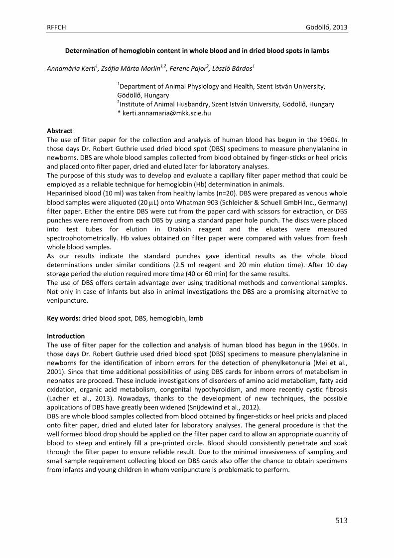
RFFCH Gödöllő, 2013
513
Determination of hemoglobin content in whole blood and in dried blood spots in lambs
Annamária Kerti1, Zsófia Márta Morlin1,2, Ferenc Pajor2, László Bárdos1
1Department of Animal Physiology and Health, Szent István University, Gödöllő, Hungary 2Institute of Animal Husbandry, Szent István University, Gödöllő, Hungary * [email protected]
Abstract The use of filter paper for the collection and analysis of human blood has begun in the 1960s. In those days Dr. Robert Guthrie used dried blood spot (DBS) specimens to measure phenylalanine in newborns. DBS are whole blood samples collected from blood obtained by finger-sticks or heel pricks and placed onto filter paper, dried and eluted later for laboratory analyses. The purpose of this study was to develop and evaluate a capillary filter paper method that could be employed as a reliable technique for hemoglobin (Hb) determination in animals. Heparinised blood (10 ml) was taken from healthy lambs (n=20). DBS were prepared as venous whole
blood samples were aliquoted (20 L) onto Whatman 903 (Schleicher & Schuell GmbH Inc., Germany) filter paper. Either the entire DBS were cut from the paper card with scissors for extraction, or DBS punches were removed from each DBS by using a standard paper hole punch. The discs were placed into test tubes for elution in Drabkin reagent and the eluates were measured spectrophotometrically. Hb values obtained on filter paper were compared with values from fresh whole blood samples. As our results indicate the standard punches gave identical results as the whole blood determinations under similar conditions (2.5 ml reagent and 20 min elution time). After 10 day storage period the elution required more time (40 or 60 min) for the same results. The use of DBS offers certain advantage over using traditional methods and conventional samples. Not only in case of infants but also in animal investigations the DBS are a promising alternative to venipuncture. Key words: dried blood spot, DBS, hemoglobin, lamb Introduction The use of filter paper for the collection and analysis of human blood has begun in the 1960s. In those days Dr. Robert Guthrie used dried blood spot (DBS) specimens to measure phenylalanine in newborns for the identification of inborn errors for the detection of phenylketonuria (Mei et al., 2001). Since that time additional possibilities of using DBS cards for inborn errors of metabolism in neonates are proceed. These include investigations of disorders of amino acid metabolism, fatty acid oxidation, organic acid metabolism, congenital hypothyroidism, and more recently cystic fibrosis (Lacher et al., 2013). Nowadays, thanks to the development of new techniques, the possible applications of DBS have greatly been widened (Snijdewind et al., 2012). DBS are whole blood samples collected from blood obtained by finger-sticks or heel pricks and placed onto filter paper, dried and eluted later for laboratory analyses. The general procedure is that the well formed blood drop should be applied on the filter paper card to allow an appropriate quantity of blood to steep and entirely fill a pre-printed circle. Blood should consistently penetrate and soak through the filter paper to ensure reliable result. Due to the minimal invasiveness of sampling and small sample requirement collecting blood on DBS cards also offer the chance to obtain specimens from infants and young children in whom venipuncture is problematic to perform.
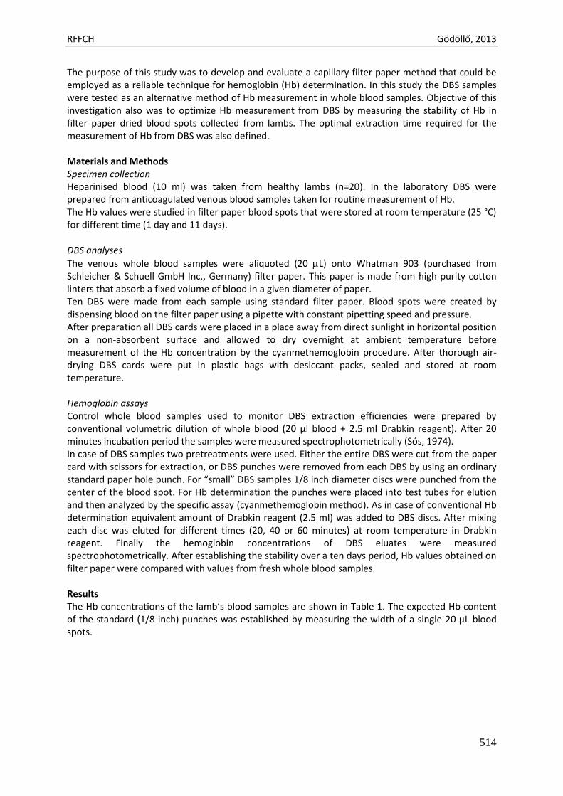
RFFCH Gödöllő, 2013
514
The purpose of this study was to develop and evaluate a capillary filter paper method that could be employed as a reliable technique for hemoglobin (Hb) determination. In this study the DBS samples were tested as an alternative method of Hb measurement in whole blood samples. Objective of this investigation also was to optimize Hb measurement from DBS by measuring the stability of Hb in filter paper dried blood spots collected from lambs. The optimal extraction time required for the measurement of Hb from DBS was also defined. Materials and Methods Specimen collection Heparinised blood (10 ml) was taken from healthy lambs (n=20). In the laboratory DBS were prepared from anticoagulated venous blood samples taken for routine measurement of Hb. The Hb values were studied in filter paper blood spots that were stored at room temperature (25 °C) for different time (1 day and 11 days). DBS analyses
The venous whole blood samples were aliquoted (20 L) onto Whatman 903 (purchased from Schleicher & Schuell GmbH Inc., Germany) filter paper. This paper is made from high purity cotton linters that absorb a fixed volume of blood in a given diameter of paper. Ten DBS were made from each sample using standard filter paper. Blood spots were created by dispensing blood on the filter paper using a pipette with constant pipetting speed and pressure. After preparation all DBS cards were placed in a place away from direct sunlight in horizontal position on a non-absorbent surface and allowed to dry overnight at ambient temperature before measurement of the Hb concentration by the cyanmethemoglobin procedure. After thorough air-drying DBS cards were put in plastic bags with desiccant packs, sealed and stored at room temperature. Hemoglobin assays Control whole blood samples used to monitor DBS extraction efficiencies were prepared by conventional volumetric dilution of whole blood (20 μl blood + 2.5 ml Drabkin reagent). After 20 minutes incubation period the samples were measured spectrophotometrically (Sós, 1974). In case of DBS samples two pretreatments were used. Either the entire DBS were cut from the paper card with scissors for extraction, or DBS punches were removed from each DBS by using an ordinary standard paper hole punch. For “small” DBS samples 1/8 inch diameter discs were punched from the center of the blood spot. For Hb determination the punches were placed into test tubes for elution and then analyzed by the specific assay (cyanmethemoglobin method). As in case of conventional Hb determination equivalent amount of Drabkin reagent (2.5 ml) was added to DBS discs. After mixing each disc was eluted for different times (20, 40 or 60 minutes) at room temperature in Drabkin reagent. Finally the hemoglobin concentrations of DBS eluates were measured spectrophotometrically. After establishing the stability over a ten days period, Hb values obtained on filter paper were compared with values from fresh whole blood samples. Results The Hb concentrations of the lamb’s blood samples are shown in Table 1. The expected Hb content of the standard (1/8 inch) punches was established by measuring the width of a single 20 μL blood spots.
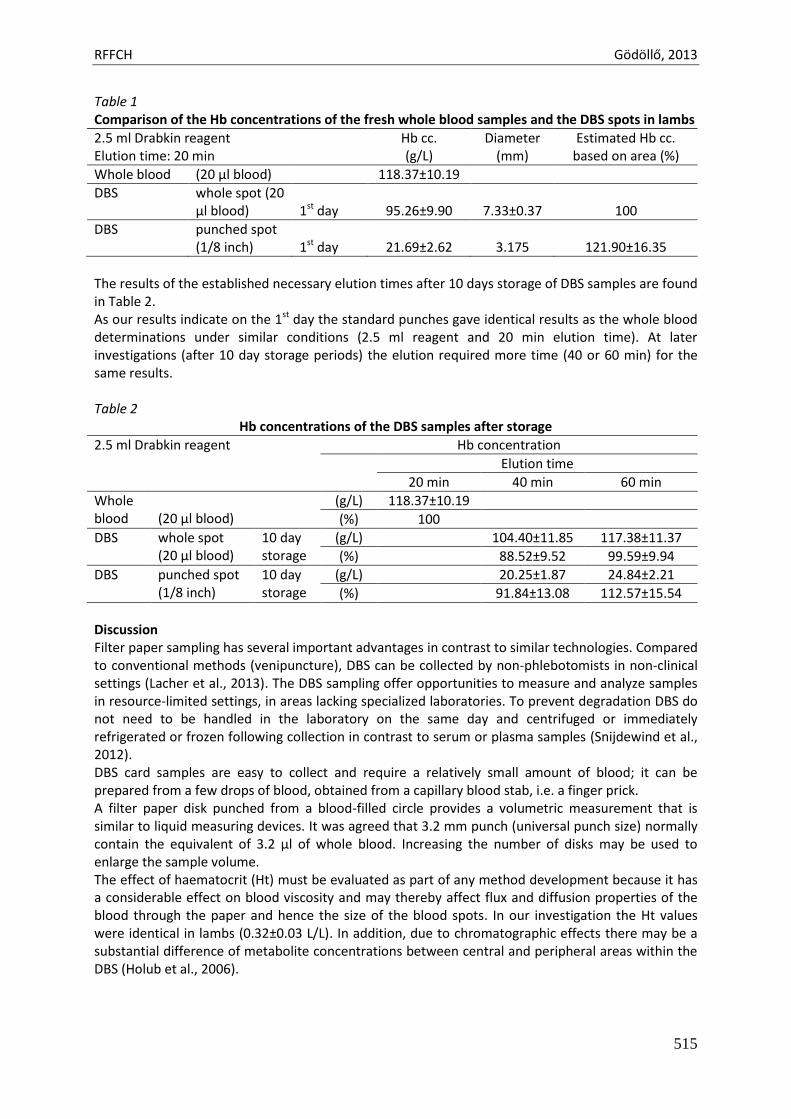
RFFCH Gödöllő, 2013
515
Table 1 Comparison of the Hb concentrations of the fresh whole blood samples and the DBS spots in lambs
2.5 ml Drabkin reagent Elution time: 20 min
Hb cc. (g/L)
Diameter (mm)
Estimated Hb cc. based on area (%)
Whole blood (20 μl blood) 118.37±10.19
DBS whole spot (20 μl blood)
1st day
95.26±9.90
7.33±0.37
100
DBS punched spot (1/8 inch)
1st day
21.69±2.62
3.175
121.90±16.35
The results of the established necessary elution times after 10 days storage of DBS samples are found in Table 2. As our results indicate on the 1st day the standard punches gave identical results as the whole blood determinations under similar conditions (2.5 ml reagent and 20 min elution time). At later investigations (after 10 day storage periods) the elution required more time (40 or 60 min) for the same results. Table 2
Hb concentrations of the DBS samples after storage
2.5 ml Drabkin reagent Hb concentration
Elution time
20 min 40 min 60 min
Whole blood
(20 μl blood)
(g/L) 118.37±10.19
(%) 100
DBS whole spot (20 μl blood)
10 day storage
(g/L) 104.40±11.85 117.38±11.37
(%) 88.52±9.52 99.59±9.94
DBS punched spot (1/8 inch)
10 day storage
(g/L) 20.25±1.87 24.84±2.21
(%) 91.84±13.08 112.57±15.54
Discussion Filter paper sampling has several important advantages in contrast to similar technologies. Compared to conventional methods (venipuncture), DBS can be collected by non-phlebotomists in non-clinical settings (Lacher et al., 2013). The DBS sampling offer opportunities to measure and analyze samples in resource-limited settings, in areas lacking specialized laboratories. To prevent degradation DBS do not need to be handled in the laboratory on the same day and centrifuged or immediately refrigerated or frozen following collection in contrast to serum or plasma samples (Snijdewind et al., 2012). DBS card samples are easy to collect and require a relatively small amount of blood; it can be prepared from a few drops of blood, obtained from a capillary blood stab, i.e. a finger prick. A filter paper disk punched from a blood-filled circle provides a volumetric measurement that is similar to liquid measuring devices. It was agreed that 3.2 mm punch (universal punch size) normally contain the equivalent of 3.2 μl of whole blood. Increasing the number of disks may be used to enlarge the sample volume. The effect of haematocrit (Ht) must be evaluated as part of any method development because it has a considerable effect on blood viscosity and may thereby affect flux and diffusion properties of the blood through the paper and hence the size of the blood spots. In our investigation the Ht values were identical in lambs (0.32±0.03 L/L). In addition, due to chromatographic effects there may be a substantial difference of metabolite concentrations between central and peripheral areas within the DBS (Holub et al., 2006).
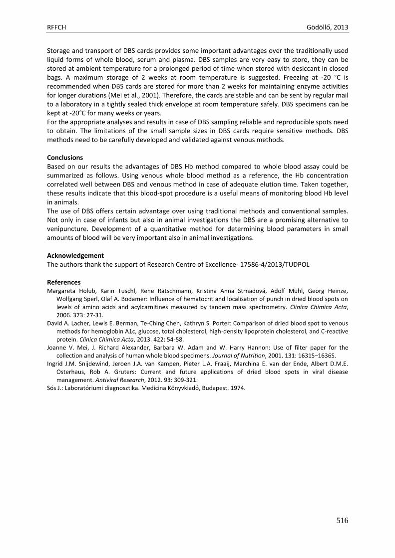
RFFCH Gödöllő, 2013
516
Storage and transport of DBS cards provides some important advantages over the traditionally used liquid forms of whole blood, serum and plasma. DBS samples are very easy to store, they can be stored at ambient temperature for a prolonged period of time when stored with desiccant in closed bags. A maximum storage of 2 weeks at room temperature is suggested. Freezing at -20 °C is recommended when DBS cards are stored for more than 2 weeks for maintaining enzyme activities for longer durations (Mei et al., 2001). Therefore, the cards are stable and can be sent by regular mail to a laboratory in a tightly sealed thick envelope at room temperature safely. DBS specimens can be kept at -20°C for many weeks or years. For the appropriate analyses and results in case of DBS sampling reliable and reproducible spots need to obtain. The limitations of the small sample sizes in DBS cards require sensitive methods. DBS methods need to be carefully developed and validated against venous methods. Conclusions Based on our results the advantages of DBS Hb method compared to whole blood assay could be summarized as follows. Using venous whole blood method as a reference, the Hb concentration correlated well between DBS and venous method in case of adequate elution time. Taken together, these results indicate that this blood-spot procedure is a useful means of monitoring blood Hb level in animals. The use of DBS offers certain advantage over using traditional methods and conventional samples. Not only in case of infants but also in animal investigations the DBS are a promising alternative to venipuncture. Development of a quantitative method for determining blood parameters in small amounts of blood will be very important also in animal investigations. Acknowledgement The authors thank the support of Research Centre of Excellence- 17586-4/2013/TUDPOL References Margareta Holub, Karin Tuschl, Rene Ratschmann, Kristina Anna Strnadová, Adolf Mühl, Georg Heinze,
Wolfgang Sperl, Olaf A. Bodamer: Influence of hematocrit and localisation of punch in dried blood spots on levels of amino acids and acylcarnitines measured by tandem mass spectrometry. Clinica Chimica Acta, 2006. 373: 27-31.
David A. Lacher, Lewis E. Berman, Te-Ching Chen, Kathryn S. Porter: Comparison of dried blood spot to venous methods for hemoglobin A1c, glucose, total cholesterol, high-density lipoprotein cholesterol, and C-reactive protein. Clinica Chimica Acta, 2013. 422: 54-58.
Joanne V. Mei, J. Richard Alexander, Barbara W. Adam and W. Harry Hannon: Use of filter paper for the collection and analysis of human whole blood specimens. Journal of Nutrition, 2001. 131: 1631S–1636S.
Ingrid J.M. Snijdewind, Jeroen J.A. van Kampen, Pieter L.A. Fraaij, Marchina E. van der Ende, Albert D.M.E. Osterhaus, Rob A. Gruters: Current and future applications of dried blood spots in viral disease management. Antiviral Research, 2012. 93: 309-321.
Sós J.: Laboratóriumi diagnosztika. Medicina Könyvkiadó, Budapest. 1974.
Animal welfare, etológia és tartástechnológia
Animal welfare, ethology and housing systems
Volume 9 Issue 3
Különszám/Special Issue
Gödöllő
2013
RFFCH Gödöllő, 2013
517
Pasta preparation by coloured natural antioxidants Zsuzsanna Kiss, Annamária Kerti, Csaba Szabó, Krisztina Karchesz, László Bárdos*
Department of Animal Physiology & Health, Szent István University, Gödöllő, Hungary *[email protected]
Abstract Our aim was to prepare pastas with different colours by using natural antioxidant containing foods as additives. The tomato and paprika contain red colours ingredients lycopene and capsanthin and capsorubin respectively. Spinach is chlorophyll rich food plant and it contains a lot of lutein too. These model pasta beside their uncommon colours have better biological activities and can be used as functional food. Wheat flour was used for the preparation of pastas. The spinach puree for the green pasta, tomato puree, and red paprika powder for the red coloured pasta. The surface colours of paste were determined by handy spectrometers (Micromatch™ Sheen Ltd) which use the international standard of CIELAB values. The coloration was successful by the additives and can be seen before and after cooking. It was a fact that the colouring by paprika has not changed. In the other cases there were more or less alterations of CIELAB values. The chlorophyll content of spinach and lycopene content of tomato puree was analyzed. Three antioxidant reactions of pastas were characterized by ABTS, DPPH and FRAP methods respectively. The measurements of antioxidant capacity of different components showed a ranking order. These additives increase the antioxidant capacity of paste that they have health protective effects. Keywords: coloured pasta, antioxidant capacity, ABTS, DPPH, FRAP Introduction Functional foods are foods that have a potentially positive effect on health beyond basic nutrition. To produce such a foods sometimes new ingredients has been added to it and the new product has a new function often related to health-promotion or disease prevention. Epidemiological and experimental evidence associating diets rich in fruits and vegetables with prevention of chronic diseases such as cancer has stimulated interest in plant food phytochemicals as physiologically active dietary components. Our aim was to prepare pastas with different colours by using natural antioxidant containing foods as additives. The tomato and paprika contain red colours ingredients (lycopene and capsanthin and capsorubin respectively. Spinach is chlorophyll rich food plant and it contains a lot of lutein too. These model pasta beside their uncommon colours have better biological activities and can be used as functional food. Lycopene is a carotenoid and phytonutrient found in red fruits and vegetables such as tomatoes, pink grapefruits, watermelons etc. It is the compound that is responsible for the red colour in these foods. Numerous studies have shown that ingesting lycopene-rich foods can result in positive health benefits. (Britton, et al. 2009). Oxidative stress is one of the major contributors to increased risk of some diseases. Lycopene is a very effective antioxidant. A diet rich in lycopene containing tomato products has been found to protect against these chronic diseases by mitigating oxidative damage. (Rao, 2004). Considerable evidence suggests that lycopene, has significant antioxidant potential in vitro and may play a role in preventing prostate cancer and cardiovascular disease in humans. (Arab and Steck 2000) Prostate cancer is the most common cancer in men population in the industrialized countries. Recent epidemiological studies have suggested a potential benefit of this carotenoid against the risk of prostate cancer (Giovannucci, 2002).
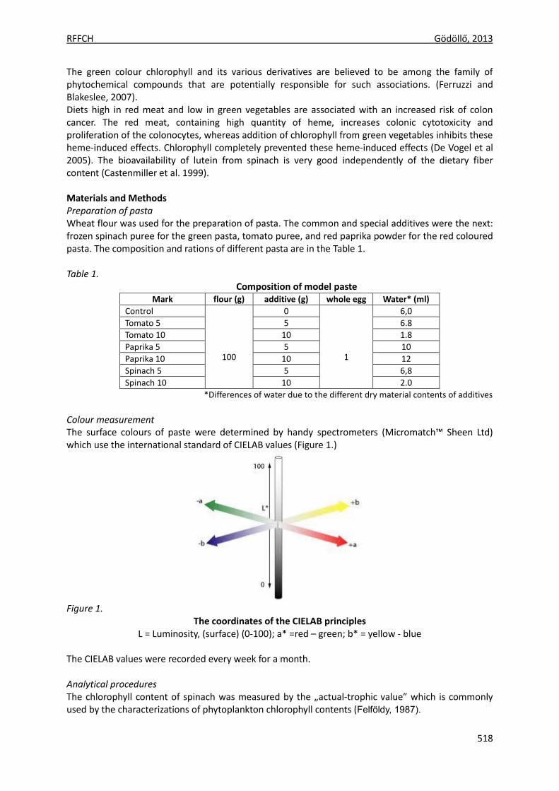
RFFCH Gödöllő, 2013
518
The green colour chlorophyll and its various derivatives are believed to be among the family of phytochemical compounds that are potentially responsible for such associations. (Ferruzzi and Blakeslee, 2007). Diets high in red meat and low in green vegetables are associated with an increased risk of colon cancer. The red meat, containing high quantity of heme, increases colonic cytotoxicity and proliferation of the colonocytes, whereas addition of chlorophyll from green vegetables inhibits these heme-induced effects. Chlorophyll completely prevented these heme-induced effects (De Vogel et al 2005). The bioavailability of lutein from spinach is very good independently of the dietary fiber content (Castenmiller et al. 1999). Materials and Methods Preparation of pasta Wheat flour was used for the preparation of pasta. The common and special additives were the next: frozen spinach puree for the green pasta, tomato puree, and red paprika powder for the red coloured pasta. The composition and rations of different pasta are in the Table 1. Table 1.
Composition of model paste Mark flour (g) additive (g) whole egg Water* (ml)
Control
100
0
1
6,0
Tomato 5 5 6.8
Tomato 10 10 1.8
Paprika 5 5 10
Paprika 10 10 12
Spinach 5 5 6,8
Spinach 10 10 2.0
*Differences of water due to the different dry material contents of additives
Colour measurement The surface colours of paste were determined by handy spectrometers (Micromatch™ Sheen Ltd) which use the international standard of CIELAB values (Figure 1.)
Figure 1.
The coordinates of the CIELAB principles L = Luminosity, (surface) (0-100); a* =red – green; b* = yellow - blue
The CIELAB values were recorded every week for a month. Analytical procedures The chlorophyll content of spinach was measured by the „actual-trophic value” which is commonly used by the characterizations of phytoplankton chlorophyll contents (Felföldy, 1987).
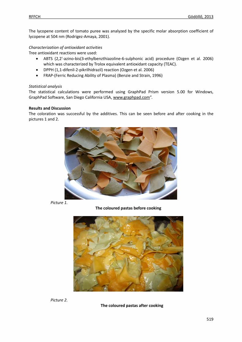
RFFCH Gödöllő, 2013
519
The lycopene content of tomato puree was analyzed by the specific molar absorption coefficient of lycopene at 504 nm (Rodrigez-Amaya, 2001). Characterization of antioxidant activities Tree antioxidant reactions were used:
ABTS (2,2'-azino-bis(3-ethylbenzthiazoline-6-sulphonic acid) procedure (Ozgen et al. 2006) which was characterized by Trolox equivalent antioxidant capacity (TEAC).
DPPH (1,1-difenil-2-pikrilhidrazil) reaction (Ozgen et al. 2006)
FRAP-(Ferric Reducing Ability of Plasma) (Benzie and Strain, 1996) Statistical analysis The statistical calculations were performed using GraphPad Prism version 5.00 for Windows, GraphPad Software, San Diego California USA, www.graphpad.com”. Results and Discussion The coloration was successful by the additives. This can be seen before and after cooking in the pictures 1 and 2.
Picture 1.
The coloured pastas before cooking
Picture 2. The coloured pastas after cooking
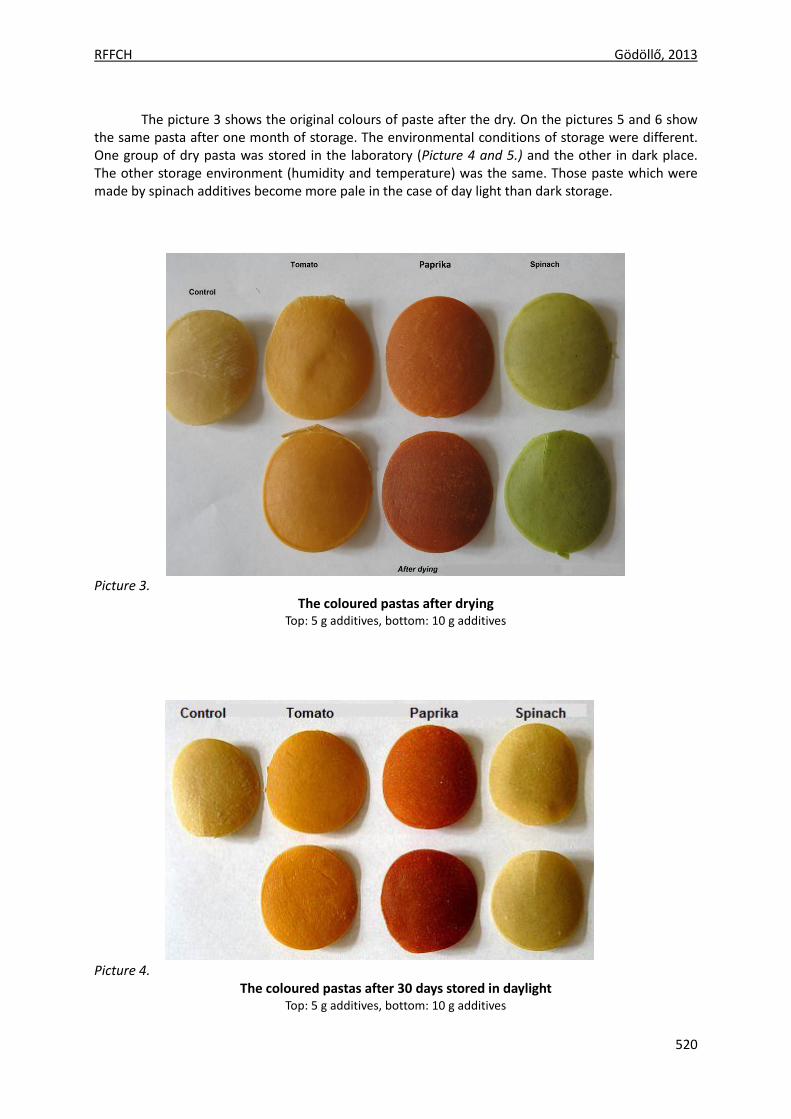
RFFCH Gödöllő, 2013
520
The picture 3 shows the original colours of paste after the dry. On the pictures 5 and 6 show
the same pasta after one month of storage. The environmental conditions of storage were different. One group of dry pasta was stored in the laboratory (Picture 4 and 5.) and the other in dark place. The other storage environment (humidity and temperature) was the same. Those paste which were made by spinach additives become more pale in the case of day light than dark storage.
Picture 3.
The coloured pastas after drying Top: 5 g additives, bottom: 10 g additives
Picture 4.
The coloured pastas after 30 days stored in daylight Top: 5 g additives, bottom: 10 g additives
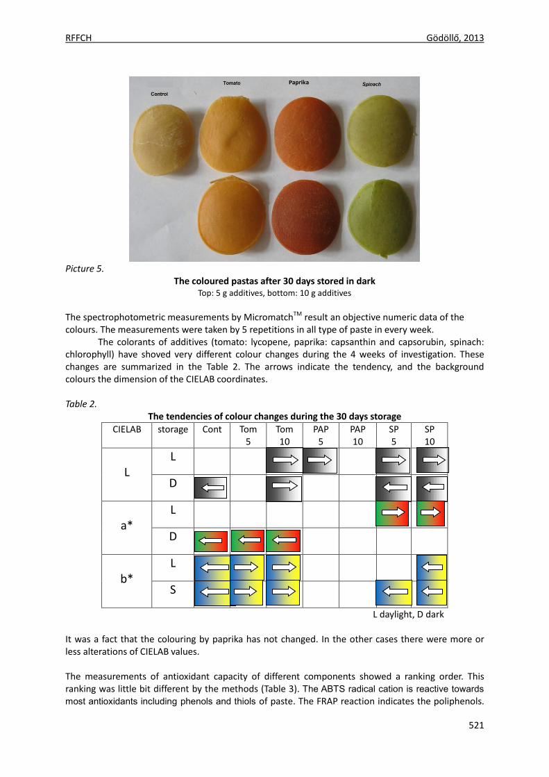
RFFCH Gödöllő, 2013
521
Picture 5.
The coloured pastas after 30 days stored in dark
Top: 5 g additives, bottom: 10 g additives
The spectrophotometric measurements by MicromatchTM result an objective numeric data of the colours. The measurements were taken by 5 repetitions in all type of paste in every week.
The colorants of additives (tomato: lycopene, paprika: capsanthin and capsorubin, spinach: chlorophyll) have shoved very different colour changes during the 4 weeks of investigation. These changes are summarized in the Table 2. The arrows indicate the tendency, and the background colours the dimension of the CIELAB coordinates. Table 2.
The tendencies of colour changes during the 30 days storage
CIELAB storage Cont Tom 5
Tom 10
PAP 5
PAP 10
SP 5
SP 10
L
L
D
a*
L
D
b*
L
S
L daylight, D dark It was a fact that the colouring by paprika has not changed. In the other cases there were more or less alterations of CIELAB values. The measurements of antioxidant capacity of different components showed a ranking order. This ranking was little bit different by the methods (Table 3). The ABTS radical cation is reactive towards
most antioxidants including phenols and thiols of paste. The FRAP reaction indicates the poliphenols.
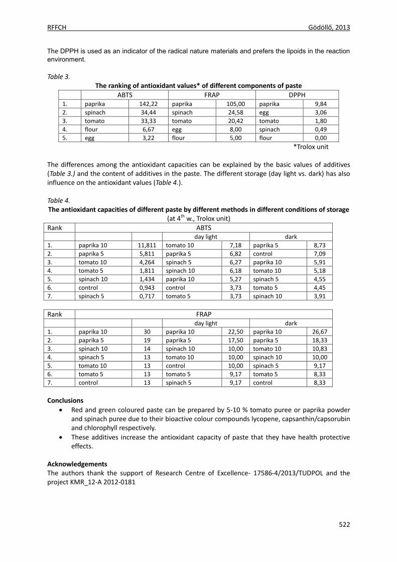
RFFCH Gödöllő, 2013
522
The DPPH is used as an indicator of the radical nature materials and prefers the lipoids in the reaction
environment. Table 3.
The ranking of antioxidant values* of different components of paste
ABTS FRAP DPPH 1. paprika 142,22 paprika 105,00 paprika 9,84
2. spinach 34,44 spinach 24,58 egg 3,06
3. tomato 33,33 tomato 20,42 tomato 1,80
4. flour 6,67 egg 8,00 spinach 0,49
5. egg 3,22 flour 5,00 flour 0,00
*Trolox unit The differences among the antioxidant capacities can be explained by the basic values of additives (Table 3.) and the content of additives in the paste. The different storage (day light vs. dark) has also influence on the antioxidant values (Table 4.). Table 4. The antioxidant capacities of different paste by different methods in different conditions of storage
(at 4th w., Trolox unit)
Rank ABTS day light dark
1. paprika 10 11,811 tomato 10 7,18 paprika 5 8,73
2. paprika 5 5,811 paprika 5 6,82 control 7,09
3. tomato 10 4,264 spinach 5 6,27 paprika 10 5,91
4. tomato 5 1,811 spinach 10 6,18 tomato 10 5,18
5. spinach 10 1,434 paprika 10 5,27 spinach 5 4,55
6. control 0,943 control 3,73 tomato 5 4,45
7. spinach 5 0,717 tomato 5 3,73 spinach 10 3,91
Rank FRAP day light dark
1. paprika 10 30 paprika 10 22,50 paprika 10 26,67
2. paprika 5 19 paprika 5 17,50 paprika 5 18,33
3. spinach 10 14 spinach 10 10,00 tomato 10 10,83
4. spinach 5 13 tomato 10 10,00 spinach 10 10,00
5. tomato 10 13 control 10,00 spinach 5 9,17
6. tomato 5 13 tomato 5 9,17 tomato 5 8,33
7. control 13 spinach 5 9,17 control 8,33
Conclusions
Red and green coloured paste can be prepared by 5-10 % tomato puree or paprika powder and spinach puree due to their bioactive colour compounds lycopene, capsanthin/capsorubin and chlorophyll respectively.
These additives increase the antioxidant capacity of paste that they have health protective effects.
Acknowledgements The authors thank the support of Research Centre of Excellence- 17586-4/2013/TUDPOL and the project KMR_12-A 2012-0181
RFFCH Gödöllő, 2013
523
References ARAB, L. , STECK, S. (2000): Lycopene and cardiovascular disease. Am J Clin Nutr; 71(suppl):1691S–1695S. BENZIE, F.F., STRAIN, J.J. (1996): The ferric reducing ability of plasma (FRAP) as a measure of “antioxidant power”:
The FRAP assay. Anal. Biochem. 239: 70-76. BLOIS M. S.(1958): Antioxidant determination by the use of a stable free radicals. Nature, 1958. (4617): 1198-
1200. BRITTON, G., LIAAEN-JENSEN, S., PFANDER, H. (2009): Carotenoids. Nutrition and Health. Birkhäuser Verlag, Basel,
Volume 5. CASTENMILLER, J.J.M., WEST, C.E., ET. AL. (1999) The Food Matrix of Spinach Is a Limiting Factor in Determining the
Bioavailability of ß-CAROTENE and to a Lesser Extent of Lutein in Humans. Journal of Nutr; 129:349-355. DE VOGEL,J., JONKER-TERMONT, D.S.M., VAN LIESHOUT, E.M.M., KATAN,M.B., VAN DER MEER, R. (2005): Green vegetables,
red meat and colon cancer: chlorophyll prevents the cytotoxic and hyperproliferative effects of haem in rat colon. Carcinogenesis 26:387—393.
FELFÖLDY L. (1987): A biológiai vízminősítés (4. javított és bővített kiadás). Vízügyi Hidrobiológia 16., 1-258. - VGI, Budapest.
FERRUZZI,M.G., BLAKESLEE, J.(2007): Digestion, absorption, and cancer preventative activity of dietary chlorophyll derivatives. Nutr. Res.,27: 1– 12.
GIOVANNUCCI, E. (2002): A Review of Epidemiologic Studies of Tomatoes, Lycopene, and Prostate Cancer. Exp.Biol. Med., 227:852-859.)
HAE-YUN CHUNG ET. AL. (2004): Lutein Bioavailability Is Higher from Lutein-Enriched Eggs than from Supplements and Spinach in Men. .J. Nutr. 134: 1887–1893.
KRINSKY, N.I., LANDRUM, J.T., BONE,R.A.: Biologic mechanisms of the protective role of lutein and zeaxanthin in the eye. Ann. Rev. Nutr., 23: 171-201. 2003.).
OZGEN, M., REESE, N.R., TULIO, A.Z., SCHEERENS, J.C., MILLER, R. (2006): Modified 2,2-Azino-bis-3-ethylbenzothiazoline-6-sulfonic Acid (ABTS) Method to Measure Antioxidant Capacity of Selected Small Fruits and Comparison to Ferric Reducing Antioxidant Power (FRAP) and 2,2¢-Diphenyl-1-picrylhydrazyl (DPPH) Methods. J. Agric. Food Chem. 54, 1151-1157
RAO, A.V. (2004): Processed Tomato Products as a Source of Dietary Lycopene:Bioavailability and Antioxidant Properties. Canad. J.Diet. Practice Res., 65: 161-165.
RODRIGEZ-AMAYA,D.B. (2001): A guide to carotenoid analysis in food, ILSI Press, Washington, D.C. WHO/FAO (2003):Diet, Nutrition and Prevention of Chronic Diseases, 2003.

Animal welfare, etológia és tartástechnológia
Animal welfare, ethology and housing systems
Volume 9 Issue 3
Különszám/Special Issue
Gödöllő
2013
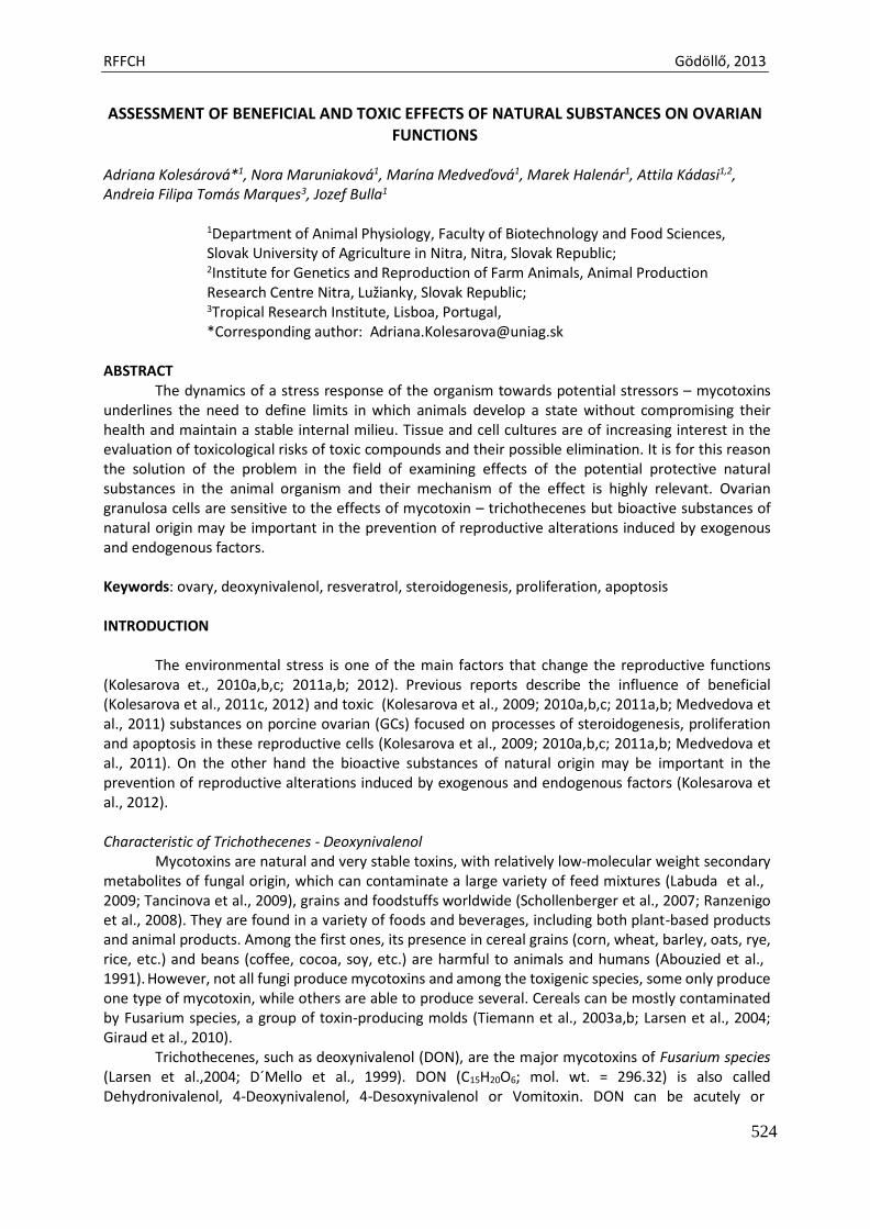
RFFCH Gödöllő, 2013
524
ASSESSMENT OF BENEFICIAL AND TOXIC EFFECTS OF NATURAL SUBSTANCES ON OVARIAN FUNCTIONS
Adriana Kolesárová*1, Nora Maruniaková1, Marína Medveďová1, Marek Halenár1, Attila Kádasi1,2, Andreia Filipa Tomás Marques3, Jozef Bulla1
1Department of Animal Physiology, Faculty of Biotechnology and Food Sciences, Slovak University of Agriculture in Nitra, Nitra, Slovak Republic; 2Institute for Genetics and Reproduction of Farm Animals, Animal Production Research Centre Nitra, Lužianky, Slovak Republic; 3Tropical Research Institute, Lisboa, Portugal, *Corresponding author: [email protected]
ABSTRACT
The dynamics of a stress response of the organism towards potential stressors – mycotoxins underlines the need to define limits in which animals develop a state without compromising their health and maintain a stable internal milieu. Tissue and cell cultures are of increasing interest in the evaluation of toxicological risks of toxic compounds and their possible elimination. It is for this reason the solution of the problem in the field of examining effects of the potential protective natural substances in the animal organism and their mechanism of the effect is highly relevant. Ovarian granulosa cells are sensitive to the effects of mycotoxin – trichothecenes but bioactive substances of natural origin may be important in the prevention of reproductive alterations induced by exogenous and endogenous factors.
Keywords: ovary, deoxynivalenol, resveratrol, steroidogenesis, proliferation, apoptosis
INTRODUCTION
The environmental stress is one of the main factors that change the reproductive functions
(Kolesarova et., 2010a,b,c; 2011a,b; 2012). Previous reports describe the influence of beneficial (Kolesarova et al., 2011c, 2012) and toxic (Kolesarova et al., 2009; 2010a,b,c; 2011a,b; Medvedova et al., 2011) substances on porcine ovarian (GCs) focused on processes of steroidogenesis, proliferation and apoptosis in these reproductive cells (Kolesarova et al., 2009; 2010a,b,c; 2011a,b; Medvedova et al., 2011). On the other hand the bioactive substances of natural origin may be important in the prevention of reproductive alterations induced by exogenous and endogenous factors (Kolesarova et al., 2012).
Characteristic of Trichothecenes - Deoxynivalenol
Mycotoxins are natural and very stable toxins, with relatively low-molecular weight secondary metabolites of fungal origin, which can contaminate a large variety of feed mixtures (Labuda et al., 2009; Tancinova et al., 2009), grains and foodstuffs worldwide (Schollenberger et al., 2007; Ranzenigo et al., 2008). They are found in a variety of foods and beverages, including both plant-based products and animal products. Among the first ones, its presence in cereal grains (corn, wheat, barley, oats, rye, rice, etc.) and beans (coffee, cocoa, soy, etc.) are harmful to animals and humans (Abouzied et al., 1991). However, not all fungi produce mycotoxins and among the toxigenic species, some only produce one type of mycotoxin, while others are able to produce several. Cereals can be mostly contaminated by Fusarium species, a group of toxin-producing molds (Tiemann et al., 2003a,b; Larsen et al., 2004; Giraud et al., 2010).
Trichothecenes, such as deoxynivalenol (DON), are the major mycotoxins of Fusarium species (Larsen et al.,2004; D´Mello et al., 1999). DON (C15H20O6; mol. wt. = 296.32) is also called Dehydronivalenol, 4-Deoxynivalenol, 4-Desoxynivalenol or Vomitoxin. DON can be acutely or
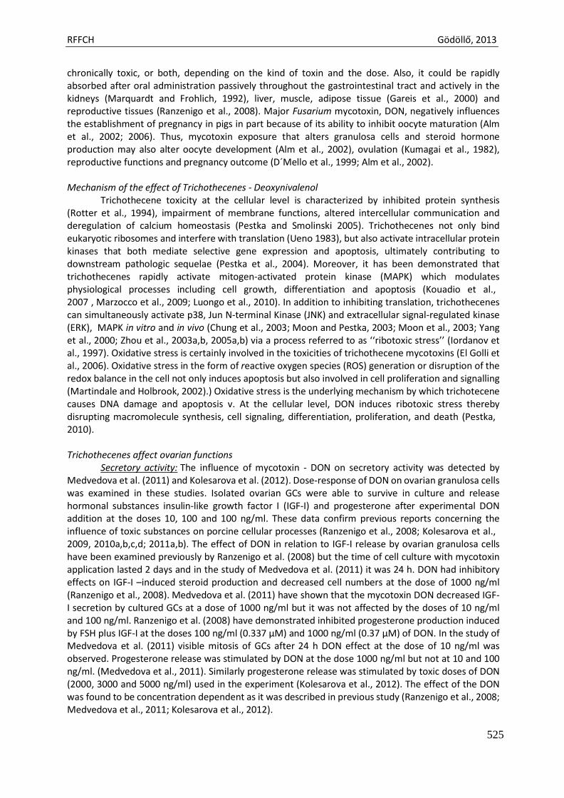
RFFCH Gödöllő, 2013
525
chronically toxic, or both, depending on the kind of toxin and the dose. Also, it could be rapidly absorbed after oral administration passively throughout the gastrointestinal tract and actively in the kidneys (Marquardt and Frohlich, 1992), liver, muscle, adipose tissue (Gareis et al., 2000) and reproductive tissues (Ranzenigo et al., 2008). Major Fusarium mycotoxin, DON, negatively influences the establishment of pregnancy in pigs in part because of its ability to inhibit oocyte maturation (Alm et al., 2002; 2006). Thus, mycotoxin exposure that alters granulosa cells and steroid hormone production may also alter oocyte development (Alm et al., 2002), ovulation (Kumagai et al., 1982), reproductive functions and pregnancy outcome (D´Mello et al., 1999; Alm et al., 2002).
Mechanism of the effect of Trichothecenes - Deoxynivalenol
Trichothecene toxicity at the cellular level is characterized by inhibited protein synthesis (Rotter et al., 1994), impairment of membrane functions, altered intercellular communication and deregulation of calcium homeostasis (Pestka and Smolinski 2005). Trichothecenes not only bind eukaryotic ribosomes and interfere with translation (Ueno 1983), but also activate intracellular protein kinases that both mediate selective gene expression and apoptosis, ultimately contributing to downstream pathologic sequelae (Pestka et al., 2004). Moreover, it has been demonstrated that trichothecenes rapidly activate mitogen-activated protein kinase (MAPK) which modulates physiological processes including cell growth, differentiation and apoptosis (Kouadio et al., 2007 , Marzocco et al., 2009; Luongo et al., 2010). In addition to inhibiting translation, trichothecenes can simultaneously activate p38, Jun N-terminal Kinase (JNK) and extracellular signal-regulated kinase (ERK), MAPK in vitro and in vivo (Chung et al., 2003; Moon and Pestka, 2003; Moon et al., 2003; Yang et al., 2000; Zhou et al., 2003a,b, 2005a,b) via a process referred to as ‘‘ribotoxic stress’’ (Iordanov et al., 1997). Oxidative stress is certainly involved in the toxicities of trichothecene mycotoxins (El Golli et al., 2006). Oxidative stress in the form of reactive oxygen species (ROS) generation or disruption of the redox balance in the cell not only induces apoptosis but also involved in cell proliferation and signalling (Martindale and Holbrook, 2002).) Oxidative stress is the underlying mechanism by which trichotecene causes DNA damage and apoptosis v. At the cellular level, DON induces ribotoxic stress thereby disrupting macromolecule synthesis, cell signaling, differentiation, proliferation, and death (Pestka, 2010).
Trichothecenes affect ovarian functions
Secretory activity: The influence of mycotoxin - DON on secretory activity was detected by Medvedova et al. (2011) and Kolesarova et al. (2012). Dose-response of DON on ovarian granulosa cells was examined in these studies. Isolated ovarian GCs were able to survive in culture and release hormonal substances insulin-like growth factor I (IGF-I) and progesterone after experimental DON addition at the doses 10, 100 and 100 ng/ml. These data confirm previous reports concerning the influence of toxic substances on porcine cellular processes (Ranzenigo et al., 2008; Kolesarova et al., 2009, 2010a,b,c,d; 2011a,b). The effect of DON in relation to IGF-I release by ovarian granulosa cells have been examined previously by Ranzenigo et al. (2008) but the time of cell culture with mycotoxin application lasted 2 days and in the study of Medvedova et al. (2011) it was 24 h. DON had inhibitory effects on IGF-I –induced steroid production and decreased cell numbers at the dose of 1000 ng/ml (Ranzenigo et al., 2008). Medvedova et al. (2011) have shown that the mycotoxin DON decreased IGF- I secretion by cultured GCs at a dose of 1000 ng/ml but it was not affected by the doses of 10 ng/ml and 100 ng/ml. Ranzenigo et al. (2008) have demonstrated inhibited progesterone production induced by FSH plus IGF-I at the doses 100 ng/ml (0.337 µM) and 1000 ng/ml (0.37 µM) of DON. In the study of Medvedova et al. (2011) visible mitosis of GCs after 24 h DON effect at the dose of 10 ng/ml was observed. Progesterone release was stimulated by DON at the dose 1000 ng/ml but not at 10 and 100 ng/ml. (Medvedova et al., 2011). Similarly progesterone release was stimulated by toxic doses of DON (2000, 3000 and 5000 ng/ml) used in the experiment (Kolesarova et al., 2012). The effect of the DON was found to be concentration dependent as it was described in previous study (Ranzenigo et al., 2008; Medvedova et al., 2011; Kolesarova et al., 2012).
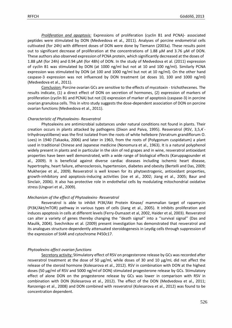
RFFCH Gödöllő, 2013
526
Proliferation and apoptosis: Expressions of proliferation (cyclin B1 and PCNA)- associated
peptides were stimulated by DON (Medvedova et al., 2011). Analyses of porcine endometrial cells cultivated (for 24h) with different doses of DON were done by Tiemann (2003a). These results point out to significant decrease of proliferation at the concentrations of 1.88 µM and 3.76 µM of DON. These authors also observed expression of PCNA protein, which significantly decreased at the doses of 1.88 µM (for 24h) and 0.94 µM (for 48h) of DON. In the study of Medvedova et al. (2011) expression of cyclin B1 was stimulated by DON (at 1000 ng/ml but not at 10 and 100 ng/ml). Similarly PCNA expression was stimulated by DON (at 100 and 1000 ng/ml but not at 10 ng/ml). On the other hand caspase-3 expression was not influenced by DON treatment (at doses 10, 100 and 1000 ng/ml) (Medvedova et al., 2011).
Conclusion: Porcine ovarian GCs are sensitive to the effects of mycotoxin - trichothecenes. The results indicate, (1) a direct effect of DON on secretion of hormones, (2) expression of markers of proliferation (cyclin B1 and PCNA) but not (3) expression of marker of apoptosis (caspase-3) in porcine ovarian granulosa cells. This in vitro study suggests the dose-dependent association of DON on porcine ovarian functions (Medvedova et al., 2011).
Characteristic of Phytoalexins- Resveratrol
Phytoalexins are antimicrobial substances under natural conditions not found in plants. Their creation occurs in plants attacked by pathogens (Dixon and Paiva, 1995). Resveratrol (RSV, 3,5,4´- trihydroxystilbene) was the first isolated from the roots of white hellebore (Veratrum grandiflorum O. Loes) in 1940 (Takaoka, 2006) and later in 1963, from the roots of (Polygonum cuspidatum) a plant used in traditional Chinese and Japanese medicine (Nonomura et al., 1963). It is a natural polyphenol widely present in plants and in particular in the skin of red grapes and in wine, resveratrol antioxidant properties have been well demonstrated, with a wide range of biological effects (Karuppagounder et al., 2009). It is beneficial against diverse cardiac diseases including ischemic heart disease, hypertrophy, heart failure, atherosclerosis, hypertension, diabetes and obesity (Bertelli and Das, 2009; Mukherjee et al., 2009). Resveratrol is well known for its phytoestrogenic, antioxidant properties, growth-inhibitory and apoptosis-inducing activities (Joe et al., 2002; Jiang et al., 2005; Baur and Sinclair, 2006). It also has protective role in endothelial cells by modulating mitochondrial oxidative stress (Ungvari et al., 2009).
Mechanism of the effect of Phytoalexins- Resveratrol
Resveratrol is able to inhibit PI3K/Akt Protein Kinase/ mammalian target of rapamycin (PI3K/Akt/mTOR) pathway in various types of cells (Jiang et al., 2005). It inhibits proliferation and induces apoptosis in cells at different levels (Ferry-Dumazet et al, 2002, Haider et al, 2003). Resveratrol can alter a variety of genes thereby changing the ‘‘death signal’’ into a ‘‘survival signal’’ (Das and Maulik, 2004). Svechnikov et al. (2009) present investigation has demonstrated that resveratrol and its analogues structure-dependently attenuated steroidogenesis in Leydig cells through suppression of the expression of StAR and cytochrome P450c17.
Phytoalexins affect ovarian functions Secretory activity: Stimulatory effect of RSV on progesterone release by GCs was recorded after
resveratrol treatment at the dose of 50 μg/ml, while doses of 30 and 10 μg/mL did not affect the release of the steroid hormone (Kolesarova et al., 2012). RSV in combination with DON at the highest doses (50 µg/ml of RSV and 5000 ng/ml of DON) stimulated progesterone release by GCs. Stimulatory effect of alone DON on the progesterone release by GCs was lower in comparison with RSV in combination with DON (Kolesarova et al., 2012). The effect of the DON (Medvedova et al., 2011; Ranzenigo et al., 2008) and DON combined with resveratrol (Kolesarova et al., 2012) was found to be concentration dependent.
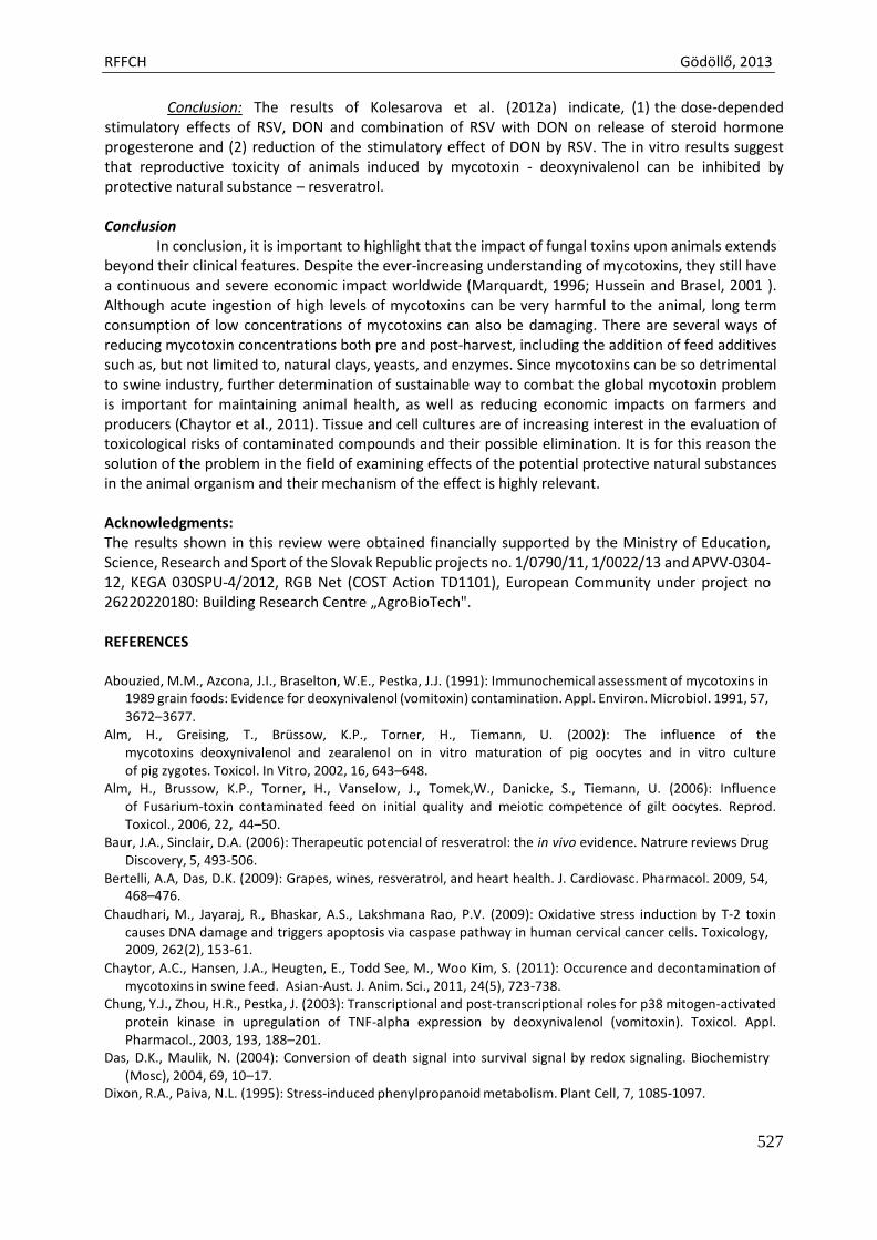
RFFCH Gödöllő, 2013
527
Conclusion: The results of Kolesarova et al. (2012a) indicate, (1) the dose-depended stimulatory effects of RSV, DON and combination of RSV with DON on release of steroid hormone progesterone and (2) reduction of the stimulatory effect of DON by RSV. The in vitro results suggest that reproductive toxicity of animals induced by mycotoxin - deoxynivalenol can be inhibited by protective natural substance – resveratrol.
Conclusion
In conclusion, it is important to highlight that the impact of fungal toxins upon animals extends beyond their clinical features. Despite the ever-increasing understanding of mycotoxins, they still have a continuous and severe economic impact worldwide (Marquardt, 1996; Hussein and Brasel, 2001 ). Although acute ingestion of high levels of mycotoxins can be very harmful to the animal, long term consumption of low concentrations of mycotoxins can also be damaging. There are several ways of reducing mycotoxin concentrations both pre and post-harvest, including the addition of feed additives such as, but not limited to, natural clays, yeasts, and enzymes. Since mycotoxins can be so detrimental to swine industry, further determination of sustainable way to combat the global mycotoxin problem is important for maintaining animal health, as well as reducing economic impacts on farmers and producers (Chaytor et al., 2011). Tissue and cell cultures are of increasing interest in the evaluation of toxicological risks of contaminated compounds and their possible elimination. It is for this reason the solution of the problem in the field of examining effects of the potential protective natural substances in the animal organism and their mechanism of the effect is highly relevant.
Acknowledgments: The results shown in this review were obtained financially supported by the Ministry of Education, Science, Research and Sport of the Slovak Republic projects no. 1/0790/11, 1/0022/13 and APVV-0304- 12, KEGA 030SPU-4/2012, RGB Net (COST Action TD1101), European Community under project no 26220220180: Building Research Centre „AgroBioTech".
REFERENCES
Abouzied, M.M., Azcona, J.I., Braselton, W.E., Pestka, J.J. (1991): Immunochemical assessment of mycotoxins in
1989 grain foods: Evidence for deoxynivalenol (vomitoxin) contamination. Appl. Environ. Microbiol. 1991, 57, 3672–3677.
Alm, H., Greising, T., Brüssow, K.P., Torner, H., Tiemann, U. (2002): The influence of the mycotoxins deoxynivalenol and zearalenol on in vitro maturation of pig oocytes and in vitro culture of pig zygotes. Toxicol. In Vitro, 2002, 16, 643–648.
Alm, H., Brussow, K.P., Torner, H., Vanselow, J., Tomek,W., Danicke, S., Tiemann, U. (2006): Influence of Fusarium-toxin contaminated feed on initial quality and meiotic competence of gilt oocytes. Reprod. Toxicol., 2006, 22, 44–50.
Baur, J.A., Sinclair, D.A. (2006): Therapeutic potencial of resveratrol: the in vivo evidence. Natrure reviews Drug Discovery, 5, 493-506.
Bertelli, A.A, Das, D.K. (2009): Grapes, wines, resveratrol, and heart health. J. Cardiovasc. Pharmacol. 2009, 54, 468–476.
Chaudhari, M., Jayaraj, R., Bhaskar, A.S., Lakshmana Rao, P.V. (2009): Oxidative stress induction by T-2 toxin causes DNA damage and triggers apoptosis via caspase pathway in human cervical cancer cells. Toxicology, 2009, 262(2), 153-61.
Chaytor, A.C., Hansen, J.A., Heugten, E., Todd See, M., Woo Kim, S. (2011): Occurence and decontamination of mycotoxins in swine feed. Asian-Aust. J. Anim. Sci., 2011, 24(5), 723-738.
Chung, Y.J., Zhou, H.R., Pestka, J. (2003): Transcriptional and post-transcriptional roles for p38 mitogen-activated protein kinase in upregulation of TNF-alpha expression by deoxynivalenol (vomitoxin). Toxicol. Appl. Pharmacol., 2003, 193, 188–201.
Das, D.K., Maulik, N. (2004): Conversion of death signal into survival signal by redox signaling. Biochemistry (Mosc), 2004, 69, 10–17.
Dixon, R.A., Paiva, N.L. (1995): Stress-induced phenylpropanoid metabolism. Plant Cell, 7, 1085-1097.
RFFCH Gödöllő, 2013
528
D'Mello, J.P.F., Placinta, C.M., Macdonald, A.M.C. (1999): Fusarium mycotoxins, a review of global implications
for animal health, welfare and productivity, Anim. Feed Sci. Technol., 1999, 80, 183–205.
El Golli, E., Hassen, W., Bouslimi, A., Bouaziz, C., Ladjimi, M.M., Bacha, H. (2006): Induction of Hsp 70 in Vero Cells in Response to Mycotoxins Cytoprotection by Sub-Lethal Heat Shock and by Vitamin E. Toxicol. Lett., 2006,
166, 122-130. Ferry-Dumazet, H., Garnier, O., Mamani-Matsuda, M., Vercauteren, J., Belloc, F., Billiard, C., Dupouy, M., Thiolat,
D., Kolb, J. P., Marit, G., Reiffers, J., Mossalayi, M. D. (2002): Resveratrol inhibits the growth and induces the apoptosis of both normal and leukemic hematopoietic cells. Carcinogenesis, 23, 8, 1327-1333.
Gareis, M., Wolff, J. Relevance of mycotoxin contaminated feed for farm animals and carryover of mycotoxins to food of animal origin. Mycoses, 2000, 43 Suppl 1:79-83.
Giraud, F., Pasquali, M., El Jarroudi, M., Vrancken, C., Brochot, C., Cocco, E., Hoffmann, L., Delfosse, P., Bohn, T. (2010): Fusarium head blight and associated mycotoxin occurrence on winter wheat in Luxembourg in 2007/2008. Food Addit. Contam. Part A, Chem. Anal. Control Expo. Risk. Assess., 2010, 26, 1–11.
Haider, U. G., Sorescu, D., Griendling, K. K., Vollmar, A. M., Dirsch, V. M. (2003): Resveratrol increases serine15- phosphorylated but transcriptionally impaired p53 and induces a reversible DNA replication block in serum- activated vascular smooth muscle cells. Molecular Pharmacology, 2003, 63, 4, 925-932.
Hussein, H.S., Brasel, J.M., 2001: Toxicity, metabolism, and impact of mycotoxins on humans and animals. Toxicology, 2001, 167, 101–134.
Iordanov, M.S., Pribnow, D., Magun, J.L., Dinh, T.H., Pearson, J.A., Chen, S.L., Magun, B.E. (1997): Ribotoxic stress response: activation of the stress-activated protein kinase JNK1 by inhibitors of the peptidyl transferase reaction and by sequence-specific RNA damage to the alpha-sarcin/ricin loop in the 28S rRNA. Mol Cell Biol., 1997, 17(6), 3373–3381.
Jiang, H., Zhang, L., Kuo, J., Kuo, K., Gautam, S.C., Groc, L., Rodriguez, A.I, Koubi, D., Hunter, T.H., Corcoran, G.B., Seidman, M.D. Levine, R.L. (2005). Resveratrol-induced apoptotic death in human U251 glioma cells. Molecular Cancer Therapy, 4, 554-561.
Joe, A.K, Liu, H., Suzui, M., Vural, M.E., Xiao, D., Weinstein, I.B. (2002): Resveratrol induces growth inhibition, S- phase arrest, apoptosis, and changes in biomarker expression in several human cancer cell lines. Clinical Cancer Research, 8, 893-903.
Karuppagounder, S.S., Pinto, J.T., Xu, H., Chen, H.L., Beal, M.F., Gibson, G.E. (2009): Dietary supplementation with resveratrol reduces plaque pathology in a transgenic model of Alzheimer’s disease. Neurochem Int., 2009, 54, 111–118.
Kolesarova, A., Slivkova, J., Sirotkin, A., Massanyi, P., Capcarova, M. (2009): The release of insulin-like growth factor-I by ovarian granulosa cells of pregnant sows after lead and mercury administration in vitro. Slovak J. Anim. Sci., 2009, 42, 35–41.
Kolesarova, A., Roychoudhury, S., Slivkova, J., Sirotkin, A., Capcarova, M., Massanyi, P. (2010a): In vitro study on the effect of lead and mercury on porcine ovarian granulosa cells. Journal of Environmental Science and Health, Part A Toxic hazardous substances and environmental engineering, 2010a, 45, 320-331.
Kolesarova, A., Capcarova, M., Sirotkin, A., Medvedova, M., Kovacik, J. (2010b): Cobalt-induced changes in the IGF-I and progesteron release, expression of proliferation- and apoptosis-related peptides in porcine ovarian granulosa cells in vitro. Journal of Environmental Science and Health, Part A, 2010b, 45, 810-817.
Kolesarova, A., Sirotkin, A., Roychoudhury, S., Capcarova, M. (2010c): Puberty related changes in hormonal levels, productive performance, carcass traits, and their interactions in Slovakian White gilts. Asian–Australian Journal of Animal Science, 2010c, 23, 182-187.
Kolesarova, A., Capcarova, M., Roychoudhury, S. (2010d): Metal induced ovarian signaling. - Nitra : Slovak Agricultural University, 2010d, 135p. ISBN 978-80-552-0456-7.
Kolesarova, A., Capcarova, M., Medvedova, M., Sirotkin, A.V., Kovacik, J. (2011a) In vitro assessment of iron effect on porcine ovarian granulosa cells: secretory activity, markers of proliferation and apoptosis. Physiol Res., 2011a, 60(3), 503-10.
Kolesarova, A., Capcarova, M., Sirotkin, A.V., Medvedova, M., Kovacik, J. (2011b): In vitro assessment of silver effect on porcine ovarian granulosa cells. J Trace Elem Med Biol., 2011b, 25(3), 166-70.
Kolesarova, A., Capcarova, M., Bakova, Z., Galik, B., Juracek, M., Simko, M., Sirotkin, A.V. (2011c) The effect of bee pollen on secretion activity, markers of proliferation and apoptosis of porcine ovarian granulosa cells in vitro. J Environ Sci Health B. 2011c, 46(3), 207-12.
Kolesarova, A.; Capcarova, M.; Maruniakova, N.; Lukac, N.; Ciereszko, R. E.; Sirotkin, A.V. (2012): Resveratrol inhibits reproductive toxicity induced by deoxynivalenol. In J Environ Science and Health, part A, 2012, 47(9), 329-34.
RFFCH Gödöllő, 2013
529
Kouadio, J. H., Sebastien, D. D., Moukha, S. (2007): Effect of combinations of fusarium mycotoxins on the
inhibition of macromolecular synthesis malondialdehyde levels, DNA methylation and fragmentation and viability in Caco-2 cells. Toxicon, 2007, 49, 306–317.
Kumagai, S., Shimizu, T. (1982): Neonatal exposure to zearalenone causes persistent anovulatory estrus in the rat. Arch. Toxicol., 1982, 50, 279–286.
Labuda, R., Parich, A., Vekiru, E., Tancinova, D. (2009): Incidence of fuminosins, moniliformin and Fusarium species in poultry feed mixtures from Slovakia. Ann. Agric. Environ. Med., 2009, 12(1), 81–86.
Larsen, J.C., Hunt, J., Perrin, I., Ruckenbauer, P. (2004): Workshop on trichothecenes with a focus on DON: summary report. Toxicol. Lett., 2004, 153(1), 1–22.
Luongo, D., Severino, L., Bergamo, P., D’arienzo, R., Rossi, M. (2010): Trichothecenes NIV and DON modulate the maturation of murine dendritic cells. Toxicon., 2010, 55, 73–80.
Marquardt, R.R., Frohlich, A.A. (1992): A review of recent advances in understanding ochratoxicosis. Journal of Animal Science, 1992, 70, 3968–3988.
Marquardt, R.R. 1996. Effects of molds and their toxins on livestock performance: a western Canadian perspective. Animal Feed Sci. Technol., 1996, 58, 77–89.
Martindale, J.L., Holbrook, N.J. (2002): Cellular response to oxidative stress: signaling for suicide and survival. J. Cell Physiol., 2002, 192, 1–15.
Marzocco, S., Russo, R., Bianco, G., Autore, G., Severino, L. (2009): Pro-apoptotic effects of nivalenol and deoxynivalenol trichothecenes in J774A.1 murine macrophages. Toxicol. Lett., 2009, 189, 21–26.
Medvedova, M., Kolesarova, A., Capcarova, M., Labuda, R., Sirotkin, A.V., Kovacik, J., Bulla, J. (2011): The effect of deoxynivalenol on the secretion activity, proliferation and apoptosis of porcine ovarian granulosa cells in vitro. J. Environ. Sci. Health B, 2011, 46(3), 213-219.
Moon, Y., And Pestka, J.J. (2003a): Deoxynivalenol-induced mitogen-activated protein kinase phosphorylation and IL-6 expression in mice suppressed by fish oil. J. Nutr. Biochem., 2003a, 14, 717–726.
Moon, Y., Uzarski, R., Pestka, J. (2003b): Relationship of trichothecene structure to COX-2 induction in the macrophage: selective action of Type B (8-keto) trichothecenes. J. Toxicol. Environ. Health, 2003b, 66, 1967– 1983.
Mukherjee, S., Lekli, I., Gurusamy, N., Bertelli, A.A., Das, D.K. (2009): Expression of the longevity proteins by both red and white wines and their cardioprotective components, resveratrol, tyrosol, and hydroxytyrosol. Free Radic. Biol. Med., 2009, 46, 573–578.
Nonomura, S., Kanagawa, H., Makimoto, A. (1963): Chemical constituents of polygonaceous plants. I. Studies on the components of Ko-jo-kon (Polygonum cuspidatum Sieb. Et Zucc.). Yakugaku Zasshi, 1963, 83, 988-990.
Pestka, J., Zhou, H.R., Moon, Y., Chung, Y.J. (2004): Cellular and molecular mechanisms for immune modulation by deoxynivalenol and other trichothecenes: unraveling a paradox. Toxicology Letters, 2004, 153, 61–73.
Pestka, J.J., Smolinski, A.T. (2005): Deoxynivalenol: toxicology and potential effects on humans. J. Toxicol. Environ. Health B Crit. Rev., 2005, 8(1), 39–69.
Pestka, J.J. (2010): Deoxynivalenol: mechanisms of action, human exposure, and toxicological relevance. Arch. Toxicol., 2010, 84(9), 663-679.
Ranzenigo, G., Caloni, F., Cremonesi, F., Aad, P.Y., Spicer, L.J. (2008): Effects of Fusarium mycotoxins on steroid production by porcine granulosa cells. Anim. Reprod. Sci., 2008, 107(1–2), 115–130.
Rotter, B.A., Thompson, B.K., Lessard, M., Trenholm, H.L., Tryphonas, H. (1994): Influence of low-level exposure to Fusarium mycotoxins on selected immunological and hematological parameters in young swine. Fundam. Appl. Toxicol., 1994, 23, 117–124.
Schollenberger, M., Muller, H.M., Rufle, M., Terry-Jara, H., Suchy, S., Plank, S., Drochner, W. (2007): Natural occurrence of Fusarium toxins in soy food marketed in Germany. Int. J. Food Microbiol., 2007, 113, 142–146.
Svechnikov, K., Spatafora, C., Svechnikova, I., Tringali, C., Soder, O. (2009): Effects of resveratrol analogs on steroidogenesis and mitochondrial function in rat Leydig cells in vitro. J. Appl. Toxicol., 2009, 29, 673-680.
Takaoka, M.J .(1940): Of the phenolic substances of white hellebore (Veratrum grandiflorum Loes. Fil.). J. Faculty Sci. Hokkaido Imperial University, 1940, 3, 1-16.
Tancinova, D., Labuda, R. (2009): Fungi on wheat bran and their toxinogenity. Ann. Agric. Environ. Med., 2009, 16(2), 325–331.
Tiemann, U., Tomek, W., Schneider, F., Vanselow, J. (2003a): Effects of the mycotoxins alpha- and beta-zearalenol on regulation of progesterone synthesis in cultured granulosa cells from porcine ovaries. Reprod. Toxicol., 2003a, 17(16), 673–681.
Tiemann, U., Viergutz, T., Jonas, L., Schneider, F. (2003b): Influence of the mycotoxins α- and β-zearalenol and deoxynivalenol on the cell cycle of cultured porcine endometrial cells. Reproductive Toxicology, 2003b, 17, 209-218.
RFFCH Gödöllő, 2013
530
UENO, Y. (1983): General toxicology. In: Ueno, Y. (Ed.), Developments in food science. Trichothecenes. Chemical,
biological and toxicological aspects. Elsevier, Tokyo, 135–146. Ungvari, Z., Labinskyy, N., Mukhopadhyay, P., Pinto, J.T., Bagi, Z., Ballabh, P., Zhang, C., Pacher, P., Csiszar, A.
(2009): Resveratrol attenuates mitochondrial oxidative stress in coronary arterial endothelial cells. Am. J. Physiol. Heart Circ. Physiol., 2009, 297, 1876–1881.
Yang, G., Jarvis, B.B., Chung, Y., Pestka, J.J. (2000): Apoptosis induction by the satratoxins and other trichothecene mycotoxins: relationship to ERK, p38 MAPK and SAPK/JNK activation. In Toxicol. Appl. Pharmacol., 2000, 164, 149–160.
Zhou, H.R., Islam, Z., Pestka, J.J. (2003a): Rapid, sequential activation of mitogen-activated protein kinases and transcription factors precedes proinflammatory cytokine mRNA expression in spleens of mice exposed to the trichothecene vomitoxin. Toxicol. Sci., 2003a, 72, 130–142.
Zhou, H.R., Lau, A.S., Pestka, J.J. (2003b): Role of double-stranded RNA-activated protein kinase (PKR) in deoxynivalenolinduced ribotoxic stress response. Toxicol. Sci., 2003b, 74, 335– 344.
Zhou, H.R., Islam, Z., Pestka J.J. (2005a): Induction of competing apoptotic and survival signaling pathways in the macrophage by the ribotoxic trichothecene deoxynivalenol. Toxicol. Sci., 2005a, 87(1), 113–122.
Zhou, H.R., Jia, Q., Pestka, J.J. (2005b): Ribotoxic stress response to the trichothecene deoxynivalenol in the macrophage involves the SRC family kinase Hck. Toxicol. Sci., 2005b, 85 (2), 916–926.

Animal welfare, etológia és tartástechnológia
Animal welfare, ethology and housing systems
Volume 9 Issue 3
Különszám/Special Issue
Gödöllő
2013
RFFCH Gödöllő, 2013
531
Analysis of Na+/K+ ATPase activity in selected brain structures of mice after intra-peritoneal injection of Streptozotocin
Kopańska M.*, Formicki G.*, Kraska K.*, Slanina T. **, Lukáčová J.**, Čupka P.**
*Department of Animal Physiology and Toxicology, Institute of Biology Pedagogical University of Cracow, ul. Podbrzezie 3, 31-054 Kraków ** Slovak University of Agriculture in Nitra, Faculty of Biotechnology and Food Sciences, Department of Animal Physiology, Tr. A. Hlinku 2, 949 76 Nitra, Slovak Republic.
ABSTRACT
The sodium-potassium pump (Na,K-ATPase) is a critical protein found in the membranes of animal cells. It functions in the active transport of potassium and sodium ions across the cell membrane against their gradients of concentrations (Morth et al., 2007). The aim of our work was to estimate the Na,K-ATPase activity in selected brain structures (right hemisphere, the left hemisphere, cerebellum and trunk of the brain ) after streptozotocin. The level of ions was measured in the mixture of supernatant on the EasyLyte Na/K/Cl analyzer. We noticed influence of streptozotocin on the sodium-potassium pump activity in selected brain structures. Although no high significant changes after each time, we noticed decreased tendency in the activity. Significantly decreased of Na,K-ATPase activity occures in diabetic patients. Key words: Na+/K+ ATPase, brain, streptozotocin, oxidative stress, diabetes INTRODUCTION
Sodium-potassium adenosine triphosphatase (Na+/K+ ATPase) is also known as Na+/K+ pump, sodium pump or sodium-potassium pump. The Na/K-ATPase is a critically and important membrane protein. This pump transports 2 K+ ions into and 3 Na+ions out of the cell against the electrochemical gradient by using the energy of the hydrolysis of 1 ATP molecule per transport cycle. Na+/K+ ATPase has many functions. It creates and maintains the transmembrane Na+ and K+gradients that contribute to the membrane excitability and potential, determining a significant fraction of the cellular metabolic rate via ATP hydrolysis and driving secondary active transport systems coupled to Na+ fluxes. 30–70 % of the cell’s ATP is used for this transporter (Hall et al., 2006). Furthermore, the Na/K-ATPase is the pharmacological receptor for cardiac glycosides.
In the last decade, many researches demonstrated different functions of the pump. In
addition to the classical ion transporting, this membrane protein can also relay extracellular ouabain-binding signalling into the cell through regulation of protein tyrosine phosphorylation. Ouabains signals activate protein kinase (MAPK) signal cascades, activation of phospholipase C (PLC) and inositol triphosphate (IP3) receptor (IP3R) as well as mitochondrial reactive oxygen species (ROS) production in different intracellular compartments (Howarth et al., 2012; Morth et al.,2007). The sodium-potassium pump was discovered by Jens Christian Skou - Danish scientist in the 1950s. It was an important step to our understanding of how ions get into and out of cells. Moreover, it has a particular significance for excitable cells such as nervous cells, which depend on this pump for transmitting impulses and responding to stimuli (Morth et al.,2007).
The aim of our work was to estimate the Na,K-ATPase activity in selected brain structures
after streptozotocin in: the: right hemisphere, the left hemisphere, cerebellum and trunk of the brain.
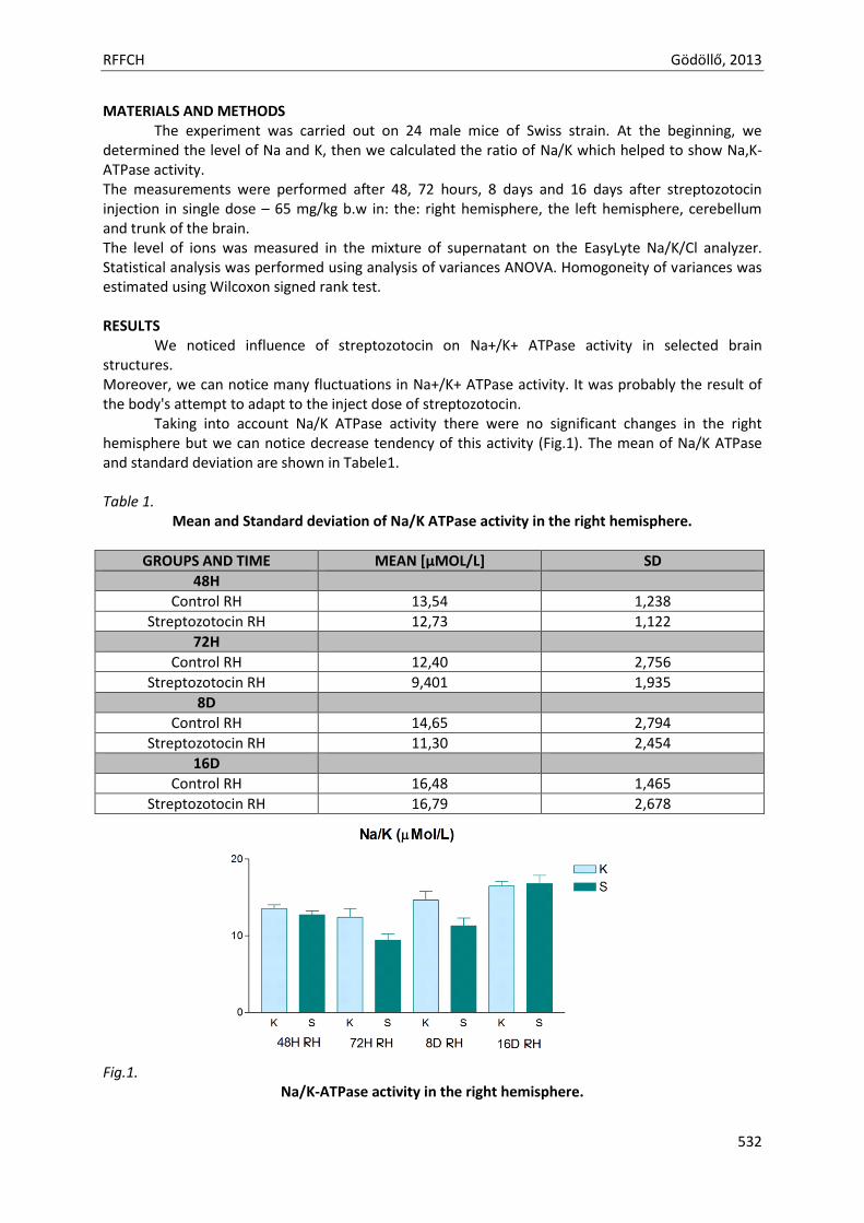
RFFCH Gödöllő, 2013
532
MATERIALS AND METHODS The experiment was carried out on 24 male mice of Swiss strain. At the beginning, we
determined the level of Na and K, then we calculated the ratio of Na/K which helped to show Na,K-ATPase activity. The measurements were performed after 48, 72 hours, 8 days and 16 days after streptozotocin injection in single dose – 65 mg/kg b.w in: the: right hemisphere, the left hemisphere, cerebellum and trunk of the brain. The level of ions was measured in the mixture of supernatant on the EasyLyte Na/K/Cl analyzer. Statistical analysis was performed using analysis of variances ANOVA. Homogoneity of variances was estimated using Wilcoxon signed rank test. RESULTS
We noticed influence of streptozotocin on Na+/K+ ATPase activity in selected brain structures. Moreover, we can notice many fluctuations in Na+/K+ ATPase activity. It was probably the result of the body's attempt to adapt to the inject dose of streptozotocin.
Taking into account Na/K ATPase activity there were no significant changes in the right hemisphere but we can notice decrease tendency of this activity (Fig.1). The mean of Na/K ATPase and standard deviation are shown in Tabele1. Table 1.
Mean and Standard deviation of Na/K ATPase activity in the right hemisphere.
GROUPS AND TIME MEAN [µMOL/L] SD
48H
Control RH 13,54 1,238
Streptozotocin RH 12,73 1,122
72H
Control RH 12,40 2,756
Streptozotocin RH 9,401 1,935
8D
Control RH 14,65 2,794
Streptozotocin RH 11,30 2,454
16D
Control RH 16,48 1,465
Streptozotocin RH 16,79 2,678
Fig.1.
Na/K-ATPase activity in the right hemisphere.
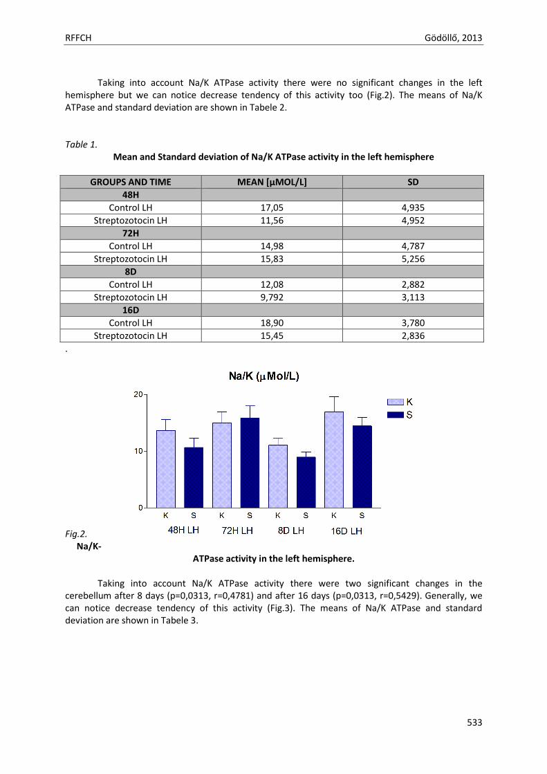
RFFCH Gödöllő, 2013
533
Taking into account Na/K ATPase activity there were no significant changes in the left
hemisphere but we can notice decrease tendency of this activity too (Fig.2). The means of Na/K ATPase and standard deviation are shown in Tabele 2. Table 1.
Mean and Standard deviation of Na/K ATPase activity in the left hemisphere
GROUPS AND TIME MEAN [µMOL/L] SD
48H
Control LH 17,05 4,935
Streptozotocin LH 11,56 4,952
72H
Control LH 14,98 4,787
Streptozotocin LH 15,83 5,256
8D
Control LH 12,08 2,882
Streptozotocin LH 9,792 3,113
16D
Control LH 18,90 3,780
Streptozotocin LH 15,45 2,836
.
Fig.2. Na/K-
ATPase activity in the left hemisphere. Taking into account Na/K ATPase activity there were two significant changes in the
cerebellum after 8 days (p=0,0313, r=0,4781) and after 16 days (p=0,0313, r=0,5429). Generally, we can notice decrease tendency of this activity (Fig.3). The means of Na/K ATPase and standard deviation are shown in Tabele 3.
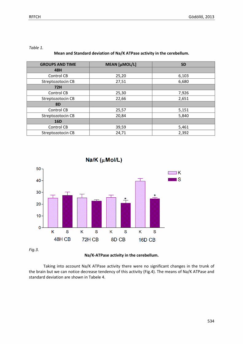
RFFCH Gödöllő, 2013
534
Table 1.
Mean and Standard deviation of Na/K ATPase activity in the cerebellum.
GROUPS AND TIME MEAN [µMOL/L] SD
48H
Control CB 25,20 6,103
Streptozotocin CB 27,51 6,680
72H
Control CB 25,30 7,926
Streptozotocin CB 22,66 2,651
8D
Control CB 25,57 5,151
Streptozotocin CB 20,84 5,840
16D
Control CB 39,59 5,461
Streptozotocin CB 24,71 2,392
Fig.3.
Na/K-ATPase activity in the cerebellum.
Taking into account Na/K ATPase activity there were no significant changes in the trunk of the brain but we can notice decrease tendency of this activity (Fig.4). The means of Na/K ATPase and standard deviation are shown in Tabele 4.
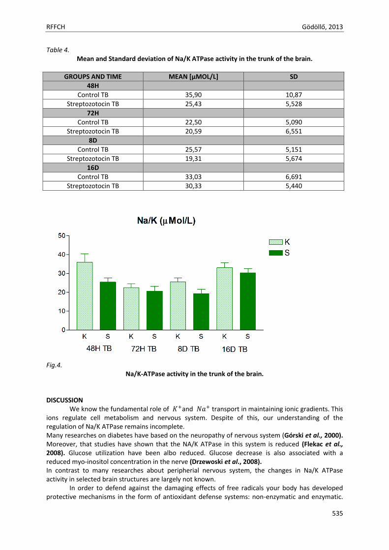
RFFCH Gödöllő, 2013
535
Table 4. Mean and Standard deviation of Na/K ATPase activity in the trunk of the brain.
GROUPS AND TIME MEAN [µMOL/L] SD
48H
Control TB 35,90 10,87
Streptozotocin TB 25,43 5,528
72H
Control TB 22,50 5,090
Streptozotocin TB 20,59 6,551
8D
Control TB 25,57 5,151
Streptozotocin TB 19,31 5,674
16D
Control TB 33,03 6,691
Streptozotocin TB 30,33 5,440
Fig.4.
Na/K-ATPase activity in the trunk of the brain. DISCUSSION We know the fundamental role of and transport in maintaining ionic gradients. This ions regulate cell metabolism and nervous system. Despite of this, our understanding of the regulation of Na/K ATPase remains incomplete. Many researches on diabetes have based on the neuropathy of nervous system (Górski et al., 2000). Moreover, that studies have shown that the NA/K ATPase in this system is reduced (Flekac et al., 2008). Glucose utilization have been albo reduced. Glucose decrease is also associated with a reduced myo-inositol concentration in the nerve (Drzewoski et al., 2008). In contrast to many researches about peripherial nervous system, the changes in Na/K ATPase activity in selected brain structures are largely not known.
In order to defend against the damaging effects of free radicals your body has developed protective mechanisms in the form of antioxidant defense systems: non-enzymatic and enzymatic.
RFFCH Gödöllő, 2013
536
The enzyme system is responsible for antioxidant defense. This system is composed of superoxide dismutase, catalase and glutathione peroxidase (Fraczek et al., 2005). It was found that hyperglycemia promotes the formation of reactive oxygen species (Mrowicka, 2011).
Diabetes is associated with an increased level of free radicals and disturbances of the enzymatic antioxidant defense system. Furthermore, these abnormalities lead to a redox imbalance called oxidative stress (Mrowicka, 2011). The activity of sodium-potassium pump is impaired by oxidative stress. It is of great importance in the pathogenesis of diabetes. In normal conditions there is a balance between the emerging reactive oxygen species (ROS) and endogenous antioxidant systems. Oxidative stress is a condition characterized by excessive reactive oxygen species. As a result of elevated blood glucose increased occurs the impairment of remove free radicals. The blood vessels are and enhances the oxidative stress. It inhibits the sodium-potassium pump. In addition, the source of reactive oxygen species in diabetes is a mitochondrial respiratory chain (Giugliano et al., 1996). It has been shown that increasing glycolysis and the Krebs cycle in response to hyperglycemia leads to a huge increase in the pool of protons, which then cause an increase in proton gradient and consequently there is a formation of free radicals. Sodium potassium pump activity is also reduced by decreasing of energy in cells (Brownlee, 2001). Hyperglycemia also causes an increased conversion of glucose to sorbitol and fructose by the polyol pathway (Dickinson et al., 2002). Decreasing ratio of reduced to oxidized form of nicotinamide adenine dinucleotide phosphate - NADPH / NADP +, and increases the ratio of nicotinamide adenine dinucleotide - NADH / NAD +, which leads to activation of growth cycle of transformations of arachidonic acid, which determines the additional production of free radicals (Brownlee, 2001).
The streptozotocin initiated first step of diabetes. It was the reason of starting production of free radicals and occurrenced oxidative stress. In our opinion, it was not enough time to induced full diabetes. We need to continue our experiment. It could be the reason of no significant changes in Na/K ATPase activity.
Although not many significant changes after each time, we noticed decreased tendency in Na/K ATPase activity. Significantly decreased of Na+/K+ ATPase occures in diabetic patients. CONCLUSIONS
The present study has confirmed other reports decreasing activity of Na/K ATPase. Further studies involving streptozotocin and much time are necessary to clarify the results of the present study.
REFERENCES BROWNLEE M. 2001. Biochemistry and molecular cell biology of diabetic complications. Nature, 2001, 414:
p.813–820. DICKINSON P.J., CARRINGTON A.L., FROST G.S., BOULTON A.J. 2002. Neurovascular disease, antioxidants and
glycation in diabetes. Diabetes Metab. Res. Rev., 2002; 18: p. 260–272 DRZEWOSKI J. 2008. Gliclazide, inflammation and atherosclerosis. Antiinflamm. Antiallergy Agents Med. Chem.,
2008; 7: p. 224–230 EVANS J.L., GOLDFINE I.D., MADDUX B.A., GRODSKY G.M. 2003. Are oxidative stress-activated signaling
pathways mediators of insulin resistance and b-cell dysfunction? Diabetes, 2003; 52: 1–8 FLEKAC M., SKRHA J., HILGERTOVA J., LACINOVA Z., JAROLIMKOVA M. 2008. Gene polymorphisms of
superoxide dismutases and catalase in diabetes mellitus. BMC Med. Genet., 2008; 9: 30 FRACZEK M., KURPISZ M. 2005. The redox system in human semen and peroxidative damage of spermatozoa.
Postępy Hig. Med. Dośw., 2005; 59: 523–534 GIUGLIANO D., CERIELLO A., PAOLISSO G. 1996. Oxidative stress and diabetic vascular complications. Diabetes
Care, 1996; 19: 257–267 GÓRSKI J., KNAPP M., MUSIAŁ W. 2000. Metabolizm substratów energetycznych w mięśniu sercowym. Wpływ
niedotlenienia i cukrzycy. Czynniki Ryzyka, 2000; 2–3: 18–23 HALL, JOHN E.; GUYTON, ARTHUR C. 2006. Textbook of medical physiology. St. Louis, Mo: Elsevier Saunders.
RFFCH Gödöllő, 2013
537
HOWARTH C, GLEESON P, ATTWELL D. 2012. Updated energy budgets for neural computation in the neocortex and cerebellum. J. Cereb. Blood Flow Metab. 2012. 32 (7): p. 1222–32.
MORTH JP, PEDERSEN BP, TOUSTRUP-JENSEN MS, SORENSEN TLM, PETERSEN J, ANDERSEN JP, VILSEN B, NISSEN P. 2007. Crystal Structure of the Sodium-Potassium Pump. Nature 2007; 450: p. 1043-1049.
YUAN Z, CAI T, TIAN J, IVANOV AV, GIOVANNUCCI DR, XIE Z. 2005. Na/K-ATPase tethers phospholipase C and IP3 receptor into a calcium-regulatory complex "Molecular Biology of the Cell 16 (9): p. 4034–45.
Animal welfare, etológia és tartástechnológia
Animal welfare, ethology and housing systems
Volume 9 Issue 3
Különszám/Special Issue
Gödöllő
2013
RFFCH Gödöllő, 2013
THE ACTIVITY OF CATALASE AND CONTENT OF REDUCED GLUTATHIONE IN TESTES OF MICE EXPOSED TO ACRYLAMIDE
Kraska K., Formicki G., Kapusta E., Goc E., Kopańska M.
Acrylamide (ACR) monomer is mutagenic and causes disturbances in male reproductive organs.
Acrylamide is used in various industries for example polymer industry, cosmetic, paper and textile
industries. It is also used in laboratory gels. Most important is the fact that acrylamide was found in
food. Daily intake of acrylamide from foodstuff was estimated in the range of 0.3-0.8 mg/kg bw per
day. The International Agency for Research on Cancer classifies acrylamide as 2A, which means that
acrylamide is a probable human carcinogen. Glutathione is in a constant state of metabolic turnover
because is actively synthesized and degraded. Reduced glutathione enters the oxidation and reduction
reactions with electrophilic reagents such as xenobiotics, free radicals, toxins, organic hyperoxides.
Catalase is an enzyme catalyzing decomposition of H202.
The aim of our work was to investigate concentration of reduced glutathione and activity of catalase
in testes of mice exposed to acrylamide. The research was conducted on SWISS mice 8 week old,
weight 26 g. Animals were fed with standard diet and grow in 12/12 light/dark photoperiod. The
animals were segregated into two experimental and one control group. Animals of experimental
groups were injected intraperitoneally with ACR in the dose of 80 mg/kg. Mice from the control group
were injected with physiological saline. The measurements were performed 24, hours and 2 weeks
after acrylamide injection.
GSH concentration decreased along with the duration the experiment. The activity of catalase also
decreased with duration of the experiment. Significant differences in GSH content occurred after two
weeks of ACR injections.
538

Animal welfare, etológia és tartástechnológia
Animal welfare, ethology and housing systems
Volume 9 Issue 3
Különszám/Special Issue
Gödöllő
2013
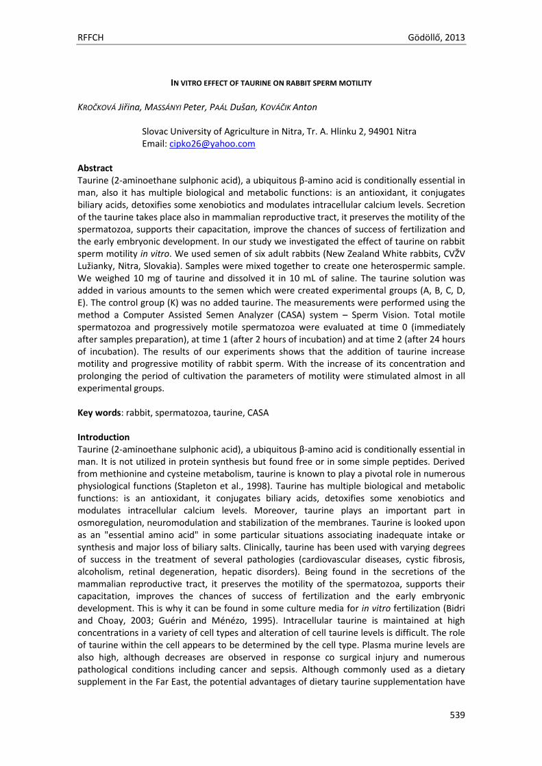
RFFCH Gödöllő, 2013
539
IN VITRO EFFECT OF TAURINE ON RABBIT SPERM MOTILITY KROČKOVÁ Jiřina, MASSÁNYI Peter, PAÁL Dušan, KOVÁČIK Anton
Slovac University of Agriculture in Nitra, Tr. A. Hlinku 2, 94901 Nitra Email: [email protected]
Abstract Taurine (2-aminoethane sulphonic acid), a ubiquitous β-amino acid is conditionally essential in man, also it has multiple biological and metabolic functions: is an antioxidant, it conjugates biliary acids, detoxifies some xenobiotics and modulates intracellular calcium levels. Secretion of the taurine takes place also in mammalian reproductive tract, it preserves the motility of the spermatozoa, supports their capacitation, improve the chances of success of fertilization and the early embryonic development. In our study we investigated the effect of taurine on rabbit sperm motility in vitro. We used semen of six adult rabbits (New Zealand White rabbits, CVŽV Lužianky, Nitra, Slovakia). Samples were mixed together to create one heterospermic sample. We weighed 10 mg of taurine and dissolved it in 10 mL of saline. The taurine solution was added in various amounts to the semen which were created experimental groups (A, B, C, D, E). The control group (K) was no added taurine. The measurements were performed using the method a Computer Assisted Semen Analyzer (CASA) system – Sperm Vision. Total motile spermatozoa and progressively motile spermatozoa were evaluated at time 0 (immediately after samples preparation), at time 1 (after 2 hours of incubation) and at time 2 (after 24 hours of incubation). The results of our experiments shows that the addition of taurine increase motility and progressive motility of rabbit sperm. With the increase of its concentration and prolonging the period of cultivation the parameters of motility were stimulated almost in all experimental groups. Key words: rabbit, spermatozoa, taurine, CASA Introduction Taurine (2-aminoethane sulphonic acid), a ubiquitous β-amino acid is conditionally essential in man. It is not utilized in protein synthesis but found free or in some simple peptides. Derived from methionine and cysteine metabolism, taurine is known to play a pivotal role in numerous physiological functions (Stapleton et al., 1998). Taurine has multiple biological and metabolic functions: is an antioxidant, it conjugates biliary acids, detoxifies some xenobiotics and modulates intracellular calcium levels. Moreover, taurine plays an important part in osmoregulation, neuromodulation and stabilization of the membranes. Taurine is looked upon as an "essential amino acid" in some particular situations associating inadequate intake or synthesis and major loss of biliary salts. Clinically, taurine has been used with varying degrees of success in the treatment of several pathologies (cardiovascular diseases, cystic fibrosis, alcoholism, retinal degeneration, hepatic disorders). Being found in the secretions of the mammalian reproductive tract, it preserves the motility of the spermatozoa, supports their capacitation, improves the chances of success of fertilization and the early embryonic development. This is why it can be found in some culture media for in vitro fertilization (Bidri and Choay, 2003; Guérin and Ménézo, 1995). Intracellular taurine is maintained at high concentrations in a variety of cell types and alteration of cell taurine levels is difficult. The role of taurine within the cell appears to be determined by the cell type. Plasma murine levels are also high, although decreases are observed in response co surgical injury and numerous pathological conditions including cancer and sepsis. Although commonly used as a dietary supplement in the Far East, the potential advantages of dietary taurine supplementation have
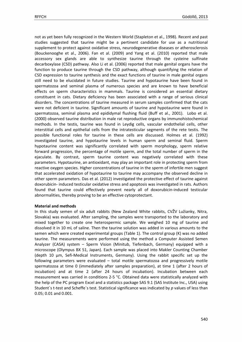
RFFCH Gödöllő, 2013
540
not as yet been fully recognized in the Western World (Stapleton et al., 1998). Recent and past studies suggested that taurine might be a pertinent candidate for use as a nutritional supplement to protect against oxidative stress, neurodegenerative diseases or atherosclerosis (Bouckenooghe et al., 2006). Fan et al. (2009) and Yang et al. (2010) reported that male accessory sex glands are able to synthesize taurine through the cysteine sulfinate decarboxylase (CSD) pathway. Also Li et al. (2006) reported that male genital organs have the function to produce taurine through the CSD pathway, although quantifying the relation of CSD expression to taurine synthesis and the exact functions of taurine in male genital organs still need to be elucidated in future studies. Taurine and hypotaurine have been found in spermatozoa and seminal plasma of numerous species and are known to have beneficial effects on sperm characteristics in mammals. Taurine is considered an essential dietary constituent in cats. Dietary deficiency has been associated with a range of serious clinical disorders. The concentrations of taurine measured in serum samples confirmed that the cats were not deficient in taurine. Significant amounts of taurine and hypotaurine were found in spermatozoa, seminal plasma and epididymal flushing fluid (Buff et al., 2001). Lobo et al. (2000) observed taurine distribution in male rat reproductive organs by immunohistochemical methods. In the testis, taurine was found in Leydig cells, vascular endothelial cells, other interstitial cells and epithelial cells from the intratesticular segments of the rete testis. The possible functional roles for taurine in these cells are discussed. Holmes et al. (1992) investigated taurine, and hypotaurine levels in human sperm and seminal fluid. Sperm hypotaurine content was significantly correlated with sperm morphology, sperm relative forward progression, the percentage of motile sperm, and the total number of sperm in the ejaculate. By contrast, sperm taurine content was negatively correlated with these parameters. Hypotaurine, an antioxidant, may play an important role in protecting sperm from reactive oxygen species. Higher concentrations of taurine in the sperm of infertile men suggest that accelerated oxidation of hypotaurine to taurine may accompany the observed decline in other sperm parameters. Das et al. (2012) investigated the protective effect of taurine against doxorubicin- induced testicular oxidative stress and apoptosis was investigated in rats. Authors found that taurine could effectively prevent nearly all of doxorubicin-induced testicular abnormalities, thereby proving to be an effective cytoprotectant. Material and methods In this study semen of six adult rabbits (New Zealand White rabbits, CVŽV Lužianky, Nitra, Slovakia) was evaluated. After sampling, the samples were transported to the laboratory and mixed together to create one heterospermic sample. We weighed 10 mg of taurine and dissolved it in 10 mL of saline. Then the taurine solution was added in various amounts to the semen which were created experimental groups (Table 1). The control group (K) was no added taurine. The measurements were performed using the method a Computer Assisted Semen Analyzer (CASA) system – Sperm Vision (Minitub, Tiefenbach, Germany) equipped with a microscope (Olympus BX 51, Japan). Each sample was placed into Makler Counting Chamber (depth 10 µm, Sefi-Medical Instruments, Germany). Using the rabbit specific set up the following parameters were evaluated – total motile spermatozoa and progressively motile spermatozoa at time 0 (immediately after samples preparation), at time 1 (after 2 hours of incubation) and at time 2 (after 24 hours of incubation). Incubation between each measurement was carried in conditions 2-5 °C. Obtained data were statistically analyzed with the help of the PC program Excel and a statistics package SAS 9.1 (SAS Institute Inc., USA) using Student`s t-test and Scheffe`s test. Statistical significance was indicated by p values of less than 0.05; 0.01 and 0.001.
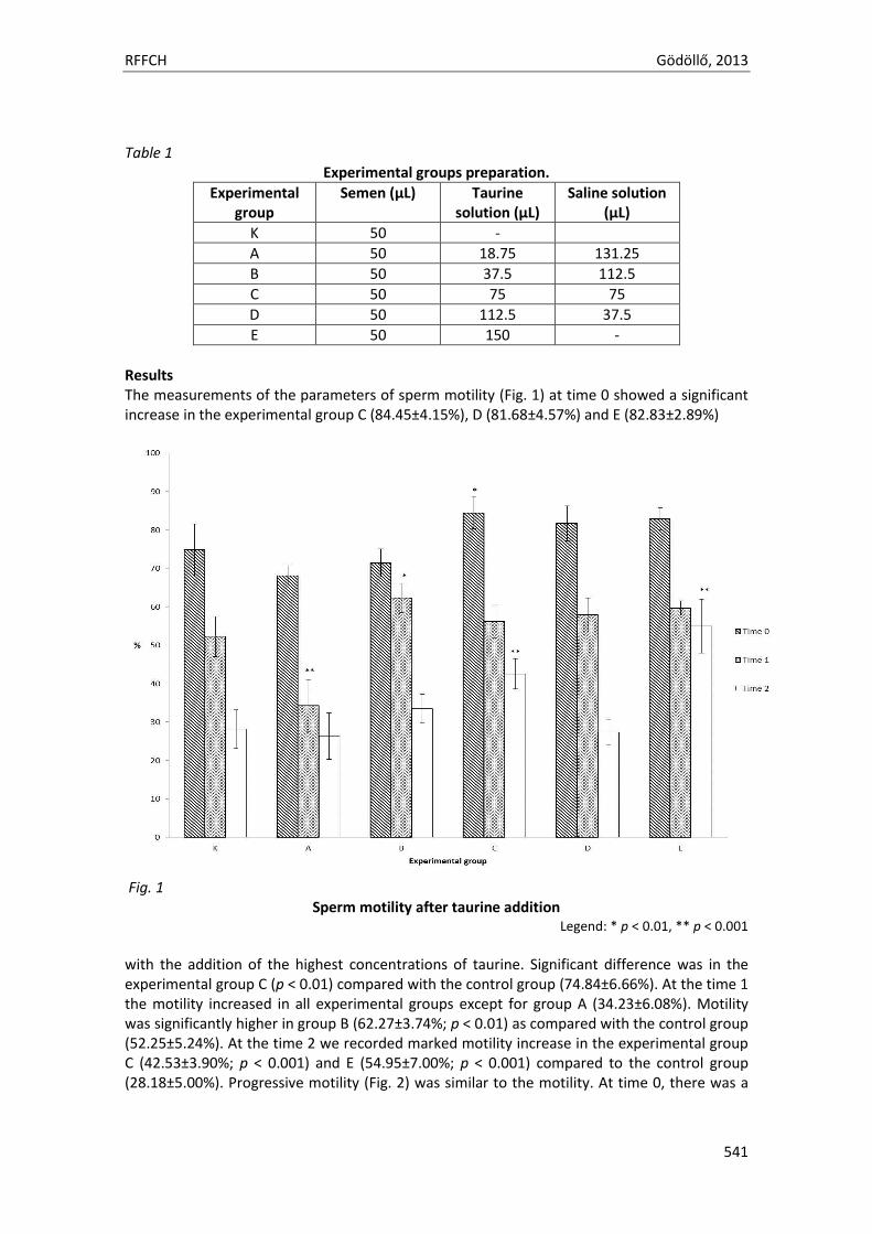
RFFCH Gödöllő, 2013
541
Table 1
Experimental groups preparation.
Experimental group
Semen (µL) Taurine solution (µL)
Saline solution (µL)
K 50 -
A 50 18.75 131.25
B 50 37.5 112.5
C 50 75 75
D 50 112.5 37.5
E 50 150 -
Results The measurements of the parameters of sperm motility (Fig. 1) at time 0 showed a significant increase in the experimental group C (84.45±4.15%), D (81.68±4.57%) and E (82.83±2.89%)
Fig. 1 Sperm motility after taurine addition
Legend: * p < 0.01, ** p < 0.001
with the addition of the highest concentrations of taurine. Significant difference was in the experimental group C (p < 0.01) compared with the control group (74.84±6.66%). At the time 1 the motility increased in all experimental groups except for group A (34.23±6.08%). Motility was significantly higher in group B (62.27±3.74%; p < 0.01) as compared with the control group (52.25±5.24%). At the time 2 we recorded marked motility increase in the experimental group C (42.53±3.90%; p < 0.001) and E (54.95±7.00%; p < 0.001) compared to the control group (28.18±5.00%). Progressive motility (Fig. 2) was similar to the motility. At time 0, there was a
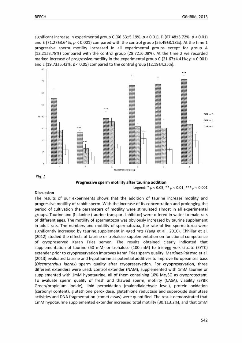
RFFCH Gödöllő, 2013
542
significant increase in experimental group C (66.53±5.19%; p < 0.01), D (67.48±3.72%; p < 0.01) and E (71.27±3.64%; p < 0.001) compared with the control group (55.49±8.18%). At the time 1 progressive sperm motility increased in all experimental groups except for group A (13.21±3.78%) compared with the control group (28.72±6.08%). At the time 2 we recorded marked increase of progressive motility in the experimental group C (21.67±4.41%; p < 0.001) and E (19.73±5.43%; p < 0.05) compared to the control group (12.19±4.25%).
Fig. 2 Progressive sperm motility after taurine addition
Legend: * p < 0.05, ** p < 0.01, *** p < 0.001
Discussion The results of our experiments shows that the addition of taurine increase motility and progressive motility of rabbit sperm. With the increase of its concentration and prolonging the period of cultivation the parameters of motility were stimulated almost in all experimental groups. Taurine and β-alanine (taurine transport inhibitor) were offered in water to male rats of different ages. The motility of spermatozoa was obviously increased by taurine supplement in adult rats. The numbers and motility of spermatozoa, the rate of live spermatozoa were significantly increased by taurine supplement in aged rats (Yang et al., 2010). Chhillar et al. (2012) studied the effects of taurine or trehalose supplementation on functional competence of cryopreserved Karan Fries semen. The results obtained clearly indicated that supplementation of taurine (50 mM) or trehalose (100 mM) to tris-egg yolk citrate (EYTC) extender prior to cryopreservation improves Karan Fries sperm quality. Martínez-Páramo et al. (2013) evaluated taurine and hypotaurine as potential additives to improve European sea bass (Dicentrarchus labrax) sperm quality after cryopreservation. For cryopreservation, three different extenders were used: control extender (NAM), supplemented with 1mM taurine or supplemented with 1mM hypotaurine, all of them containing 10% Me2SO as cryoprotectant. To evaluate sperm quality of fresh and thawed sperm, motility (CASA), viability (SYBR Green/propidium iodide), lipid peroxidation (malondialdehyde level), protein oxidation (carbonyl content), glutathione peroxidase, glutathione reductase and superoxide dismutase activities and DNA fragmentation (comet assay) were quantified. The result demonstrated that 1mM hypotaurine supplemented extender increased total motility (30.1±3.2%), and that 1mM
*
RFFCH Gödöllő, 2013
543
taurine extender produced higher velocity (18.1±2.6μm/s) and linearity (46.0±4.8%) than the control extender (21.8±3.2%, 15.5±1.3μm/s, 41.8±2.4%, respectively). Cell viability, lipid peroxidation and protein oxidation were not statistically different between treatments. Similar results were obtained for glutathione peroxidase and superoxide dismutase activities. Only glutathione reductase showed differential activity before and after freezing, increasing its activity in thawed sperm. Regarding the comet assay results, taurine and hypotaurine significantly reduced DNA fragmentation (52.8±0.9% and 51.8±0.9%, respectively) in comparison to the control (55.7±0.8%). Authors reported that extenders supplemented with 1mM taurine and 1mM hypotaurine improved some parameters of sperm quality after thawing, resulting in better motility and lower DNA damage than the control, two very important factors related to fertilization success Alam et al. (2011) treated mice with taurine orally (100. mg/kg. b.wt.) for nine days. Treatment with taurine showed increases sperm count and motility, and decreases the incidence of sperm abnormalities. Das et al. (2009) reported that oral administration of taurine (at a dose of 100 mg/kg body weight for 5 days) to rats was found to be effective in counteracting arsenic-induced oxidative stress, attenuation of testicular damages and amelioration of apoptosis in testicular tissue. Taurine was also found to play similar beneficial role via mitochondrial dependent pathways in arsenic-induced testicular damages leading to apoptotic cell death. Cabrita et al. (2011) analyzed the effect of extender supplementation with taurine on post-thawed sperm motility, viability and DNA integrity of two commercial species, gilthead seabream (Sparus aurata) and European seabass (Dicentrarchus labrax). Sperm was cryopreserved in ten different extenders containing taurine (1 and 10. mM). Taurine slightly increased the percentage of motile cells and significantly reduced both DNA fragmentation parameters, protecting DNA against strand breaks. Conclusion At the end of our experiment, we observed that the addition of small amounts of taurine in rabbit sperm has a stimulating effect on sperm motility. The increasing of taurine concentrations and extension of its period of exposure, taurine has positive effect on sperm motion parameters. Acknowledgments We would like to thank Ing. Ondruška from CVŽV Lužianky for providing of samples. References
Alam, S.S., Hafiz, N.A., Abd El-Rahim, A.H. 2011. Protective role of taurine against genotoxic damage in mice treated with methotrexate and tamoxfine. Environmental Toxicology and Pharmacology, 31, 143-152.
Bidri, M., Choay, P. 2003. Taurine: A particular aminoacid with multiple functions ( Short Survey ) [La taurine: Un aminoacide particulier aux fonctions multiples]. Annales Pharmaceutiques Francaises, 61, 385-391.
Bouckenooghe, T., Remacle, C., Reusens, B. 2006. Is taurine a functional nutrient? ( Review ). Current Opinion in Clinical Nutrition and Metabolic Care, 9, Issue 6, 728-733.
Buff, S., Donzé, A., Guérin, P., Guillaud, J., Fontbonne, A., Ménézo, Y. Taurine and hypotaurine in spermatozoa and epididymal fluid of cats. Journal of reproduction and fertility. Supplement, 57, 93-95.
Cabrita, E., Ma, S., Diogo, P., Martínez-Páramo, S., Sarasquete, C., Dinis, M.T. 2011. The influence of certain aminoacids and vitamins on post-thaw fish sperm motility, viability and DNA fragmentation. Animal Reproduction Science, 125, 189-195.
Chhillar, S., Singh, V.K., Kumar, R., Atreja, S.K. 2012. Effects of Taurine or Trehalose supplementation on functional competence of cryopreserved Karan Fries semen. Animal Reproduction Science, 135, 1-7.
RFFCH Gödöllő, 2013
544
Das, J., Ghosh, J., Manna, P., Sil, P.C. 2012. Taurine protects rat testes against doxorubicin-induced oxidative stress as well as p53, Fas and caspase 12-mediated apoptosis. Amino Acids, 42, 1839-1855.
Das, J., Ghosh, J., Manna, P., Sinha, M., Sil, P.C. 2009. Taurine protects rat testes against NaAsO2-induced oxidative stress and apoptosis via mitochondrial dependent and independent pathways. Toxicology Letters, 187, 201-210.
Fan, J.J., Zhou, J.L., Li, J.H., Cui, S. 2009. Accessory sex glands of male mice have the ability to synthesize taurinevia the cysteine sulfinate decarboxylase pathway. Cell Biology International, 33, 684-689.
Guérin, P., Ménézo, Y. 1995. Hypotaurine and taurine in gamete and embryo environments: de novo synthesis via the cysteine sulfinic acid pathway in oviduct cells. Zygote (Cambridge, England), 3, 333-343.
Holmes, R.P., Goodman, H.O., Hurst, C.H., Shihabi, Z.K., Jarrow, J.P. 1992. Hypotaurine in male reproduction. In: Lombardini, J.B., Schaffer, S.W., Azuma, J. Taurine. First edit. New York : Plenum Press, 437–441 p.
Li, J.H., Ling, Y.Q., Fan, J.J., Zhang, X.P., Cui, S. 2006. Expression of cysteine sulfinate decarboxylase (CSD) in male reproductive organs of mice. Histochemistry and Cell Biology, 125, 607-613.
Lobo, M.D.V.T., Alonso, F.J.M., Martín Del Río, R. 2000. Immunohistochemical localization of taurine in the male reproductive organs of the rat. Journal of Histochemistry and Cytochemistry, 48, 313-320.
Martínez-Páramo, S., Diogo, P., Dinis, M.T., Soares, F., Sarasquete, C., Cabrita, E. 2013. Effect of two sulfur-containing amino acids, taurine and hypotaurine in European sea bass (Dicentrarchus labrax) sperm cryopreservation. Cryobiology, 66, 333-338.
Stapleton, P.P., O'Flaherty, L., Redmond, H.P., Bouchier-Hayes, D.J. 1998. Host defense - A role for the amino acid taurine? ( Review ). Journal of Parenteral and Enteral Nutrition, 22, 42-48.
Yang, J., Wu, G.,
Feng,
Y., Lv, Q.,
Lin, S., Hu, J.
2010. Effects of taurine on male reproduction in rats of
different ages. J Biomed Sci., 17, 9.

Animal welfare, etológia és tartástechnológia
Animal welfare, ethology and housing systems
Volume 9 Issue 3
Különszám/Special Issue
Gödöllő
2013
RFFCH Gödöllő, 2013
545
EFFECTS OF ANTIOXIDANTS ON ACROLEIN-INDUCED OXIDATIVE PROTEIN DAMAGE IN THE YEAST SACCHAROMYCES CEREVISAE
Magdalena KWOLEK-MIREK*, Renata ZADRĄG-TĘCZA
Department of Biochemistry and Cell Biology, Faculty of Biology and Agriculture, University of Rzeszow, Zelwerowicza 4, 35-601 Rzeszow, Poland *Corresponding author: [email protected]
Abstract Acrolein is an important environmental and endogenous toxin. It is an electrophile showing high reactivity with biological nucleophiles leading to a decrease in the level of glutathione as well as protein and DNA modification. Our previous results showed that 1-h incubation of both
wild-type and sod1 mutant cells with allyl alcohol (precursor of acrolein) caused concentration-dependent increase in the level of carbonyl groups. The aim of this study was to determine if antioxidants are able to protect proteins from oxidation. We discovered that several thiol antioxidants such as glutathione, N-acetylcysteine, cysteine and dithiothreitol can decrease the level of carbonyl groups in yeast cells exposed to acrolein. Similar effect wasn’t observed in case of ascorbic acid and Tempo. Key words: acrolein, allyl alcohol, oxidative stress, protein carbonyls, antioxidant, yeast
Abbreviations: AA, allyl alcohol; ASC, ascorbic acid; CYS, cysteine; DTT, dithiothreitol; GSH, glutathione; NAC, N-acetylcysteine; Tempo, 2,2,6,6-tetramethylpiperidine-1-oxyl
Introduction Acrolein is the simplest, unsaturated aldehyde commonly occurring in the environment. It is formed during incomplete combustion of petrol, coal, wood, cotton and plastic materials. It is a by-product in many branches of the chemical industry and is also a constituent of tobacco smoke. Acrolein can be also formed endogenously as a product of lipid peroxidation, during degradation of threonine and polyamines as well as during activation of cytostatic drugs. Being electrophilic, acrolein shows high reactivity with biological nucleophiles leading to a decrease in the level of glutathione as well as protein and DNA modification (Stevens and Maier, 2008). The studies on the effect of acrolein on cells are carried out using various experimental models. Our team has proposed a simple eukaryotic model for such studies, viz. the baker’s yeast Saccharomyces cerevisiae (Bilinski et al., 2005). A definite advantage of this model is the lack of occurrence of lipid peroxidation, which is the main endogenous source of acrolein. This allows to track acrolein and its metabolites in the cell and better define the toxic and lethal doses for cells. Our previous results (Bilinski et al., 2005; Kwolek-Mirek et al., 2009; Kwolek-Mirek and Bartosz, 2011) show that the yeast strains defective in antioxidant defense: Cu,Zn-superoxide
dismutase (sod1), γ-glutamylcysteine ligase (gsh1), thioredoxins 1 and 2 (trx1trx2) and
transcription factor Yap1p (yap1) are hypersensitive to acrolein which is formed in cells from allyl alcohol. The proposal of replacement of direct introduction of acrolein with its metabolic precursor allyl alcohol has been put forward by our team and allows to eliminate extracellular effects of acrolein resulting from its high reactivity. The data obtained demonstrate that incubation of yeast cells with allyl alcohol causes a range of negative effects: a decrease of glutathione content, a decrease of total antioxidant capacity and an increase of the content of reactive oxygen species. The metabolic activation of allyl alcohol causes also activation and relocation of Yap1p from cytoplasm to the nucleus, which evidences a stress reaction. Other
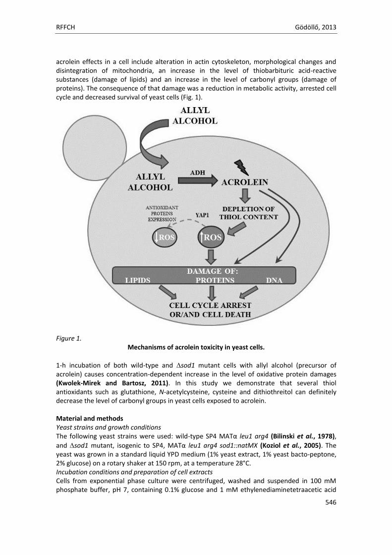
RFFCH Gödöllő, 2013
546
acrolein effects in a cell include alteration in actin cytoskeleton, morphological changes and disintegration of mitochondria, an increase in the level of thiobarbituric acid-reactive substances (damage of lipids) and an increase in the level of carbonyl groups (damage of proteins). The consequence of that damage was a reduction in metabolic activity, arrested cell cycle and decreased survival of yeast cells (Fig. 1).
Figure 1.
Mechanisms of acrolein toxicity in yeast cells.
1-h incubation of both wild-type and sod1 mutant cells with allyl alcohol (precursor of acrolein) causes concentration-dependent increase in the level of oxidative protein damages (Kwolek-Mirek and Bartosz, 2011). In this study we demonstrate that several thiol antioxidants such as glutathione, N-acetylcysteine, cysteine and dithiothreitol can definitely decrease the level of carbonyl groups in yeast cells exposed to acrolein. Material and methods Yeast strains and growth conditions The following yeast strains were used: wild-type SP4 MATα leu1 arg4 (Bilinski et al., 1978),
and sod1 mutant, isogenic to SP4, MATα leu1 arg4 sod1::natMX (Koziol et al., 2005). The yeast was grown in a standard liquid YPD medium (1% yeast extract, 1% yeast bacto-peptone, 2% glucose) on a rotary shaker at 150 rpm, at a temperature 28°C. Incubation conditions and preparation of cell extracts Cells from exponential phase culture were centrifuged, washed and suspended in 100 mM phosphate buffer, pH 7, containing 0.1% glucose and 1 mM ethylenediaminetetraacetic acid
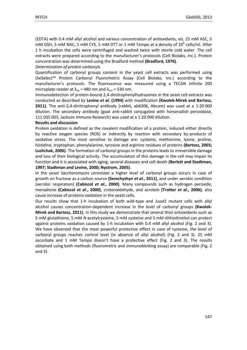
RFFCH Gödöllő, 2013
547
(EDTA) with 0.4 mM allyl alcohol and various concentration of antioxidants, viz. 25 mM ASC, 5 mM GSH, 5 mM NAC, 5 mM CYS, 5 mM DTT or 1 mM Tempo at a density of 108 cells/ml. After 1 h incubation the cells were centrifuged and washed twice with sterile cold water. The cell extracts were prepared according to the manufacturer’s protocols (Cell Biolabs, Inc.). Protein concentration was determined using the Bradford method (Bradford, 1976). Determination of protein carbonyls Quantification of carbonyl groups content in the yeast cell extracts was performed using OxiSelect™ Protein Carbonyl Fluorometric Assay (Cell Biolabs, Inc.) according to the manufacturer’s protocols. The fluorescence was measured using a TECAN Infinite 200 microplate reader at λex = 480 nm and λem = 530 nm. Immunodetection of protein-bound 2,4-dinitrophenylhydrazones in the yeast cell extracts was conducted as described by Levine et al. (1994) with modification (Kwolek-Mirek and Bartosz, 2011). The anti-2,4-dinitrophenyl antibody (rabbit, ab6306, Abcam) was used at a 1:20 000 dilution. The secondary antibody (goat anti-rabbit conjugated with horseradish peroxidase, 111 035 003, Jackson Immuno Research) was used at a 1:20 000 dilution. Results and discussion Protein oxidation is defined as the covalent modification of a protein, induced either directly by reactive oxygen species (ROS) or indirectly by reaction with secondary by-products of oxidative stress. The most sensitive to damage are: cysteine, methionine, lysine, proline, histidine, tryptophan, phenylalanine, tyrosine and arginine residues of proteins (Bartosz, 2003; Lushchak, 2006). The formation of carbonyl groups in the proteins leads to irreversible damage and loss of their biological activity. The accumulation of this damage in the cell may impair its function and it is associated with aging, several diseases and cell death (Berlett and Stadtman, 1997; Stadtman and Levine, 2000; Nystrom, 2005). In the yeast Saccharomyces cerevisiae a higher level of carbonyl groups occurs in case of growth on fructose as a carbon source (Semchyshyn et al., 2011), and under aerobic condition (aerobic respiration) (Cabiscol et al., 2000). Many compounds such as hydrogen peroxide, menadione (Cabiscol et al., 2000), crotonaldehyde, and acrolein (Trotter et al., 2006), also cause increase of proteins oxidation in the yeast cells.
Our results show that 1-h incubation of both wild-type and sod1 mutant cells with allyl alcohol causes concentration-dependent increase in the level of carbonyl groups (Kwolek-Mirek and Bartosz, 2011). In this study we demonstrate that several thiol antioxidants such as 5 mM glutathione, 5 mM N-acetylcysteine, 5 mM cysteine and 5 mM dithiothreitol can protect against proteins oxidation caused by 1-h incubation with 0.4 mM allyl alcohol (Fig. 2 and 3). We have observed that the most powerful protective effect in case of cysteine, the level of carbonyl groups reaches control level (in absence of allyl alcohol) (Fig. 2 and 3). 25 mM ascorbate and 1 mM Tempo doesn’t have a protective effect (Fig. 2 and 3). The results obtained using both methods (fluorometric and immunoblotting assay) are comparable (Fig. 2 and 3).
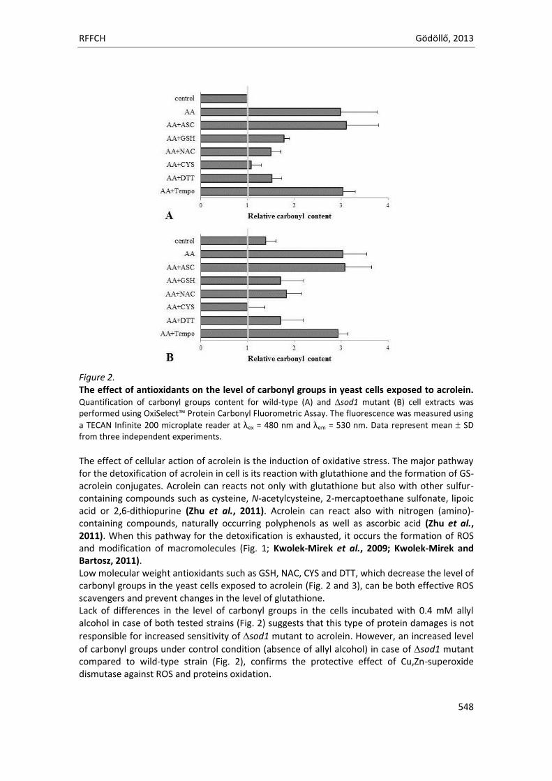
RFFCH Gödöllő, 2013
548
Figure 2. The effect of antioxidants on the level of carbonyl groups in yeast cells exposed to acrolein. Quantification of carbonyl groups content for wild-type (A) and sod1 mutant (B) cell extracts was performed using OxiSelect™ Protein Carbonyl Fluorometric Assay. The fluorescence was measured using
a TECAN Infinite 200 microplate reader at λex = 480 nm and λem = 530 nm. Data represent mean SD from three independent experiments. The effect of cellular action of acrolein is the induction of oxidative stress. The major pathway for the detoxification of acrolein in cell is its reaction with glutathione and the formation of GS-acrolein conjugates. Acrolein can reacts not only with glutathione but also with other sulfur-containing compounds such as cysteine, N-acetylcysteine, 2-mercaptoethane sulfonate, lipoic acid or 2,6-dithiopurine (Zhu et al., 2011). Acrolein can react also with nitrogen (amino)-containing compounds, naturally occurring polyphenols as well as ascorbic acid (Zhu et al., 2011). When this pathway for the detoxification is exhausted, it occurs the formation of ROS and modification of macromolecules (Fig. 1; Kwolek-Mirek et al., 2009; Kwolek-Mirek and Bartosz, 2011). Low molecular weight antioxidants such as GSH, NAC, CYS and DTT, which decrease the level of carbonyl groups in the yeast cells exposed to acrolein (Fig. 2 and 3), can be both effective ROS scavengers and prevent changes in the level of glutathione. Lack of differences in the level of carbonyl groups in the cells incubated with 0.4 mM allyl alcohol in case of both tested strains (Fig. 2) suggests that this type of protein damages is not
responsible for increased sensitivity of sod1 mutant to acrolein. However, an increased level
of carbonyl groups under control condition (absence of allyl alcohol) in case of sod1 mutant compared to wild-type strain (Fig. 2), confirms the protective effect of Cu,Zn-superoxide dismutase against ROS and proteins oxidation.
RFFCH Gödöllő, 2013
549
Figure 3. The effect of antioxidants on the level of carbonyl groups in yeast cells exposed to acrolein. Image A and B presents the results for wild-type, C and D for sod1 mutant cell extracts. Line M – Prestained Protein Molecular Weight Marker, line 1 – control cells (in absence of allyl alcohol), line 2 – cells incubated with 0.4 mM AA, line 3 – cells incubated with 0.4 mM AA and 25 mM ASC, line 4 – cells incubated with 0.4 mM AA and 5 mM GSH, line 5 – cells incubated with 0.4 mM AA and 5 mM NAC, line 6 – cells incubated with 0.4 mM AA and 5 mM CYS, line 7 – cells incubated with 0.4 mM AA and 5 mM DTT and line 8 – cells incubated with 0.4 mM AA and 1mM Tempo. Image A and B Coomassie Brilliant Blue staining PVDF membranes, C and D Immunoblotting analysis using anty-2,4-dinitrophenyl antibodies. The figure shows representative images recorded during one of the two independent experiments.
Acknowledgements This study was supported by the Grant Iuventus Plus No. 0289/IP1/2011/71 of Polish Ministry of Science and Higher Education. References BARTOSZ, G. 2003. The other face of oxygen, Published by the PWN. BERLETT, B.S., STADTMAN, E.R. 1997. Protein oxidation in aging, disease, and oxidative stress. J Biol
Chem, 272(33), 20313-16. BILINSKI, T., KWOLEK, M., SAS, E., KRYNICKA, M., KOZIOL, S., OWSIAK-TELEON, A., KRZEPILKO, A.,
BARTOSZ, G. 2005. A novel test identifying genes involved in aldehyde detoxification in the yeast. Increased sensitivity of superoxide-deficient yeast to aldehydes and their metabolic precursors. Biofactors, 24, 59-65.
BILINSKI, T., LUKASZKIEWICZ, J., SLEDZIEWSKI, A. 1978. Demonstration of anaerobic catalase synthesis in the cz1 mutant of Saccharomyces cerevisiae. Biochem Biophys Res Comm, 83, 1225-33.
BRADFORD, M.M. 1976. A rapid and sensitive method for the quantitation of microgram quantities of protein utilizing the principle of protein-dye binding. Anal Biochem, 72, 248-54.
CABISCOL, E., PIULATS, E., ECHAVE, P. 2000. Oxidative stress promotes specific protein damage in Saccharomyces cerevisiae. J Biol Chem, 275, 27393-98.
RFFCH Gödöllő, 2013
550
KOZIOL, S., ZAGULSKI, M., BILINSKI, T., BARTOSZ, G. 2005. Antioxidants protect the yeast Saccharomyces cerevisiae against hypertonic stress. Free Radic Res, 39, 365-71.
KWOLEK-MIREK, M., BARTOSZ, G. 2011. Biochemical and cellular effects of acrolein toxicity in the yeast Saccharomyces cerevisiae. Xenobiotics. Environmental exposure, toxicity and physiological response, Published by the University of Rzeszow, 271-81.
KWOLEK-MIREK, M., BEDNARSKA S., BARTOSZ, G., BILINSKI, T. 2009. Acrolein toxicity involves oxidative stress caused by glutathione depletion in the yeast Saccharomyces cerevisiae. Cell Biol Toxicol, 25(4), 363-78.
LEVINE, R.L., WILLIAMS, J.A., STADTMAN, E.R., SHACTER, E. 1994. Carbonyl assays for determination of oxidatively modified proteins. Methods Enzymol, 233, 346-57.
LUSHCHAK, V.I. 2006. Budding yeast Saccharomyces cerevisiae as a model to study oxidative modification of proteins in eukaryotes. Acta Biochim Pol, 53, 679-84.
NYSTROM, T. 2005. Role of oxidative carbonylation in protein quality control and senescence. EMBO J, 24, 1311-17.
SEMCHYSHYN, H.M., LOZINSKA, L.M., MIEDZOBRODZKI, J., LUSHCHAK, V.I. 2011. Fructose and glucose differentially affect aging and carbonyl/oxidative stress parameters in Saccharomyces cerevisiae cells. Carbohydrate Research, 346, 933-38.
STADTMAN, E.R., LEVINE, R.L. 2000. Protein oxidation. Annals of the New York Academy of Science, 899, 191-208.
STEVENS, J.F., MAIER, C.S. 2008. Acrolein: sources, metabolism, and biomolecular interactions relevant to human health and disease. Mol Nutr Food Res, 52, 7-25.
TROTTER, E.W., COLLINSON, E.J., DAWES, I.W., GRANT, C.M. 2006. Old yellow enzymes protect against toxicity in the yeast Saccharomyces cerevisiae. Appl Environ Microbiol, 72(7), 4885-92.
ZHU, Q., SUN, Z., JIANG, Y., CHEN, F., WANG, M. 2011. Acrolein scavengers: reactivity, mechanism and impact on health. Mol Nutr Food Res, 55, 1375-90.

Animal welfare, etológia és tartástechnológia
Animal welfare, ethology and housing systems
Volume 9 Issue 3
Különszám/Special Issue
Gödöllő
2013
RFFCH Gödöllő, 2013
551
Effect of α-tocopherol in ovo supplementation on chick embryo development Lis M.W., Cichy A., Trzeciak K.B., Pawlak K., Dżugan M. and Niedziółka J.W.
1Department of Poultry and Fur Animal Breeding and Animal Hygiene, University of Agriculture in Krakow, Poland
2Department of Chemistry and Food Toxicology, University of Rzeszów, Rzeszów, Poland α-tocopherol (vitamin E) effects on the reproductive processes, participating in the synthesis of sex hormones. Vitamin E is not synthesised by intestinal microflora of birds and its deficiency can disturb of pre- and post- hatch chicken development. However, in the available literature there is lack of data concerning the effect of high dose of α-tocopherol on embryogenesis and endocrine glands in avian species. In 4th day of incubation (E4) the fertilized chicken eggs were injected with 0, 0.5 and 5 mg/egg of α-tocopherol, dissolved in 50 μl peanut oil. In E14 and E20 the blood samples and gonads, heart and liver were collected from 20 embryos of each group. The organs were weighted and frozen in -80°C to further histological measures while blood samples were centrifuged and blood plasma was stored in -20°C for the determination of testosterone, progesterone and estradiol by RIA. The results of experiment indicate that α-tocopherol in ovo supplementation on the E4 decreased embryo mortality between E4-E6 but increased during hatching period (P≤0.05). Moreover there was disturbed in sex ratio which ranged as 7 males to 3 females. Simultaneously, supplementation α-tocopherol at a dose of 0.5 and 5 mg per egg decreased about 30% of estradiol concentration in the blood female embryos in E20. Concluded, in ovo supplementation of α-tocopherol can disturb embryogenesis of chicken embryos.

Animal welfare, etológia és tartástechnológia
Animal welfare, ethology and housing systems
Volume 9 Issue 3
Különszám/Special Issue
Gödöllő
2013
RFFCH Gödöllő, 2013
552
APPLICATION OF THERMOGRAPHY IN COWS PRODUCTIVITY EVALUATION: THE CORRELATION BETWEEN THE BODY
TEMPERATURE AND MILK YIELD Lisowska-Lis A1., Lis M.W.2
1Polytechnical Institute, State Higher Vocational School in Tarnow, ul. Mickiewicza 8, 33-100 Tarnow. Poland. [email protected] 2Department of Poultry and Fur Animal Breeding and Animal Hygiene, Agricultural University of Kraków. al. Mickiewicza 24/28, 30-059 Kraków, Poland. [email protected]
The aim of this work was an attempt of using thermography measurements to monitoring cows productivity. The changes in the temperature of the cow's udder were analysed. The temperature measurements as thermograms for the individual cows (HF breed) were taken before and after the milking, in the milking parlor (364 thermograms). Measurements were taken by InfraCAM SD (FLIR system) camera. Thermograms were analysed using the FLIR-QUICK-REPORT program. Thermal analysis results for every individual were compared with the stage of lactation, the number of lactation and the milk yield (productivity). Statistical analysis were done using SIGMASTAT 3.5 (two-way ANOVA, Tukey test).
Results of experiment showed no differences between the temperature (measured for the
cows` udder) at the beginning and at the end of milking. Temperature at the beginning was T=
37,8±0,59°C, and at the end of milking was T= 37,8±0,59°C (P>0,05). Temperature did not depend on
the cows age (number of lactation), (P>0,05). It was observed correlation between temperature and
month of lactation(P≤0,05). The highest temperature was observed in the second part of the first
month of lactation. The high temperature of the cows udder was observed since the second part of
the first month till the seventh month of the lactation. Cows productivity for the first month of
lactation was between 30 – 45 l milk per day, and in the seventh month it ranged between 15 – 40 l.
At the seventh month of lactation the rapid decrease of the temperature was observed. The lower
temperature of the udder was typical for the cows between 7th up to 12th month of lactation (or
more). It is probably correlated with the natural decrease of cows productivity at that stage of the
lactation.
CONCLUSION The obtained results confirms possible application of thermography as an noninvasive method of assessing the productivity of farm animals.
Animal welfare, etológia és tartástechnológia
Animal welfare, ethology and housing systems
Volume 9 Issue 3
Különszám/Special Issue
Gödöllő
2013
RFFC Gödöllő, 2013
553
CONCENTRATION OF MALONDIALDEHYDE (MDA) IN MEAT PRODUCTS DURING PRODUCTION
Lukáčová Anetta*, Tvrdá Eva, Golian Jozef Slovak University of Agriculture in Nitra, Faculty of Biotechnology and Food Sciences, Tr. Andreja Hlinku 2, 949 76 Nitra, Slovak Republic; *[email protected] Phone: +421 37 6415826.
Abstract Lipid oxidation is one of the major causes of quality deterioration in meat. Lipid oxidation is induced by oxy- and/or lipid free radical generation and results in the generation of toxic compounds such as the malondialdehyde and cholesterol oxidation products. The aim of this study was carried out to determine concentrations of malondialdehyde (MDA) in meat products (salami “Malokarpatska” and “Lovecka”, frankfurter, Selected ham) during technological process. This study, we have demonstrated the presence of malondialdehyde a deleterious by product of lipid peroxidation in meat products. The results unequivocally showed that adding spice to homogenized samples significantly increased the formation of lipid-peroxidation products. Also in all samples were increased concentrations of MDA in beef as raw materials. The beef samples had higher MDA than the pork, pork bacon and leathery emulsion samples.
Key words: Malondialdehyde, meat, meat products, lipid oxidation
Introduction Food lipids are principally triacylglycerides, phospoholipids and sterolsfound naturally in most biological materials consumed as food and added as functional ingredients in many processed foods. Lipids contribute many desirable qualities to foods, including attributes of texture, structure, mouth feel, flavour and colour. However, lipids are also one of the most chemically unstable food components and will readily undergo free-radical chain reactions that only deteriorate the lipids but also: produce oxidative fragments, some of which are volatile and are perceived as the off-flavors of rancidity (German, 1999). Lipid oxidation is one of the major causes of quality deterioration in meat (Jo and Ahn, 1998). Lipid oxidation is often responsible for quality loss via formation of rancid flavour and is affected by the duration and temperature of storage of meat. Lipid oxidation is the major form of deterioration in stored muscle foods. Oxidative reactions in meat are the most important factor in quality losses, including flavour, texture, nutritive value and colour. Lipid oxidation is induced by oxy- and/or lipid free radical generation and results in the generation of toxic compounds such as the malondialdehyde and cholesterol oxidation products (Sun et al., 2002). Over the past 30 y, there has been accumulating evidence that lipid oxidation can play an important role in the processes of atherogenesis and carcinogenesis (Li et al., 2010). Fogelman et al. (1980) reported that malondialdehyde an obligate product of the oxidation of arachidonic acid by lipoxygenase pathways. Dietary and environmental chemicals such as N-nitrosamines and their precursors (nitrate and nitrite) and malondialdehyde involved in the etiology of cancer and other related disease conditions are part of the challenges still facing the world today (Okafor et al., 2007). One of the most important preservation methods for meat and meat products since compared with other methods is freezing, it leads to a minimal loss of quality during long-term storage (Soyer et al., 2010). Since the development of oxidation process in meat is important, related to its quality and consumer´s acceptance, the aim of this study was carried out to determine concentrations of malondialdehyde (MDA) in meat products (salami “Malokarpatska” and Lovecka”, frankfurter, Selected ham) during technological process.
Materials and method Sampling was done so that the samples were representative that to have the average composition and characteristics of the goods from which they were collected. The collection sample during the manufacturing process was carried out under the following scheme. “Lovecka salami” - was collected basic raw (beef, pork and pork bacon); than samples mixed meat with additives (salt,
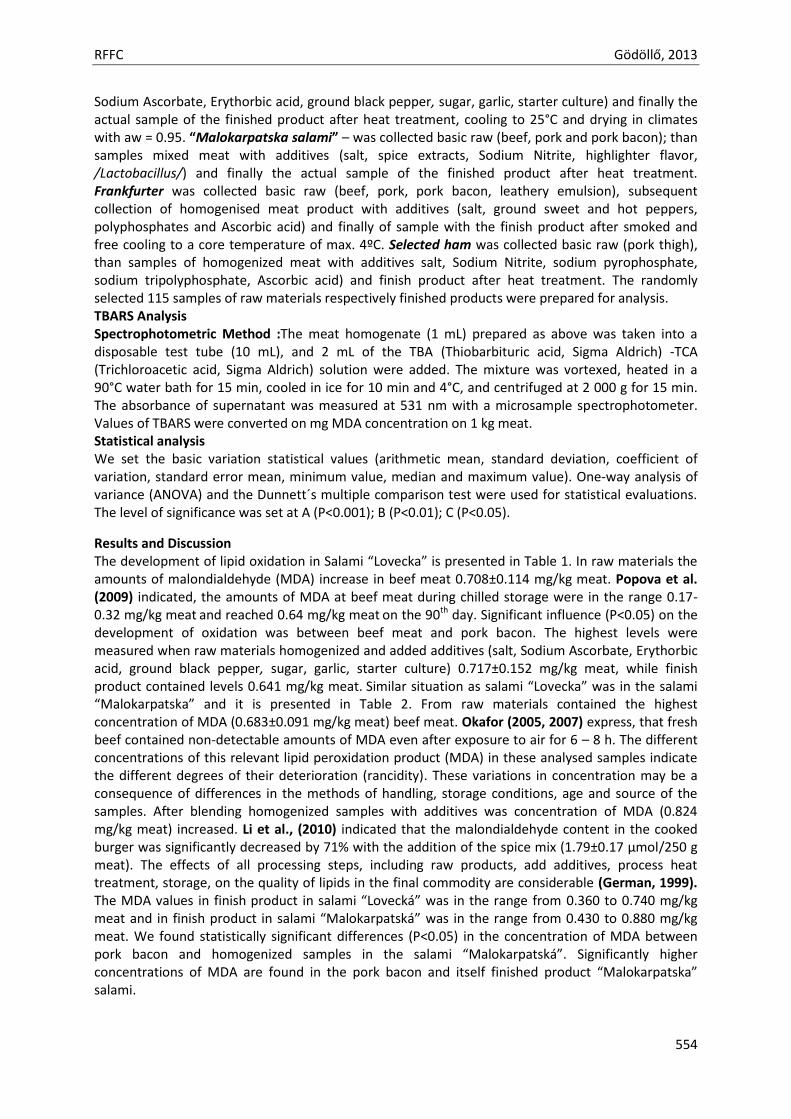
RFFC Gödöllő, 2013
554
Sodium Ascorbate, Erythorbic acid, ground black pepper, sugar, garlic, starter culture) and finally the actual sample of the finished product after heat treatment, cooling to 25°C and drying in climates with aw = 0.95. “Malokarpatska salami” – was collected basic raw (beef, pork and pork bacon); than samples mixed meat with additives (salt, spice extracts, Sodium Nitrite, highlighter flavor, /Lactobacillus/) and finally the actual sample of the finished product after heat treatment. Frankfurter was collected basic raw (beef, pork, pork bacon, leathery emulsion), subsequent collection of homogenised meat product with additives (salt, ground sweet and hot peppers, polyphosphates and Ascorbic acid) and finally of sample with the finish product after smoked and free cooling to a core temperature of max. 4ºC. Selected ham was collected basic raw (pork thigh), than samples of homogenized meat with additives salt, Sodium Nitrite, sodium pyrophosphate, sodium tripolyphosphate, Ascorbic acid) and finish product after heat treatment. The randomly selected 115 samples of raw materials respectively finished products were prepared for analysis. TBARS Analysis Spectrophotometric Method :The meat homogenate (1 mL) prepared as above was taken into a disposable test tube (10 mL), and 2 mL of the TBA (Thiobarbituric acid, Sigma Aldrich) -TCA (Trichloroacetic acid, Sigma Aldrich) solution were added. The mixture was vortexed, heated in a 90°C water bath for 15 min, cooled in ice for 10 min and 4°C, and centrifuged at 2 000 g for 15 min. The absorbance of supernatant was measured at 531 nm with a microsample spectrophotometer. Values of TBARS were converted on mg MDA concentration on 1 kg meat. Statistical analysis We set the basic variation statistical values (arithmetic mean, standard deviation, coefficient of variation, standard error mean, minimum value, median and maximum value). One-way analysis of variance (ANOVA) and the Dunnett´s multiple comparison test were used for statistical evaluations. The level of significance was set at A (P<0.001); B (P<0.01); C (P<0.05).
Results and Discussion The development of lipid oxidation in Salami “Lovecka” is presented in Table 1. In raw materials the amounts of malondialdehyde (MDA) increase in beef meat 0.708±0.114 mg/kg meat. Popova et al. (2009) indicated, the amounts of MDA at beef meat during chilled storage were in the range 0.17-0.32 mg/kg meat and reached 0.64 mg/kg meat on the 90th day. Significant influence (P<0.05) on the development of oxidation was between beef meat and pork bacon. The highest levels were measured when raw materials homogenized and added additives (salt, Sodium Ascorbate, Erythorbic acid, ground black pepper, sugar, garlic, starter culture) 0.717±0.152 mg/kg meat, while finish product contained levels 0.641 mg/kg meat. Similar situation as salami “Lovecka” was in the salami “Malokarpatska” and it is presented in Table 2. From raw materials contained the highest concentration of MDA (0.683±0.091 mg/kg meat) beef meat. Okafor (2005, 2007) express, that fresh beef contained non-detectable amounts of MDA even after exposure to air for 6 – 8 h. The different concentrations of this relevant lipid peroxidation product (MDA) in these analysed samples indicate the different degrees of their deterioration (rancidity). These variations in concentration may be a consequence of differences in the methods of handling, storage conditions, age and source of the samples. After blending homogenized samples with additives was concentration of MDA (0.824 mg/kg meat) increased. Li et al., (2010) indicated that the malondialdehyde content in the cooked burger was significantly decreased by 71% with the addition of the spice mix (1.79±0.17 µmol/250 g meat). The effects of all processing steps, including raw products, add additives, process heat treatment, storage, on the quality of lipids in the final commodity are considerable (German, 1999). The MDA values in finish product in salami “Lovecká” was in the range from 0.360 to 0.740 mg/kg meat and in finish product in salami “Malokarpatská” was in the range from 0.430 to 0.880 mg/kg meat. We found statistically significant differences (P<0.05) in the concentration of MDA between pork bacon and homogenized samples in the salami “Malokarpatská”. Significantly higher concentrations of MDA are found in the pork bacon and itself finished product “Malokarpatska” salami.
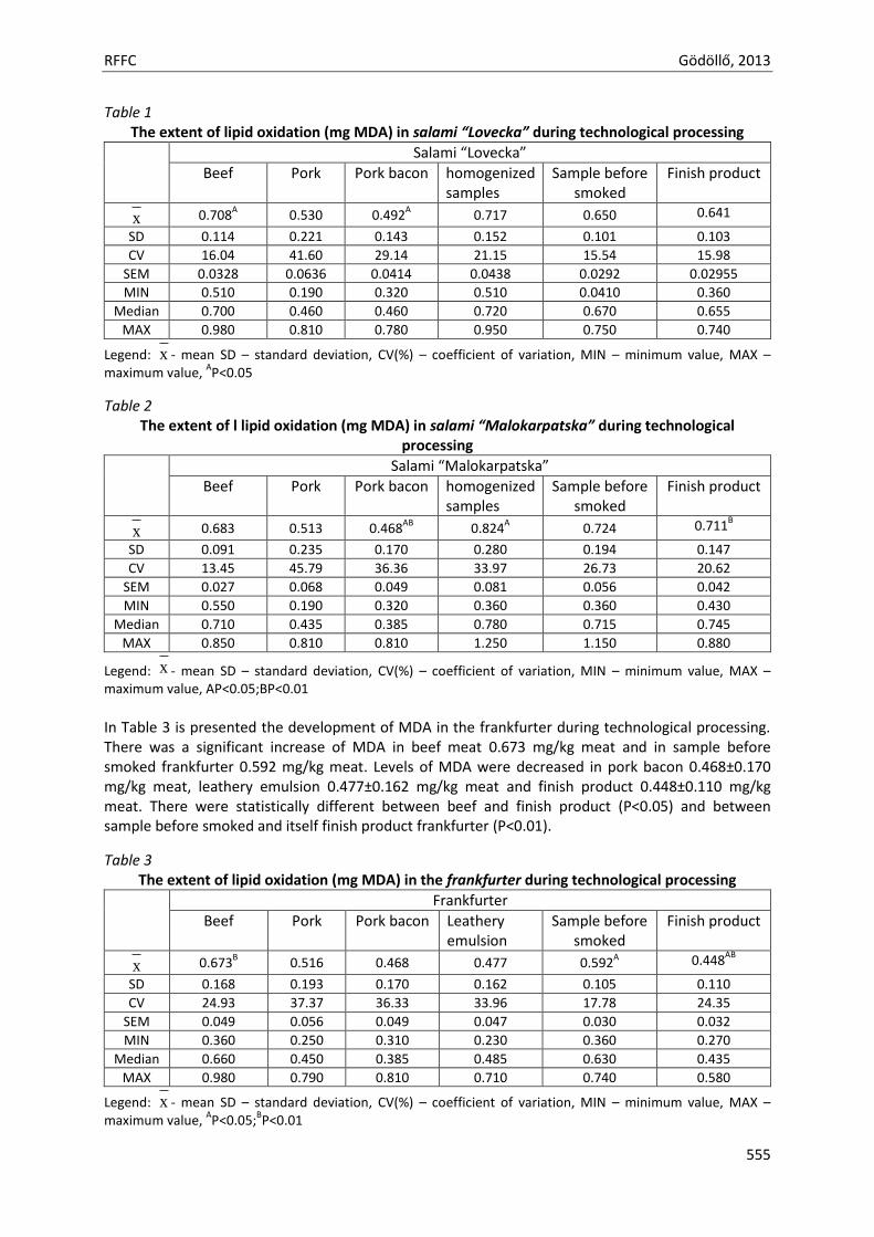
RFFC Gödöllő, 2013
555
Table 1 The extent of lipid oxidation (mg MDA) in salami “Lovecka” during technological processing
Salami “Lovecka”
Beef Pork Pork bacon homogenized samples
Sample before smoked
Finish product
x 0.708A 0.530 0.492
A 0.717 0.650 0.641
SD 0.114 0.221 0.143 0.152 0.101 0.103
CV 16.04 41.60 29.14 21.15 15.54 15.98
SEM 0.0328 0.0636 0.0414 0.0438 0.0292 0.02955
MIN 0.510 0.190 0.320 0.510 0.0410 0.360
Median 0.700 0.460 0.460 0.720 0.670 0.655
MAX 0.980 0.810 0.780 0.950 0.750 0.740
Legend: x - mean SD – standard deviation, CV(%) – coefficient of variation, MIN – minimum value, MAX – maximum value,
AP<0.05
Table 2 The extent of l lipid oxidation (mg MDA) in salami “Malokarpatska” during technological
processing
Salami “Malokarpatska”
Beef Pork Pork bacon homogenized samples
Sample before smoked
Finish product
x 0.683 0.513 0.468AB
0.824A 0.724 0.711
B
SD 0.091 0.235 0.170 0.280 0.194 0.147
CV 13.45 45.79 36.36 33.97 26.73 20.62
SEM 0.027 0.068 0.049 0.081 0.056 0.042
MIN 0.550 0.190 0.320 0.360 0.360 0.430
Median 0.710 0.435 0.385 0.780 0.715 0.745
MAX 0.850 0.810 0.810 1.250 1.150 0.880
Legend: x - mean SD – standard deviation, CV(%) – coefficient of variation, MIN – minimum value, MAX – maximum value, AP<0.05;BP<0.01
In Table 3 is presented the development of MDA in the frankfurter during technological processing. There was a significant increase of MDA in beef meat 0.673 mg/kg meat and in sample before smoked frankfurter 0.592 mg/kg meat. Levels of MDA were decreased in pork bacon 0.468±0.170 mg/kg meat, leathery emulsion 0.477±0.162 mg/kg meat and finish product 0.448±0.110 mg/kg meat. There were statistically different between beef and finish product (P<0.05) and between sample before smoked and itself finish product frankfurter (P<0.01).
Table 3 The extent of lipid oxidation (mg MDA) in the frankfurter during technological processing
Frankfurter
Beef Pork Pork bacon Leathery emulsion
Sample before smoked
Finish product
x 0.673B 0.516 0.468 0.477 0.592
A 0.448
AB
SD 0.168 0.193 0.170 0.162 0.105 0.110
CV 24.93 37.37 36.33 33.96 17.78 24.35
SEM 0.049 0.056 0.049 0.047 0.030 0.032
MIN 0.360 0.250 0.310 0.230 0.360 0.270
Median 0.660 0.450 0.385 0.485 0.630 0.435
MAX 0.980 0.790 0.810 0.710 0.740 0.580
Legend: x - mean SD – standard deviation, CV(%) – coefficient of variation, MIN – minimum value, MAX – maximum value,
AP<0.05;
BP<0.01
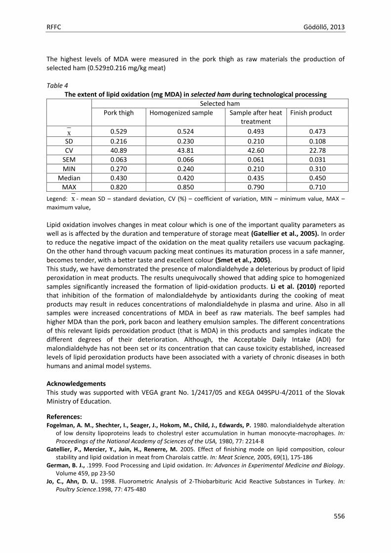
RFFC Gödöllő, 2013
556
The highest levels of MDA were measured in the pork thigh as raw materials the production of selected ham (0.529±0.216 mg/kg meat) Table 4
The extent of lipid oxidation (mg MDA) in selected ham during technological processing
Selected ham
Pork thigh Homogenized sample Sample after heat treatment
Finish product
x 0.529 0.524 0.493 0.473
SD 0.216 0.230 0.210 0.108
CV 40.89 43.81 42.60 22.78
SEM 0.063 0.066 0.061 0.031
MIN 0.270 0.240 0.210 0.310
Median 0.430 0.420 0.435 0.450
MAX 0.820 0.850 0.790 0.710
Legend: x - mean SD – standard deviation, CV (%) – coefficient of variation, MIN – minimum value, MAX –
maximum value, Lipid oxidation involves changes in meat colour which is one of the important quality parameters as well as is affected by the duration and temperature of storage meat (Gatellier et al., 2005). In order to reduce the negative impact of the oxidation on the meat quality retailers use vacuum packaging. On the other hand through vacuum packing meat continues its maturation process in a safe manner, becomes tender, with a better taste and excellent colour (Smet et al., 2005). This study, we have demonstrated the presence of malondialdehyde a deleterious by product of lipid peroxidation in meat products. The results unequivocally showed that adding spice to homogenized samples significantly increased the formation of lipid-oxidation products. Li et al. (2010) reported that inhibition of the formation of malondialdehyde by antioxidants during the cooking of meat products may result in reduces concentrations of malondialdehyde in plasma and urine. Also in all samples were increased concentrations of MDA in beef as raw materials. The beef samples had higher MDA than the pork, pork bacon and leathery emulsion samples. The different concentrations of this relevant lipids peroxidation product (that is MDA) in this products and samples indicate the different degrees of their deterioration. Although, the Acceptable Daily Intake (ADI) for malondialdehyde has not been set or its concentration that can cause toxicity established, increased levels of lipid peroxidation products have been associated with a variety of chronic diseases in both humans and animal model systems. Acknowledgements This study was supported with VEGA grant No. 1/2417/05 and KEGA 049SPU-4/2011 of the Slovak Ministry of Education.
References: Fogelman, A. M., Shechter, I., Seager, J., Hokom, M., Child, J., Edwards, P. 1980. malondialdehyde alteration
of low density lipoproteins leads to cholestryl ester accumulation in human monocyte-macrophages. In: Proceedings of the National Academy of Sciences of the USA, 1980, 77: 2214-8
Gatellier, P., Mercier, Y., Juin, H., Renerre, M. 2005. Effect of finishing mode on lipid composition, colour stability and lipid oxidation in meat from Charolais cattle. In: Meat Science, 2005, 69(1), 175-186
German, B. J., .1999. Food Processing and Lipid oxidation. In: Advances in Experimental Medicine and Biology. Volume 459, pp 23-50
Jo, C., Ahn, D. U.. 1998. Fluorometric Analysis of 2-Thiobarbituric Acid Reactive Substances in Turkey. In: Poultry Science.1998, 77: 475-480
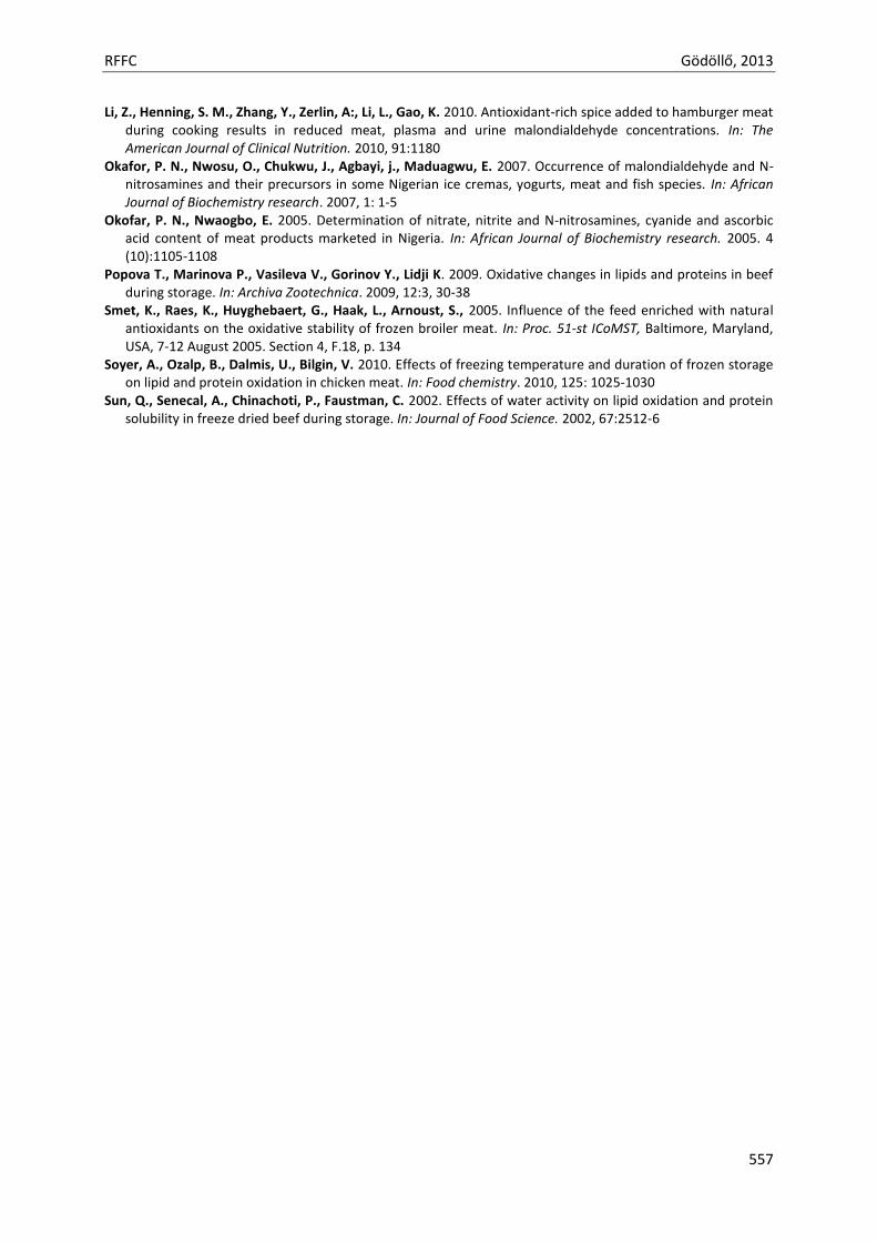
RFFC Gödöllő, 2013
557
Li, Z., Henning, S. M., Zhang, Y., Zerlin, A:, Li, L., Gao, K. 2010. Antioxidant-rich spice added to hamburger meat during cooking results in reduced meat, plasma and urine malondialdehyde concentrations. In: The American Journal of Clinical Nutrition. 2010, 91:1180
Okafor, P. N., Nwosu, O., Chukwu, J., Agbayi, j., Maduagwu, E. 2007. Occurrence of malondialdehyde and N-nitrosamines and their precursors in some Nigerian ice cremas, yogurts, meat and fish species. In: African Journal of Biochemistry research. 2007, 1: 1-5
Okofar, P. N., Nwaogbo, E. 2005. Determination of nitrate, nitrite and N-nitrosamines, cyanide and ascorbic acid content of meat products marketed in Nigeria. In: African Journal of Biochemistry research. 2005. 4 (10):1105-1108
Popova T., Marinova P., Vasileva V., Gorinov Y., Lidji K. 2009. Oxidative changes in lipids and proteins in beef during storage. In: Archiva Zootechnica. 2009, 12:3, 30-38
Smet, K., Raes, K., Huyghebaert, G., Haak, L., Arnoust, S., 2005. Influence of the feed enriched with natural antioxidants on the oxidative stability of frozen broiler meat. In: Proc. 51-st ICoMST, Baltimore, Maryland, USA, 7-12 August 2005. Section 4, F.18, p. 134
Soyer, A., Ozalp, B., Dalmis, U., Bilgin, V. 2010. Effects of freezing temperature and duration of frozen storage on lipid and protein oxidation in chicken meat. In: Food chemistry. 2010, 125: 1025-1030
Sun, Q., Senecal, A., Chinachoti, P., Faustman, C. 2002. Effects of water activity on lipid oxidation and protein solubility in freeze dried beef during storage. In: Journal of Food Science. 2002, 67:2512-6

Animal welfare, etológia és tartástechnológia
Animal welfare, ethology and housing systems
Volume 9 Issue 3
Különszám/Special Issue
Gödöllő
2013
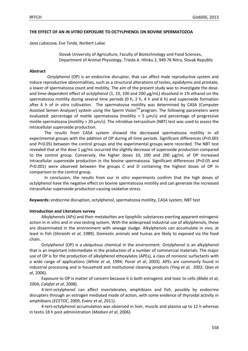
RFFCH Gödöllő, 2013
558
THE EFFECT OF AN IN VITRO EXPOSURE TO OCTYLPHENOL ON BOVINE SPERMATOZOA Jana Lukacova, Eva Tvrda, Norbert Lukac
Slovak University of Agriculture, Faculty of Biotechnology and Food Sciences, Department of Animal Physiology, Trieda A. Hlinku 2, 949 76 Nitra, Slovak Republic
Abstract
Octylphenol (OP) is an endocrine disruptor, that can affect male reproductive system and induce reproductive abnormalities, such as a structural alterations of testes, epididymis and prostate, a lower of spermatozoa count and motility. The aim of the present study was to investigate the dose- and time-dependent effect of octylphenol (1, 10, 100 and 200 µg/mL) dissolved in 1% ethanol on the spermatozoa motility during several time periods (0 h, 2 h, 4 h and 6 h) and superoxide formation after 6 h of in vitro cultivation. The spermatozoa motility was determined by CASA (Computer Assisted Semen Analyzer) system using the Sperm VisionTM program. The following parameters were evaluated: percentage of motile spermatozoa (motility > 5 µm/s) and percentage of progressive motile spermatozoa (motility > 20 µm/s). The nitroblue-tetrazolium (NBT) test was used to assess the intracellular superoxide production.
The results from CASA system showed the decreased spermatozoa motility in all experimental groups with the addition of OP during all time periods. Significant differences (P<0.001 and P<0.05) between the control groups and the experimental groups were recorded. The NBT test revealed that at the dose 1 µg/mL occurred the slightly decrease of superoxide production compared to the control group. Conversely, the higher doses 10, 100 and 200 µg/mL of OP increased intracellular superoxide production in the bovine spermatozoa. Significant differences (P<0.05 and P<0.001) were observed between the groups C and D containing the highest doses of OP in comparison to the control group.
In conclusion, the results from our in vitro experiments confirm that the high doses of octylphenol have the negative effect on bovine spermatozoa motility and can generate the increased intracellular superoxide production causing oxidative stress. Keywords: endocrine disruption, octylphenol, spermatozoa motility, CASA system, NBT test Introduction and Literature survey
Alkylphenols (APs) and their metabolites are lipophilic substances exerting apparent estrogenic action in in vitro and in vivo testing system. With the widespread industrial use of alkylphenols, these are disseminated in the environment with sewage sludge. Alkylphenols can accumulate in vivo, at least in fish (Shiraishi et al, 1989). Domestic animals and humas are likely to exposed via the food chain.
Octylphenol (OP) is a ubiquitous chemical in the environment. Octylphenol is an alkylphenol that is an important intermediate in the production of a number of commercial materials. The major use of OP is for the production of alkylphenol ethoxylates (APEs), a class of nonionic surfactants with a wide range of applications (White et al, 1994; Pocar et al, 2003). APEs are commonly found in industrial processing and in household and institutional cleaning products (Ying et al, 2002; Qian et al, 2006).
Exposure to OP is matter of concern because it is both estrogenic and toxic to cells (Blake et al, 2004; Calafat et al, 2008).
4-tert-octylphenol can affect invertebrates, amphibians and fish, possibly by endocrine disrupters through an estrogen mediated mode of action, with some evidence of thyroidal activity in amphibians (ECETOC, 2009, Evans et al, 2011).
4-tert-octylphenol accumulation was observed in liver, muscle and plasma up to 12 h whereas in testis 18 h post administration (Madsen et al, 2006).
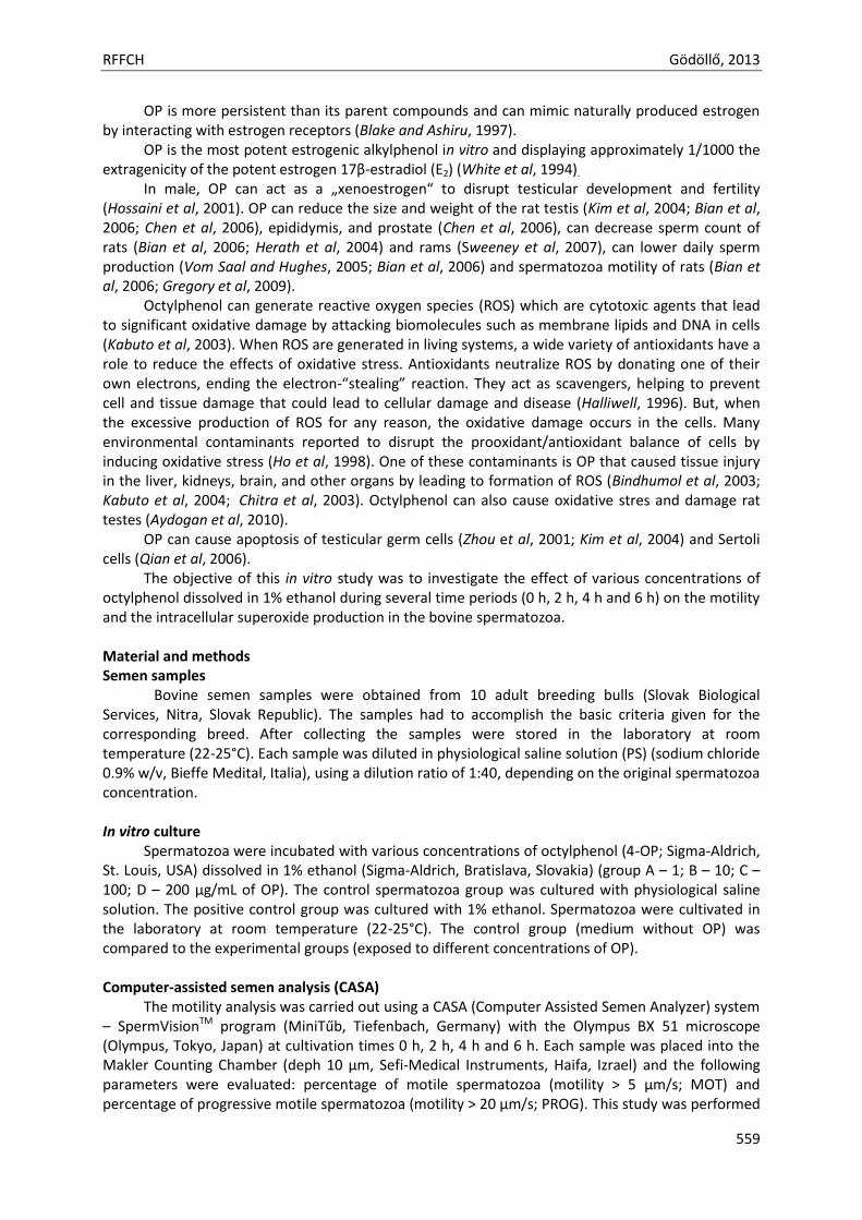
RFFCH Gödöllő, 2013
559
OP is more persistent than its parent compounds and can mimic naturally produced estrogen by interacting with estrogen receptors (Blake and Ashiru, 1997).
OP is the most potent estrogenic alkylphenol in vitro and displaying approximately 1/1000 the extragenicity of the potent estrogen 17β-estradiol (E2) (White et al, 1994).
In male, OP can act as a „xenoestrogen“ to disrupt testicular development and fertility (Hossaini et al, 2001). OP can reduce the size and weight of the rat testis (Kim et al, 2004; Bian et al, 2006; Chen et al, 2006), epididymis, and prostate (Chen et al, 2006), can decrease sperm count of rats (Bian et al, 2006; Herath et al, 2004) and rams (Sweeney et al, 2007), can lower daily sperm production (Vom Saal and Hughes, 2005; Bian et al, 2006) and spermatozoa motility of rats (Bian et al, 2006; Gregory et al, 2009).
Octylphenol can generate reactive oxygen species (ROS) which are cytotoxic agents that lead to significant oxidative damage by attacking biomolecules such as membrane lipids and DNA in cells (Kabuto et al, 2003). When ROS are generated in living systems, a wide variety of antioxidants have a role to reduce the effects of oxidative stress. Antioxidants neutralize ROS by donating one of their own electrons, ending the electron-“stealing” reaction. They act as scavengers, helping to prevent cell and tissue damage that could lead to cellular damage and disease (Halliwell, 1996). But, when the excessive production of ROS for any reason, the oxidative damage occurs in the cells. Many environmental contaminants reported to disrupt the prooxidant/antioxidant balance of cells by inducing oxidative stress (Ho et al, 1998). One of these contaminants is OP that caused tissue injury in the liver, kidneys, brain, and other organs by leading to formation of ROS (Bindhumol et al, 2003; Kabuto et al, 2004; Chitra et al, 2003). Octylphenol can also cause oxidative stres and damage rat testes (Aydogan et al, 2010).
OP can cause apoptosis of testicular germ cells (Zhou et al, 2001; Kim et al, 2004) and Sertoli cells (Qian et al, 2006).
The objective of this in vitro study was to investigate the effect of various concentrations of octylphenol dissolved in 1% ethanol during several time periods (0 h, 2 h, 4 h and 6 h) on the motility and the intracellular superoxide production in the bovine spermatozoa.
Material and methods Semen samples
Bovine semen samples were obtained from 10 adult breeding bulls (Slovak Biological Services, Nitra, Slovak Republic). The samples had to accomplish the basic criteria given for the corresponding breed. After collecting the samples were stored in the laboratory at room temperature (22-25°C). Each sample was diluted in physiological saline solution (PS) (sodium chloride 0.9% w/v, Bieffe Medital, Italia), using a dilution ratio of 1:40, depending on the original spermatozoa concentration. In vitro culture
Spermatozoa were incubated with various concentrations of octylphenol (4-OP; Sigma-Aldrich, St. Louis, USA) dissolved in 1% ethanol (Sigma-Aldrich, Bratislava, Slovakia) (group A – 1; B – 10; C – 100; D – 200 µg/mL of OP). The control spermatozoa group was cultured with physiological saline solution. The positive control group was cultured with 1% ethanol. Spermatozoa were cultivated in the laboratory at room temperature (22-25°C). The control group (medium without OP) was compared to the experimental groups (exposed to different concentrations of OP). Computer-assisted semen analysis (CASA)
The motility analysis was carried out using a CASA (Computer Assisted Semen Analyzer) system – SpermVisionTM program (MiniTűb, Tiefenbach, Germany) with the Olympus BX 51 microscope (Olympus, Tokyo, Japan) at cultivation times 0 h, 2 h, 4 h and 6 h. Each sample was placed into the Makler Counting Chamber (deph 10 µm, Sefi-Medical Instruments, Haifa, Izrael) and the following parameters were evaluated: percentage of motile spermatozoa (motility > 5 µm/s; MOT) and percentage of progressive motile spermatozoa (motility > 20 µm/s; PROG). This study was performed
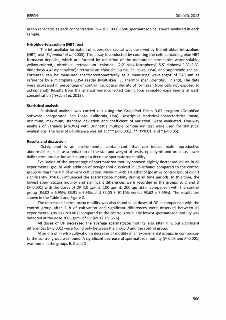
RFFCH Gödöllő, 2013
560
in ten replicates at each concentration (n = 10). 1000-1500 spermatozoa cells were analyzed in each sample. Nitroblue-tetrazolium (NBT) test
The intracellular formation of superoxide radical was observed by the nitroblue-tetrazolium (NBT) test (Esfandiari et al, 2003). This assay is conducted by counting the cells containing blue NBT formazan deposits, which are formed by reduction of the membrane permeable, water-soluble, yellow-colored, nitroblue tetrazolium chloride (2,2´-bis(4-Nitrophenyl)-5,5´-diphenyl-3,3´-(3,3´-dimethoxy-4,4´-diphenylene)ditetrazolium chloride; Sigma, St. Louis, USA) and superoxide radical. Formazan can be measured spectrophotometrically at a measuring wavelenght of 570 nm as reference by a microplate ELISA reader (Multiskan FC, ThermoFisher Scientific, Finland). The data were expressed in percentage of control (i.e. optical density of formazan from cells not exposed to octylphenol). Results from the analysis were collected during four repeated experiments at each concentration (Tvrdá et al, 2013).
Statistical analysis
Statistical analysis was carried out using the GraphPad Prism 3.02 program (GraphPad Software Incorporated, San Diego, California, USA). Descriptive statistical characteristics (mean, minimum, maximum, standard deviation and coefficient of variation) were evaluated. One-way analysis of variance (ANOVA) with Dunnett´s multiple comparison test were used for statistical evaluations. The level of significance was set at *** (P<0.001); ** (P<0.01) and * (P<0.05). Results and discussion
Octylphenol is an environmental contaminant, that can induce male reproductive abnormalities, such as a reduction of the size and weight of testis, epididymis and prostate, lower daily sperm production and count or a decrease spermatozoa motility.
Evaluation of the percentage of spermatozoa motility showed slightly decreased values in all experimental groups with addition of octylphenol dissolved in 1% ethanol compared to the control group during time 0 h of in vitro cultivation. Medium with 1% ethanol (positive control group) didn´t significantly (P>0.05) influenced the spermatozoa motility during all time periods. In this time, the lowest spermatozoa motility and significant differences were recorded in the groups B, C and D (P<0.001) with the doses of OP (10 µg/mL; 100 µg/mL; 200 µg/mL) in comparison with the control group (84.02 ± 6.95%; 83.91 ± 9.96% and 82.00 ± 10.10% versus 93.62 ± 3.39%). The results are shown in the Table 1 and Figure 2.
The decreased spermatozoa motility was also found in all doses of OP in comparison with the control group after 2 h of cultivation and significant differences were observed between all experimental groups (P<0.001) compared to the control group. The lowest spermatozoa motility was detected at the dose 200 µg/mL of OP (69.12 ± 9.65%).
All doses of OP decreased the average spermatozoa motility also after 4 h, but significant differences (P<0.001) were found only between the group D and the control group.
After 6 h of in vitro cultivation a decrease of motility in all experimental groups in comparison to the control group was found. A significant decrease of spermatozoa motility (P<0.05 and P<0.001) was found in the groups B, C and D.
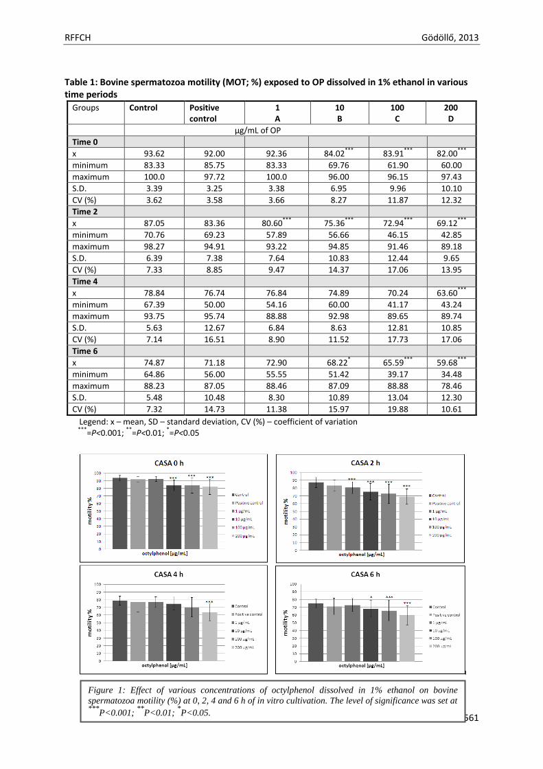
RFFCH Gödöllő, 2013
561
Table 1: Bovine spermatozoa motility (MOT; %) exposed to OP dissolved in 1% ethanol in various time periods
Groups Control
Positive control
1 A
10 B
100 C
200 D
µg/mL of OP
Time 0
x 93.62 92.00 92.36
84.02***
83.91***
82.00***
minimum 83.33 85.75 83.33 69.76 61.90 60.00
maximum 100.0 97.72 100.0 96.00 96.15 97.43
S.D. 3.39 3.25 3.38 6.95 9.96 10.10
CV (%) 3.62 3.58 3.66 8.27 11.87 12.32
Time 2
x 87.05 83.36 80.60***
75.36***
72.94***
69.12***
minimum 70.76 69.23 57.89 56.66 46.15 42.85
maximum 98.27 94.91 93.22 94.85 91.46 89.18
S.D. 6.39 7.38 7.64 10.83 12.44 9.65
CV (%) 7.33 8.85 9.47 14.37 17.06 13.95
Time 4
x 78.84 76.74 76.84 74.89 70.24 63.60***
minimum 67.39 50.00 54.16 60.00 41.17 43.24
maximum 93.75 95.74 88.88 92.98 89.65 89.74
S.D. 5.63 12.67 6.84 8.63 12.81 10.85
CV (%) 7.14 16.51 8.90 11.52 17.73 17.06
Time 6
x 74.87 71.18 72.90 68.22*
65.59***
59.68***
minimum 64.86 56.00 55.55 51.42 39.17 34.48
maximum 88.23 87.05 88.46 87.09 88.88 78.46
S.D. 5.48 10.48 8.30 10.89 13.04 12.30
CV (%) 7.32 14.73 11.38 15.97 19.88 10.61
Legend: x – mean, SD – standard deviation, CV (%) – coefficient of variation ***
=P<0.001; **
=P<0.01; *=P<0.05
Figure 1: Effect of various concentrations of octylphenol dissolved in 1% ethanol on bovine
spermatozoa motility (%) at 0, 2, 4 and 6 h of in vitro cultivation. The level of significance was set at
***P<0.001;
**P<0.01;
*P<0.05.
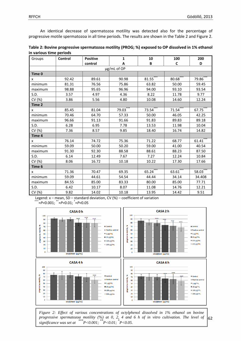
RFFCH Gödöllő, 2013
562
An identical decrease of spermatozoa motility was detected also for the percentage of progressive motile spermatozoa in all time periods. The results are shown in the Table 2 and Figure 2. Table 2: Bovine progressive spermatozoa motility (PROG; %) exposed to OP dissolved in 1% ethanol in various time periods
Groups Control
Positive control
1 A
10 B
100 C
200 D
µg/mL of OP
Time 0
x 92.42 89.61 90.98 81.55***
80.68***
79.86***
minimum 81.31 76.56 75.86 63.82 50.00 59.45
maximum 98.88 95.65 96.96 94.00 93.10 93.54
S.D. 3.57 4.97 4.36 8.22 11.78 9.77
CV (%) 3.86 5.56 4.80 10.08 14.60 12.24
Time 2
x 85.45 81.04 79.03***
73.54***
71.54***
67.75***
minimum 70.46 64.70 57.33 50.00 46.05 42.25
maximum 96.66 91.13 91.66 91.83 89.83 89.18
S.D. 6.28 6.95 7.78 13.53 11.98 10.04
CV (%) 7.36 8.57 9.85 18.40 16.74 14.82
Time 4
x 76.14 74.72 75.36 71.22 68.77 61.41***
minimum 59.09 50.00 50.20 59.00 41.00 40.54
maximum 91.30 92.30 88.58 88.61 88.23 87.50
S.D. 6.14 12.49 7.67 7.27 12.24 10.84
CV (%) 8.06 16.72 10.18 10.22 17.30 17.66
Time 6
x 71.36 70.47 69.35 65.24***
63.61***
58.03***
minimum 59.09 44.61 54.54 44.44 34.14 34.408
maximum 84.55 85.00 83.33 80.00 85.00 77.71
S.D. 6.42 10.17 8.07 11.08 14.76 12.21
CV (%) 9.82 14.02 10.18 13.95 14.42 9.51
Legend: x – mean, SD – standard deviation, CV (%) – coefficient of variation ***
=P<0.001; **
=P<0.01; *=P<0.05
Figure 2: Effect of various concentrations of octylphenol dissolved in 1% ethanol on bovine
progressive spermatozoa motility (%) at 0, 2, 4 and 6 h of in vitro cultivation. The level of
significance was set at ***
P<0.001; **
P<0.01; *P<0.05.
RFFCH Gödöllő, 2013
563
Many in vivo and in vitro studies suggest that octylphenol induces abnormities in male reproductive system. The data obtained from our study indicate, that octylphenol has also a negative in vitro inhibitory effect on spermatozoa motility during short-term cultivation. Our results showed a decreased spermatozoa motility in all concentrations of OP (1, 10, 100 and 200 µg/mL) dissolved during all time periods. The similar results reached a study by Bian et al. (2006), that demonstrated the toxic effect of various concentrations of 4-tert-octylphenol (50, 150 and 450 mg/kg/d) on testicular functions of rats and found out, that the size and weight of the testis, epididymis and prostate were reduced in all the three dosages. The dose of 150 mg/kg/d of OP significantly lowered sperm motility, raising the dose to 450 mg/kg/d also significantly decreased testicular sperm count and daily sperm production.
Gregory et al. (2009) evaluated the effects of chronic exposure to OP on male reproduction and recorded a significant decrease in body weight in the 125 mg/kg group after 60 day of treatment. Total percent sperm motility was significantly lower in rats exposed to the intermediate dose (50 mg/kg of body weight).
Sweeney et al. (2007) determined if maternal exposure to octylphenol pre- and/or postnatally influenced FSH concentrations and semen quantity and quality in postpubertal rams. They found out, that the exposure to OP from birth to weaning increased the number of morphologically abnormal sperm cells in the ejaculates of these rams.
Herath et al. (2004) also confirmed that OP can reduce sperm counts resulting from lowered plasma testosterone in male rats just after puberty.
Vom Saal et al. (1998) found that mice exposed to OP experienced significantly lower daily sperm production.
Blake and Boockfor (1997) demonstrated that chronic administration of 4-tert-octylphenol (OP) to adult male rats resulted in lower levels of LH, FSH, prolactin and testosterone, and proposed that OP inhibited the secretion of reproductive hormones by mimicking estrogen. The same authors also established that chronic administration of 80 mg/kg OP caused shrinkage of the testis. Male accessory sex organs and spermatogenesis were disrupted, spermatozoa deformities were observed (Boockfor and Blake, 1996).
A study of Kim et al. (2004) looked at the effects of OP on the testicular development of prepubertal rats and showed that OP is able to reduce the size and function of the male reproductive organs due to increased apoptosis of testicular germ cells and the decreased biosynthesis of testosterone.
Raychoudhury et al. (1999) observed, that octylphenol is directly toxic to cultured rat spermatogenic cells and Sertoli cells and suggested that this toxic effect in Sertoli cells is exerted through Ca(2+)- independent apoptosis.
The NBT test revealed after 6 h of in vitro cultivation with observed endocrine disruptor that the doses 10, 100 and 200 µg/mL of OP increased intracellular superoxide production in the bovine spermatozoa (Figure 3). However, significant differences (P<0.05 and P<0.001) were found out only between groups C and D containing the highest doses of OP in comparison to the control group. At the dose 1 µg/mL we recorded slightly decrease of superoxide production compared to the control group.
Aydogan et al. (2010) demonstrated that OP at dose 25 mg/kg/day orally administered to rats
three times a week for 45 days can generate reactive oxygen species that cause oxidative damage in testes of Wistar rats.
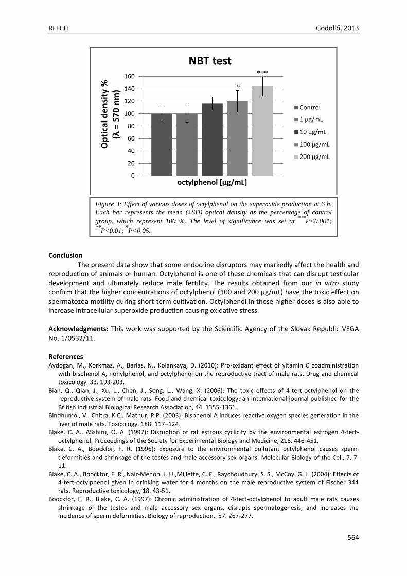
RFFCH Gödöllő, 2013
564
Conclusion
The present data show that some endocrine disruptors may markedly affect the health and reproduction of animals or human. Octylphenol is one of these chemicals that can disrupt testicular development and ultimately reduce male fertility. The results obtained from our in vitro study confirm that the higher concentrations of octylphenol (100 and 200 µg/mL) have the toxic effect on spermatozoa motility during short-term cultivation. Octylphenol in these higher doses is also able to increase intracellular superoxide production causing oxidative stress. Acknowledgments: This work was supported by the Scientific Agency of the Slovak Republic VEGA No. 1/0532/11. References Aydogan, M., Korkmaz, A., Barlas, N., Kolankaya, D. (2010): Pro-oxidant effect of vitamin C coadministration
with bisphenol A, nonylphenol, and octylphenol on the reproductive tract of male rats. Drug and chemical toxicology, 33. 193-203.
Bian, Q., Qian, J., Xu, L., Chen, J., Song, L., Wang, X. (2006): The toxic effects of 4-tert-octylphenol on the reproductive system of male rats. Food and chemical toxicology: an international journal published for the British Industrial Biological Research Association, 44. 1355-1361.
Bindhumol, V., Chitra, K.C., Mathur, P.P. (2003): Bisphenol A induces reactive oxygen species generation in the liver of male rats. Toxicology, 188. 117–124.
Blake, C. A., ASshiru, O. A. (1997): Disruption of rat estrous cyclicity by the environmental estrogen 4-tert-octylphenol. Proceedings of the Society for Experimental Biology and Medicine, 216. 446-451.
Blake, C. A., Boockfor, F. R. (1996): Exposure to the environmental pollutant octylphenol causes sperm deformities and shrinkage of the testes and male accessory sex organs. Molecular Biology of the Cell, 7. 7-11.
Blake, C. A., Boockfor, F. R., Nair-Menon, J. U.,Millette, C. F., Raychoudhury, S. S., McCoy, G. L. (2004): Effects of 4-tert-octylphenol given in drinking water for 4 months on the male reproductive system of Fischer 344 rats. Reproductive toxicology, 18. 43-51.
Boockfor, F. R., Blake, C. A. (1997): Chronic administration of 4-tert-octylphenol to adult male rats causes shrinkage of the testes and male accessory sex organs, disrupts spermatogenesis, and increases the incidence of sperm deformities. Biology of reproduction, 57. 267-277.
0
20
40
60
80
100
120
140
160
Op
tica
l de
nsi
ty %
(λ
= 5
70
nm
)
octylphenol [µg/mL]
NBT test
Control
1 µg/mL
10 µg/mL
100 µg/mL
200 µg/mL
***
*
Figure 3: Effect of various doses of octylphenol on the superoxide production at 6 h.
Each bar represents the mean (±SD) optical density as the percentage of control
group, which represent 100 %. The level of significance was set at ***
P<0.001; **
P<0.01; *P<0.05.
RFFCH Gödöllő, 2013
565
Calafat, A. M., Ye, X., Wong, L.-Y., Reidy, J. A., Needham, L. L. (2008): Exposure of the U.S. Population to Bisphenol A and 4-tertiary-Octylphenol: 2003–2004. Environmental Health Perspectives, 116. 39-44.
ECETOC: Guidance on Identifying Endocrine Disrupting Effects, (2009): Technical report No. 206, 1-137. Esfandiari, N., Sharma, R. K., Saleh, R. A., Thomas, A. J. Jr., Agarwal, A. (2003): Utility of the nitroblue
tetrazolium reduction test for assessment of reactive oxygen species production by seminal leukocytes and spermatozoa. Journal of Andrology, 24. 862-870.
Evans, R., Kortenkamp, A., Martin, O., McKinlay, R., Orton, F., Rosivatz, E. (2011): State of the Art Assesment of Endocrine Disrupters 2nd Interim Report, 135.
Gregory, M., Lacroix, A., Haddad, S., Devine, P., Charbonneau, M., Tardif, R., Krishan, K., Cooke, G. M., Schrader, T., Cyr, D. G. (2009): Effects of chronic exposure to octylphenol on the male rat reproductive system. Journal of toxicology and environmental health. Part A, 72. 1553-1560.
Halliwell, B. (1996): Antioxidants in human health and disease. Annual Review of Nutrition, 16. 33–50. Herath, C.B., Jin, W., Watanabe, G., Arai, K., Suzuki, A.K., TAYA, K. (2004): Adverse effects of environmental
toxicants, octylphenol and bisphenol A, on male reproductive functions in pubertal rats. Endocrine, 25. 163-172.
Ho, Y.S., Magnenat, J.-L., Gargano, M., Cao, J.(1998): The nature of antioxidant defense mechanisms: a lesson from transgenic studies. Environmental Health Perspectives, 106. 1219–1228.
Hossaini, A., Dalgaard, M.,Vinggaard, A. M., Frandsen, H., Larsen, J. J. (2001): In utero reproductive study in rats exposed to nonylphenol. Reproductive toxicology, 15. 537-543.
Chen, B., Duan, J. C., Mai, B. X., Luo, X. J., Yang, Q. S., Sheng, G. Y., Fu, J. M. (2006): Distribution of alkylphenols in the Pearl River Delta and adjacent northern South China Sea, China. Chemosphere, 63. 652-661.
Chitra, K.C., Latchoumycandane, C., Mathur, PP. (2003): Induction of oxidative stress by bisphenol A in the epididymal sperm of rats. Toxicology, 185. 119-27.
Kim, S. K., Lee, H. J., Yang, H., Kim, H. S., Yoon, Y. D. (2004): Prepubertal exposure to 4-tert-octylphenol induces apoptosis of testicular germ cells in adult rat. Archives of andrology, 50. 427-441.
Kabuto, H., Hasuike, S., Minagawa, N., Shishibori. T. (2003): Effects of bisphenol A on the active oxygen species in mouse tissues. Environmental Research, 93. 31–35.
Kabuto, H., Amakawa, M., Shishibori. T. (2004): Exposure to bisphenol A during embryonic/fetal life and infancy increases oxidative injury and causes underdevelopment of the brain and testis in mice. Life Science. 74. 2931–2940.
Madsen, L.L., Korsgaard, B., Bjerregaard, P. (2006): Oral single pulse exposure of flounder Platichthys flesus to 4-tert-octylphenol: Relations between tissue levels and estrogenic effects. Marine environmental research, 61. 352-362.
Pocar, P., Augustin, R., Gandolfi, F., Fisher, B. (2003): Toxic effects of in vitro exposure to p-tert-octylphenol on bovine oocyte maturation and developmental competence. Biology of reproduction, 69. 462-468.
Qian, J., Bian, Q., Cui, L., Chen, J., Song, L., Wang, X. (2006): Octylphenol induces apoptosis in cultured rat Sertoli cells. Toxicology letters, 166. 178-186.
Raychoudhury, S., Blake, C., Millette, C. (1999): Toxic effects of octylphenol on cultured rat spermatogenic cells and Sertolli cells. Toxicology and applied pharmacology, 157. 192-202.
Shiraishi, H., Carter, D.S., Hites, R.A. (1989): Identification and determination of tert-alkylphenols in carp from the Trenton Channel of the Detroit River, Michigan, USA. Biomedical and environmental mass spectrometry, 18. 478-483.
Sweeney, T., Fox, J., Robertson, L., Kelly, G., Duffy, P., Lolergan, P., O'Doherty, J., Roche, J.F., Evans, N.P. (2007): Postnatal exposure to octylphenol decreases semen quality in the adult ram. Theriogenology, 67. 1068-75.
Tvrdá, E., Lukáč, N., Lukáčová, J., Kňažická, Z., Massányi, P. (2013): Stimulating and protective effects of vitamin E on bovine spermatozoa. Journal of Microbiology, Biotechnology and Food Sciences, 2. 1386-1395.
vom Saal, F.S., Cooke, P.S., Buchanan, D.L., Palanza, P., Thayar, K.A., Nagel, S.C., Parmigiani, S., Welshons, W.V. (1998): A physiologically based approach to the study of bisphenol A and other estrogenic chemicals on the size of reproductive organs, daily sperm production, and behavior. Toxicology and Industrial Health, 14. 239-260.
vom Saal, F.S., Hughes, C. (2005): An Extensive New Literature Concerning Low-Dose Effects of Bisphenol A Shows the Need for a New Risk Assessment. Environmental Health Perspectives, 113. 926-933.
RFFCH Gödöllő, 2013
566
White, R., Jobling, S., Hoare, S. A., Sumpter, J. P., Parker, M. G. (1994): Environmentally persistent alkylphenolic compounds are estrogenic. Endocrinology, 135. 175-182. Ying, G. G., Williams, B., Kookana, R. (2002): Environmental fate of alkylphenols and alkylphenol ethoxylates--a review. Environment international, 28. 215-226. Zhou, X. C., Wei, P., Hu, Z. Y., Gao, F., Zhou, R. J., Liu, Y. X. (2001): Role of Fas/FasL genes in azoospermia or oligozoospermia induced by testosterone undecanoate in rhesus monkey. Acta Pharmacologica Sinica, 22. 1028-1033.

Animal welfare, etológia és tartástechnológia
Animal welfare, ethology and housing systems
Volume 9 Issue 3
Különszám/Special Issue
Gödöllő
2013
RFFCH Gödöllő, 2013
567
DIAGNOSTICS OF TOXOPLASMA GONDII IN MILK SAMPLES OF SHEEP FROM EASTERN SLOVAKIA Lenka LUPTÁKOVÁ*1, Eva PETROVOVÁ 2, Ľubica ŠŤAVOVÁ1
1University of Veterinary Medicine and Pharmacy, Department of Biology and Genetics, Komenského 73, 041 81 Kosice, Slovakia 2University of Veterinary Medicine and Pharmacy, Department of Anatomy, Histology and Physiology, Komenského 73, 041 81 Kosice, Slovakia * MVDr. Lenka Luptáková, PhD, [email protected]
Abstract The paper presents the results of examination of 62 sheep from flocks of eastern Slovakia for the presence of Toxoplasma gondii antibodies by complement fixation test. Thirteen samples were found to be positive at the titre of 1:64, 10 were positive at 1:128 and two at 1:256 dilution. Subsequently, DNA was isolated from blood samples of these animals. PCR analysis with specific primers was then used in search for T. gondii DNA. In PCR analysis, 5 of 25 (20%) samples tested positive. Key words: milk, real time PCR, sheep, Toxoplasma gondii Introduction Toxoplasmosis is one of the most common parasitic zoonoses of mammals and birds transmissible to humans. The agent of the disease is Toxoplasma gondii (T. gondii), the definitive hosts of which are the representatives of the family F e l i d a e, infected with oocysts from the environment and with tachyzoites and bradyzoites from intermediate hosts. Intermediate hosts are all vertebrates including man (Frenkel, 2000). It is a panthropic cosmopolite, a facultative heteroxenic coccidium. Sheep were in fact the first mammals in which congenital toxoplasmosis were proven with abortions, dead-born fetuses and frequent manifestations of infection including infertility. The first case of manifest toxoplasmosis in sheep with symptoms of encephalomyelitis and tachycardia was described by Olafson and Monlux. Sheep play a significant role in the economy of many countries since they are important source of meat and milk products. Toxoplasmosis is the major parasitic disease affecting sheep. It is important for veterinary medicine, animal science and public health since it causes reproductive and economic losses in the herd, as well as damaging human health due to consumption of contaminated meat and milk, which can facilitate zoonotic transmission (Camossi et al., 2011). Material and methods Samples: A total of 50 milk samples of adult sheep were examined by real time PCR. Sheep originated from farm located 30 km of Kosice in eastern Slovakia. In the farm proper sanitary conditions were respected and sheep were regularly pastured on grassland where they could contamined with T. gondii. Molecular diagnostics: Examination was carried out on samples of sheep milk. Isolation of DNA: DNA was isolated from 50 milk samples by commercially available kit (DNA-sorb-B, Amplisens, Russia). DNA was isolated according to the manufacturer´s protocol and stored at -20 °C for futher examination. Detection of DNA by real time PCR: Amplification of the isolated DNA was carried out by the real time PCR with SYBR green as a detection system from the T. gondii gene region TGR1E, repeated in the genome 30-35 times, using the specific primers TGR1E-1 and TGR1E-2 (Cristina et al., 1991). Cloned T. gondii TGR gene (GenExpress, Germany) diluted to 104-109 was used for the calibration curve. In each reaction, a melting analysis (comparison of the melting temperature (Tm) of PCR products) was determined to differentiate specific and non-specific PCR products. The reaction volume was 25 μl, which contained commercial FastStart Universal SYBR Green Master (Roche,
RFFCH Gödöllő, 2013
568
Germany) and 0.2 μM primers (TGR1-1 and TGR1E-2). Real-time PCR was completed using a thermocycler Line GeneK with the software Line GeneK Fluorescent Quantitative Detection system (BIOER Technology, China). After incubation at 50°C for 2 minutes and initial denaturation at 95°C for 10 minutes, 40 amplification cycles were performed (95°C for 15 s, 60°C for 1 minute). Melting analysis was carried out at temperatures ranging from 60°C to 95°C, in which the temperature was gradually increased by 0.5°C and the period of measurement at individual steps was 15 s. Every PCR run included a control without DNA (containing the reaction mix alone and nuclease-free water). Results Using quantitative real-time PCR the presence of T. gondii DNA was detected and the number of copies quantified in the 50 milk samples of sheep. Using real-time PCR, T. gondii DNA was detected and quantified in 5 samples of milk. Standards with the known dilution of Toxoplasma DNA were used to determine the detection limit of a modified real-time PCR and to create a calibration curve that ranged from 109-104 copies of Toxoplasma DNA. The correlation coefficient of the calibration curve was 0.998. As SYBR Green a fluorescent dye, was used as a detection system, a melting analysis was a part of the real-time PCR to distinguish between specific and non-specific products. During the melting analysis, the melting temperature (Tm) of a positive control and positive samples was 84°C. In quantifying the examined samples within a 40-cycle protocol for the real-time PCR, the number of copies detected in the positive samples ranged from 1.1 x10² to 1.30 x10⁵ (Table 1). Table 1
The number of DNA copies in positive samples
sample DNA copies Ct value
Sample1 1.1 x 10² 19,50
Sample 5 1.3 x 10⁵ 18,72
Sample 23 5.7 x 10⁴ 19,37
Discussion Consumption of raw food, especially among immunocompromised persons and pregnant women, poses a potential threat to public health. The milk of sheep and goats represent risk factors as they are consumed raw (Spisak at al., 2010). Consumption of unpasteurized goat, sheep or cow milk or its products represents a risk because tachyzoites, the stage most likely present in milk are thought to be immediately destroyed by the gastric juice (Dubey, 1998). However, evidence indicates that the part of ingested tachyzoites is not destroyed by gastric juice and it can cause infection (Cook et al., 2000). Toxoplasmosis transmission by unpasteurized or inadequately processed milk or fresh cheese, important food sources in rural areas, can be a significant means of contamination by this agent (Hiramoto et al., 2001) In the period of 1999 – 2002, studies conducted in Sardinia analyzed 9639 samples of sera and 815 samples from abortions (670 aborted fetus and 145 placentas) obtained from 964 sheep and goat farms. The sampled sera were examined for presence of IgG and IgM specific antibodies against Toxoplasma gondii by means of indirect immunofluorescence method while fetus and placenta were examined by PCR. Specific antibodies against IgG were diagnosed in 652 (9%) sheep. of the total number of 2471 samples of sheep fetuses including muscles, liver, abomasums, spleen, brain and placenta, PCR proved positivity in 271 (11.1%) cases. on the one hand, these
RFFCH Gödöllő, 2013
569
results indicate relatively high seroprevalence and, on the other hand point to the important role of PCR diagnostics related to sheep abortions (Masala et al., 2003). In southern Italy, region Campania, 117 sheep flocks kept on pasture were examined. Samples of blood and milk were taken from 10 adult sheep (> 18 months old) from each flock. Thus altogether 1170 sheep were subjected to examination. Blood sera were examined for the presence of IgG antibodies by indirect immunofluorescence. Of the examined samples collected from 1170 sheep 333 samples were positive (28.5%) and PCR proved presence of Toxoplasma gondii in 4 samples of milk, which represents 3.4% of the total analyzed samples, demonstrating the transmission potential of ovine milk and dairy products (Fusco et al., 2007). The tachyzoites presented in sheep or goat milk are generally not considered as an important source of oral transmission of T. gondii because they are rapidly killed outside the host and because they are considered sensitive to proteolytic enzymes (Powell et al., 2001). But the contamination of milk should not be underestimated since it can represent a critical point in food safety. Some local home-made cheeses deriving from mass-milk production and destined to be consumed fresh can represent a risk factor for public health if they are produced in small family-based farms without previous milk pasteurization.
Acknowledgement The paper is a result of research work done within the frames of grant projects VEGA of the Ministry of Education of the Slovak Republic No. 1/0063/13.
References Frenkel J.K. 2000. Biology of Toxoplasma gondii. In: Ambroise-Thomas P, Peterse E. (ed): Congenital
toxoplasmosis: scientific background, clinical management and control. Springer-Verlag, Paris, pp. 9-25. Olafson, P., Monlux, W.S. 1942. Toxoplasma infections in animals. Cornell Veterinarian, 32, 16-190. Camossi, L.G., Greca-Júnior, H., Correa, A.P.F.L., Richini-Pereira, V.B., Silva, R.C., Da Silva, A.V., Langoni, H.
2011. Detection of Toxoplasma gondii DNA in the milk of naturally infected ewes. Veterinary Parasitology, 177, 256-261.
Cristina, N., Liaud, M.F., Santoro, F., Oury, B., Ambroise-Thomas, P. 1991. A family of repeated DNA sequences in Toxoplasma gondii: cloning, sequence analysis, and use in strain characterization. Experimental Parasitology, 1991, 73, 73-81
Spisak, F., Turcekova, L., Reiterova, K., Spilovska, S., Dubinsky P. 2010. Prevalence estimation and genotypization of Toxoplasma gondii in goats. Biologia, 65, 670-674.
Dubey, J.P. 1998. Re-examination of resistance of Toxoplasma gondii tachyzoites and bradyzoites to pepsin and trypsin digestion. Journal of Parasitology, 116, 43-50.
Cook, A.J.C., Gilbert, R.E., Buffolano, W., Zufferey, J., Peterse, E., Jenum, P.A., Foulon, W., Semprini, A.E., Dunn, D.T. 2000. Sources of Toxoplasma infection in pregnant women: European multicentre case-control study. BMJ, 321, 142-147.
Hiramoto, R.M., Mayrbaurl-Borges, M., Galisteo, A.J., Meireles, L.R., Macre, M.S., Andrade, Jr., H.F. 2001. Infectivity of cystis of the ME-49 Toxoplasma gondii strain in bovine milk and homemade cheese. Revista de Saúde Pública, 35, 113-118.
Masala G, Porcu R, Madau L, Tanda A, Ibba B, Satta G, Tola S. 2003. Survey of ovine and caprine toxoplasmosis by IFAT and PCR assays in Sardinia, Italy. Veterinary Parasitology 117, 15-21.
Fusco G, Rinaldi L, Guarino A, Proroga YTR, Pesce A, Giuseppina DM, Cringoli G. 2007. Toxoplasma gondii in sheep from the Campania region (Italy). Periodical Veterinary parasitology 149, 1-4.
Powell, C.C., Brewer, M., Lappin, M.R. 2001. Detection of Toxoplasma gondii in the milk of experimentally infecting lacting cats. Veterinary Parasitology, 102, 29-33.
Animal welfare, etológia és tartástechnológia
Animal welfare, ethology and housing systems
Volume 9 Issue 3
Különszám/Special Issue
Gödöllő
2013
RFFCH Gödöllő, 2013
570
STRUCTURAL CHANGES IN THE POSTERIOR KIDNEY OF FISH EXPOSED TO MANCOZEB Hanna LUTNICKA1 , Agnieszka LUDWIKOWSKA1 , Anna PECIO2, Bartosz BOJARSKI1
University of Agriculture in Krakow, Faculty of Animal Science, Department of Poultry and Fur Animal Breeding and Animal Hygiene, Poland1 Jagiellonian University, Institute of Zoology, Department of Comparative Anatomy, Poland2
ABSTRACT Fungicides, including mancozeb, are widely used in agriculture in the world. The low or very low concentrations of these pesticides are noted in surface waters - the main source for fish culture. They can influent fish inner organs causing pathological changes observed only on a cell's level. A kidney is an important organ secreting toxins metabolites. Fish posterior kidney have to different parts: haematopoietic tissue and renal parts (excretory tissue). The aim of the study was to get to know if the fungicide mancozeb cause the ultrastructural lesions in posterior kidney of fish. Key words: fungicide, mancozeb, fish posterior kidney, ultrastructural changes Material and methods Carp Cyprinus carpio L. (mean weight ± sd, 50 ± 10 g) was exposed for 14 days, in aquaria conditions, to the fungicide mancozeb in concentration 1 mg L-1 . After exposure fish were transferred to the clean water for the next 30 days for a possible recovery. Kidneys were sampled after 14 days of exposure and at the end of the recovery period. Ultrastructural changes were studied by transmission electron microscopy. Results The fungicide mancozeb caused pathological lesions in carp kidney. After the exposure the most important lesions were observed in hematopoietic tissue. The maturing blood cells were separated from themselves. They had different, not natural shapes/forms because of the cell's fragmentation process. In the excretory part the epithelial cells adhesion could be seen. The structure of the most of mitochondria of the cells was correct. Only some of them were locally damaged or a process of deliquescence was observed. After the recovery period (30 days) in the hematopoietic tissue the same lesions as noticed after exposure was observed. In epithelial cells increase of RER could be seen in relation to the RER observed after exposure to the fungicide. Intensive RER fragmentation process was observed, too. Numerous RER was pure in ribosomes . The recovery time was too short for the completely tissue repair. The work has been finacially supported by N N 304 279440
Animal welfare, etológia és tartástechnológia
Animal welfare, ethology and housing systems
Volume 9 Issue 3
Különszám/Special Issue
Gödöllő
2013
RFFCH Gödöllő, 2013
571
Introduction of the Ecotoxicologist Masters’ degree at SZIU Péter Nagy*, Gábor Bakonyi
Department of Zoology and Animal Ecology Szent István University, Faculty of Agricultural and Environmental Sciences Gödöllő, Hungary *[email protected]
A wide range of environmental disturbances by human activities constitute massive threats nowadays to living organisms from individual levels to ecological systems. Consequently, it is becoming more and more important to educate experts capable of handling a wide range of environmental risks with a sound scientific and methodological background. The above needs and the lack of concerted education in the region, justified the establishment of a relevant educational tool as a response. The Ecotoxicologist major was proposed, the first time in Hungary, in 2008 by Professor Gábor Bakonyi of the SZIU Department of Zoology and Animal Ecology in order to fulfil the above need. Since this proposal was accepted, the responsibility of coordination is taken by our department and the organizational tasks are also performed here. The major comprises of four semesters supplemented by a practical week and a professional practical period of 4 weeks. The placement of the professional practical ranges over several types of partner organizations, from research institutes to companies interested in receiving our students, considered often as futures employees. The list of subjects is very wide, covering sciences from toxicological and ecotoxicological nature to courses on environmental risk assessment, planning scientific projects and fund raising as well as scientific publication strategies. This way, our students obtain education on problems and possibly solutions all along different natural and anthropogenic food chains as well. The teachers of the courses were selected by merit, specialization and experience, so the proportion of external lecturers is rather high in this major. Several of our students have gained prices in National Scientific Students’ Competitions. Since its establishment in 2010, a new class of 11-17 students could be recruited every year. So the third class is preparing for graduation this December. So far, almost all of our graduates have succeeded in obtaining a job, most of them being employed in jobs relevant to their degree. As for the future, we are confident in reserving the high standards of the education that should maintain the attraction of our degree. Furthermore, we hope to be able to introduce this major in English, so that we can extend this service to foreign students. Acknowledgements The authors thank the support of Research Centre of Excellence- 17586-4/2013/TUDPOL

Animal welfare, etológia és tartástechnológia
Animal welfare, ethology and housing systems
Volume 9 Issue 3
Különszám/Special Issue
Gödöllő
2013
RFFCH Gödöllő, 2013
572
THE CONTENT OF RISK ELEMENTS IN BONES OF BANK VOLE (MYODES GLAREOLUS) AND WOOD MOUSE (APODEMUS SYLVATICUS) FROM KOLÍŇANY AREA (SLOVAKIA)
Radoslav OMELKA1, Mária BAUEROVÁ1, Robert STAWARZ2, Grzegorz FORMICKI2, Ivan BALÁŽ3, Monika MARTINIAKOVÁ4
1Department of Botany and Genetics, Constantine the Philosopher University, 949 74 Nitra, Slovakia 2Institute of Biology, Krakow Pedagogical University, Krakow 31 054, Poland 3Department of Ecology and Environmentalistics, Constantine the Philosopher University, 949 01 Nitra, Slovakia 4Department of Zoology and Anthropology, Constantine the Philosopher University, 949 74 Nitra, Slovakia Corresponding author: [email protected]
ABSTRACT
The content of selected risk elements (Fe, Cu, Zn, Ni, Mg, Na, K) in the femora of bank vole (Myodes glareolus) and wood mouse (Apodemus sylvaticus) from Kolíňany area (Slovakia) which is characterized by a high degree of environmental pollution was investigated in the present study. The rodents were trapped in this locality in February 2011. Higher concentrations of Mg and Na were detected in the bones of wood mouse in comparison with those of bank vole (P<0.05). Moreover, the concentrations of Cu, Zn and Ni were significantly higher in the bones of both species in our study in comparison with the values which were obtained in the same animal species on the same site in February 2007. Therefore, our results demonstrate increased accumulation of these elements in the femora of Myodes glareolus and Apodemus sylvaticus and thus give an evidence of still ongoing contamination of this locality. Keywords: Risk elements. Bone. Bank vole. Wood mouse. Atomic absorption spectrophotometry. INTRODUCTION
Many investigations have been performed to determine risk elements accumulation in selected organs of various animal species (Stawarz et al., 2003; Jancova et al., 2006; Kolesarova et al., 2008; Gasparik et al., 2012). In general, rodents are ideal for monitoring environmental pollution as well as for evaluating the exposure risk for people living in contaminated areas. The bank vole (Myodes glareolus, formerly Clethrionomys glareolus) is a small microtine rodent that is common throughout Europe and it is one of the most common woodland rodents in Slovakia. Seeds, fruits and green vegetation constitute about 44% of their food, insects, earthworms and other invertebrates between 9 and 23%, depending on the season, and in winter they add tree bark to their food (Zemanek 1972; Gdula-Argasinska et al., 2004). This species has been used to monitor environmental pollution from a variety of technogenic sources up to date (Milton et al., 2003; Topolska et al., 2004). The wood mouse (Apodemus sylvaticus) belongs to the most dominant rodent species in Slovakia. The diet of the wood mouse consists of roots, grains, seeds, berries, nuts, grasses, grain kernels, fruits and insects (Nowak, 1991). The content of many elements in various organs has also been investigated in this species (Milton et al., 2002; Milton et al., 2004).
Since bone can serve as a good biomarker of long-term accumulation of various xenobiotics, we analysed concentrations of selected risk elements (Fe, Cu, Zn, Mg, Na, K) in the femora of two rodent species: bank vole and wood mouse. The rodents were trapped at
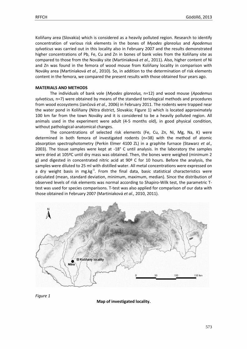
RFFCH Gödöllő, 2013
573
Kolíňany area (Slovakia) which is considered as a heavily polluted region. Research to identify concentration of various risk elements in the bones of Myodes glareolus and Apodemus sylvaticus was carried out in this locality also in February 2007 and the results demonstrated higher concentrations of Pb, Fe, Cu and Zn in bones of bank voles from the Kolíňany site as compared to those from the Nováky site (Martiniaková et al., 2011). Also, higher content of Ni and Zn was found in the femora of wood mouse from Kolíňany locality in comparison with Nováky area (Martiniaková et al., 2010). So, in addition to the determination of risk elements content in the femora, we compared the present results with those obtained four years ago.
MATERIALS AND METHODS
The individuals of bank vole (Myodes glareolus, n=12) and wood mouse (Apodemus sylvaticus, n=7) were obtained by means of the standard teriological methods and procedures from wood ecosystems (Jančová et al., 2006) in February 2011. The rodents were trapped near the water pond in Kolíňany (Nitra district, Slovakia; Figure 1) which is located approximately 100 km far from the town Nováky and it is considered to be a heavily polluted region. All animals used in the experiment were adult (4-5 months old), in good physical condition, without pathological-anatomical changes.
The concentrations of selected risk elements (Fe, Cu, Zn, Ni, Mg, Na, K) were determined in both femora of investigated rodents (n=38) with the method of atomic absorption spectrophotometry (Perkin Elmer 4100 ZL) in a graphite furnace (Stawarz et al., 2003). The tissue samples were kept at -18° C until analysis. In the laboratory the samples were dried at 105ºC until dry mass was obtained. Then, the bones were weighed (minimum 2 g) and digested in concentrated nitric acid at 90º C for 10 hours. Before the analysis, the samples were diluted to 25 ml with distilled water. All metal concentrations were expressed on a dry weight basis in mg.kg-1. From the final data, basic statistical characteristics were calculated (mean, standard deviation, minimum, maximum, median). Since the distribution of observed levels of risk elements was normal according to Shapiro-Wilk test, the parametric T-test was used for species comparisons. T-test was also applied for comparison of our data with those obtained in February 2007 (Martiniaková et al., 2010, 2011).
Figure 1
Map of investigated locality.
RFFCH Gödöllő, 2013
574
RESULTS AND DISCUSSION The concentrations of selected risk elements (Fe, Cu, Zn, Ni, Mg, Na, K) in the femora of Myodes glareolus and Apodemus sylvaticus from Kolíňany area are listed in Table 1. Higher concentrations of all investigated elements were detected in the bones of wood mouse; however, significant differences were observed only for concentrations of Mg and Na (P<0.05). We observed higher concentrations of Zn in the femora of Myodes glareolus compared with the data reported by Milton et al. (2003). These investigators analysed Zn concentration (173 ± 5.1 μg g−1 dry weight) in the femora of bank voles trapped at the contaminated, unused Pb mine at Frongoch in west Wales. In contrast, Zn concentration in the femora of bank voles from Kolíňany area was lower as value reported by Milton and Johnson (1999) who analysed femora of laboratory-bred bank voles exposed to increased levels of dietary Zn.
The concentrations of Cu, Zn and Ni were significantly higher in the bones of both species Myodes glareolus and Apodemus sylvaticus in our study in comparison with the values which were obtained in the same animal species on the same site (Kolíňany) in February 2007 (Table 2). In addition, the bones of wood mouse also included a significantly higher content of Fe as compared to the value from 2007. Therefore, our results demonstrate increased accumulation of these elements in the femora of both rodents investigated and thus give an evidence of still ongoing contamination of this locality. This fact can be explained by intensive agricultural production and subsequent contamination of soil, water, and food, by road traffic pollution as well as by various factories in industrial zones in western Slovakia known from the history affecting also nowadays situation (e.g. production of Ni in Sereď and its damping place, Gasparik et al., 2012). Intensive agricultural production and the use of chemicals are characteristic for the whole region of Nitra. It is generally known that an application of agricultural chemicals can lead to a higher accumulation of specific elements, including Fe, Cu and Zn into the soil. In addition, there is heavy road traffic near the capture locality, which is also considered to be a significant source of risk elements that have a potential ability to be transported by air flow over large distances. There is also a possibility of falling dust being transported in the air from large industrial regions, such as Bratislava, Vienna, Budapest, or factories nearby Nitra region. This hypothesis may be supported by studies indicating the possibility of the long range transportation of various xenobiotics (Coggins et al., 2006).
CONCLUSION The accumulation of selected risk elements (Fe, Cu, Zn, Ni, Mg, Na, K) in the femora of bank vole (Myodes glareolus) and wood mouse (Apodemus sylvaticus) from Kolíňany area was investigated in the present study. We detected higher concentrations of Mg and Na in the bones of wood mouse in comparison with those of bank vole (P<0.05). The concentrations of Cu, Zn and Ni were significantly higher in the bones of both species in our study in comparison with the values which were obtained in the same animal species on the same site four years ago. Our results give an evidence of still ongoing contamination of this locality.
Acknowledgments: This study was supported by the grants KEGA 035UKF-4/2013 and VEGA 1/0790/11.
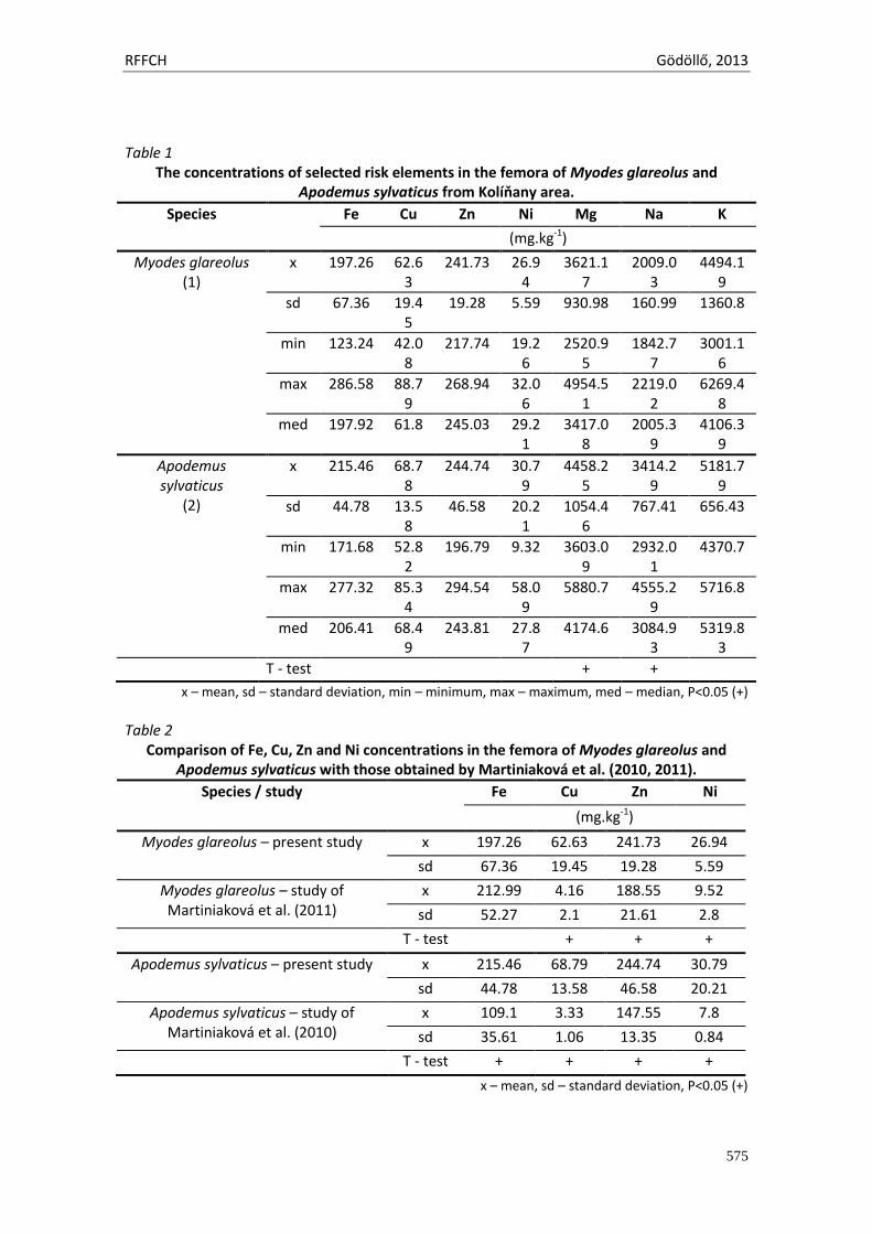
RFFCH Gödöllő, 2013
575
Table 1
The concentrations of selected risk elements in the femora of Myodes glareolus and Apodemus sylvaticus from Kolíňany area.
Species Fe Cu Zn Ni Mg Na K
(mg.kg-1)
Myodes glareolus (1)
x 197.26 62.63
241.73 26.94
3621.17
2009.03
4494.19
sd 67.36 19.45
19.28 5.59 930.98 160.99 1360.8
min 123.24 42.08
217.74 19.26
2520.95
1842.77
3001.16
max 286.58 88.79
268.94 32.06
4954.51
2219.02
6269.48
med 197.92 61.8 245.03 29.21
3417.08
2005.39
4106.39
Apodemus sylvaticus
(2)
x 215.46 68.78
244.74 30.79
4458.25
3414.29
5181.79
sd 44.78 13.58
46.58 20.21
1054.46
767.41 656.43
min 171.68 52.82
196.79 9.32 3603.09
2932.01
4370.7
max 277.32 85.34
294.54 58.09
5880.7 4555.29
5716.8
med 206.41 68.49
243.81 27.87
4174.6 3084.93
5319.83
T - test + +
x – mean, sd – standard deviation, min – minimum, max – maximum, med – median, P<0.05 (+)
Table 2
Comparison of Fe, Cu, Zn and Ni concentrations in the femora of Myodes glareolus and Apodemus sylvaticus with those obtained by Martiniaková et al. (2010, 2011).
Species / study Fe Cu Zn Ni
(mg.kg-1)
Myodes glareolus – present study x 197.26 62.63 241.73 26.94
sd 67.36 19.45 19.28 5.59
Myodes glareolus – study of Martiniaková et al. (2011)
x 212.99 4.16 188.55 9.52
sd 52.27 2.1 21.61 2.8
T - test + + +
Apodemus sylvaticus – present study x 215.46 68.79 244.74 30.79
sd 44.78 13.58 46.58 20.21
Apodemus sylvaticus – study of Martiniaková et al. (2010)
x 109.1 3.33 147.55 7.8
sd 35.61 1.06 13.35 0.84
T - test + + + +
x – mean, sd – standard deviation, P<0.05 (+)
RFFCH Gödöllő, 2013
576
REFERENCES ARBON, K. S., C. M. CHRISTENSEN, W. A. HARVEY and S. J. HEGGLAND, 2012. Cadmium exposure
activates the ERK signaling pathway leading to altered osteoblast gene expression and apoptotic death in Saos-2 cells. In: Food Chem. Toxicol. Vol. 50, pp. 198-205.
COGGINS, A. M., S. G. JENNINGS and R. EBINGHAUS, 2006. Accumulation rates of the heavy metals lead, mercury and cadmium in ombrotrophic peatlands in the west of Ireland. In: Atmospheric Environ. Vol. 40, pp. 260-278.
GASPARIK, J., M. DOBIAS, M. CAPCAROVA, P. SMEHYL, J. SLAMECKA, J. BUJKO and J. GASPARIK, JR, 2012. Concentration of cadmium, mercury, zinc, copper and cobalt in the tissues of wild boar (Sus scrofa) hunted in the western Slovakia. In: J. Environ. Sci. Health A Tox. Hazard Subst. Environ. Eng. Vol. 47, pp. 1212-6.
GDULA-ARGASINSKA, J., J. APPLETON, K. SAWICKA-KAPUSTA and B. SPENCE, 2004. Further investigation of the heavy metal content of the teeth of the bank vole as an exposure indicator of environmental pollution in Poland. In: Environ. Pollut. Vol. 131, pp. 71-79.
JANČOVÁ, A., P. MASSÁNYI, P. NAĎ, B. KORÉNEKOVÁ, M. SKALICKÁ, J. DRÁBEKOVÁ and I. BALÁŽ, 2006. Accumulation of heavy metals in selected organs of yellow necked mouse (Apodemus flavicollis). In: Ekol. Bratislava Vol. 25, pp. 19-26.
KOLESAROVA, A., J. SLAMECKA, R. JURCIK, F. TATARUCH, N. LUKAC, J. KOVACIK, M. CAPCAROVA, M. VALENT and P. MASSANYI, 2008. Environmental levels of cadmium, lead and mercury in brown hares and their relation to blood metabolic parameters. In: J. Environ. Sci. Health A Tox. Hazard Subst. Environ. Eng. Vol. 43, pp. 646-50.
MARTINIAKOVÁ, M., R. OMELKA, A. JANČOVÁ, R. STAWARZ and G. FORMICKI, 2010. Heavy metal content in the femora of yellow-necked mouse (Apodemus flavicollis) and wood mouse (Apodemus sylvaticus) from different types of polluted environment in Slovakia. In: Environ. Monit. Assess. Vol. 171, pp. 651-660.
MARTINIAKOVÁ, M., R. OMELKA, A. JANČOVÁ, R. STAWARZ and G. FORMICKI, 2011. Concentrations of selected heavy metals in bones and femoral bone structure of bank (Myodes glareolus) and common (Microtus arvalis) voles from different polluted biotopes in Slovakia. In: Arch. Environ. Contam. Toxicol. Vol. 60, pp. 524-532.
MILTON, A. and M. JOHNSON, 1999. Biomonitoring of contaminated mine tailings through age accumulation of trace metals in the bank vole (Clethrionomys glareolus). In: J. Environ. Monit. Vol. 1, pp. 219–225.
MILTON, A., M. S. JOHNSON and J. A. COOKE, 2002. Lead within ecosystems on metalliferous mine tailings in Wales and Ireland. In: Sci. Total Environ. Vol. 299, pp. 177–190.
MILTON, A., J. A. COOKE and M. S. JOHNSON, 2003. Accumulation of lead, zinc, and cadmium in a wild population of Clethrionomys glareolus from an abandoned lead mine. In: Arch. Environ. Contam. Toxicol. Vol. 44, pp. 405-411.
MILTON, A., J. A. COOKE, and M. S. JOHNSON, 2004. A comparison of cadmium in ecosystems on metalliferous mine tailings in Wales and Ireland. In: Water Air Soil Poll. Vol. 153, pp. 157–172.
NOWAK, R. M., 1991. Walker’s mammals of the world (Vol II). Baltimore: Johns Hopkins University Press. STAWARZ, R., M. ZAKRZEWSKI, A. MARENČÍK and Š. HRAŠKA, 2003. Heavy metal concentration in the
toad Bufo Bufo from a region of Mochovce, Slovakia. In: Ekol. Bratislava, Vol. 22, pp. 292-297 TOPOLSKA, K., K. SAWICKA-KAPUSTA and E. CIESLIK, 2004. The effect of contamination of the Krakow
region on heavy metals content in the organs of bank voles (Cleithrionomys glareolus, Schreber, 1780). In: Pol. J. Environ. Stud. Vol. 13, pp. 103-109.
ZEMANEK, M., 1972. Food and feeding habits of rodents in a deciduous forest. In: Acta Theriol. Vol. 23, pp. 315-325.
Animal welfare, etológia és tartástechnológia
Animal welfare, ethology and housing systems
Volume 9 Issue 3
Különszám/Special Issue
Gödöllő
2013
RFFCH Gödöllő, 2013
578
FIBROTIC CHANGES IN THE RABBIT LIVER AFTER PESTICIDE ADMINISTRATION
PETROVOVA, E.1, MAZENSKY, D.1, LUPTAKOVA, L.2
1Department of Anatomy, Histology and Physiology, University of Veterinary Medicine and Pharmacy, Komenskeho 73, 04011 Kosice, Slovakia 2Department of Biology and Genetics, University of Veterinary Medicine and Pharmacy, Komenskeho 73, 04011 Kosice, Slovakia
Abstract The aim of our study was to observe fibrotic changes of the rabbit liver tissue treated with bendiocarb, mostly in the area of the central vein and portal tract. Animals were divided into groups (control, days 10, 20, 30 of administration), and each group comprised 14 animals. Rabbits in all experimental groups received carbamate pesticide (bendiocarb) per os in a dose 5 mg/kg per day and after day 11 in a same dose per 48 hours. Picro-sirius red staining was used for visualization of collagen fibers and staging of hepatic fibrosis. The central vein wall thickness is increased in thickness at 10 and 30 days, but not significantly. Further, at the same days the fibrous septa of variable lengths are spreading from portal tract into the lobules and perisinusoidal space. In view of the data of the present study, it can be concluded that bendiocarb has moderately toxic effect on the rabbit liver tissue, regardless of gender after short-term (10 days) and long-term (30 days) bendiocarb administration.
Introduction Pesticides are the significant contaminants of environment which are used in agriculture and households. Their widely using cause the question of toxicity pesticides to non-target organisms, their persistence, accumulation and combined effect with other chemicals. The annual application of synthetic pesticides to food crops in the EU exceeds 140,000 tones per year. The wide use of pesticides are mainly organophosphates (OPs), carbamates and pyrethroids. Further, over 25% of fruits, vegetables, and cereals grown in the European Union contain detectable residues of at least two pesticides while their total quantity, which Europe's population is exposed to is unknown (Bjørling-Poulsen a kol., 2008). Carbamate insecticides, like the OPs, inhibit acetylcholinesterase (AChE) and elicit cholinergic hyperstimulation. However, carbamates cause only reversible inhibition of AChE which lasts only minutes or 24 hours (Kamel and Hoppin, 2004). Bendiocarb is a broad spectrum pesticide belonging to the N-methylcarbamate group (Pacioni and Veglia, 2007). Using of bendiocarb can dramatically reduce the risk of insect-borne diseases which are transmitted by arthropod vectors such as mosquitoes (malaria, dengue fever, yellow fever, encephalitis, filariasis, West Nile fever and chikungunya), ticks (e.g. Lyme disease) and sandflies (leishmaniasis; Nauen, 2007). Bendiocarb is degraded in the liver, which has a major role in the biotransformation and excretion of carbamate pesticides from the body. Reactions of detoxication take place in hepatocytes by the enzymatic system situated in the endoplasmic reticulum (Schenck and Kolb, 1990), and bendiocarb is excreted as a sulphate and β-glucuronide conjugates of the phenol derivatives. In a study with rats, 90% of an oral bendiocarb dose was excreted in the urine, 1-3% in expired air, and 3-8% in the feces. Excretion was complete within 24 hours (Challis and Adcock, 1981). The aim of our study was to observe fibrotic changes of the rabbit liver tissue treated with bendiocarb, mostly in the area of the central vein and portal tract.
Material and Methods In our experiment were used 56 adult rabbits (age = 84 days) of breed HY+, 28 males and 28 females, with average weight 2,5 kg from accredited animal farm (Nitra, Slovakia). Animals
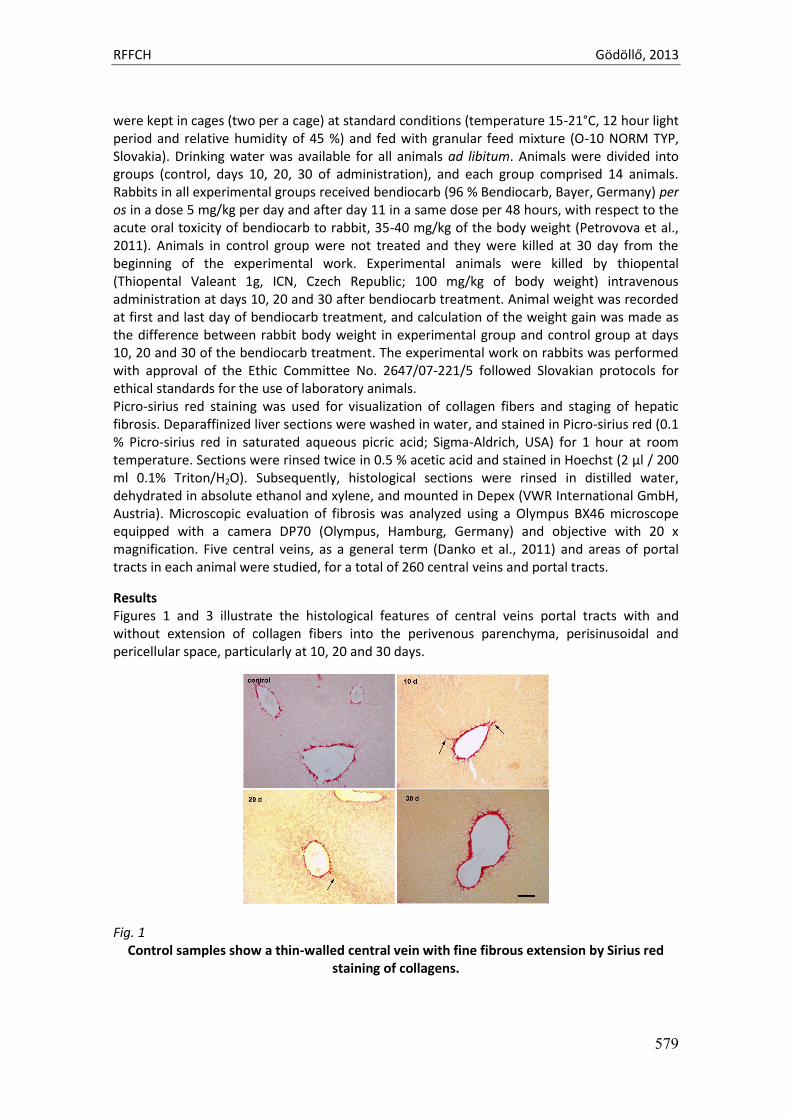
RFFCH Gödöllő, 2013
579
were kept in cages (two per a cage) at standard conditions (temperature 15-21°C, 12 hour light period and relative humidity of 45 %) and fed with granular feed mixture (O-10 NORM TYP, Slovakia). Drinking water was available for all animals ad libitum. Animals were divided into groups (control, days 10, 20, 30 of administration), and each group comprised 14 animals. Rabbits in all experimental groups received bendiocarb (96 % Bendiocarb, Bayer, Germany) per os in a dose 5 mg/kg per day and after day 11 in a same dose per 48 hours, with respect to the acute oral toxicity of bendiocarb to rabbit, 35-40 mg/kg of the body weight (Petrovova et al., 2011). Animals in control group were not treated and they were killed at 30 day from the beginning of the experimental work. Experimental animals were killed by thiopental (Thiopental Valeant 1g, ICN, Czech Republic; 100 mg/kg of body weight) intravenous administration at days 10, 20 and 30 after bendiocarb treatment. Animal weight was recorded at first and last day of bendiocarb treatment, and calculation of the weight gain was made as the difference between rabbit body weight in experimental group and control group at days 10, 20 and 30 of the bendiocarb treatment. The experimental work on rabbits was performed with approval of the Ethic Committee No. 2647/07-221/5 followed Slovakian protocols for ethical standards for the use of laboratory animals. Picro-sirius red staining was used for visualization of collagen fibers and staging of hepatic fibrosis. Deparaffinized liver sections were washed in water, and stained in Picro-sirius red (0.1 % Picro-sirius red in saturated aqueous picric acid; Sigma-Aldrich, USA) for 1 hour at room temperature. Sections were rinsed twice in 0.5 % acetic acid and stained in Hoechst (2 µl / 200 ml 0.1% Triton/H2O). Subsequently, histological sections were rinsed in distilled water, dehydrated in absolute ethanol and xylene, and mounted in Depex (VWR International GmbH, Austria). Microscopic evaluation of fibrosis was analyzed using a Olympus BX46 microscope equipped with a camera DP70 (Olympus, Hamburg, Germany) and objective with 20 x magnification. Five central veins, as a general term (Danko et al., 2011) and areas of portal tracts in each animal were studied, for a total of 260 central veins and portal tracts.
Results Figures 1 and 3 illustrate the histological features of central veins portal tracts with and without extension of collagen fibers into the perivenous parenchyma, perisinusoidal and pericellular space, particularly at 10, 20 and 30 days.
Fig. 1 Control samples show a thin-walled central vein with fine fibrous extension by Sirius red
staining of collagens.
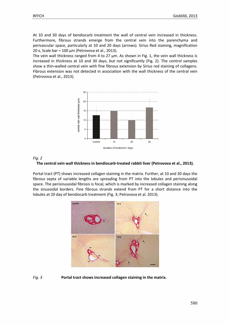
RFFCH Gödöllő, 2013
580
At 10 and 30 days of bendiocarb treatment the wall of central vein increased in thickness. Furthermore, fibrous strands emerge from the central vein into the parenchyma and perivascular space, particularly at 10 and 20 days (arrows). Sirius Red staining, magnification 20 x, Scale bar = 100 µm (Petrovova et al., 2013). The vein wall thickness ranged from 4 to 27 µm. As shown in Fig. 1, the vein wall thickness is increased in thickness at 10 and 30 days, but not significantly (Fig. 2). The control samples show a thin-walled central vein with fine fibrous extension by Sirius red staining of collagens. Fibrous extension was not detected in association with the wall thickness of the central vein (Petrovova et al., 2013).
Fig. 2
The central vein wall thickness in bendiocarb-treated rabbit liver (Petrovova et al., 2013). Portal tract (PT) shows increased collagen staining in the matrix. Further, at 10 and 30 days the fibrous septa of variable lengths are spreading from PT into the lobules and perisinusoidal space. The perisinusoidal fibrosis is focal, which is marked by increased collagen staining along the sinusoidal borders. Fine fibrous strands extend from PT for a short distance into the lobules at 20 day of bendiocarb treatment (Fig. 3; Petrovova et al. 2013). Fig. 3 Portal tract shows increased collagen staining in the matrix.
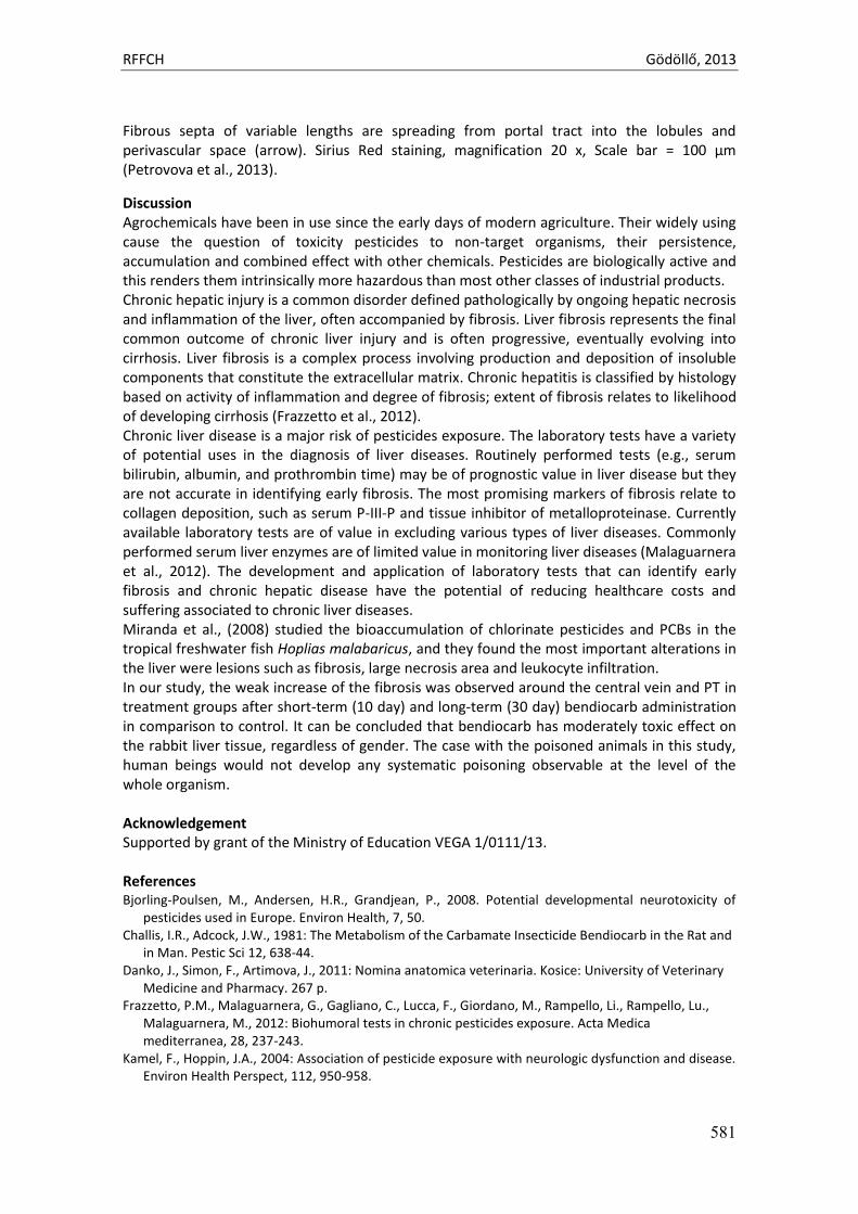
RFFCH Gödöllő, 2013
581
Fibrous septa of variable lengths are spreading from portal tract into the lobules and perivascular space (arrow). Sirius Red staining, magnification 20 x, Scale bar = 100 µm (Petrovova et al., 2013).
Discussion Agrochemicals have been in use since the early days of modern agriculture. Their widely using cause the question of toxicity pesticides to non-target organisms, their persistence, accumulation and combined effect with other chemicals. Pesticides are biologically active and this renders them intrinsically more hazardous than most other classes of industrial products. Chronic hepatic injury is a common disorder defined pathologically by ongoing hepatic necrosis and inflammation of the liver, often accompanied by fibrosis. Liver fibrosis represents the final common outcome of chronic liver injury and is often progressive, eventually evolving into cirrhosis. Liver fibrosis is a complex process involving production and deposition of insoluble components that constitute the extracellular matrix. Chronic hepatitis is classified by histology based on activity of inflammation and degree of fibrosis; extent of fibrosis relates to likelihood of developing cirrhosis (Frazzetto et al., 2012). Chronic liver disease is a major risk of pesticides exposure. The laboratory tests have a variety of potential uses in the diagnosis of liver diseases. Routinely performed tests (e.g., serum bilirubin, albumin, and prothrombin time) may be of prognostic value in liver disease but they are not accurate in identifying early fibrosis. The most promising markers of fibrosis relate to collagen deposition, such as serum P-III-P and tissue inhibitor of metalloproteinase. Currently available laboratory tests are of value in excluding various types of liver diseases. Commonly performed serum liver enzymes are of limited value in monitoring liver diseases (Malaguarnera et al., 2012). The development and application of laboratory tests that can identify early fibrosis and chronic hepatic disease have the potential of reducing healthcare costs and suffering associated to chronic liver diseases. Miranda et al., (2008) studied the bioaccumulation of chlorinate pesticides and PCBs in the tropical freshwater fish Hoplias malabaricus, and they found the most important alterations in the liver were lesions such as fibrosis, large necrosis area and leukocyte infiltration. In our study, the weak increase of the fibrosis was observed around the central vein and PT in treatment groups after short-term (10 day) and long-term (30 day) bendiocarb administration in comparison to control. It can be concluded that bendiocarb has moderately toxic effect on the rabbit liver tissue, regardless of gender. The case with the poisoned animals in this study, human beings would not develop any systematic poisoning observable at the level of the whole organism. Acknowledgement Supported by grant of the Ministry of Education VEGA 1/0111/13. References Bjorling-Poulsen, M., Andersen, H.R., Grandjean, P., 2008. Potential developmental neurotoxicity of
pesticides used in Europe. Environ Health, 7, 50. Challis, I.R., Adcock, J.W., 1981: The Metabolism of the Carbamate Insecticide Bendiocarb in the Rat and
in Man. Pestic Sci 12, 638-44. Danko, J., Simon, F., Artimova, J., 2011: Nomina anatomica veterinaria. Kosice: University of Veterinary
Medicine and Pharmacy. 267 p. Frazzetto, P.M., Malaguarnera, G., Gagliano, C., Lucca, F., Giordano, M., Rampello, Li., Rampello, Lu.,
Malaguarnera, M., 2012: Biohumoral tests in chronic pesticides exposure. Acta Medica mediterranea, 28, 237-243.
Kamel, F., Hoppin, J.A., 2004: Association of pesticide exposure with neurologic dysfunction and disease. Environ Health Perspect, 112, 950-958.
RFFCH Gödöllő, 2013
582
Malaguarnera, G., Cataudella, E., Giordano, M., Nunnari, G., Chisari, G., Malaguarnera, M., 2012: Toxic hepatitis in occupational exposure to solvents. World J Gastroenterol, 18, 2756-2766.
Miranda, A.L., Roche, H., Randi, M.A.F., Menezes, M.L., Oliveira Ribeiro, C.A., 2008: Bioaccumulation of chlorinate pesticides and PCBs in the tropical freshwater fish Hoplias Malabaricus: histopathological, physiological, and immunological findings. Environ Int 34,939-949.
Nauen, R., 2007: Perspective insecticide resistance in disease vectors of public health importance. Pest Manag Sci 63,628-633.
Pacioni, N.L., Veglia, A.V., 2007: Determination of poorly fluorescent carbamate pesticides in water, bendiocarb and promecarb, using cyclodextrin nanocavities and related media. Anal Chim Acta 583,63-71.
Petrovova, E., Massanyi, P., Capcarova, M., Zivcak, J., Stodola, L., 2011: Structural alterations in rabbit spleen after bendiocarb administration. J Environ Sci Health B 46,788-792.
Petrovova, E., Purzyc, H., Mazensky, D., Luptakova, L., Torma, N., Sopoliga, I., Sedmera, D., 2013: Morphometric alterations, steatosis, fibrosis and active caspase-3 detection in carbamate bendiocarb treated rabbit liver. Environmental Toxicology, In Press
Schenck, M., Kolb, E., 1990: Basics of physiological chemistry. Bratislava: Priroda. 666 p
Animal welfare, etológia és tartástechnológia
Animal welfare, ethology and housing systems
Volume 9 Issue 3
Különszám/Special Issue
Gödöllő 2013
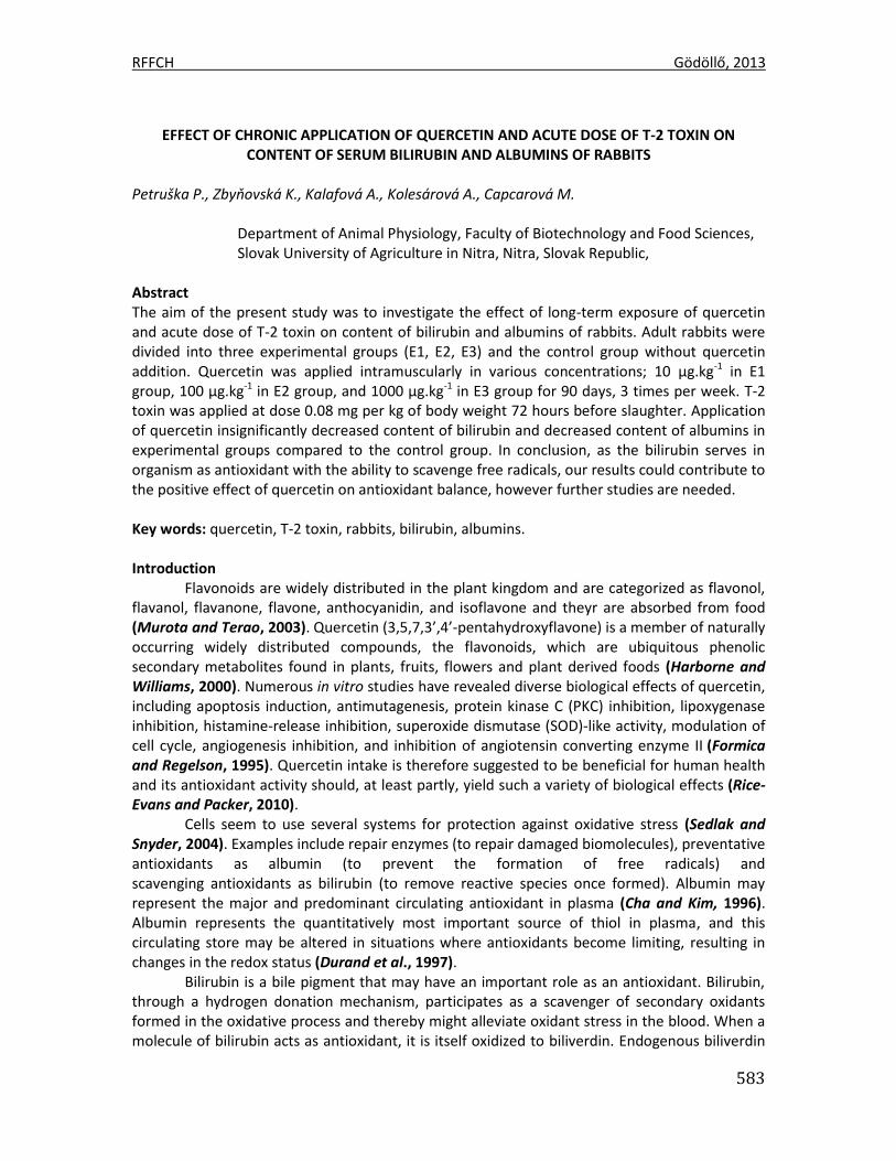
RFFCH Gödöllő, 2013
583
EFFECT OF CHRONIC APPLICATION OF QUERCETIN AND ACUTE DOSE OF T-2 TOXIN ON
CONTENT OF SERUM BILIRUBIN AND ALBUMINS OF RABBITS
Petruška P., Zbyňovská K., Kalafová A., Kolesárová A., Capcarová M.
Department of Animal Physiology, Faculty of Biotechnology and Food Sciences, Slovak University of Agriculture in Nitra, Nitra, Slovak Republic,
Abstract The aim of the present study was to investigate the effect of long-term exposure of quercetin and acute dose of T-2 toxin on content of bilirubin and albumins of rabbits. Adult rabbits were divided into three experimental groups (E1, E2, E3) and the control group without quercetin addition. Quercetin was applied intramuscularly in various concentrations; 10 µg.kg-1 in E1 group, 100 µg.kg-1 in E2 group, and 1000 µg.kg-1 in E3 group for 90 days, 3 times per week. T-2 toxin was applied at dose 0.08 mg per kg of body weight 72 hours before slaughter. Application of quercetin insignificantly decreased content of bilirubin and decreased content of albumins in experimental groups compared to the control group. In conclusion, as the bilirubin serves in organism as antioxidant with the ability to scavenge free radicals, our results could contribute to the positive effect of quercetin on antioxidant balance, however further studies are needed. Key words: quercetin, T-2 toxin, rabbits, bilirubin, albumins. Introduction
Flavonoids are widely distributed in the plant kingdom and are categorized as flavonol, flavanol, flavanone, flavone, anthocyanidin, and isoflavone and theyr are absorbed from food (Murota and Terao, 2003). Quercetin (3,5,7,3’,4’-pentahydroxyflavone) is a member of naturally occurring widely distributed compounds, the flavonoids, which are ubiquitous phenolic secondary metabolites found in plants, fruits, flowers and plant derived foods (Harborne and Williams, 2000). Numerous in vitro studies have revealed diverse biological effects of quercetin, including apoptosis induction, antimutagenesis, protein kinase C (PKC) inhibition, lipoxygenase inhibition, histamine-release inhibition, superoxide dismutase (SOD)-like activity, modulation of cell cycle, angiogenesis inhibition, and inhibition of angiotensin converting enzyme II (Formica and Regelson, 1995). Quercetin intake is therefore suggested to be beneficial for human health and its antioxidant activity should, at least partly, yield such a variety of biological effects (Rice-Evans and Packer, 2010).
Cells seem to use several systems for protection against oxidative stress (Sedlak and Snyder, 2004). Examples include repair enzymes (to repair damaged biomolecules), preventative antioxidants as albumin (to prevent the formation of free radicals) and scavenging antioxidants as bilirubin (to remove reactive species once formed). Albumin may represent the major and predominant circulating antioxidant in plasma (Cha and Kim, 1996). Albumin represents the quantitatively most important source of thiol in plasma, and this circulating store may be altered in situations where antioxidants become limiting, resulting in changes in the redox status (Durand et al., 1997).
Bilirubin is a bile pigment that may have an important role as an antioxidant. Bilirubin, through a hydrogen donation mechanism, participates as a scavenger of secondary oxidants formed in the oxidative process and thereby might alleviate oxidant stress in the blood. When a molecule of bilirubin acts as antioxidant, it is itself oxidized to biliverdin. Endogenous biliverdin
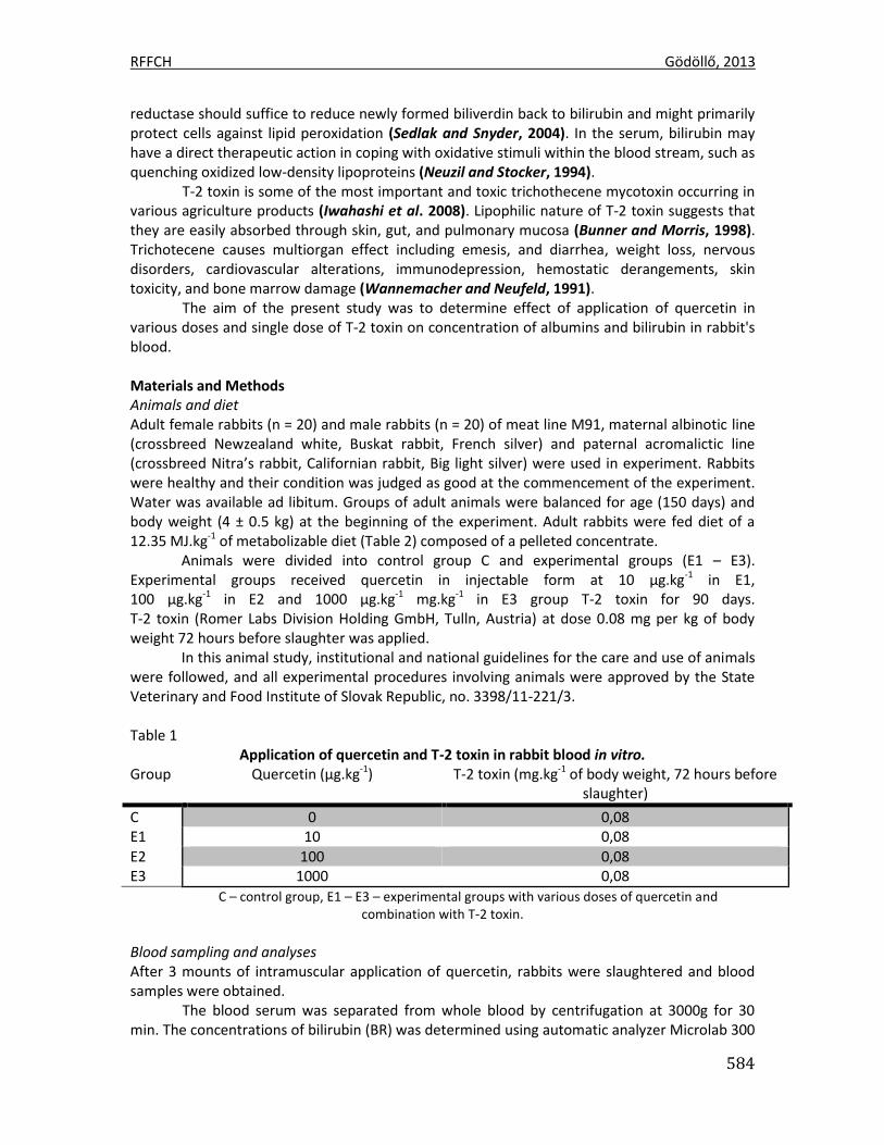
RFFCH Gödöllő, 2013
584
reductase should suffice to reduce newly formed biliverdin back to bilirubin and might primarily protect cells against lipid peroxidation (Sedlak and Snyder, 2004). In the serum, bilirubin may have a direct therapeutic action in coping with oxidative stimuli within the blood stream, such as quenching oxidized low-density lipoproteins (Neuzil and Stocker, 1994).
T-2 toxin is some of the most important and toxic trichothecene mycotoxin occurring in various agriculture products (Iwahashi et al. 2008). Lipophilic nature of T-2 toxin suggests that they are easily absorbed through skin, gut, and pulmonary mucosa (Bunner and Morris, 1998). Trichotecene causes multiorgan effect including emesis, and diarrhea, weight loss, nervous disorders, cardiovascular alterations, immunodepression, hemostatic derangements, skin toxicity, and bone marrow damage (Wannemacher and Neufeld, 1991).
The aim of the present study was to determine effect of application of quercetin in various doses and single dose of T-2 toxin on concentration of albumins and bilirubin in rabbit's blood. Materials and Methods Animals and diet Adult female rabbits (n = 20) and male rabbits (n = 20) of meat line M91, maternal albinotic line (crossbreed Newzealand white, Buskat rabbit, French silver) and paternal acromalictic line (crossbreed Nitra’s rabbit, Californian rabbit, Big light silver) were used in experiment. Rabbits were healthy and their condition was judged as good at the commencement of the experiment. Water was available ad libitum. Groups of adult animals were balanced for age (150 days) and body weight (4 ± 0.5 kg) at the beginning of the experiment. Adult rabbits were fed diet of a 12.35 MJ.kg-1 of metabolizable diet (Table 2) composed of a pelleted concentrate.
Animals were divided into control group C and experimental groups (E1 – E3). Experimental groups received quercetin in injectable form at 10 µg.kg-1 in E1, 100 µg.kg-1 in E2 and 1000 µg.kg-1 mg.kg-1 in E3 group T-2 toxin for 90 days. T-2 toxin (Romer Labs Division Holding GmbH, Tulln, Austria) at dose 0.08 mg per kg of body weight 72 hours before slaughter was applied.
In this animal study, institutional and national guidelines for the care and use of animals were followed, and all experimental procedures involving animals were approved by the State Veterinary and Food Institute of Slovak Republic, no. 3398/11-221/3.
Table 1
Application of quercetin and T-2 toxin in rabbit blood in vitro. Group Quercetin (µg.kg-1) T-2 toxin (mg.kg-1 of body weight, 72 hours before
slaughter)
C 0 0,08 E1 10 0,08
E2 100 0,08 E3 1000 0,08
C – control group, E1 – E3 – experimental groups with various doses of quercetin and combination with T-2 toxin.
Blood sampling and analyses After 3 mounts of intramuscular application of quercetin, rabbits were slaughtered and blood samples were obtained.
The blood serum was separated from whole blood by centrifugation at 3000g for 30 min. The concentrations of bilirubin (BR) was determined using automatic analyzer Microlab 300
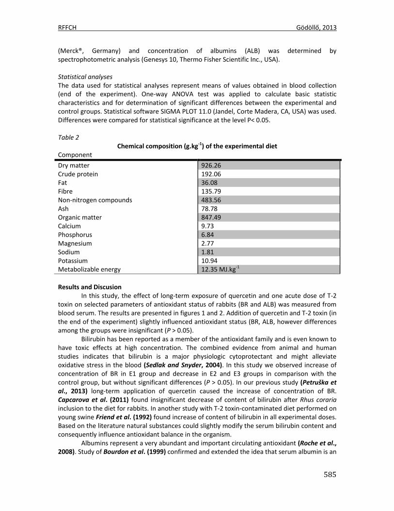
RFFCH Gödöllő, 2013
585
(Merck®, Germany) and concentration of albumins (ALB) was determined by spectrophotometric analysis (Genesys 10, Thermo Fisher Scientific Inc., USA). Statistical analyses The data used for statistical analyses represent means of values obtained in blood collection (end of the experiment). One-way ANOVA test was applied to calculate basic statistic characteristics and for determination of significant differences between the experimental and control groups. Statistical software SIGMA PLOT 11.0 (Jandel, Corte Madera, CA, USA) was used. Differences were compared for statistical significance at the level P< 0.05. Table 2
Chemical composition (g.kg-1) of the experimental diet Component
Dry matter 926.26 Crude protein 192.06 Fat 36.08 Fibre 135.79 Non-nitrogen compounds 483.56 Ash 78.78 Organic matter 847.49 Calcium 9.73 Phosphorus 6.84 Magnesium 2.77 Sodium 1.81 Potassium 10.94 Metabolizable energy 12.35 MJ.kg-1
Results and Discusion
In this study, the effect of long-term exposure of quercetin and one acute dose of T-2 toxin on selected parameters of antioxidant status of rabbits (BR and ALB) was measured from blood serum. The results are presented in figures 1 and 2. Addition of quercetin and T-2 toxin (in the end of the experiment) slightly influenced antioxidant status (BR, ALB, however differences among the groups were insignificant (P > 0.05).
Bilirubin has been reported as a member of the antioxidant family and is even known to have toxic effects at high concentration. The combined evidence from animal and human studies indicates that bilirubin is a major physiologic cytoprotectant and might alleviate oxidative stress in the blood (Sedlak and Snyder, 2004). In this study we observed increase of concentration of BR in E1 group and decrease in E2 and E3 groups in comparison with the control group, but without significant differences (P > 0.05). In our previous study (Petruška et al., 2013) long-term application of quercetin caused the increase of concentration of BR. Capcarova et al. (2011) found insignificant decrease of content of bilirubin after Rhus coraria inclusion to the diet for rabbits. In another study with T-2 toxin-contaminated diet performed on young swine Friend et al. (1992) found increase of content of bilirubin in all experimental doses. Based on the literature natural substances could slightly modify the serum bilirubin content and consequently influence antioxidant balance in the organism.
Albumins represent a very abundant and important circulating antioxidant (Roche et al., 2008). Study of Bourdon et al. (1999) confirmed and extended the idea that serum albumin is an
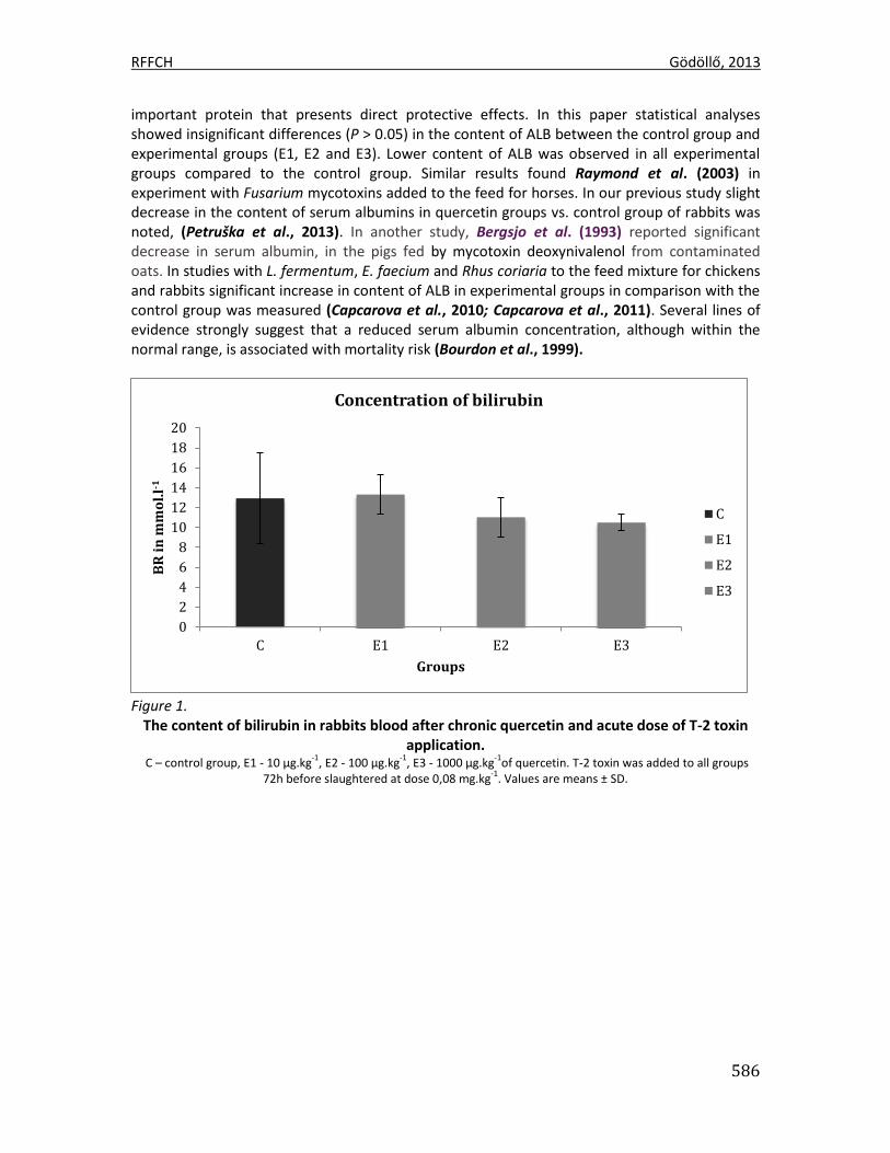
RFFCH Gödöllő, 2013
586
important protein that presents direct protective effects. In this paper statistical analyses showed insignificant differences (P > 0.05) in the content of ALB between the control group and experimental groups (E1, E2 and E3). Lower content of ALB was observed in all experimental groups compared to the control group. Similar results found Raymond et al. (2003) in experiment with Fusarium mycotoxins added to the feed for horses. In our previous study slight decrease in the content of serum albumins in quercetin groups vs. control group of rabbits was noted, (Petruška et al., 2013). In another study, Bergsjo et al. (1993) reported significant decrease in serum albumin, in the pigs fed by mycotoxin deoxynivalenol from contaminated oats. In studies with L. fermentum, E. faecium and Rhus coriaria to the feed mixture for chickens and rabbits significant increase in content of ALB in experimental groups in comparison with the control group was measured (Capcarova et al., 2010; Capcarova et al., 2011). Several lines of evidence strongly suggest that a reduced serum albumin concentration, although within the normal range, is associated with mortality risk (Bourdon et al., 1999).
Figure 1.
The content of bilirubin in rabbits blood after chronic quercetin and acute dose of T-2 toxin application.
C – control group, E1 - 10 µg.kg-1
, E2 - 100 µg.kg-1
, E3 - 1000 µg.kg-1
of quercetin. T-2 toxin was added to all groups 72h before slaughtered at dose 0,08 mg.kg
-1. Values are means ± SD.
0
2
4
6
8
10
12
14
16
18
20
C E1 E2 E3
BR
in
mm
ol.
l-1
Groups
Concentration of bilirubin
C
E1
E2
E3
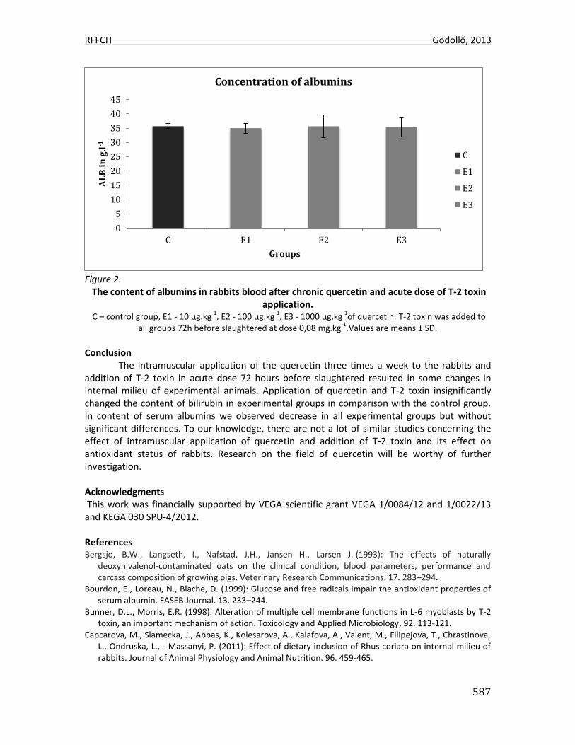
RFFCH Gödöllő, 2013
587
Figure 2.
The content of albumins in rabbits blood after chronic quercetin and acute dose of T-2 toxin application.
C – control group, E1 - 10 µg.kg-1
, E2 - 100 µg.kg-1
, E3 - 1000 µg.kg-1
of quercetin. T-2 toxin was added to all groups 72h before slaughtered at dose 0,08 mg.kg
-1.Values are means ± SD.
Conclusion
The intramuscular application of the quercetin three times a week to the rabbits and addition of T-2 toxin in acute dose 72 hours before slaughtered resulted in some changes in internal milieu of experimental animals. Application of quercetin and T-2 toxin insignificantly changed the content of bilirubin in experimental groups in comparison with the control group. In content of serum albumins we observed decrease in all experimental groups but without significant differences. To our knowledge, there are not a lot of similar studies concerning the effect of intramuscular application of quercetin and addition of T-2 toxin and its effect on antioxidant status of rabbits. Research on the field of quercetin will be worthy of further investigation. Acknowledgments This work was financially supported by VEGA scientific grant VEGA 1/0084/12 and 1/0022/13 and KEGA 030 SPU-4/2012. References Bergsjo, B.W., Langseth, I., Nafstad, J.H., Jansen H., Larsen J. (1993): The effects of naturally
deoxynivalenol-contaminated oats on the clinical condition, blood parameters, performance and carcass composition of growing pigs. Veterinary Research Communications. 17. 283–294.
Bourdon, E., Loreau, N., Blache, D. (1999): Glucose and free radicals impair the antioxidant properties of serum albumin. FASEB Journal. 13. 233–244.
Bunner, D.L., Morris, E.R. (1998): Alteration of multiple cell membrane functions in L-6 myoblasts by T-2 toxin, an important mechanism of action. Toxicology and Applied Microbiology, 92. 113-121.
Capcarova, M., Slamecka, J., Abbas, K., Kolesarova, A., Kalafova, A., Valent, M., Filipejova, T., Chrastinova, L., Ondruska, L., - Massanyi, P. (2011): Effect of dietary inclusion of Rhus coriara on internal milieu of rabbits. Journal of Animal Physiology and Animal Nutrition. 96. 459-465.
0
5
10
15
20
25
30
35
40
45
C E1 E2 E3
AL
B i
n g
.l-1
Groups
Concentration of albumins
C
E1
E2
E3
RFFCH Gödöllő, 2013
588
Capcarova, M., Weis, J., Hrncar, C., Kolesarova, A., Pal, G. (2010): Effect of Lactobacillus fermentum and enterococcus faecium strains on internal milieu, antioxidant status and body weight of broiler chickens. Journal of Animal Physiology and Animal Nutrition. 94. 215-224.
Durand, P., Lussier-Caca, S., Blache, D. (1997): Acute methionine load-induced hyperhomocysteinemia enhances platelet aggregation, thromboxane biosynthe- sis and macrophage-derived tissue factor activity in rats. FASEB Journal 11, 1157–1168.
Formica, J.V., Regelson, W. (1995): Review of the biology of quercetin and related bioflavonoids. Food and Chemical Toxcology, 33, 1061–1080.
Friend, D.W., Thompson, B.K., Trenholm, H.L., Boermans, H.J., Hartin, K.E., Panich, P.L. (1992): Toxicity of T-2 toxin and its interaction with deoxynivalenol when fed to young pigs. Canadien Journal of Science. 72. 703-711.
Harborne, J.B., Williams, C.A. (2000): Advances in flavonoid research since 1992. Phytochemistry. 55.481-504.
Cha, M.K., Kim, I.H. (1996): Glutathione-linked thiol peroxidase activity of human serum albumin: a possible antioxidant role of serum albumin in blood plasma. Biochemical and Biophysical Research Communications. 222. 619–625.
Iwahashi, Y., Kitagawa, E., Iwahashi, H. (2008): Analysis of mechanisms of T-2 toxin toxicity using yeast DNA Microarrays. International Journal of Molecular Sciences. 9. 2585-2600.
Murota, K. Terao, J. (2003): Antioxidative flavonoid quercetin: implication of its intestinal absorbtion and metabolism. Archives of Biochemistry and Biophysics. 417. 12-17.
Neuzil, J., Stocker, R., (1994): Free and albumin bound bilirubin are efficient co-antioxidants for alpha-tocopherol, inhibiting plasma and low density lipoproteinlipid peroxidation. Journal of Biological Chemistry. 269. 16712–16719.
Petruška, P., Kalafová, A., Kolesárová, A., Zbyňovská, K., Latacz, A., Capcarová, M. (2013): Effect of cronic application of quercetin on content of serum bilirubin and albumins od rabbits. (In press).
Raymond, S.L., Smith, T.K. Swamy, H.V.L.N. (2003): Effects of feeding a blend of grains naturally contamined with Fusarium mycotoxins on feed intake, serum chemistry, and hematology of horses, and the efficacy of a polymeric glucomannan mycotoxin adsorbent. Journal of Animal Science. 81. 2123-2130.
Rice-Evans, C.A. Miller, N.J. Paganga, G. (1996): Structure-antioxidant activity relationships of flavonoids and phenolic acids. Free Radical Biology and Medicine. 20. 933–956.
Roche, M. Rondeau, P., Singh, N.R., Tarnus, E., Bourdon, E. (2008): The antioxidant properties of serum albumin. FEBS Letters. 582. 1783–1787.
Sedlak, T.W., SNYDER, S.H. (2004): Bilirubin benefits: cellular protection by a biliverdin reductase antioxidant cycle. Pediatrics. 113. 1776–1782.
Wannemacher, R.W.Jr., Bunner, D.L., Neufeld, H.A. (1991): Toxicity of trichothecenes and other related mycotoxins in laboratory animals, Smith JE, Henderson RS, eds. Mycotoxins and Animal Foods. Boca Raton, Fla: CRC Press 499–552.

1
Animal welfare, etológia és tartástechnológia
Animal welfare, ethology and housing systems
Volume 9 Issue 3
Különszám/Special Issue
Gödöllő
2013
589
POROVNANIE OBSAHU MINERÁLNYCH LÁTOK V RÔZNYCH KRMIVÁCH PRE DOJNICE COMPARISON OF THE MINERAL CONTENT IN DIFFERENT RUMINANT FEEDS.
SKALICKÁ Magdaléna, MASKAĽOVÁ Iveta
Ústav výživy, dietetiky a krmovinárstva, Univerzita veterinárskeho lekárstva a farmácie v Košiciach, Komenského 73, 041 81 Košice, Slovenská republika
Abstract The aim was evaluation of the quality of feeds for the cows on the micro mineral content (Cu, Zn, Mn) of the different geographical areas of Slovakia. Feed samples – corn silage, alfalfa silage and grass silage, TMR – total mixed ration, and alfalfa came from high - production dairy farms. In samples of plant origin was determined average levels of copper, zinc, and manganese according to the methodology used by the Official lists methods and laboratory diagnosis of food and feed (Bulletin of the Ministry of Agriculture of the Slovak Republic, 2004). The detected levels of mineral contents in feed for dairy cows were compared with the recommended values according to NRC (2001). Key words: feeds – corn silage, grass silage and alfalfa silage, alfalfa (Medicago sativa), zinc, copper, manganese, AAS Úvod
Významnú úlohu vo výžive vysoko produkčných dojníc zohrávajú mikroprvky – meď, zinok a mangán. Zúčastňujú sa na aktivácii enzýmov dôležitých pre látkovú výmenu. Ich využiteľnosť z krmiva môže byť zmenená rozdielnym zastúpením tráv, ďatelinovín a bylín, ale tiež intenzitou a spôsobom hnojenia porastov. Koncentrácia minerálov v rastlinách závisí od štyroch faktorov: genotyp rastlín, pôdne prostredie, klíma a štádium zrelosti. Zinok, meď a mangán patria medzi esenciálne prvky, ktoré sú súčasťou viacerých životne dôležitých metabolických dejov. V rámci intermediálneho metabolizmu majú tieto prvky vzájomne negatívne aj pozitívne vzťahy. Pozitívnych väzieb ( Mn - Zn) je menej, než antagonistických väzieb (Zn-Cu), vápnik inhibuje absorpciu zinku a mangánu (Shajhalal a kol., 2008, Khan a kol., 2008, Wilde, 2006, Slavík a kol., 2005). Dostupnosť zinku pre rastliny výrazne závisí na textúre pôdy, zastúpení organickej hmoty, hodnote pH, množstve fosforu a aktuálnom počasí. Častý je výskyt sekundárneho deficitu zinku u zvierat pri nadmernom obsahu Ca, P a kyseliny fytínovej v kŕmnych dávkach. Nedostatok mangánu v pôde sa vyskytuje po silnom vápnení. Do rastlín sa mangán dostáva z pôdy vo forme katiónov. Pri nedostatku medi v rastlinných bunkách sa zvyšuje obsah voľných dusíkatých látok. Dôležitú úlohu má meď pri syntéze lignínu, ktorý spevňuje bunkovú stenu. Hospodárske zvieratá obvykle získavajú väčšinu minerálnych látok z krmív a ich príjem je ovplyvnený faktormi, ktoré určujú obsah minerálov z rastlín a ich semien. Význam týchto faktorov je ovplyvnený interakciami s uvedenými faktormi a aspektmi plodín alebo pastviny, vrátane použitia hnojív, pôdnych zmien, zavlažovanie, striedanie plodín (Suttle , 2010). Pri hodnotení krmív nás zaujímalo aj hodnotenie obsahu minerálnych živín v objemových krmivách určených pre kŕmne účely hovädzieho dobytka. Materiál a metodika
V sledovaných chovoch vysoko-produkčných dojníc boli analyzované vzorky objemových krmív - žľabové vzorky TMR, kukuričná siláž, senáž lucernová, senáž trávna a lucerna siata na obsah medi, zinku a mangánu. Na stanovenie minerálnych látok vo vzorkách rastlinného materiálu bola použitá metodika, ktorú uvádza Zoznam úradných metód a laboratórnej diagnostiky potravín a krmív (Vestník MPSR, 2004).Vzorky krmiva boli spracované technológiou mikrovlnného rozkladu za
RFFCH Gödöllő, 2013
590
prítomnosti nízko vriacich kyselín(HCl, HNO3 ) v mineralizačnom systéme MLS-1 200 MEGA, fy Milestone. Cu, Zn a Mn sa analyzovali použitím plameňovej metódy na AAS, Solar 939, fy Unicam. Výsledky a diskusia
Kvalita objemových krmív je rozhodujúcej miere ovplyvnená poveternostnými podmienkami, pôdnym potenciálom a štádiom zrelosti jednotlivých druhov rastlín. Odobraté vzorky objemových krmív z vybraných lokalít boli podrobené laboratórnej analýze na stanovenie minerálnych látok – Cu, Zn, Mn . Obsah stopových prvkov v krmivách sa riadi geochemickým charakterom pôdy a druhu rastlín, okrem klimaticko poľnohospodárkych podmienok. Viacerí autori sa zaoberali výskytom stopových prvkov v krmivách a sérach dobytka z rôznych klimatických podmienok (Shahjalal a kol., 2008, Mandal a kol., 2004, Ramana a kol 2001) a zistili nízky obsah medi v trávach a senách.
Zistené priemerné množstvá medi, zinku a mangánu v kukuričnej, lucernovej, trávnej siláži a lucerne siatej sú uvedené v tabuľkách č. 1 a 2. Tab.č.1.
Zistený priemerný obsah Cu, Zn a Mn (ppm) v objemových krmivách v roku 2011
Cu
Zn
Mn
9.80 ± 4.70 9.67 ± 4.39 8.10 ± 4.83 15.38 ± 3.13
Kukričná siláž Lucernová siláž Trávna siláž Lucerna siata
55.99 ± 34.61 57.52 ± 27.51 53.60 ± 30.10 121.38 ± 33.01
40.44 ± 18.76 69.89 ± 23.98 84.03 ± 28.88 33.14 ± 6.55
x ± s x ± s x ± s x ± s
Kukuričná siláž má nezastupiteľne miesto v kŕmnych dávkach pre dojnice. Patrí medzi ľahko
stráviteľné sacharidové krmivo. V kukuričnej siláži sa analyzované obsahy medi, zinku a mangánu pohybovali v rozpätí: meď 3,85 – 20,34 ppm, zinok 8,69 – 187,18 ppm a mangán 11,52 – 84,99 ppm. Nami zistené priemerné obsahy medi (9,80 a 11,02 ppm), zinku (55,99 a 51,77 ppm) a mangánu (40,44 a 41,98 ppm) v kukuričnej siláži sú v porovnaní s uvedenými hodnotami v NRC (2001) vyššie (24 ppm Zn, 36 ppm Mn, 6 ppm Cu). Rogers a kol. (2001) v írskej kukuričnej siláži zistili nižšie množstvá medi (5,11 ppm), zinku (26,61 ppm) a mangánu (25,61 ppm) v porovnaní s hodnotami, ktoré sme zistili v kukuričnej siláži zo slovenských chovoch dojníc. Tab.č.2.
Zistený priemerný obsah Cu, Zn a Mn (ppm) v objemových krmivách v roku 2012
Cu
Zn
Mn
Kukričná siláž Lucernová siláž Trávna siláž Lucerna siata
x ± s x ± s x ± s x ± s
11.02 ± 6.07 13.78 ± 6.16 13.02 ± 6.88 15.52 ± 7.31
51.77 ± 41.94 75.04 ± 49.30 58.13 ± 31.68 54.06 ± 27.80
54.29 ± 13.85 41.98 ± 14.95 101.40 ± 77.20 107.2 ± 67.00
Lucerna siata patrí medzi naše najcennejšie krmoviny. Z hľadiska pomeru živín sa radí k
bielkovinovým krmivám. Jej kvalita vo veľkej miere závisí od fenologickej fázy rastu v čase zberu a podielu listov a byli. Listy majú relatívne konštantné zloženie až do začiatku kvitnutia a obsahujú viac minerálnych látok ako byle. Pokosenú zelenú hmotu lucerny je možné využiť ako na priame skrmovanie, tak na výrobu siláže resp. sena. Najnižšie množstvo medi v sledovaných silážach bolo zistené v lucernovej siláži 3,13 ppm a maximálne zistené množstvo medi bolo v lucerne siatej 31,07 ppm. V lucernovej, trávnej siláži a lucerne boli zistené maximálne množstvá zinku 157,20 ppm, 105,40 ppm a 118,90 ppm. Prítomnosť mangánu v lucernovej a trávnej siláži bol rozpätí 69,89 - 107,20 ppm. Lucerna siata vykazovala oveľa nižšie hodnoty mangánu (33,14-51,29 ppm sušiny) v porovnaní so silážami. Podobné priemerné množstvá medi (10,36 ppm) a mangánu (103,45 ppm) zaznamenali Rogers a kol. (2001) v írskych trávových silážach. Juráček a Bíro (2010) uvádzajú vyšší
RFFCH Gödöllő, 2013
591
obsah Cu s nižší obsah Zn a Mn u lucernových siláží po prídavku chemického konzervanta - kyseliny mravčej. Shahjalal a kol. 2008 prezentovali vo svojich prácach mikro minerálny obsah u krmív z rôznych klimatický podmienok, kde zistili nízky obsah medi. Ich zistené priemerné hodnoty Cu, Zn a Mn boli 6,12ppm, 291,3 ppm a 224,9 ppm. Obsahy Zn a Mn boli vyššie v porovnaní nami zistenými hodnotami. Vyššie priemerné koncentrácie Cu (27,9 ppm), Zn (31,00 ppm) a Mn (101,0) v trávach zaznamenali Ramana a kol. (2001).
Výsledky minerálnych rozborov kompletných miešaných kŕmnych dávok (TMR) pre produkčné dojnice podľa fáz produkcie sú uvedené v tabuľke 3 a 4. Príprava krmiva je dôležitý moment, ktorý rozhoduje o efektívnosti chovu. Každá zo základných živín vrátane minerálnych látok má svoju dôležitosť a môže byť limitujúcim prvkom úspešnosti chovu. Reakcie organizmu dojníc na prípadné nedostatky či prebytky, nepomery živín sa prejavia oveľa neskôr alebo sa prehliadnu. Priemerné zistené obsahy medi v roku 2011 a 2012 v TMR vo fáze pred otelením boli 18,02 a 20,12 ppm, po otelení 22,68 a 19,95 ppm, v TMR vo vrchole laktácie bol obsah 22,49 a 19,80 ppm. Vo fáze pred otelením bolo zistené v TMR priemerné množstvo zinku (70,93 a 100,43ppm) a mangánu (84,81 a 103,24 ppm). Mierne zvýšenie priemerného obsahu zinku a mangánu v porovnaní s tolerančnými hodnotami udávanými NRC (2001) sa zistili v TMR vo fáze po otelení (Zn 95,10 a 109,35ppm , Mn 85,87 a 101,22 ppm). V TMR určenej pre dojnice vo vrchole laktácie sa obsahy zinku (95,24 a 405,29 ppm) a mangánu (91,06 a 97,63 ppm) pohybovali v tolerančných hodnotách podľa NRC (2001). Skalická a kol. (2010) zistili skoro rovnaké priemerné množstvá medi a mangánu zistili v analyzovaných TMR v produkčných fázach, a vyššie hodnoty zinku (pred otelením 117,59 ppm , po otelení 134,79 ppm a vo vrchole 132,49 ppm) v porovnaní s rokmi 2011 a 2012. Tab.č.3.
Priemerný obsah medi, zinku a mangánu (ppm) v TMR podľa fáz produkcie (2011).
Cu
Zn
Mn
TMRpred otelením po otelení vrchol laktácie
x ± s x ± s x ± s
18.02 ± 11.91 22.68 ± 17.56 22.49 ± 17.93
70.93 ± 27.45 95.10 ± 18.01 95.24 ± 23.05
84.81 ± 30.81 85.87 ± 23.44 91.06 ± 23.44 Tab.č.4.
Priemerný obsah medi, zinku a mangánu (ppm) v TMR podľa fáz produkcie (2012).
Cu
Zn
Mn
20.12 ± 10.95 19.95 ± 10.60 19.80 ± 5.14
x ± s
100.43 ± 55.56 109.35 ± 47.89 105.29 ± 38.92
103.24 ± 49.98 101.22 ± 39.60 97.63 ± 38.84
TMRpred otelením po otelení vrchol laktácie
x ± s x ± s
Záver V sledovaných rokoch sme zaznamenali vyšší obsah zinku a mangánu v sledovaných silážach - kukuričná, lucernová a trávna. Podobne aj v lucerne siatej sme zistili vyššie množstvo medi, zinku a mangánu v porovnaní doporučenými hodnotami podľa NRC 2001. Priemerné zistené obsahy medi, zinku a mangánu v TMR sa pohybovali v tolerančných hodnotách podľa NRC (2001). Chov dojníc je ovplyvňovaný kvalitou spotrebovaných krmív. Správne zladený program fázovej výživy minimalizuje metabolické problémy dojníc. Výživarské tabuľky uvádzajú potrebu jednotlivých marko a mikroprvkov pre zvieratá. Kvôli antagonistickým vzťahom a vysokému zaťaženiu organizmu vysoko produkčných dojníc by sa mala vo výžive dojníc doporučovať dávka minerálnych látok. Literatúra
RFFCH Gödöllő, 2013
592
Vestník MP SR, 2004: Zoznam úradných metód a laboratórnej diagnostiky potravín a krmív, XXXVI, Január 9, 3 – 339.
Illek, J., 2006: Současné trendy diagnostiky poruch zdraví a produkčních onemocnení dojníc. Zborník prednášok Dni výživy a veterinárnej dietetik VII, Sept.13 – 14, Košice, 27-30.
Juráček, M., Bíro, D., 2010: Vplyv chemických aditív na obsah minerálnych látok v lucernových silážach. Zborník prednášok Lazarove dni výživy a veterinárnej dietetiky IX., Sept. 3, Košice, CD, 21-25.
Khan, Z.I., Ashraft, M., Ahmaad, K., Javed, I.,Valeem, E:E., 2008: A comparative study on mineral status blood plasma of small ruminanta and pasture in Punjab – Pakistan. Pakistan Journal of Botany vol.40, 1439 – 1151.
Mandal, A.B., Yadav, P.S., Kapoor, V., 2004: Mineral prodile and their retention in lactating cows in relation to soil, fodder and feed in Kamrup district of Assam. Indian Journal Animal Science, 71,421-429.
NRC 2001, Nutrient of requirements of dairy cattle. Seventh revised edition. 2001, National Academy Press. Washington, D.C. , pp: 401, ISBN 0-309-06997-1.
Suttle, N.F., 2010: Mineral nutrition in livestock, 4th edition, British Library, London, UK, p. 5-113. Ramana, J.V., Prasad, C.S., Gowda, N.K., 2001: Mineral prodile of soil, feed, fodders and blood samples of
animals in northern dry and northern transition zones of Karnataka. Indian Journal Dairy Science,54,40-46.
Rogers, P., Murphy, J., Kavanagh, S., 2001: Bovine mineral – vitamin balalncers for Irish maize silage. Grange research centre, Dunsay, Co. Meath, Ireland. Dostupné na internet http://homepage.eircom.net/~progers/maizesil.htm
Shahjalal, M., Khaleduzzaman, A.B.M., Khandaker, Z.H., 2008: Micro mineral profile of cattle in four selected areas of Mymensingh district. Bang. Journal of Animal Science, 37,44-52.
Slavík P., Škorič M., Illek J., Zelený J., Brožková M., 2005: Zásobení mikroprvky u masného skotu v regionu Šumava. Veterinářství 55, : 636-641
Skalická, M., Maskaľová, I.,Vajda, V., 2010: Vzťah úrovne výživy a úrovne minerálneho metabolizmu v peripartálnej a produkčnej fáze dojníc. Zborník prednášok Lazarove dni výživy a veterinárnej dietetiky IX., Sept. 9, Košice, 73-76.
Wilde, D., 2006: Influence of macro and micro minerals in the peri – parturient period on fertility in dairy cattle. Animal Reproduction Science, vol. 96: 204 – 249.
Práca bola realizovaná podpory projektu VEGA 1/0773/11
Animal welfare, etológia és tartástechnológia
Animal welfare, ethology and housing systems
Volume 9 Issue 3
Különszám/Special Issue
Gödöllő
2013
RFFCH Gödöllő, 2013
593
THE EFFECT OF CRUDE GLYCEROL ON CANINE SPERMATOZOA MOTILITY
Tomáš Slanina1, Leila Kichi1, Michal Miškeje2, Ida Petrovičová2, Anton Kováčik1, Grzegorz Formicki3, Peter Massányi1
1Slovak University of Agriculture, Faculty of Biotechnology and Food Sciences, Department of Animal Physiology, Tr. A. Hlinku 2, SK–949 76 Nitra, Slovak Republic 2Constantine the Philosopher University, Faculty of Natural Sciences, Department of Zoology and Anthropology, Nábrežie mládeže 91, SK–949 74 Nitra, Slovak Republic 3Pedagogical University, Institute of Biology, Department of Animal Physiology and Toxicology, ul. Podchorążych 2, PL – 30-084 Kraków, Poland [email protected]
Abstract The target of this study was to analyse the effect of different crude glycerol concentrations: GA – 0.11 ml.ml-1, GB – 0.027 ml.ml-1, GC – 0.0069 ml.ml-1, GD – 0.0017 ml.ml-1 on the canine spermatozoa motility parameters after an in vitro cultivation at 5°C. Semen samples diluted with physiological solution were used as the control. Individual motility parameters were recorded at five time periods: 0, 1, 2, 3, 4 hours and evaluated using Computer Assisted Semen Analyzer (CASA) system. A significantly lower (p<0.001) values of spermatozoa motility were detected at the beginning of cultivation only in the samples GA. At times of 1, 2, 3, 4 significant decrease (p<0.001) of spermatozoa motility was observed in all experimental groups compared to the control. Spermatozoa progressive motility followed the tendency of spermatozoa motility. Progressive motility in the control sample ranged between 68.80 and 81.46%. In the experimental groups, progressive motility ranged from 0 to 72.39%. The parameter of velocity curved line showed significantly (P<0.001) decreasing values between experimental samples and the control (P<0.001) from time 1 to the end of cultivation to. Results of this work clearly describe the negative effect of high glycerol concentrations on canine spermatozoa motility parameters. Key words: dog, spermatozoa, motility, CASA, glycerol
Introduction
Glycerol (CH3H8O3), a highly permeable polyhydric alcohol, is the cryoprotector most
frequently used in semen freezing in different species (Silva et al., 2003). However, this cryoprotector exhibits toxic effects on spermatozoa, such as physicochemical alterations that can lead to rupture of the plasma membrane or removal of important membrane proteins, as well as cause acrosomal damage, which will be reflected in reduced fertility (Curry, 2000; Holt, 2000). Since glycerol has a relative high molecular weight (92 kDa) and does not readily penetrate into the spermatozoa membrane, it causes tremendous osmotic stress during the thawing procedure, because it is easily removed from the cell membranes (Squires et al., 2004).
The ideal concentration of glycerol in the extender represents a balance between its toxicant
and protecting effects; high concentrations can also affect the fertilizing capacity of the spermatozoa (England, 1993; Silva et al., 2003). However, the sensitivity of spermatozoa to glycerol is highly species dependent (Schäfer-Somi et al., 2006).
The aim of this study was to analyse the influence of different crude glycerol concentrations
on canine spermatozoa motility parameters during short in vitro cultivation.
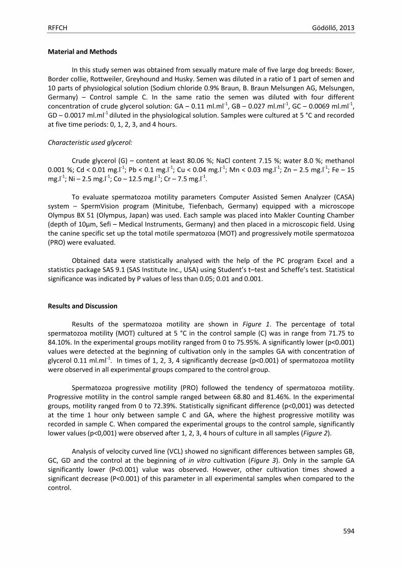
RFFCH Gödöllő, 2013
594
Material and Methods In this study semen was obtained from sexually mature male of five large dog breeds: Boxer,
Border collie, Rottweiler, Greyhound and Husky. Semen was diluted in a ratio of 1 part of semen and 10 parts of physiological solution (Sodium chloride 0.9% Braun, B. Braun Melsungen AG, Melsungen, Germany) – Control sample C. In the same ratio the semen was diluted with four different concentration of crude glycerol solution: GA – 0.11 ml.ml-1, GB – 0.027 ml.ml-1, GC – 0.0069 ml.ml-1, GD – 0.0017 ml.ml-1 diluted in the physiological solution. Samples were cultured at 5 °C and recorded at five time periods: 0, 1, 2, 3, and 4 hours. Characteristic used glycerol:
Crude glycerol (G) – content at least 80.06 %; NaCl content 7.15 %; water 8.0 %; methanol
0.001 %; Cd < 0.01 mg.l-1; Pb < 0.1 mg.l-1; Cu < 0.04 mg.l-1; Mn < 0.03 mg.l-1; Zn – 2.5 mg.l-1; Fe – 15 mg.l-1; Ni – 2.5 mg.l-1; Co – 12.5 mg.l-1; Cr – 7.5 mg.l-1.
To evaluate spermatozoa motility parameters Computer Assisted Semen Analyzer (CASA)
system – SpermVision program (Minitube, Tiefenbach, Germany) equipped with a microscope Olympus BX 51 (Olympus, Japan) was used. Each sample was placed into Makler Counting Chamber (depth of 10μm, Sefi – Medical Instruments, Germany) and then placed in a microscopic field. Using the canine specific set up the total motile spermatozoa (MOT) and progressively motile spermatozoa (PRO) were evaluated.
Obtained data were statistically analysed with the help of the PC program Excel and a
statistics package SAS 9.1 (SAS Institute Inc., USA) using Student’s t–test and Scheffe’s test. Statistical significance was indicated by P values of less than 0.05; 0.01 and 0.001. Results and Discussion
Results of the spermatozoa motility are shown in Figure 1. The percentage of total
spermatozoa motility (MOT) cultured at 5 °C in the control sample (C) was in range from 71.75 to 84.10%. In the experimental groups motility ranged from 0 to 75.95%. A significantly lower (p<0.001) values were detected at the beginning of cultivation only in the samples GA with concentration of glycerol 0.11 ml.ml-1. In times of 1, 2, 3, 4 significantly decrease (p<0.001) of spermatozoa motility were observed in all experimental groups compared to the control group.
Spermatozoa progressive motility (PRO) followed the tendency of spermatozoa motility.
Progressive motility in the control sample ranged between 68.80 and 81.46%. In the experimental groups, motility ranged from 0 to 72.39%. Statistically significant difference (p<0,001) was detected at the time 1 hour only between sample C and GA, where the highest progressive motility was recorded in sample C. When compared the experimental groups to the control sample, significantly lower values (p<0,001) were observed after 1, 2, 3, 4 hours of culture in all samples (Figure 2).
Analysis of velocity curved line (VCL) showed no significant differences between samples GB,
GC, GD and the control at the beginning of in vitro cultivation (Figure 3). Only in the sample GA significantly lower (P<0.001) value was observed. However, other cultivation times showed a significant decrease (P<0.001) of this parameter in all experimental samples when compared to the control.
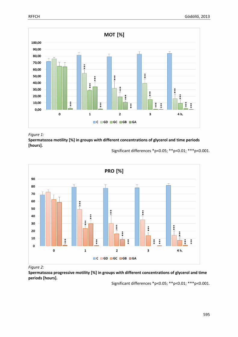
RFFCH Gödöllő, 2013
595
Figure 1: Spermatozoa motility [%] in groups with different concentrations of glycerol and time periods [hours].
Significant differences *p<0.05; **p<0.01; ***p<0.001.
Figure 2: Spermatozoa progressive motility [%] in groups with different concentrations of glycerol and time periods [hours].
Significant differences *p<0.05; **p<0.01; ***p<0.001.
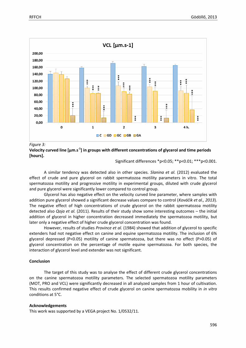
RFFCH Gödöllő, 2013
596
Figure 3: Velocity curved line [μm.s-1] in groups with different concentrations of glycerol and time periods [hours].
Significant differences *p<0.05; **p<0.01; ***p<0.001.
A similar tendency was detected also in other species. Slanina et al. (2012) evaluated the effect of crude and pure glycerol on rabbit spermatozoa motility parameters in vitro. The total spermatozoa motility and progressive motility in experimental groups, diluted with crude glycerol and pure glycerol were significantly lower compared to control group.
Glycerol has also negative effect on the velocity curved line parameter, where samples with addition pure glycerol showed a significant decrease values compare to control (Kováčik et al., 2013). The negative effect of high concentrations of crude glycerol on the rabbit spermatozoa motility detected also Qoja et al. (2011). Results of their study show some interesting outcomes – the initial addition of glycerol in higher concentration decreased immediately the spermatozoa motility, but later only a negative effect of higher crude glycerol concentration was found.
However, results of studies Province et al. (1984) showed that addition of glycerol to specific extenders had not negative effect on canine and equine spermatozoa motility. The inclusion of 6% glycerol depressed (P<0.05) motility of canine spermatozoa, but there was no effect (P>0.05) of glycerol concentration on the percentage of motile equine spermatozoa. For both species, the interaction of glycerol level and extender was not significant. Conclusion
The target of this study was to analyse the effect of different crude glycerol concentrations
on the canine spermatozoa motility parameters. The selected spermatozoa motility parameters (MOT, PRO and VCL) were significantly decreased in all analyzed samples from 1 hour of cultivation. This results confirmed negative effect of crude glycerol on canine spermatozoa mobility in in vitro conditions at 5°C. Acknowledgements This work was supported by a VEGA project No. 1/0532/11.
RFFCH Gödöllő, 2013
597
References Curry, M.R. (2000): Cryopreservation of semen from domestic livestock. Reproduction, 5:46-52. England, G.C (1993): Cryopreservation of dog semen: a review. Journal of Reproduction and fertility,
Supplement. 47:243-255. Holt, W.V. (2000): Fundamental aspects of sperm cryobiology: The importance of species and individual
differences. Theriogenology, 53:47-58. Kováčik, A., Kichi, L., Slanina, T., Massányi, P., Kročková, J., Bulla, J., Čupka, P. (2013): Use of Computer Assisted
Sperm Analysis (CASA) for Monitoring the Effects of Glycerol Solutions on Rabbit Spermatozoa Motility. Scientific papers Animal science and biotechnologies, 46.
Province, C.A., Amann, R.P., Pickett, B.W, Squiers, E.L. (1984): Extenders for preservation of canine and equine spermatozoa at 5°C. Theriogenology, 22:409-415.
Qoja, A.O., Rafajová, M., Massányi, P. The effect of crude glycerol on rabbit spermatozoa motility in vitro. Animal Physiology 2011 : proceedings of scientific publication, Nitra 2011, 457–462.
Schäfer-Somi, S., Kluger, S., Knapp, E., Klein, D. and Aurich, Ch. (2006): Effects of semen extender and semen processing on motility and viability of frozen-thawed dog spermatozoa. Theriogenology, 66: 173-182.
Silva, A.R., Cardoso, R.C.S., Uchoa, D.C. and Silva, L.D.M. (2003): Quality of canine semen submitted to single or fractionated glycerol addition during the freezing process. Theriogenology, 59: 821-829.
Slanina, T., Bíziková, K., Massányi, P. (2012): The effect of glycerol on rabbit spermatozoa motility. Animal Physiology 2012 : proceedings of international conference, Lednice 2012, 187-191.
Squires, E.L., Keith, S.L. and Graham J.K. (2004): Evaluation of alternative cryoprotectants for preserving stallion spermatozoa. Theriogenology, 62:1056–65.
[Ide írhatja a szöveget]
Animal welfare, etológia és tartástechnológia
Animal welfare, ethology and housing systems
Volume 9 Issue 3
Különszám/Special Issue
Gödöllő
2013
RFFCH Gödöllő, 2013
598
THE EFFECT OF BISPHENOL A ON SHEEP LACTATE DEHYDROGENASE ENZYME SYSTEM IN VITRO Irena ŠUTIAKOVÁ*1, Natália KOVALKOVIČOVÁ
2, Drahomíra SOPKOVÁ2, Zuzana KRAVCOVÁ2, Mária TULENKOVÁ
1.
1University of Prešov, Ul. 17. Novembra 1, 081 16 Prešov, Slovak Republic; e-mail: [email protected] 2University of Veterinary Medicine and Pharmacy, Komenského 73, 041 81 Košice, Slovak Republic; e-mail: [email protected] *Corresponding author: Assoc. prof. RNDr. Irena ŠUTIAKOVÁ, PhD., Department of Biology, Faculty of Humanities & Natural Sciences, University of Prešov, St.17. Novembra 1, 081 16 Prešov, Slovak Republic, phone number: 098804744, e-mail: [email protected]
ABSTRACT Lactate dehydrogenase (LDH) and its isoenzymes should serve both as the indicators of cell membrane integrity, as well as cell viability one, in vitro. Our experiments and observations of the cytotoxic effects of bisphenol A (BPA) in the culture medium of sheep (48 treatment time) were carried out at the concentrations of 1x10⁻⁴, 1x10⁻⁵, 1x10⁻⁶ and 1x10⁻⁷ mol.L-1. LDH and its isozymes of the culture medium were used as an endpoint for cytotoxicity studies. The Anova tested data of our experiments showed no significant differences in the both total activity of LDH (µkat.L─1) and also its isoenzymes (µkat.L─1) in the culture medium of our control and experimental group after 48 exposure to BPA. Our results showed that LDH should serve as potential marker of cell injury. Keywords: in vitro, culture medium, sheep, lactate dehydrogenase, bisphenol A, cytotoxicity Bisphenol A (BPA), 2, 2,-bis (4-hydroxyphenyl) propane, is an industrial chemical that is made by combining acetone and phenol. It is extensively used as a monomer in the production of polycarbonate plastics and as a precursor of epoxy resins. Polycarbonate (PC) is widely used in the manufacture of food containers (e.g., milk, water and infant bottles) and epoxy resins are used as an interior protective lining for food and beverage cans (Aschberger et al., 2010). BPA is studied mainly in relation to human and animal health. The presence of BPA in the environment can cause serious health problems (endocrine disruptions, neurotoxic, genotoxic and other problems) (Wu et al., 2013; Renz et al., 2013; Loganathan and Kannan, 2011). Yan et al., (2013) investigated the acute effects of BPA on ventricular arrhythmias and infarction in rat hearts. Authors stated that in addition to endogenous pathophysiological conditions, have also important role the environmental chemicals in these diseases. There are different methodological approaches in toxicological research to identify hazards and to verify the processes which influence the expression of response of living organism to pesticides, different xenobiotics and food-associated chemicals (Eisenbrand et al., 2002). The damages caused by xenobiotics depend on their bioavailability, metabolism and on other factors (e.g. as is tissue turn-over rate etc.). At low and intermediate concentration of xenobiotics they may impair specific functions. In contrast at high concentration of agents they cause general cytotoxicity and cell death (Camatini et al., 1996). In vitro toxicological studies are very important and relevant endpoints. These endpoints have been established in many cell types which determine, for example the membrane permeability changes. Intracellular lactate dehydrogenase (EC. 1.1.1.27), L-(+)-lactate: NAD+ oxidoreductase (LDH) leakage is a well known indicator of cell membrane integrity and cell viability (Legrand et al., 1992). LDH catalyses the intraconversion of pyruvate and lactate and is involved in both the catabolism and anabolism of carbohydrates. In addition to metabolic roles in cells includes in a number of other biological processes (Powers et al., 1991). In animals LDH was found to be a tetrameric molecule, which exists in five different isoenzymes LDH 1 – LDH 5. There are at least two gene duplication events which have led to three genetically subunits. LDH-A or M
RFFCH Gödöllő, 2013
599
(muscle)-type, LDH-B or H (heart)-type and LDH-C or X-type are known in vertebrates and each species has different physico-chemical properties, biological function and development regulations (Kopperschläger and Kirchberger, 1996). The present studies comprised in vitro analyses of the potential cytotoxic effect of BPA in the blood of sheep on the basis of LDH and isoenzymes, released into culture medium. MATERIALS AND METHODS Chemicals – culture medium BPA, synonyms: [2, 2-Bis (4-hydroxyphenyl) propane], 4, 4′-Isopropylidene-diphenol, with formula (CH3)2C(C6H4OH)2, molecular weight 228.29, CAS No. 80-05-7 (Sigma-Aldrich, USA). BPA was dissolved in dimethylsulfoxide (DMSO) and was freshly prepared before each experiment at concentrations of: 1x10⁻⁴, 1x10⁻⁵, 1x10⁻⁶ and 1x10⁻⁷ mol.L-1. The final concentration of DMSO in the cultures was 0.5%. The tested substances were added at 24 h after culture initiation and left until the end of cultivation (48 h treatment time). Cytochalasin-B (Cyt-B, CAS No. 14930-96-2), mytomycin C (MMC, CAS No. 50-07-7), DMSO, (CAS No. 67-68-5), and penicillin G (benzyl penicillin sodium salt; CAS No. 200-710-2) were obtained from Sigma, USA. Streptomycin (streptomycin sulphate salt; CAS No. 3810-74-0) was obtained from Sigma-Aldrich, USA. Cyt-B was dissolved in DMSO. MMC at a final concentration of 0.25 μg.mL-1 was dissolved in distilled water and served as the positive control to ensure the validity of the assay. Chromosome medium serum-free Panserine 701, CAS No. P0-710701 M, and 7.5% NaHCO3 were obtained from PAN Systems GmbH, Biotechnologische Producte, Germany. Heparin (heparinum natricum) was obtained from Zentiva, a. s., Czech Republic. Assay of Total LDH activity The concentration of total LDH (µkat.L─1) activity was assessed using commercial diagnostic kits (Randox, United Kingdom) with automatic biochemical analyser Alizé (Lisabio, France). Before determination we mixed 300 µl of reagents (phosphate buffer with concentration 50 mmol.L-1 and pH 7.5; pyruvate with concetration 0.6 mmol.L-1; NADH with concentration 0.18 mmol.L-1) with 5 µl of sample and measuring absorbance at 340 nm. LDH isoenzyme activity For electrophoretic study, 10 µl of culture supernatant was used for each separation. Hydrasys device (Sebia, France) was used for the determination of LDH isoenzymes activity (µkat. L─1). The samples were separated using commercial electrophoretic kits Hydragel 7 ISO-LDH (Ecomed, Žilina) on alkaline buffered (pH 8.4) agarose gels. Each gel contained agarose (0.8g/dL) and alkaline buffer (pH 8.40±0.05). Substrate contained phenazine methosulfate (PMS), nicotinamide adenine dinucleotide (NAD) and nitro blue tetrazolium (NBT). Blocking solution contained acetic acid (5%) and citric acid (0.5%). For determination apply 10 µl of serum into the applicators, and then place the applicators into the wet storage chamber for 5 minutes. Put the gel plate into the device and start the migration at 20 °C for 8 minutes. Apply substrate solution (45 °C/ 20 minutes) and cool (20 °C/ 5 minutes). Remove the remaining substrate solution and pipette the blocking solution with incubation at 20 °C for 10 minutes. Remove the blocking solution and apply one thick filter paper and dry. The dried gels were prepared for visual examination and densitometry to obtain accurate relative quantification of individual zones. Then photographs of the gels were taken. Qualitative evaluations of the gels were done directly from the electrophoretograms and the densitometric curves of the separations were created by means of Epson Perfection V 700 Photo densitometer scanning at 570 nm. The visualization of LDH isoenzymes was performed according to the following reactions: LDH Lactate + NAD —> Pyruvate + NADH2
NADH2 + PMS —> Reduced PMS + NAD Reduced PMS + NBT —> PMS + Reduced NBT (precipitate of Formosan-blue) The amount of resulting Formosan precipitate was proportional to the LDH enzymatic activity.
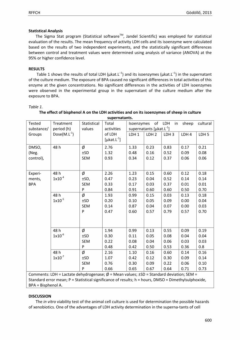
RFFCH Gödöllő, 2013
600
Statistical Analysis The Sigma Stat program (Statistical softwareTM, Jandel Scientific) was employed for statistical evaluation of the results. The mean frequency of activity LDH cells and its isoenzyme were calculated based on the results of two independent experiments, and the statistically significant differences between control and treatment values were determined using analysis of variance (ANOVA) at the 95% or higher confidence level. RESULTS Table 1 shows the results of total LDH (µkat.L─1) and its isoenzymes (µkat.L─1) in the supernatant of the culture medium. The exposure of BPA caused no significant differences in total activities of this enzyme at the given concentrations. No significant differences in the activities of LDH isoenzymes were observed in the experimental group in the supernatant of the culture medium after the exposure to BPA. Table 1.
The effect of bisphenol A on the LDH activities and on its isoenzymes of sheep in culture supernatants.
Tested substance/ Groups
Treatment period (h) Dose(M.L-1)
Statistical values
Total activities of LDH [μkat.L-1]
Isoenzymes of LDH in sheep cultural supernatants [μkat.L-1]
LDH 1 LDH 2 LDH 3 LDH 4 LDH 5
DMSO, (Neg. control),
48 h Ø ±SD SEM
2.76 1.32 0.93
1.33 0.48 0.34
0.23 0.16 0.12
0.83 0.52 0.37
0.17 0.09 0.06
0.21 0.08 0.06
Experi- ments, BPA
48 h 1x10⁻⁴
Ø ±SD, SEM P
2.26 0.47 0.33 0.84
1.23 0.23 0.17 0.91
0.15 0.04 0.03 0.60
0.60 0.52 0.37 0.60
0.12 0.14 0.01 0.50
0.18 0.14 0.01 0.70
48 h 1x10⁻⁵
Ø ±SD SEM P
1.93 0.20 0.14 0.47
0.99 0.10 0.87 0.60
0.15 0.05 0.04 0.57
0.03 0.09 0.07 0.79
0.13 0.00 0.00 0.57
0.18 0.04 0.03 0.70
48 h 1x10⁻⁶
Ø ±SD SEM P
1.94 0.30 0.22 0.48
0.99 0.11 0.08 0.42
0.13 0.05 0.04 0.50
0.55 0.08 0.06 0.53
0.09 0.04 0.03 0.36
0.19 0.04 0.03 0.8
48 h 1x10⁻⁷
Ø ±SD SEM P
2.16 1.07 0.76 0.66
1.10 0.42 0.30 0.65
0.16 0.12 0.09 0.67
0.60 0.30 0.22 0.64
0.14 0.09 0.06 0.71
0.16 0.14 0.10 0.73
Comments: LDH = Lactate dehydrogenase; Ø = Mean values; ±SD = Standard deviation; SEM = Standard error mean; P = Statistical significance of results; h = hours, DMSO = Dimethylsulphoxide, BPA = Bisphenol A.
DISCUSSION The in vitro viability test of the animal cell culture is used for determination the possible hazards of xenobiotics. One of the advantages of LDH activity determination in the superna-tants of cell
RFFCH Gödöllő, 2013
601
cultures is based on the fact that the examination reveals changes in cell viability and not in cell metabolism (Racher et al., 1990). Our results suggest no significant release of this enzyme from the ovine blood cells into the culture medium. The assumptions are made that intracellular enzymes are only released after damage to the cell membrane and all activity is released from damaged cells into culture medium (Goergen et al., 1993). We observed that at the concentrations of BPA decreased total activity of LDH. But on the other hand there are also other parameters that may influence the intracellular activity of LDH from other cells, as well as the pH, chemical composition of the medium and some other factors (Geaguey et al., 1990). Kending and Tarloff, (2007) used LDH as an endpoint for cytotoxicity studies induced by several chemicals. Their studies suggest that LDH is released to the culture medium, and is susceptible to degradation when reactive chemicals are present. In our case, in the culture media were present such chemicals as is cytochalasin and also antibiotics. We simultaneously studied also the cytotoxic effect of BPA by calculating nuclear division index. In the case of nuclear division index BPA did not affect also cytotoxic index at all treatment concentrations (Šutiaková et al., 2013). It is known that LDH is a highly sensitive but not specific marker. The function of specific indicators is performed by LDH isoenzymes, which allow us to identify the damage either to cells and tissues by different xenobiotics. The examination intracellular isoenzyme patterns allow also detect the interspecies cross-contamina-tion in culture medium (Steube et al., 1995; Nims et al., 1998). However our results did not record significant differences between control and experimental groups at the different concentrations of bisphenol A in the medium. Biochemical and molecular parameters that are specific and sensitive may be useful for identifying their influences in biosystems through certain biomarkers also in situations when they are not statistically significant (Anderson and Barton, 1998). Further studies are necessary in this research because the presence of such xenobiotics as is BPA may affect the health and production of animals and health of human. Acknowledgments This research was supported by VEGA Grant No. 1/0287/11, ( Slovakia). REFERENCES ANDERSEN M.E., BARTON, H.A. 1998. The use of biochemical and molecular parameters to estimate dose-
response relationships at low levels of exposure. In Environ. Health Perspect., Suppl. 1, 1998, p. 349-355. ASCHBERGER, A., CASTELLO, P., HOEKSTRA, E., KARAKITSIOS, S., MUNN, S., PAKALIN, S., SARAGIANNIS, D.:
Bisphenol A and baby bottles: challenges and perspectives. JRC Scientific and Technical Reports, European Commission, EUR 24389 EN- Joint Research Centre, Institute for Health and Consumer Protection, 2010, 55p. webuhttp://publications.jrc.ec.europa.eu/repository/bitstream/111111111/14221/1/eur%2024389_bpa%20%20baby%20bottles_chall%20%20persp%20%282%29.pdf
CAMATINI, M., COLOMBO, A., BONFANTI, P., DOLDI, M., URANI, C., DIBISCEGLIA, M., NAGELKERKE, J.F. 1996. In vitro biological systems as models to evaluate the toxicity of pesticides. In Int. J. Environ. Anal. Chem., Vol. 65, 1996, p. 153-167.
EISENBRAND, G., POOL-ZOBEL, B., BAKER, V., BALLS, M., BLAAUBOER, B.J., BOOIS, A., CARERE, A., KEVEKORDES, S., LHUGUENOT, J.-C., PIETERS, R., KLEINE, J., MARTIAL, A., MARC, A., WUDTKE, M., KRETZMER, G., R., J. 2002. Methods of in vitro toxicology. In Food Chem. Toxicol., Vol. 40, 2002, p. 193-236.
GEAUGEY, V., PASCAL, F., ENGASSER, J.M., MARC, A. 1990. Influence of the culture oxygenation on the release of LDH by hybridoma cells. In Biotechnol. Techn., Vol. 4, p. 257 – 262.
GOERGEN, J.L. MARC A., ENGASSER, J.M. 1993. Determination of cell lysis and death kinetics in continuous hybridoma cultures from the measurement of lactate dehydrogenase release. In Cytotechnol., Vol. 11, 1993, p. 189-195.
KENDING, D.M., TARLOFF, J.B.: 2007. Inactivation of lactate dehydrogenase by several chemicals: Implications for in vitro toxicology studies. In Toxicol. in Vitro, Vol. 21, 2007, p. 125-132.
KOPPERSCHLÄGER, G., KIRCHBERGER, J. 1996. Methods for the separation of lactate dehydrogenase and clinical significance of the enzyme. In J. Chromat., B, Vol. 684, 1996, p. 25- 49.
RFFCH Gödöllő, 2013
602
LEGRAND, C., BOUR, J.M., JACOB, C., CAPIAUMONT, J., MARTIAL, A., MARC, A., WUDTKE, M., KRETZMER, G., DEMANGEL, C., DUVAL, D., HACHE, J. 1992. Lactate dehydrogenase (LDH) activity of the number of dead cells in the medium of cultured eukaryotic cells as marker. In J. Biotechnol., Vol. 25, 1992, p. 231- 243.
LOGANATHAN, S.N., KANNAN, K. 2011. Occurrence of bisphenol A in indoor dust from two locations in the Eastern United States and implications for human exposures. In Arch. Environ. Contam. Toxicol., Vol. 61, 2011, p. 68-73.
NIMS, R.W., SHOEMAKER, A.P., BAUERNSCHUB, M.A., REC, L.J., HARBELL, J.W. 1998. Sensitivity o isoenzyme analysis for the detection of interspecies cell line cross-contamination. In In vitro Cell De. Biol. Animal, Vol. 34, 1998, p. 35-39.
POWERS, D.A., LAUERMAN, T., CRAWFORD, D., DIMICHELE, L. 1991. Genetic mechanisms for adapting to a changing environment. In Ann. Rev. Genet., Vol. 25, 1991, 629-59.
RACHER, A.J., LOOBY, D., GRIFITHS, J.B. 1990. Use of lactate dehydrogenase release to assess changes in culture viability. In Cytotechnol., Vol. 3, 1990, p. 301-307.
RENZ, L., VOLZ, C., MICHANOWICZ, D., FERRAR, K., CHRISTIAN, CH., LENZNER, D., EL-HEFMAWY, T. 2013. A study of parabens and bisphenol A in surface water and fish brain tissue from the greater Pittsburgh area. In Ecotoxicol., Vol. 22, 2013, p. 632-641.
STEUBE, K.S., GRUNICKE, D., DREXLER, H.G. 1995. Isoenzyme analysis as a rapid method for the examination of the species identity of cell cultures. In In Vitro Cell. Dev. Biol., Vol. 31, 1995, p. 115-119.
ŠUTIAKOVÁ, I., KOVALKOVIČOVÁ, N., TULENKOVÁ, M. 2013. In vitro evaluation of the bisphenol genotoxicity in bovine peripheral lymphocytes. In 33
rd International Symposium „Industrial Toxicology 2013”, June 19-21.
Accepted. YAN, S., SONG, W., CHEN, Y., HONG, K., RUBINSTEIN, J., WANG, H-S. 2013. Low-dose bisphenol A and
estrogen increase ventricular arrhythmias following ischemia-reperfusion in female rat hearts. In Food Chem. Toxicol., Vol. 56, 2013, p. 75-80.
WU, H-J., LIU, CH., DUAN, W-X., XU, S-CH., HE, M-D., CHEN, CH-H., WANG, Y., ZHOU, Z., YU, Z-P., ZHANG, L., CHEN, Y. 2013. Melatonin ameliorates bisphenol A-induced DNA damage in the germ cells of adult male rats. In Mutat. Res./ Genetic Toxicol. Environ. Mutagen., Vol. 752, 1-2, 2013, p. 57-67.

Animal welfare, etológia és tartástechnológia
Animal welfare, ethology and housing systems
Volume 9 Issue 3
Különszám/Special Issue
Gödöllő
2013
RFFCH Gödöllő, 2013
603
The immunological effect of heat stress in laying hens with use of apple cider vinegar, betaine and their combination in drinking water
Szabó, Cs.1*, Kerti, A1., Pajor, F. 2, Póti, P.2, Bárdos, L.1
1Department of Animal Physiology and Health, Szent István University, Gödöllő, Hungary 2Institute of Animal Husbandry, Szent István University, Gödöllő, Hungary
*[email protected] Egg production and some blood parameters were investigated during high temperature in laying hens. Tetra-SL hens were kept in four groups (15 animals in each) for 47 days in a hot summer period. For the prevention of the possible heat stress additives were added to the water. The Group A was treated with apple cider vinegar (1 L 5% vinegar/100 L drinking water). The Group B with betaine (10 g/10 L drinking water) and both additives were mixed into the water of third the Group AB. The fourth group received only tap water and served as control (Group K). The environmental and pen temperature was registered. Before, during and after the heat stress period (the temperature was 15-20 oC higher than the technological adequate for a week) the number of egg laid, and some blood value (packed cell volume, PCV and titre of immunoglobulin IgY) were determined.
PCV (L / L) values decreased in all treatment groups compared to blood samples before the heat stress period. After statistical analysis, only the control group (before HS 28.4% after 25.7% HS) and apple cider vinegar-betain in the case of treatment (before HS 28.8%, 24.4% after HS) had significantly decreasing value. The hens responded to increased water consumption in the warm environment for thermoregulation. This is caused moderate hemodilution.
The 10 days of extreme heat causes decreased in all groups of IgY titers. Differences in
samples taken before and after heat stress titers in betaine group had the smallest. However, significance was detected only in group AB (0.147 OD before HS after HS OD 0.127). The vinegar and betaine have no synergism in our case. The Tetra-SL hybrid can tolerate the heat stress conditions which characterised 15-20 oC higher values than the technological adequate. Key words: heat stress, laying hen, IgY, immune status, apple cider vinegar, betain, PCV, ELISA Acknowledgement The authors thank the support of Research Centre of Excellence- 17586-4/2013/TUDPOL and the project KMR_12-A 2012-0181

Animal welfare, etológia és tartástechnológia
Animal welfare, ethology and housing systems
Volume 9 Issue 3
Különszám/Special Issue
Gödöllő
2013
RFFCH Gödöllő, 2013
604
CHANGES OF BLOOD PARAMETERS ASSOCIATED WITH NICKEL ADMINISTRATION IN RATS
Toman Robert1*, Lukáč Norbert2, Massányi Peter2, Hajková Zuzana1
1Slovak University of Agriculture, Department of Veterinary Disciplines, Tr. A. Hlinku 2, 94976 Nitra, Slovakia 2Slovak University of Agriculture, Department of Animal Physiology, Tr. A. Hlinku 2, 94976 Nitra, Slovakia *[email protected]
Abstract The aim of this work was to analyze the biochemical parameters of blood plasma in rats after
intraperitoneal and peroral administration of nickel. Experimental group E1 was administered by nickel at a dose of 15 mg/kg, group E2 with 25 mg/kg and the group E3 with 35 mg/kg b.w. intraperitoneally (i.p.). After 48 hours, the blood samples were collected for biochemical analysis. Group E4 was dosed with nickel at a concentration of 100 mg/L in drinking water for 90 days. The parameters of mineral profile (Ca, P, Mg, Na. K and chlorides) and other parameters of energy, nitrogen and enzymatic profile (glucose, cholesterol, total proteins, triglycerides, urea, bilirubine, AST, ALT, ALP, GLDH) were measured. The most affected elements were potassium and calcium. Calcium concentration decreased significantly in group E1, E3 (P<0.05) and E4 (P<0.01). Concentrations of potassium decreased significantly in groups E1, E2 and E3 (P<0.05) and E4 (P<0.01). Mg concentration decreased in group E2 and E4 (P<0.05) and chlorides decreased only in i.p. groups E1 (P<0.05) and E3 (P<0.01). Analysis of nitrogen and energy profile showed a significant increase in concentrations of urea (group E1, P<0.05 and group E3, P<0.01), glucose (groups E2, E3, E4, P<0.05), and decrease in total proteins (group E1, P<0.01 and group E4, P<0.01), cholesterol (groups E2 and E4, P<0.05), bilirubine (all experimental groups, P<0.05) and triglycerides (group E1, E3, P<0.05 and group E2, P<0.01). Changes in enzymatic activity were as follows: activity increased in AST (group E3, P<0.05), ALT (group E1, P<0.05 and group E3, P<0.01), ALP (group E1, P<0.01 and groups E2, E3, E4, P<0.05), and GLDH (group E4, P<0.05). The results showed that nickel may have negative effects on the metabolism due to the disruption of certain metabolic processes. Decreased concentrations of mineral elements along with the parameters of energy and nitrogen profile suggest the renal dysfunction. Influence of enzymatic activity is sign of hepatocellular injury and liver functions. Nickel is capable to affect the metabolic processes and organ functions after an intraperitonal and also peroral exposure.
Keywords: nickel, blood serum, biochemical profile, rat
Introduction Nickel (Ni) is the 24th most abundant element and the earth’s core is composed of 6 % of this metal. It
is used in stainless steel products, nickel plating, to color ceramics, in batteries and other industrial applications (ATSDR, 2005). Anke et al. (1984) discussed the nickel essentiality for living systems and stated that low nickel offers reduce growth particularly during an intra-uterine development. Nickel deficiency is accompanied by histological and biochemical changes and reduced iron resorption and leads to anaemia. Nickel therefore performs a vital function in metabolism. However, the increasing utilization of heavy metals in modern industries leads to an increase in the environmental burden.
Nickel represents a good example of a metal whose use is widening in modern technologies. Its accumulation in the environment may represent a serious hazard to human health. Food is the major source of exposure to nickel but only about 1% is absorbed after the nickel intake in food. A human study shows that 40 more nickel was absorbed from the gastrointestinal tract when NiSO4 (nickel sulphate) was given in the drinking water than when it was given in food (Sunderman et al., 1989). When administered in water, significant elevations in nickel levels were found in the small intestine of mouse after 8 weeks of exposure. In
RFFCH Gödöllő, 2013
605
the kidneys, the nickel levels were only significantly higher than controls after 20 weeks of administration (Radike et al., 2002). Skin allergies, lung fibrosis, kidney diseases, cardiovascular system damage, hematopoiesis disorder and stimulation of neoplastic transformation are the well-known health effects of nickel (Denkhaus and Salnikow, 2002, Oller et al., 2008, Hfaiedh et al., 2008, Adjroud and Mouffok, 2009, Adjroud, 2013).
Nickel compounds have been implicated in the etiology of human lung cancer. They activate hypoxia signaling pathways. The mechanism of this effect involves the ability of either soluble or insoluble nickel compounds to block iron uptake leading to cellular iron depletion or/and directly affect iron containing enzymes. This can lead to activation of hypoxic signaling (Costa et al., 2005). The relative carcinogenic activity of nickel compounds is related to their water solubility (Coogan et al., 1989). One of the major intracellular targets of nickel ions in the cell is the iron- and 2-oxoglutarate-dependent dioxygenase family of enzymes (Chervona et al., 2012).
Nickel chloride also acts on cellular metabolism and in particular on erythrocyte glutathione peroxidase with consequent increase in oxidative stress. A significant decrease in intracellular glutathione in both human (25%) and fish erythrocytes (18%) after treatment with nickel chloride has been reported (De Luca et al., 2007). Alteration in blood biochemistry caused by nickel peroral exposure was found in hens. Associations between nickel and calcium, magnesium, triglycerides and alanine transaminase (ALT) in blood serum were recorded (Kolesarova et al., 2008a). Ni induced liver damage was clearly shown by the increased activities of serum hepatic enzymes along with increased elevation of lipid peroxidation indices. The toxic effect of Ni was also indicated by significantly decreased levels of enzymatic antioxidants superoxide dismutase (SOD), catalase (CAT), glutathione peroxidase (GPx) and glutathione S-transferase (GST) and non-enzymatic antioxidants (glutathione, vitamin C and vitamin E) (Pari and Prasath, 2008). The nickel related changes in liver and also kidney functions were observed in nickel deficient animals. The activity of SDH and ALD, as well as the level of urea-N was significantly lower in the serum of Ni-deficient animals than in the control. Ni-deficient animals also had significantly lower enzyme activities in the heart (succinate dehydrogenase - SDH, hydroxybutyrate dehydrogenase - HBDH, aspartate aminotransferase - AST, ALT, aldolase - ALD and creatine kinase - CK), liver (sorbitol dehydrogenase - SDH and cholinesterase - CHE) and kidneys (HBDH and CK) (Szilágyi et al., 1991). However, Kolesarova et al. (2008b) were no table to find the significant correlations between levels of nickel in internal organs and biochemical parameters.
The aim of this study was to determine the effect of intraperitoneal and peroral nickel exposure on the biochemical parameters in Wistar rats.
Materials and Methods Fifty males Wistar rats were divided to 5 groups: nickel-treated groups (E1, E2, E3, E4) and control group (C), each containing 10 males. The males were housed individually in a plastic cages. The animals had unlimited access to drinking water and feed. The rats in the nickel (NiCl2,, Sigma, MO, USA) exposed groups were intraperitoneally dosed with 15 mgNi/kg b.w. (E1), 25 mgNi/kg b.w. (E2), 35 mgNi/kg b.w. (E3), and perorally at the concentration of 100 mg/L (E4) in drinking water for 90 days. Ten males (group C) served as the untreated control group without nickel treatment. 48 hours after an intraperitoneal administration of nickel (groups E1, E2, E3) and 90 days after the nickel intake in group E4, the rats were euthanized and blood samples were collected by cardiac puncture in glass tubes with heparin for analyses. The blood serum was separated by centrifugation at 3000 rpm for 30 minutes and stored at -18°C. The parameters of mineral profile (calcium, phosphorus, magnesium, sodium. potassium and chlorides) and other parameters of energy, nitrogen and enzymatic profile (glucose, cholesterol, total proteins, triglycerides, urea, bilirubine, aspartate aminotransferase – AST, alanine aminotransferase – ALT, alkaline phosphatase – ALP, glutamate dehydrogenase – GLDH) were measured by automatic analyzer Microlab 300 (Vital Scientific, Dieren, Netherland) and analyzer EasyLite (Medica, Bedford,
RFFCH Gödöllő, 2013
606
USA). The statistical analysis of variance and t-test were calculated using the Microsoft Excel software (Microsoft, USA).
Results and discussion Nickel is known due its effects on haematological parameters leading to anaemia. Also it acts on metabolism and decreases the erythrocyte glutathione peroxidase with consequent increase in oxidative stress in human and fish (De Luca et al., 2007). In this work, the biochemical parameters of blood serum were analyzed. The average concentrations and activities of biochemical parameters measured in rat blood serum are shown in table 1 – 3. The most affected elements were potassium and calcium. Potassium concentration significantly decreased in all experimental groups regardless of way of administration. The most significant decline (P<0.01) was observed after a peroral intake of nickel during 90 days of experiment. Calcium concentrations also significantly decreased in group E1, E3 and E4 and the most significant change was again found in peroral group (2,16 mmol/L compared with the control 3.00 mmol/L). Significant decreases in calcium concentration were observed after nickel chloride administration in hens in dose of 2.0 mg/L of drinking water (Kolesarova et al., 2008a). Hayashi et al. (2003) suggested that the increased excretion level of Ca is the result of renal dysfunction. The similar effect was described after peroral administration of cadmium during 3 months in pheasants (Toman et al., 2006). However, Kalafova et al. (2010) were not able to find the significant changes in the serum concentrations of calcium in rabbits after nickel administration (17.5 and 35 mg/kg) for 30-90 days. On the contrary, Hirano et al. (1994) found increase in calcium concentration in blood serum in rats. Authors exposed the animals with NiSO4 via inhalation. Phosphorus is present in blood serum mainly in form of inorganic phosphate, which is easy utilized in the chemical reactions. In our experiments, slight decrease in P concentrations was observed but the differences between control and experimental groups were not significant. Similarly, the sodium concentrations did not change significantly. Magnesium concentration in blood plasma decreased significantly in group E2 (25 g NiCl2/kg i.p.) and E4 (100 mg NiCl2/L p.o.). In experiment with hens, Kolesarova et al. (2008a) were not able to find any significant changes, even if the Mg concentration decreased. Another significantly affected element in our experiment was potassium. It decreased in all experimental groups significantly. Significant depression of potassium in liver following nickel treatment in rats observed Sidhu et al. (2004). Changes in potassium blood levels may negatively affect the blood pressure as reviewed by Haddy et al. (2006). The higher decrease in K concentration was observed in peroral group (E4), so we can conclude that nickel may be an effective element in potassium decrease in blood after long-term intake. Potassium depletion can affect renal tubular cell function. Chronic potassium depletion can also cause a form of nephropathy known as hypokalaemic nephropathy, in which hypokalaemia itself is believed to cause a proximal tubular lesion associated with interstitial inflammation and fibrosis, leading to a fall in glomerular filtration rate (Walsh et al., 2011). Chlorides play a major role in maintaining the pH levels of the body and also in metabolism to produce energy. Low plasma chloride levels are indicative of a lack of oxygen in the body or respiratory and metabolic acidosis. Hypochloridemia was found as most common chloride concentration change in stroke (Alam et al., 2012). In our experiments, chloride concentrations decreased significantly in group dosed with 15 and 35 mgNi/kg (95.32 and 94.34 mmol/L, respectively) when compared to the control value (100.33 mmol/L). Kidney and liver failure disturbs the metabolism of many compounds like minerals, nutrients and enzymes. It leads to changes in the blood plasma constituents. The changes in plasma mineral elements in our experiments suggest the kidney function failure.
RFFCH Gödöllő, 2013
607
Table 1:
Mineral profile of the control and experimental rats
Element Group (x±SD)
Control (n=10)
E1 – 15 mg/kg (n=10)
E2 – 25 mg/kg (n=10)
E3 – 35 mg/kg (n=10)
E4 – 100 mg/L (n=10)
Ca (mmol/L) 3.00±0.35 2.48±0.28* 3.21±0.47 2.42±0.23* 2.16±0.18**
P (mmol/L) 3.97±0.98 2.94±0.40 2.63±0.19 3.36±0.49 2.70±0.26
Mg (mmol/L) 1.57±0.50 1.25±0.26 0.88±0.23* 1.55±0.08 1.03±0.09*
Na (mmol/L) 140.9±1.60 141.20±2.55 141.29±1.82 141.41±3.69 140.95±2.74
K (mmol/L) 6.72±0.74 5.32±0.82* 5.48±0.61* 5.27±0.26* 4.93±0.45**
Cl- (mmol/L) 100.33±1.74 95.32±3.09* 100.12±4.46 94.34±2.67** 98.76±2.54
x = mean, SD = standard deviation, *= P<0.05, **= P<0.01
A significant increase in urea concentration is evident after an intraperitoneal administration of nickel but not after a peroral exposure (Table 2). The mean concentration of urea increased from the control level 8.05 mmol/L to 24.10 mmol/L in group E3 (P<0.01). Urea is synthesized in the liver from ammonia, which is in turn generated as a break down product of ingested and endogenous protein. It is completely filtered by the kidney, but is also reabsorbed. Tubular reabsorption of urea is increased when tubular flow rates and volumes are decreased. Increases in serum urea may signal the onset of renal disease which has been shown in many other studies (Prabu et al., 2010, Zaki and Mohamed, 2012). Nickel increases the protein catabolism. The total plasma proteins decreased significantly in the lowest-dose intraperitoneally exposed group E1 and in perorally exposed group E4 from the control 77.67 g/L to 69.97 and 65.45 g/L, respectively. Decreases in total plasma proteins are indicative of edema. It causes fluids to leave the circulatory system and localize in tissue spaces. Blood urea concentration reflects the balance between protein catabolism and clearance (Kemp et al., 2001). Effect of nickel on the glucose metabolism is known (Clary, 1975, Chaudhry and Nath, 1984). In our study, the concentration of glucose increased significantly (P<0.05) in two high-dose groups E2 and E3, as well in peroral group E4 form the control 8.33 mmol/L to 9.26; 10.99 and 10.95 mmol/L, respectively. No significant changes in serum glucose concentration were found after nickel administration in fish (Ptashynski et al., 2002), goats (Yousuf, 2002) and hens (Kolesarova et al., 2008a). Total cholesterol and triglycerides showed decrease in mean concentration. Cholesterol decreased in group E2 and E4 from the control 2.80 mmol/L to 1.62 and 1.63 mmol/L, respectively. It means that both intraperitoneal and peroral administration of nickel affected the cholesterol concentration in blood serum. Our results do not correspond with those observed by Das et al. (2006). Authors found increase in total cholesterol and triglycerides after an intraperitoneal administration of nickel sulphate (2.0 mg/100 g b.w.) in rats. Lind et al. (2012) confirmed the relationship between nickel exposure and cholesterol and triglycerides in blood of old humans. Decrease in cholesterol concentrations in broiler chicken after addition of 50 mgNi/kg to feed mixture but no changes in cholesterol and increase in triglycerides concentrations were found in rabbits after the dose of 50 and 500 mgNi/kg in feed (Bersényi et al., 2004). The bilirubine formed from breakdown of red blood cells is transported in plasma bound to albumin, so the decrease in bilirubine level is a result of reduction in the natural breakdown of red blood cells (Ogbe et al., 2012). It therefore seems that nickel might provide protection to oxidative breakdown of red blood cells membranes.
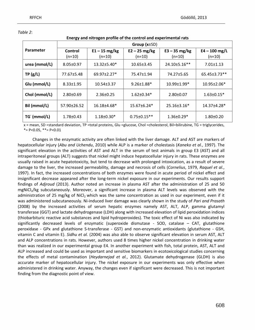
RFFCH Gödöllő, 2013
608
Table 2: Energy and nitrogen profile of the control and experimental rats
Parameter Group (x±SD)
Control (n=10)
E1 – 15 mg/kg (n=10)
E2 – 25 mg/kg (n=10)
E3 – 35 mg/kg (n=10)
E4 – 100 mg/L (n=10)
urea (mmol/L) 8.05±0.97 13.32±5.40* 10.65±3.45 24.10±5.16** 7.01±1.13
TP (g/L) 77.67±5.48 69.97±2.27* 75.47±1.94 74.27±5.65 65.45±3.73**
Glu (mmol/L) 8.33±1.95 10.54±3.37 9.26±1.88* 10.99±1.99* 10.95±2.06*
Chol (mmol/L) 2.80±0.69 2.36±0.25 1.62±0.34* 2.80±0.07 1.63±0.15*
Bil (mmol/L) 57.90±26.52 16.18±4.68* 15.67±6.24* 25.16±3.16* 14.37±4.28*
TG- (mmol/L) 1.78±0.43 1.18±0.30* 0.75±0.15** 1.36±0.29* 1.80±0.20
x = mean, SD =standard deviation, TP =total proteins, Glu =glucose, Chol =cholesterol, Bil=bilirubine, TG = triglycerides,
*= P<0.05, **= P<0.01
Changes in the enzymatic activity are often linked with the liver damage. ALT and AST are markers of hepatocellular injury (Abu and Uchendu, 2010) while ALP is a marker of cholestasis (Kaneko et al., 1997). The significant elevation in the activities of AST and ALT in the serum of test animals in group E3 (AST) and all intraperitoneal groups (ALT) suggests that nickel might induce hepatocellular injury in rats. These enzymes are usually raised in acute hepatotoxicity, but tend to decrease with prolonged intoxication, as a result of severe damage to the liver, the increased permeability, damage and necrosis of cells (Cornelius, 1979, Raquel et al., 1997). In fact, the increased concentrations of both enzymes were found in acute period of nickel effect and insignificant decrease appeared after the long-term nickel exposure in our experiments. Our results support findings of Adjroud (2013). Author noted an increase in plasma AST after the administration of 25 and 50 mgNiCl2/kg subcutaneously. Moreover, a significant increase in plasma ALT levels was observed with the administration of 25 mg/kg of NiCl2 which was the same concentration as used in our experiment, even if it was administered subcutaneously. Ni-induced liver damage was clearly shown in the study of Pari and Prasath (2008) by the increased activities of serum hepatic enzymes namely AST, ALT, ALP, gamma glutamyl transferase (GGT) and lactate dehydrogenase (LDH) along with increased elevation of lipid peroxidation indices (thiobarbituric reactive acid substances and lipid hydroperoxides). The toxic effect of Ni was also indicated by significantly decreased levels of enzymatic (superoxide dismutase - SOD, catalase – CAT, glutathione peroxidase - GPx and glutathione S-transferase - GST) and non-enzymatic antioxidants (glutathione - GSH, vitamin C and vitamin E). Sidhu et al. (2004) was also able to observe significant elevation in serum AST, ALT and ALP concentrations in rats. However, authors used 8 times higher nickel concentration in drinking water than was realized in our experimental group E4. In another experiment with fish, total protein, AST, ALT and ALP increased and could be used as important and sensitive biomarkers in ecotoxicological studies concerning the effects of metal contamination (Heydarnejad et al., 2012). Glutamate dehydrogenase (GLDH) is also accurate marker of hepatocellular injury. The nickel exposure in our experiments was only effective when administered in drinking water. Anyway, the changes even if significant were decreased. This is not important finding from the diagnostic point of view.
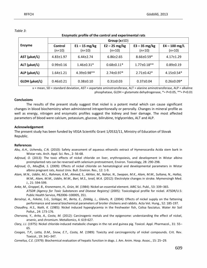
RFFCH Gödöllő, 2013
609
Table 3:
Enzymatic profile of the control and experimental rats
Enzyme Group (x±SD)
Control (n=10)
E1 – 15 mg/kg (n=10)
E2 – 25 mg/kg (n=10)
E3 – 35 mg/kg (n=10)
E4 – 100 mg/L (n=10)
AST (µkat/L) 4.83±1.97 6.44±2.74 6.86±2.65 8.66±0.59* 4.17±1.29
ALT (µkat/L) 0.99±0.16 1.46±0.31* 0.68±0.11* 1.77±0.18** 0.89±0.19
ALP (µkat/L) 1.64±1.21 4.39±0.98** 2.74±0.97* 2.71±0.42* 4.15±0.54*
GLDH (µkat/L) 0.46±0.21 0.38±0.10 0.31±0.03 0.37±0.04 0.26±0.09*
x = mean, SD = standard deviation, AST = aspartate aminotransferase, ALT = alanine aminotransferase, ALP = alkaline
phosphatase, GLDH = glutamate dehydrogenase, *= P<0.05, **= P<0.01
Conclusions The results of the present study suggest that nickel is a potent metal which can cause significant changes in blood biochemistry when administered intraperitoneally or perorally. Changes in mineral profile as well as energy, nitrogen and enzymatic profiles suggest the kidney and liver damage. The most affected parameters of blood were calcium, potassium, glucose, bilirubine, triglycerides, ALT and ALP. Acknowledgement The present study has been funded by VEGA Scientific Grant 1/0532/11, Ministry of Education of Slovak Republic. References Abu, A.H., Uchendu, C.N. (2010): Safety assessment of aqueous ethanolic extract of Hymenocardia Acida stem bark in
Wistar rats. Arch. Appl. Sci. Res., 2: 56-68. Adjroud, O. (2013): The toxic effects of nickel chloride on liver, erythropoiesis, and development in Wistar albino
preimplanted rats can be reversed with selenium pretreatment, Environ. Toxicology, 28: 290-298. Adjroud, O., Mouffok, S. (2009): Effects of nickel chloride on hematological and developmental parameters in Wistar
albino pregnant rats, Assiut Univ. Bull. Environ. Res., 12: 1-9. Alam, M.N., Uddin, M.J., Rahman, K.M., Ahmed, S., Akhter, M., Nahar, N., Swapan, M.K., Alam, M.M., Sultana, N., Hallaz,
M.M., Alam, M.M., Uddin, M.M., Bari, M.S., Israil, M.A. (2012): Electrolyte changes in stroke. Mymensingh Med. J., 21: 594-599.
Anke, M., Groppel, B., Kronemann, H., Grün, M. (1984): Nickel-an essential element. IARC Sci. Publ., 53: 339–365. ATSDR (Agency for Toxic Substances and Disease Registry) (2005): Toxicological profile for nickel. ATSDR/U.S: Public Health Service, PB2006–100005, 351.
Bersényi, A., Fekete, S.G., Szilágyi, M., Berta, E., Zöldág, L., Glávits, R. (2004): Effects of nickel supply on the fattening performance and several biochemical parameters of broiler chickens and rabbits. Acta Vet. Hung., 52: 185-197.
Chaudhry, H.S., Nath, K. (1985): Nickel induced hyperglycemia in the freshwater fish, Colisa fasciatus. Water Air Soil Pollut., 24: 173-176.
Chervona, Y., Arita, A., Costa, M. (2012): Carcinogenic metals and the epigenome: understanding the effect of nickel, arsenic, and chromium. Metallomics, 4: 619-627.
Clary, J.J. (1975): Nickel chloride-induced metabolic changes in the rat and guinea pig. Toxicol. Appl. Pharmacol., 31: 55–65.
Coogan, T.P., Latta, D.M., Snow, E.T., Costa, M. (1989): Toxicity and carcinogenicity of nickel compounds. Crit. Rev. Toxicol., 19: 341–347.
Cornelius, C.E. (1979): Biochemical evaluation of hepatic function in dogs. J. Am. Anim. Hosp. Assoc., 15: 25–29.
RFFCH Gödöllő, 2013
610
Costa, M., Davidson, T.L., Chen, H., Ke, Q., Zhang, P., Yan, Y., Huang, C., Kluz, T. (2005): Nickel carcinogenesis: epigenetics and hypoxia signaling. Mutat. Res., 592: 79-88.
Das, K.K., Gupta, A.D., Dhundasi, S.A., Patil, A.M., Das, S.N., Ambekar, J.G. (2006): Effect of L-ascorbic acid on nickel-induced alterations in serum lipid profiles and liver histopathology in rats. J. Basic. Clin. Physiol. Pharmacol., 17: 29-44.
De Luca, G., Gugliotta, T., Parisi, G., Romano, P., Geraci, A., Romano, O., Scuteri, A., Romano, L. (2007): Effects of nickel on human and fish red blood cells. Biosci. Rep., 27: 265-273.
Denkhaus, E., Salnikow, K. (2002): Nickel essentiality, toxicity, and carcinogenicity. Crit. Rev. Oncol. Hematol., 42: 35–56. Haddy, F.J., Vanhoutte, P.M., Feletou, M. (2006): Role of potassium in regulating blood flow and blood pressure. Am. J.
Physiol. Regul. Integr. Comp. Physiol., 290: 546-552. Hayashi, Y., Kobayashi, E., Okubo, Y., Suwazono, Y., Kido, T., Nogawa, K. (2003): Excretion levels of urinary calcium and
phosphorus among the inhabitants of Cd-polluted Kakehashi River basin of Japan. Biol. Trace Elem. Res., 91: 45-55.
Heydarnejad, M.S., Khosravian-Hemamai, M., Nematollahi, A. (2013): Effects of cadmium at sub-lethal concentration on growth and biochemical parameters in rainbow trout (Oncorhynchus mykiss). Ir. Vet. J., 66: 11.
Hfaiedh, N., Allagui, M.S., Hfaiedh, M., Feki, A.E., Zourgui, L., Croute, F. (2008): Protective effect of cactus (Opuntia ficus indica) cladode extract upon nickel-induced toxicity in rats. Food Chem. Toxicol., 46: 3759–3763.
Hirano, S., Shimada, T., Osugi, J., Kodama, N., Suzuki, K.T. (1994): Pulmonary clearance and inflammatory potency of intratracheally instilled or acutely inhaled nickel sulfate in rats. Arch. Toxicol., 68: 548-554.
Kalafová, A., Kováčik, J., Capcarová, M., Massányi, P., Kolesárová, A., Chrastinová, Ľ., Lukáč, N., Schneidgenová, M., Čupka, P., Jurčík, R. (2010): Effect of nickel and zinc peroral administration on serum calcium, phosphorus and magnesium of rabbit female. Potravinárstvo, 4: 301-307.
Kaneko, J.J., Harvey, J.W., Bruss, M.L. (1997): Clinical biochemistry of domestic animals. 5th Edition, Academic Press, San Diego, 932.
Kemp, H.J., Parnham, A., Tomson, C.R. (2001): Urea kinetic modeling: a measure of dialysis adequacy. Ann. Clin. Biochem., 38: 20-27.
Kolesarova, A., Capcarova, M., Arpasova, H., Kalafova, A., Massanyi, P., Lukac, N., Kovacik, J., Schneidgenova, M. (2008a): Nickel-induced blood biochemistry alterations in hens after an experimental peroral administration. J. Environ. Sci. Health B, 43: 625-632.
Kolesarova, A., Slamecka, J., Jurcik, R., Tataruch, F., Lukac, N., Kovacik, J., Capcarova, M., Valent, M., Massanyi, P. (2008b): Environmental levels of cadmium, lead and mercury in brown hares and their relation to blood metabolic parameters. J. Environ. Sci. Health A, 43: 646-650.
Lind, P.M., Olsén, L., Lind, L. (2012): Circulating levels of metals are related to carotid atherosclerosis in elderly. Sci. Total Environ., 416: 80–88.
Ogbe, R.J., Adenkola, A.Y., Anefu E. (2012): Aqueous ethanolic extract of Mangifera indica stem bark effect on the biochemical and haematological parameters of albino rats. Arch. Appl. Sci. Res., 4: 1618-1622.
Oller, A.R., Kirkpatrick, D.T., Radovsky, A., Bates, H.K. (2008): Inhalation carcinogenicity study with nickel metal powder in Wistar rats. Toxicol. Appl. Pharmacol., 233: 262–275.
Pari, L., Prasath, A. (2008): Efficacy of caffeic acid in preventing nickel induced oxidative damage in liver of rats. Chem. Biol. Interact., 173: 77-83.
Prabu, S.M., Shagirtha, K., Renugadevi, J. (2010): Quercetin in combination with vitamins (C and E) improves oxidative stress adrenal injury in cadmium intoxicated rats. Eur. Rew. Med. Pharmacol. Sci., 14: 903-914.
Ptashynski, M.D., Pedlar, R.M., Evans, R.E., Baron, C.L., Klaverkamp, J.F. (2002): Toxicology of dietary nickel in lake whitefish (Coregonus clupeaformis). Aquat. Toxicol., 58: 229-247.
Radike, M., Warshawsky, D., Caruso, J., Goth-Goldstein, R., Reilman, R., Collins, T., Yaeger, M., Wang, J., Vela, N., Olsen, L., Schneider, J. (2002): Distribution and accumulation of a mixture of arsenic, cadmium, chromium, nickel and vanadium in mouse small intestine, kidneys, pancreas, and femur following oral administration in water or feed. J. Toxicol. Environ. Health A, 65: 2029–2052.
Schlattner, U., Tokarska-Schlattner, M., Wallimann, T. (2006): Mitochondrial creatine kinase in human health and disease. Biochim. Biophys. Acta, 1762: 164 – 180.
Sidhu, P., Garg, M.L., Morgenstern, P., Vogt, J., Butz, T., Dhawan, D.K. (2004): Role of zinc in regulating the levels of hepatic elements following nickel toxicity in rats. Biol. Trace Elem. Res., 102: 161-172.
RFFCH Gödöllő, 2013
611
Sunderman, F.W., Hopfer, S.M., Sweeney, K.R., Marcus, A.H., Most, B.M., Creason, J. (1989): Nickel absorption and kinetics in human volunteers. Proc. Soc. Exp. Biol. Med., 191: 5–11.
Szilágyi, M., Anke, M., Balogh, I. (1991): Effect of nickel deficiency on biochemical variables in serum, liver, heart and kidneys of goats. Acta Vet. Hung., 39: 231-238.
Toman, R., Massányi, P., Zakrzewski, M., Stawarz, R., Hluchý, S., Lukáč, N., Mindek, S., Formicki, G. (2006): Effect of cadmium on blood parameters of pheasants (Phasianum colchicus) after long-term administration in drinking water In: J. Physiol. Pharmacol., 57: 235.
Yousuf, M.B. (2002): Effect of high dietary intake of nickel in the West African dwarf goat. Ghana Jnl. Agric. Sci., 35: 147-151.
Zaki, M.S., Mohamed, M.I. (2012): Some studies in Baraki sheep intoxicated with cadmium. Life Sci. J., 9: 786-790. Zoroddu, M.A., Schinocca, L., Kowalik-Jankowska, T., Kozlowski, H., Salnikow, K., Costa, M. (2002): Molecular mechanisms
in nickel carcinogenesis: modeling Ni(II) binding site in histone H4. Environ. Health Perspect., 110: 719-723. Walsh, S.B., Unwin, E., Vargas-Poussou, R., Houillier, P., Unwin, R. (2011): Does hypokalaemia cause nephropathy? an
observational study of renal function in patients with Bartter or Gitelman syndrome. QJM: Int. J. Med., 104: 939-944.

Animal welfare, etológia és tartástechnológia
Animal welfare, ethology and housing systems
Volume 9 Issue 3
Különszám/Special Issue
Gödöllő
2013
RFFCH Gödöllő, 2013
USING OF RAPID ELECTRONIC TESTS FOR MASTITIS DETECTION
Peter TONGEL1*
1 Animal Production Research Centre, Nitra, Slovakia, *[email protected]
Abstract
Even in the best dairy farms there are many cows with health problems. There are for example mastitis,
laminitis, and etc. Mastitis, or inflammation of the mammary gland, is the most common and the most
expensive disease of dairy cows throughout the world. Because these health problems have a great
influence on economy of milk production each farmer has to know about them. The question is: How can
farmer get to know which cow is ill? In our Institute we are developing under projects: "MLIEKO No.
26220220098" supported by the Operational Program Research and Development funded from the
European Regional Development Fund and project APVV-0632-10 of the Slovak Research and
Development Agency, Bratislava different rapid electronic tests for health disorders of dairy cows. For
detection of mastitis we have developed two rapid electronic mastitis tests. One of them is: SCC counter
(somatic cell counter) which works on the basis of milk viscosity measurement and another one: REM
test (Rapid electronic mastitis test) which works on the basis of milk electrical conductivity
measurement. In the first experiment the accuracy of REM test was tested in laboratory conditions, and
then in practical conditions. The results have shown the accuracy was excellent (1 % from measurement
range). The statistical value of electrical conductivity of healthy cows was 4.8 mS/cm (sd 0.4 mS/cm) and
cows with health disorders was 6.4 mS/cm (sd 0.9 mS/cm). The difference between them was significant
(p<0.01). In the second experiment we tested SCC counter for its accuracy and reliability in practical
conditions as well. Samples of milk were taken during milking. From each sample the control one was
sent to the certificated laboratories for SCC analysis. Total fifty samples have been measured. The
correlation coefficient between SCC from SCC counter and SCC from laboratories was computed (r =
0.96, p< 0.01). It was found out that the SCC counter can measure SCC in a range 150 000 to 3 000 000
with sufficient accuracy. Based on our previous experiments, we can say, that these two electronic
mastitis tests are good tools for mastitis prevention. REM test is suitable for quick monitoring of the
whole herd of dairy cows whereas the SCC counter can serve as quick and cheap equipment for SCC
measurement.
Keywords: Mastitis, SCC measurement, electrical conductivity measurement, rapid electronic tests, dairy cow
612

Animal welfare, etológia és tartástechnológia
Animal welfare, ethology and housing systems
Volume 9 Issue 3
Különszám/Special Issue
Gödöllő
2013
RFFCH Gödöllő, 2013
613
THE EFFECT OF VARIOUS MANGANESE SOURCES ON EGG QUALITY PARAMETERS OF LAYING HENS
Katarína VENGLOVSKÁ1, Klaudia ČOBANOVÁ2, Ľubomíra GREŠÁKOVÁ2
1Institute of Biology and Ecology, Faculty of Science, Pavol Jozef Šafárik University, Moyzesova 11, Košice, 040 01, Slovak Republic 2Institute of Animal Physiology, Slovak Academy of Sciences, Šoltésovej 4-6, Košice, 040 01, Slovak Republic
Abstract This study was conducted to investigate the effect of feed supplementation with different manganese (Mn) sources on performance and egg quality parameters of Lohmann Brown hens. Ninety six 20-weeks-old laying hens were allocated to four dietary treatments. Hens were fed maize-soybean basal diets (BD) which differed only in the form of Mn supplemented. Group 1 were fed unsupplemented BD during the whole experiment. Diets for groups 2, 3 and 4 consisted of BD supplemented with a Mn dose of 30 mg.kg-1 in the form of Mn sulphate, Mn chelate of protein hydrolysate (Mn-Pro) and Mn-glycine chelate (Mn-Gly), respectively. During the experiment performance parameters, eggshell qualitative parameters, concentration of manganese and zinc in egg yolks and oxidative stability of egg yolk lipids were determined. During the entire experiment, neither daily feed intake, egg weight nor feed to egg mass ratio were affected by dietary treatments. Egg production was significantly increased in groups supplemented with Mn-Pro and Mn-Gly. Regardless of source, the Mn supplementation to feed significantly increased the relative eggshell weight and eggshell thickness. The proportion of cracked eggs was significantly decreased in groups fed organic sources of Mn. Intake of BD enriched with Mn-sulphate resulted in significantly lower concentration of malondialdehyde in yolk of eggs compared to group fed Mn-Gly. The storage time significantly worsened the oxidative stability of yolk lipids in the group 1, 3 and 4. Our experiment showed that supplementation of hen´s diet with Mn has positive effect on eggshell quality. Diet supplementation with inorganic form could prevent the oxidative damage of yolk lipids during the storage time.
Introduction For the egg producers are very important to increase egg production, egg weight and
improve quality of eggshell because these features are related with economic profitability of poultry industry. Eggshell quality is one of the most important egg parameters. Eggs of poor shell quality cause significant losses to producers of consumer eggs. Nonstandard eggs usually account for 3 to 12% of the total yield (Jelínek, 1996; Ledvinka et al., 2000). High breaking strength of eggshell and absence of shell defects are essential for protection against the penetration of pathogenic bacteria such as Salmonella sp. into eggs (Swiatkiewicz et al., 2010). Eggshell quality is most often expressed as the proportion of the eggshell in egg weight, and by thickness. It is well known that main biological factors affecting eggshell quality are internal factors such as genetics, hen’s age, production cycle stage, egg weight, intensity and persistence of production, time of laying, combined with external factors such as the nutritional level, farm system and microclimatic parameters. From nutritional point of view is the quality of eggshell often connected to macrominerals (Ca, P) and vitamin D3 influence, but nowadays it is well known that trace elements are also very important in mineralization process. Manganese and zinc, as cofactors of metaloenzymes responsible for carbonate and mucopolysaccharides synthesis, play an important role in eggshell formation (Swiatkiewicz and Koreleski, 2008). Mabe et al. (2003) suggested that trace elements as Mn, Zn and Cu could
RFFCH Gödöllő, 2013
614
affect mechanical properties of eggshell by effect on calcite crystal formation and modifying crystallographic structure of eggshell. Mn may affect eggshell quality, because activates the glycosyl transferases involved in the formation of mucopolysaccharides, which are components of proteoglycans (Leach, 1976). Mn also playes crucial role in antioxidant protection, because it is integral part of Mn-superoxide dismutase (Mn-SOD) (Underwood and Suttle, 1999).
In recent years, studies on the efficacy of organic trace minerals in animals have received increasing attention. Compared with inorganic minerals, organic minerals have several benefits including: protect from unwanted chemical reaction in gastrointestinal tract; easily pass intact through intestine wall; and be absorbed by different routes (Mateos et al., 2005). Regarding the use of organic sources the results of the studies are still inconsistent. Some studies suggested that use of organic sources of Mn substantially affects laying performance and eggshell quality (Yildiz et al., 2011; Sun et al., 2012), whereas others authors claimed that there is no difference between inorganic and organic sources of Mn (Lim and Paik, 2003; Mabe et al., 2003; Swiatkiewicz and Koreleski, 2008).
The objective of this study was to compare the efficacy of dietary organic Mn sources and MnSO4.H2O by investigation their effects on egg quality parameters, laying performance, lipid oxidation of egg yolks during storage and mineral retention in egg yolks of laying hens.
Material and methods A total of 96 hens of laying strain Lohmann Brown, 20 weeks old were randomly allotted
into 4 equal groups according to the dietary treatment. Each group consisted of 6 replicates with 4 hens in each. For three weeks of adaptation period, all experimental groups were fed with a basal diet (BD) based on maize and soybean meal. During next 8 weeks the birds were fed the same BD which differed only in the form of Mn supplemented. Group 1 continued on unsupplemented BD (analyzed Mn content 52 mg.kg-1 DM). Diets for groups 2, 3 and 4 consisted of BD supplemented with a Mn dose of 30 mg.kg-1 in the form of Mn sulphate, Mn chelate of protein hydrolysate (Mn-Pro) and Mn-glycine chelate (Mn-Gly), respectively. The experiment lasted for 11 weeks. During the whole experiment the layers were housed in room with a controlled ventilation and lighting (15hours/day) at 2 hens per cage. During experiment hens had constant access to water and feed. All experimental procedures were in accordance with established standards for the care and use of animals for research purposes.
Birds were individually weighed at the beginning and at the end of the experiment. Egg production, egg weight, number of cracked eggs were recorded daily during the 8 weeks of experimental period. Feed intake (per cage) was registered weekly. Using collected data, basic production parameters (egg mass, feed to egg mass ratio, daily feed intake) were calculated. At 31th week of age three eggs from each replicate (18 eggs/treatment/day), for two consecutive days were collected to determine the egg quality parameters (eggshell weight (g), eggshell thickness (mm), eggshell proportion (%), yolk and albumen weight (g), yolk and albumen proportion (%), eggshape index (%)). Eggs were broken, and the albumens and yolks were separated and weighed. Eggshell weight was measured after washing the interior egg membrane and after drying for 48 hours at 60°C. Eggshell thickness was measured using micrometer (Model 7313, Mitutoyo corp., Japan) as the average of both ends (air cell and sharp end) and at the middle, without the shell membranes. The proportion of eggshell (ES), albumen (A) and yolk (Y) were calculated ((ES or A or Y weight/egg weight)x100). The short and long diameters of the eggs were measured with a micrometer to determine egg shape index.
At the end of experimental period 3 eggs per replicate were collected and yolks were pooled for analysis of Mn and Cu concentration by flame atomic absorption spectrophotometry using AAS Shimadzu (Kyoto, Japan) Model AA-700.
RFFCH Gödöllő, 2013
615
The malondialdehyde concentration in the yolks (3 eggs per replicate, total 18 eggs/treatment) of fresh eggs and eggs stored for 10, 20, 30, 40 days at 4°C was measured by the modified fluorometric method in accordance with Jo and Ahn (1998).
Statistical analysis of the differences between the groups was carried out using one-way analysis of variance with the post hoc Tukey multiple comparison test. Differences were considered significant at P<0.05. All statistical analysis were performed with GraphPad Prism 5.0 software.
Results and discussion The effects of dietary Mn supplementation on laying performance parameters are
presented in Table 1. The Mn supplementation source has not statistically significant effect on the weight gain, feed intake, egg weight and on the feed to egg mass ratio. By contrast, the egg production was significantly increased in laying hens supplemented with organic sources of Mn. Yildiz et al. (2011) reported that body weight gains and the egg weights were significantly enhanced when Mn was used as organic form (Mn-Pro). On the contrary Swiatkiewicz and Koreleski (2008) observed no beneficial effects of organic Mn sources on the laying performance parameters. The National Research Council (1994) estimated the requirement for manganese as 20 mg.kg-1 per hen per day. Maurice and Whisenhut (1980) , and Sazzad et al. (1994) reported no significant differences in egg production, egg weight and feed conversion with increasing manganese level in the diet. Similar results observed also Luo et al. (2003) who reported no effect of supplemental Mn (30-120 mg.kg-1) on egg production in brown layers. However, in this study, hen-day egg production was affected by supplementing 30 mg.kg-1 Mn with both organic forms. The basal diet containing 52 mg.kg-1 Mn provided sufficient amount for the other laying performance parameters.
Table 1
Treatment effects on performance of laying hens
Item Treatment
Basal diet (BD) BD + Mn sulphate BD + Mn-Pro BD + Mn-Gly Weight gain 20-31 wk (g)
186.5 ± 18.31 208.6 ± 43.6 153.9 ± 11.93 151,0 ± 41.91
Feed intake (g/hen/day)
114.2 ± 1.13 116.3 ± 0.98 114.3 ± 0.97 113.6 ± 1.13
Egg production (egg/hen/day)
0.97 ± 0.005a
0.98 ± 0.003ab
0.99 ± 0.004b
0.99 ± 0.003b
Egg weight (g/egg)
59.20 ± 0.41 58.32 ± 0.47 58.62 ± 0.57 59.77 ± 0.49
Feed to egg mass ratio (g feed/g egg)
2.00 ± 0.03 2.04 ± 0.02 1.98 ± 0.03 1.92 ± 0.03
a,bmeans in the same row with different superscripts differ significantly (P ≤ 0.05)
Trace elements may affect eggshell quality by their catalytic properties as key enzymes involved in the process of membrane and formation of eggshells or by interaction directly with the calcite crystals in the formation of eggshells (Mabe et al., 2003). The increased eggshell thickness in our experiment could be the effect of higher Mn dose in the diets compared to control group. In this study, all investigated parameters of the eggshell quality were significantly influenced by Mn intake (Table 2). The eggshell weight and thickness have been significantly elevated in all supplemented groups, regardless of the Mn source. Similar results were found by Mabe et al. (2003) who reported no differences in eggshell proportion and eggshell density between hens fed diet supplemented with inorganic and organic sources of
RFFCH Gödöllő, 2013
616
Mn, Zn and Cu. In contrary Bunesova (1999) and Klecker et al. (2002) found positive effect of partial substitution of inorganic Mn and Zn sources with their organic forms on eggshell weight and eggshell thickness. The proportions of the cracked eggs has appeared to be reduced in laying hens receiving the organic Mn sources than in those fed unsupplemented diet (P < 0.05). Similar results were found by Yildiz et al. (2011). The eggshell proportion was significantly increased in groups supplemented with Mn sulphate and Mn-proteinate.
Table 2
Effects of dietary manganese supplementation on parameters of egg quality
Item Treatment Basal diet (BD) BD + Mn sulphate BD + Mn-Pro BD + Mn-Gly
Egg weight (g) 60.54 ± 0.91 59.32 ± 1.07 61.51 ± 1.36 61.01 ± 0.43
Albumen weight (g) 40.31 ± 0.98 39.06 ± 0.94 40.51 ± 1.11 40.19 ± 0.51
Yolk weight (g) 14.79 ± 0.16 14.39 ± 0.08 14.99 ± 0.21 15.02 ± 0.20
Eggshell weight (g) 5.45 ± 0.06a
5.89 ± 0.13b
6.02 ± 0.12b
5.86 ± 0.05b
Albumen proportion (%) 66.54 ± 0.65 65.80 ± 0.37 65.81 ± 0.39 65.85 ± 0.43
Yolk proportion (%) 24.5 ± 0.49 24.3 ± 0.35 24.4 ± 0.27 24.6 ± 0.42
Eggshell proportion (%) 9.01 ± 0.16a
9.93 ± 0.15b
9.79 ± 0.17b
9.61 ± 0.12ab
Eggshell thickness (mm) 0.36 ± 0.004a
0.38 ± 0.003b
0.39 ± 0.006b
0.38 ± 0.003b
Egg shape index (%) 78.4 ± 0.53 77.7 ± 0.56 77.9 ± 0.50 78.6 ± 0.15
Cracked eggs (%) 3.63 ± 0.66a
2.31 ± 0.46ab
1.35 ± 0.33b
0.78 ± 0.30b
a,bmeans in the same row with different superscripts differ significantly (P ≤ 0.05)
The malondialdehyde (MDA) values of egg yolks, as a marker of lipid peroxidation are presented in Table 3. A moderate increase in the concentration of products of lipid peroxidation measured as MDA in the yolk was observed in all experimental groups during the refrigerated storage of eggs. The MDA values were almost equivalent in all treated groups, although after 40th day of storage the lipid peroxidation in yolk of eggs in group 4 was significantly increased compared to the group supplemented with inorganic form. The information about the dietary Mn supplementation on the oxidative stability of egg yolk lipids are insufficient. We can only suppose that lipid peroxidation in egg yolk may be affected by altered activity of superoxide dismutase. Wawrzykowski and Kankofer (2011) found that the activity of superoxide dismutase in egg yolk did not change during 6 days storage but between 6th and 9th day, it decreased significantly.
Table 3 Malondialdehyde concentration in egg yolks during storage periods (mg/kg yolk)
Storage period
Treatment
Basal diet (BD) BD + Mn sulphate BD + Mn-Pro BD + Mn-Gly
0th
day 0.70 ± 0.05a 0.83 ± 0.06 0.76 ± 0.06
a 0.78 ± 0.04
a
10th
day 0.82 ± 0.04ac
0.99 ± 0.03 0.88 ± 0.05ab
0.95 ± 0.05ab
20th
day
1.07 ± 0.09b 1.01 ± 0.13 1.05 ± 0.06
b 0.88 ± 0.06
a
30th
day
0.95 ± 0.04bc
1.06 ± 0.02 1.08 ± 0.08b 1.10 ± 0.06
b
40th
day
1.03 ± 0.03bcAB
0.96 ± 0.02A 1.03 ± 0.06
bAB 1.13 ± 0.03
bB
a,bmeans in the same column with different superscripts differ significantly (P ≤ 0.05)
A,Bmeans in the same row with different superscripts differ significantly (P ≤ 0.05)
In our experiment the addition of various forms of manganese at the dose of 30 mg/kg into diet did not significantly influenced the deposition of Mn and Zn in egg yolks .

RFFCH Gödöllő, 2013
617
Table 4 Treatment effects on mineral concentration in egg yolks (mg/kg DM)
Item Treatment
Basal diet (BD) BD + Mn sulphate
BD + Mn-Pro BD + Mn-Gly
Manganese 1.65 ± 0.16 1.77 ± 0.08 1.66 ± 0.12 1.73 ± 0.16
Zinc 78.6 ± 2.73 84.4 ± 1.67 83.5 ± 3.69 80.3 ± 1.93
Conclusion Based on this study we concluded that the Mn supplementation with different forms
(organic vs. inorganic) in the diet for laying hens positively affect quality of eggshell. Intake of BD with organic Mn sources positively affect the egg production and decreased the proportion of cracked eggs. Diet supplementation with inorganic form could reduce the negative effect of oxidative damage of yolk lipids during the storage time. References BUNESOVA, A. 1999. Chelated trace minerals (Zn, Mn) in nutrition of hens. Zeszyty Naukowe Przegladu
Ho- dowlanego, 45, 309–317. JELÍNEK, K. 1996. Defective shell – one of the problems in the production of eggs. Czech Journal of
Animal Science, 41, 375–379. JO, C., AHN, D.U. 1998. Fluorimetric analysis of 2-thiobarbituric acid reactive substances in turkey.
Poultry Science, 77, 457–480. KLECKER, D., ZEMAN, L., JELÍNEK, P., BUNESOVA, A. 2002. Effect of manganese and zinc chelates on the
quality of eggs. Acta Universitatis Agriculturae et Silviculturae Mendelianae Brunensis, 50, 59–68. LEACH, R.M., Jr. 1976. Metabolism and function of manganese. Trace Elements in Human Health and
Disease. Pages 235–247 in Vol. II, A. S. Prasad, ed., Academic Press, New York. LEDVINKA, Z., TŮMOVÁ, E., ARENT, E., HOLOUBEK, J., KLESALOVÁ, L. 2000. Egg shell quality in some
white-egged and brown-egged crossbreed combinations of dominant hens. Czech Journal of Animal Science, 45, 285–288.
LIM, H.S., PAIK, I.K. 2003. Effects of supplementary mineral methionine chelates (Zn, Cu, Mn) on the performance and eggshell quality of laying hens. Asian-Australasian Journal of Animal Sciences, 16, 1804-1805.
LUO, X.G., GUO, X.Q., LIU, B., ZHANG, X.C. et al. 2003. Study on an optimal manganese level in practical diet for brown-egg layers. Acta Veterinaria et Zootechnica Sinica, 34, 105–111.
MABE, I., RAPP, C., BAIN, M.M., NYS, Y. 2003. Supplementation of a corn-soybean meal diet with manganese, copper, and zinc from organic or inorganic sources improves eggshell quality in aged laying hens. Poultry Science, 82, 1903-1913.
MATEOS, G.G., LAZARO, R., ASTILLERO, J.R., SERRANO, M.P. 2005. Trace minerals: what text books don’t tell you. Redefining Mineral Nutrition (Taylor-Pickard J and Tuker L eds.). Pages 41-43 in Nottingham University Press. Nottingham.
MAURICE, D.V., WHISENHUT, J.E. 1980. Response of eggshell quality to dietary manganese supplementation. Poultry Science, 59, 1567–1568.
NATIONAL RESEARCH COUNCIL, 1994. Nutrient Requirements of Poultry, 9th rev. edn. National Academy Press, Washington, DC.
SAZZAD, H.M., BERTECHINI, A.G., NOBRE, P.T.C. 1994. Egg production, tissue deposition and mineral metabolism in two strains of commercial layers with various levels of manganese in the diet. Animal Feed Science and Technology, 46, 271–275.
SUN, Q., GUO, Y., LI, J., ZHANG, T., WEN, J. 2012. Effects of methionine hydroxy analog chelated Cu/Mn/Zn on laying performance, egg quality, enzyme activity and mineral retention of laying hens. Journal of Poultry Science, 49, 20-25.
SWIATKIEWICZ, S., KORELESKI J. 2008. The effect of zinc and manganese source in the diet for laying hens on eggshell and bones quality. Veterinarni Medicina, 53, 555-563.
RFFCH Gödöllő, 2013
618
SWIĄTKIEWICZ, S., KORELESKI, J., ARCZEWSKA, A. 2010. Laying performance and eggshell quality in laying hens fed diets supplemented with prebiotics and organic acids, . Czech Journal of Animal Science, 2010, 294–306.
UNDEWOOD, E. J., SUTTLE, N.F. 1999. The mineral nutrition of livestock. 3rd Edition. CABI, Publishing, New York.
WAWRZYKOWSKI, J., KANKOFER M. 2011. Changes in activity during storage and characteristics of superoxide dismutase from hen eggs (Gallus Gallus domesticus). European Food Research Technology, 232, 479-484.
YILDIZ, A.O., CUFADAR, Y., OLGUN, O. 2011. Effects of dietary organic and inorganic manganese supplementation on performance, egg quality and bone mineralisation in laying hens. Revue de Médecine Vétérinaire, 162, 482-488.
Acknowledgement This study was supported by Grant Agency for Science, VEGA of Slovak Republic, Grant No. 2/0045/12.
Animal welfare, etológia és tartástechnológia
Animal welfare, ethology and housing systems
Volume 9 Issue 3
Különszám/Special Issue
Gödöllő
2013
RFFC Gödöllő, 2013
HAZARDOUS METALS AND BIOACTIVE COMPOUNDS IN CRANBERRY FRUITS IN THE FIRST AND THE SECOND HARVEST
Vollmannová Alenaa, Krížová Líviaa, Poláková Zuzanaa, Daniel Jánb, Medvecký Michalb
aSlovak University of Agriculture in Nitra, Slovak Republic bPlant Production Research Center in Piešťany, Slovak Republic
ABSTRACT The aim of this work was to compare the contents of hazardous metals and bioactive components of cranberries obtained in the first and second harvest during one growing season. The average contents of macroelements (Mg, K, Na), microelements (Fe, Cr, Cu, Zn) and hazardous metals (Cd, Pb) were 492.4; 6326.4; 5.7; 12.0; 0.4; 2.7; 6.6; 0.1 and 0.4 mg.kg-1 DM respectively. The content of Mg and K was higher in cranberries from the second harvest in comparison with the first one by 0.2-8% and 0.9-4%; Cr, Cu and Zn by 12-200%, 57-325% and 92-267% respectively. On other hand, the content of Cd and Pb in cranberries from the second harvest were lower in comparison with the first one by 11- 42% and 14-83% respectively. The average values of the total content of anthocyanins and polyphenols were 540.0 and 2261.9 mg.kg-1FM respectively. The statistically significant differences between the first and the second harvest were confirmed only in values of total anthycyanin as well as polyphenol contents.
Key words: cranberries, hazardous metals. polyphenols, anthocyanins, antioxidant capacity
INTRODUCTION Even though cranberry has been historically associated with positive health benefits, scientific investigation into positive health benefits of cranberry (Vaccinium macrocarpon) has recently received more attention (Marwan and Nagel, 1986). Cranberry (Vaccinium vitis-idaea L.) is one of few kinds of fruit, those selected cultivars give the harvest twice during the vegetation. The first harvest usually matures in the second half of August and the second one in the first half of October. This fruit is popular not only for its specific taste, but mainly for its very positive effects on the human organism. Effect of cranberries is antiseptic and anti- inflammatory, especially on the urinary tract. Cranberries are used as a supportive treatment for diabetes and rheumatism (Hričovský et al., 2002). . They are a rich source of bioactive components, mainly polyphenols with strong antioxidant and antimicrobial effects, which inhibit the growth of pathogenic bacteria such as Escherichia coli, Helicobacter pylori and other pathogens (Wang, Jiao, 2000, Lin et al., 2005; Vattem et al. , 2005, Borowska et al., 2009). Cranberries belong to fruit species with the high antioxidant quality and quantity (Vinson et al., 2001, Ruel, Couillard, 2007). They are rich in phenolic acids and flavonoids, which inhibit oxidative processes, mainly oxidation of LDL cholesterol (Porter et al. 2001, Yan et al., 2002, Ruel, Couillard, 2007; Tumbas et al., 2007) Anticancer properties of cranberries make them a popular ingredient in dietary prevention of cancer (Seeram et al., 2006; Neto, 2007, Borowska et al ., 2009). The aim of this work was to compare the contents of bioactive components of cranberry fruits (macro-and microelements content, contents of hazardous metals, total polyphenols and anthocyanins) as well as the total antioxidant activity of cranberries obtained in the first and second harvest during one growing season.
MATERIAL AND METHODS Plant material Samples of three cranberry cultivars able to give yield twice in one vegetation period (Ida, Koralle, Runo Bielawski) were obtained from research breeding station in Kriva in Orava. Locality of cranberry cultivation is characterised by altitude 700 m, average yearly temperature 6º C and precipitation 800 till 900 mm. From manually collected cranberries 100g samples were weighted and stored in PE bags in freezing box at temperature – 18 ºC.
RFFC Gödöllő, 2013
620
Extract preparation From manually collected cranberries 100g samples were weighted vzorky and stored in PE bags in freezing box at temperature – 18 ºC. From cranberries samples 50 g were homogenised and extracted by 100 ml 80% ethanol during 12 hours. In obtained extracts total polyphenols and anthocyanins contents and antioxidant capacity were spectrophotometrically determined.
Determination of total polyphenol content (TP) The total polyphenol content was estimated using Folin-Ciocalteau reagent (Merck, Germany) according Lachman et al. (2003). Sample extract (0.05 to 1 mL to the expected polyphenol content),
2.5 mL Folin-Ciocalteau reagent and 3 – 5 mL H2O were added to a 50 mL flask. After 3 min. 7.5 mL of 20% Na2CO3 (Sigma-Aldrich, USA) were added to the flask and diluted to 50 mL with H2O. The mixture was incubated for 2 h at laboratory temperature and the absorbance was measured at 765 nm on the spectrophotometer Shimadzu 710 (Shimadzu, Japan) against the blank sample. The total polyphenol content was expressed as gallic acid equivalents in mg.kg-1 DM (dry matter).
Determination of total antioxidant capacity (TAC) For the analysis of free radical scavenging activity 2,2-diphenyl-1-picrylhydrazyl (DPPH) was used according to the protocol in Brand –Williams et al. (1995). To obtain a stock solution. 0.025 g of DPPH (Sigma-Aldrich, USA) was diluted to 100 mL with methanol and kept in a cool and dark place. Immediately before the analysis, a 1:10 dilution of the stock was made with methanol. For the analysis, 3.9 mL of the DPPH working solution was added to a cuvette and the absorbance at 515 nm was measured (A0) with a Shimadzu 710 spectrophotometer (Shimadzu, Japan). Subsequently, 0.1 mL of the extract was added to the cuvette with DPPH, and the absorbance was measured after 10 min (A10). An increasing amount of antioxidants present in the methanol extract of the sample reduced DPPH and faded the colour of the solution in a correlation proportional to the antioxidant concentration. The percentage of DPPH inhibition was measured according to the following equation:
Inhibition (%) = [(A0-A10)/A0] x 100 DPPH inhibition % means the quatitative ability of observed component to scavenge the radicals in given time.
Determination of total anthocyanin content (TA) Total anthocyanins content was determined by modified method LAPORNIK et al. (2005). Into two tubes 1 cm3 of extract was pipetted and 1 cm3 0,01% HCl in 80% ethanol was added. Then 10 cm3 14% HCl into the first tube and 10 cm3 McIlvain agens (pH 3.5) into another tube were added. Absorbance was measured at 520 nm against blank sample using Shimadzu 710 spectrophotometer (Shimadzu, Japan).
Determination of macro- and microelements and hazardous metals content The contents of macro- and microelements and risky metals were in cranberries samples determined by AAS method (AAS Varian AA Spectr DUO 240FS/240Z/UltrAA ) after previous microwave decomposition of samples.
Statistical analysis Statistical processing of the results was carried out Statgraphics. Multifactorial ANOVA was used. Mean comparisons between investigated parameters were done by the LSD test.
RESULTS AND DISCUSSION The selected parameters of cranberry harvests are presented in Table 1. Based on the yield from one cranberry plant it can be created following cultivar order: Koralle Runo Bielawski Ida.
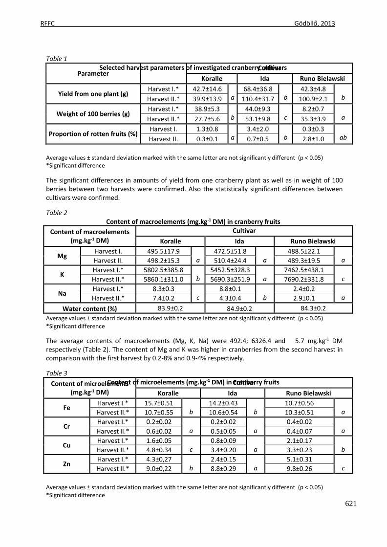
RFFC Gödöllő, 2013
621
Parameter Cultivar
Koralle Ida Runo Bielawski
Yield from one plant (g) Harvest I.* 42.7±14.6
a 68.4±36.8
b 42.3±4.8
b Harvest II.* 39.9±13.9 110.4±31.7 100.9±2.1
Weight of 100 berries (g) Harvest I.* 38.9±5.3
b 44.0±9.3
c 8.2±0.7
a Harvest II.* 27.7±5.6 53.1±9.8 35.3±3.9
Proportion of rotten fruits (%) Harvest I. 1.3±0.8
a 3.4±2.0
b 0.3±0.3
ab Harvest II. 0.3±0.1 0.7±0.5 2.8±1.0
Content of macroelements (mg.kg-1 DM)
Cultivar
Koralle Ida Runo Bielawski
Mg Harvest I. 495.5±17.9
a 472.5±51.8
a 488.5±22.1
a Harvest II. 498.2±15.3 510.4±24.4 489.3±19.5
K Harvest I.* 5802.5±385.8
b
5452.5±328.3 a
7462.5±438.1 c Harvest II.* 5860.1±311.0 5690.3±251.9 7690.2±331.8
Na Harvest I.* 8.3±0.3
c 8.8±0.1
b 2.4±0.2
a Harvest II.* 7.4±0.2 4.3±0.4 2.9±0.1
Water content (%) 83.9±0.2 84.9±0.2 84.3±0.2
Content of microelements (mg.kg-1 DM)
Cultivar
Koralle Ida Runo Bielawski
Fe Harvest I.* 15.7±0.51
b 14.2±0.43
b 10.7±0.56
a Harvest II.* 10.7±0.55 10.6±0.54 10.3±0.51
Cr Harvest I.* 0.2±0.02
a
0.2±0.02 a
0.4±0.02 a Harvest II.* 0.6±0.02 0.5±0.05 0.4±0.07
Cu Harvest I.* 1.6±0.05
c 0.8±0.09
a 2.1±0.17
b Harvest II.* 4.8±0.34 3.4±0.20 3.3±0.23
Zn Harvest I.* 4.3±0,27
b 2.4±0.15
a 5.1±0.31
c Harvest II.* 9.0±0,22 8.8±0.29 9.8±0.26
Table 1 Selected harvest parameters of investigated cranberry cultivars
Average values ± standard deviation marked with the same letter are not significantly different (p < 0.05) *Significant difference
The significant differences in amounts of yield from one cranberry plant as well as in weight of 100 berries between two harvests were confirmed. Also the statistically significant differences between cultivars were confirmed.
Table 2 Content of macroelements (mg.kg-1 DM) in cranberry fruits
Average values ± standard deviation marked with the same letter are not significantly different (p < 0.05) *Significant difference
The average contents of macroelements (Mg, K, Na) were 492.4; 6326.4 and 5.7 mg.kg-1 DM respectively (Table 2). The content of Mg and K was higher in cranberries from the second harvest in comparison with the first harvest by 0.2-8% and 0.9-4% respectively.
Table 3 Content of microelements (mg.kg-1 DM) in cranberry fruits
Average values ± standard deviation marked with the same letter are not significantly different (p < 0.05) *Significant difference
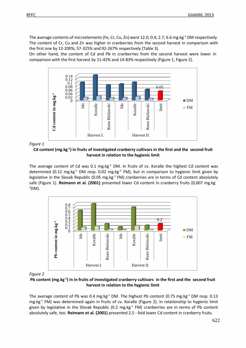
RFFC Gödöllő, 2013
622
Cd
co
nte
nt
in m
g.k
g-1
P
b c
on
ten
t in
mg
.kg
-1
Ida
Ida
Ko
rall
e
Ko
rall
e
Ru
no
Bie
law
ski
Ru
no
Bie
law
ski
Ida
Ida
Ko
rall
e
Ko
rall
e
Ru
no
Bie
law
ski
Ru
no
Bie
law
ski
lim
it
lim
it
The average contents of microelements (Fe, Cr, Cu, Zn) were 12.0; 0.4; 2.7; 6.6 mg.kg-1 DM respectively. The content of Cr, Cu and Zn was higher in cranberries from the second harvest in comparison with the first one by 12-200%, 57-325% and 92-267% respectively (Table 3). On other hand, the content of Cd and Pb in cranberries from the second harvest were lower in comparison with the first harvest by 11-42% and 14-83% respectively (Figure 1, Figure 2).
0.14 0.12
0.1 0.08 0.06 0.04 0.02
0
0.05
DM
FM
Harvest I. Harvest II.
Figure 1 Cd content (mg.kg-1) in fruits of investigated cranberry cultivars in the first and the second fruit
harvest in relation to the hygienic limit
The average content of Cd was 0.1 mg.kg-1 DM. In fruits of cv. Koralle the highest Cd content was determined (0.12 mg.kg-1 DM resp. 0.02 mg.kg-1 FM), but in comparison to hygienic limit given by legislative in the Slovak Republic (0.05 mg.kg-1 FM) cranberries are in terms of Cd content absolutely safe (Figure 1). Reimann et al. (2001) presented lower Cd content in cranberry fruits (0,007 mg.kg-
1DM).
0.8 0.7 0.6 0.5 0.4 0.3 0.2 0.1
0
0.2
DM
FM
Figure 2
Harvest I. Harvest II.
Pb content (mg.kg-1) in in fruits of investigated cranberry cultivars in the first and the second fruit harvest in relation to the hygienic limit
The average content of Pb was 0.4 mg.kg-1 DM. The highest Pb content (0.75 mg.kg-1 DM resp. 0.13 mg.kg-1 FM) was determined again in fruits of cv. Koralle (Figure 2). In relationship to hygienic limit given by legislative in the Slovak Republic (0.2 mg.kg-1 FM) cranberries are in terms of Pb content absolutely safe, too. Reimann et al. (2001) presented 2.5 - fold lower Cd content in cranberry fruits.
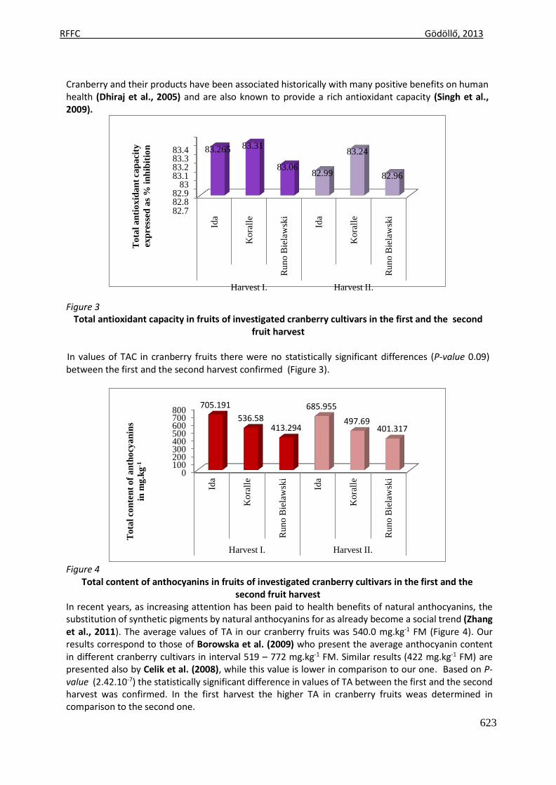
RFFC Gödöllő, 2013
623
To
tal
con
ten
t o
f a
nth
ocy
an
ins
in m
g.k
g-1
T
ota
l a
nti
ox
ida
nt
cap
aci
ty
exp
ress
ed a
s %
in
hib
itio
n
Ida
Ida
Ko
rall
e
Ko
rall
e
Ru
no
Bie
law
ski
Ru
no
Bie
law
ski
Ida
Ida
Ko
rall
e
Ko
rall
e
Ru
no
Bie
law
ski
Ru
no
Bie
law
ski
Cranberry and their products have been associated historically with many positive benefits on human health (Dhiraj et al., 2005) and are also known to provide a rich antioxidant capacity (Singh et al., 2009).
83.4 83.3 83.2 83.1
83 82.9 82.8 82.7
83.265 83.31
83.06
82.99
83.24
82.96
Figure 3
Harvest I. Harvest II.
Total antioxidant capacity in fruits of investigated cranberry cultivars in the first and the second fruit harvest
In values of TAC in cranberry fruits there were no statistically significant differences (P-value 0.09) between the first and the second harvest confirmed (Figure 3).
800 700 600 500 400 300 200 100
0
705.191
536.58
413.294
685.955 497.69
401.317
Harvest I. Harvest II.
Figure 4 Total content of anthocyanins in fruits of investigated cranberry cultivars in the first and the
second fruit harvest In recent years, as increasing attention has been paid to health benefits of natural anthocyanins, the substitution of synthetic pigments by natural anthocyanins for as already become a social trend (Zhang et al., 2011). The average values of TA in our cranberry fruits was 540.0 mg.kg-1 FM (Figure 4). Our results correspond to those of Borowska et al. (2009) who present the average anthocyanin content in different cranberry cultivars in interval 519 – 772 mg.kg-1 FM. Similar results (422 mg.kg-1 FM) are presented also by Celik et al. (2008), while this value is lower in comparison to our one. Based on P- value (2.42.10-7) the statistically significant difference in values of TA between the first and the second harvest was confirmed. In the first harvest the higher TA in cranberry fruits weas determined in comparison to the second one.
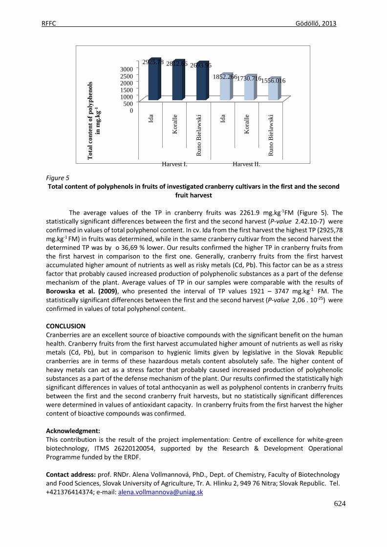
RFFC Gödöllő, 2013
624
To
tal
con
ten
t o
f p
oly
ph
eno
ls
in m
g.k
g-1
Ida
Ko
rall
e
Ru
no
Bie
law
ski
Ida
Ko
rall
e
Ru
no
Bie
law
ski
3000 2500 2000 1500 1000
500 0
2925.78 2812.66 2693.96
1852.2661730.7161556.016
Figure 5
Harvest I. Harvest II.
Total content of polyphenols in fruits of investigated cranberry cultivars in the first and the second fruit harvest
The average values of the TP in cranberry fruits was 2261.9 mg.kg-1FM (Figure 5). The
statistically significant differences between the first and the second harvest (P-value 2.42.10-7) were confirmed in values of total polyphenol content. In cv. Ida from the first harvest the highest TP (2925,78 mg.kg-1 FM) in fruits was determined, while in the same cranberry cultivar from the second harvest the determined TP was by o 36,69 % lower. Our results confirmed the higher TP in cranberry fruits from the first harvest in comparison to the first one. Generally, cranberry fruits from the first harvest accumulated higher amount of nutrients as well as risky metals (Cd, Pb). This factor can be as a stress factor that probably caused increased production of polyphenolic substances as a part of the defense mechanism of the plant. Average values of TP in our samples were comparable with the results of Borowska et al. (2009), who presented the interval of TP values 1921 – 3747 mg.kg-1 FM. The statistically significant differences between the first and the second harvest (P-value 2,06 . 10-25) were confirmed in values of total polyphenol content.
CONCLUSION Cranberries are an excellent source of bioactive compounds with the significant benefit on the human health. Cranberry fruits from the first harvest accumulated higher amount of nutrients as well as risky metals (Cd, Pb), but in comparison to hygienic limits given by legislative in the Slovak Republic cranberries are in terms of these hazardous metals content absolutely safe. The higher content of heavy metals can act as a stress factor that probably caused increased production of polyphenolic substances as a part of the defense mechanism of the plant. Our results confirmed the statistically high significant differences in values of total anthocyanin as well as polyphenol contents in cranberry fruits between the first and the second cranberry fruit harvests, but no statistically significant differences were determined in values of antioxidant capacity. In cranberry fruits from the first harvest the higher content of bioactive compounds was confirmed.
Acknowledgment: This contribution is the result of the project implementation: Centre of excellence for white-green biotechnology, ITMS 26220120054, supported by the Research & Development Operational Programme funded by the ERDF.
Contact address: prof. RNDr. Alena Vollmannová, PhD., Dept. of Chemistry, Faculty of Biotechnology and Food Sciences, Slovak University of Agriculture, Tr. A. Hlinku 2, 949 76 Nitra; Slovak Republic. Tel. +421376414374; e-mail: [email protected]
RFFC Gödöllő, 2013
625
REFERENCES
BOROWSKA, E. J. – MAZUR, B. – KOPCIUCH, R. G. – BUSZEWSKI, B. 2009. Polyphenol, anthocyanin and resveratrol massfractions and antioxidant properties of cranberry cultivars. In Food Technology and Biotechnology, roč. 47, 2009, č. 1, s. 56-61.
BRAND-WILLIAMS, W. – CUVELIER, M. E. – BERSET, C. 1995. Use of free radical method to evaluate antioxidant activity. In Lebensmittel Wissenschaft und Technologie, roč. 28, 1995, s. 25-30.
CELIK, H. – OZGEN, M. – SERCE, S. – KAYA, C. 2008. Phytochemical accumulation and antioxidant capacity at four maturity stages of cranberry fruit. In Scientia Horticulturae, roč. 117, 2008, s. 345-348.
DHIRAJ A. V. - REZA G. - KALIDAS S. 2005.Enhancing health benefits of berries through phenolic antioxidant enrichment: focus on cranberry. In Asia Pac J Clin Nutr 2005;14 (2):120-130
HRIČOVSKÝ, I. – CAGÁŇOVÁ, I. – HORČIN, V. – ŠIMALA, D. 2002. Drobné ovocie a menej známe druhy ovocia. 1.vyd., Bratislava: Príroda, 2002, 104 s., ISBN 80-07-00986-8.
LACHMAN, J. – HAMOUZ, K. – ORSÁK, M. 2005. Červeně a modře zbarvené brambory – významný zdroj antioxidantů v lidské výživě. In Chemické listy, roč. 99, 2005, č. 7, s. 474-482.
LAPORNIK, B. – PROŠEK, M. – GOLC-WONDRA, A. 2005. Comparison of extracts prepared from plant by- products using different solvents and extraction time. In Journal of Food Engineering, roč. 71, 2005, s. 214-222.
LIN, Y. T. – KWON, Y. I. – LABBE, R. G. – SHETTY, K. 2005. Inhibition of Helicobacter pylori and associate durease by oregano and cranberry phytochemical synergies. In Applied and Environmental Microbiology, roč. 71, 2005, s. 8558-8564.
MARWAN A.G. - NAGEL C. W. 1986. Characterization of cranberry benzoates and their antimicrobial properties. In J Food Sci 1986; 51: 1069-1070.
NETO, C. C. 2007. Cranberry and its phytochemicals: A review of in vitro anticancer studies. In Journal of Nutrition, roč. 137, 2007, č. 1, s. 186-193.
PORTER, M. L. – KRUEGER, C. G. – WIEBE, D. A. – CUNNINGHAM, D. G. – REED, J. D. 2001. Cranberry proantocyanidins associate with low-density lipoprotein and inhibit in vitro Cu2+-induced oxidation. In Journal of the Science of Food and Agriculture, roč. 81, 2001, s. 1306-1313.
REIMANN, C. – KOLLER, F. – KASHULINA, G. – NISKAVAARA, H. – ENGLMAIER, P. 2001. Influence of extreme pollution on the inorganic chemical composition of some plants. In Environmental Pollution, roč. 115, 2001, s. 239-252.
RUEL, G. – COUILLARD, CH. 2007. Evidences of the cardioprotective potencial of fruits: The case of cranberries. In Molecular Nutrition and Food Research, roč. 51, 2007, s. 692-701.
SEERAM, N. S. – ADAMS, L. S. – ZHANG, Y. – LEE, R. – SAND, D. – SHEULLER, H. S. – HEBER, D. 2006. Blackberry, black raspberry, blueberry, cranberry, red raspberry, and strawberry extracts inhibit growth and stimulate apoptosis of human cancer cells in vitro. In Journal of Agricultural and Food Chemistry, roč. 54, 2006, s. 9329-9339.
SINGH A. P. – WILSON T. – KALK J. A. – CHEONG J. – VORSA N. 2009. Isolation of Specific Cranberry Flavonoids for Biological Activity Assessment. Food Chem. 2009; 116(4): 963–968:
TUMBAS, V. T. – DILAS, S. M. – ČANADANOVIĆ-BRUNET, J. M. – CETKOVIC, G. S. – SAVATOVIC, S. M. 2007. Solid-phase extraction of antioxidant compounds from commercial cranberry extract and its antiradical activity. In Acta Periodica Technologica, roč. 37, 2007, č. 38, s. 157-164.
VATTEM, D. A. – GHAEDIAN, R. – SHETTY, K. 2005. Enhancing health benefits of berries through phenolic antioxidant enrichment: Focus on cranberry. In Asia Pacific Journal of Clinical Nutrition, roč. 14, 2005, č. 2, s. 120-130.
VINSON, J. A. – SU, X. – ZUBIK, L. – BOSE, P. 2001. Phenol antioxidant quantity and quality in foods: Fruits. In Journal of Agricultural and Food Chemistry, roč. 49, 2001, s. 5315-5321.
WANG, S. Y. – JIAO, H. 2000. Scavenging capacity of berry crops on superoxide radicals, hydrogen peroxide, hydroxyl radicals and singlet oxygen. In Journal of Agricultural and Food Chemistry, roč. 48, 2000, s. 5677-5684.
YAN, X. – MURPHY, B. T. – HAMMOND, G. B. – VINSON, J. A. – NETO, C. C. 2002. Antioxidant activities and antitumor screening of extracts from cranberry fruit (Vaccinium macrocarpon). In Journal of Agricultural and Food Chemistry, roč. 50, 2002, s. 5844-5849.
ZHANG W. - HE J. - PAN Q. - HAN F. - DUAN C. 2011. Separation and character analysis of anthocyanins from mulberry (Morus alba L.) pomace. In Czech J. Food Sci., 29 (2011): 268-276
Animal welfare, etológia és tartástechnológia
Animal welfare, ethology and housing systems
Volume 9 Issue 3
Különszám/Special Issue
Gödöllő
2013
RFFCH Gödöllő, 2013
626
EFFECT OF PROBIOTIC SUPPLEMENTATION ON PERFORMANCE OF DIFFERENT BROILER GENOTYPES
Ján Weis*, Cyril Hrnčár
Address*: doc. Ing. Ján Weis, CSc., Department of Poultry Science and Small Animal Husbandry, Faculty of Agrobiology and Food Resources, Slovak University of Agriculture, Tr. A. Hlinku 2, 949 76 Nitra, Slovakia, e-mail: Já[email protected], phone number: + 42137/641 4438
Abstract This experiment was conducted to investigate the effects of probiotic strain Lactobacillus fermentum supplementation on the performance and carcass parameters of different genotypes of broiler chickens. Totally 60 one-day-old chickens of Cobb 500, 60 one-day-old chickens of Hubbard JV and 60 one-day-old chickens of Ross 308 were divided in two groups: control chicken group (n=30) received drinking water without any additives and experimental chickens group (n=30) received probiotic strain Lactobacillus fermentum CCM 7158 with concentration of 1x10⁹ colony forming units (CFU) in drinking water from day 1 to day 42 of fattening. Individual body weights of all birds, feed conversion ratio per group were determined in 21 and 42 day, total mortality rate we recorded in 42 day of fattening period. Carcass quality of broiler chickens was determined at the end of the experiment. The supplementation of probiotic affected positively body weight (P<0.05) in all broiler chicken genotypes in 21 and 42 day of fattening. Feed conversion ratio in 21 and 42 day was similar in control and experimental groups. The probiotic no significant (P≥0.05) affected percentage of breast and thighs form carcass body, weight of giblets and carcass yield. The probiotic addition significantly (P<0.05) reduced the abdominal fat content of the chicken meat. Key words: Lactobacillus fermentum, body weight, feed utility, mortality, carcass parameters INTRODUCTION The consumption of chicken meat is of increasing importance in the EU and per head consumption is still rising. One reason for this case might be a more health-conscious nutrition by the consumers. Other reasons like the BSE problem or antibiotic abuse in pig production might also be important for an increase in consumption of chicken meat (Schedle et al., 2006). Research studies have reported feed residues in chicken meat products and the development of bacterial resistance to antibiotics used both in human medicine and poultry production (Edens, 2003). On 1st January 2006 the European Union introduced a complete ban on the use of antibiotic growth promoters in feeds for animals for consumption. The ban was introduced at the same time in all Member States. Since then, antibiotics have been allowed to be used as medicines only in medical feeds or prophylactic additives. Resolution No 1831/2003 EC of the European Parliament and Council of 22nd August 2003 devoted to the issue of additives used in feeding animals described probiotics as alternative feed additives to antibiotic growth promoters (Casewell et al., 2003; Patterson and Burkholder, 2003; Berghmann et al., 2005).
RFFCH Gödöllő, 2013
627
Probiotics are additives that can be used to replace antibiotics in poultry nutrition (Griggs and Jacob, 2005). The term probiotic stems from the Greek and means “in favor of life”; its antonym is antibiotics, which means “against life” (Coppola and Turnes, 2004). Probiotics are defined as live microbial food supplements, which beneficially influence only not human (Songisepp et al., 2005), but also poultry health and performance, chickens (Haščík et al., 2005; Weis et al., 2010), hens (Capcarova et al., 2010), turkeys (Capcarova, 2008), pigeons (Malíková et al., 2013) and waterfowl (Weis et al., 2008; Hrnčár et al., 2013). Probiotics in poultry maintaining normal intestinal microflora by competitive exclusion and antagonism (Kabir et al., 2005; Kizerwetter-Swida and Binek, 2009), alter metabolism by increasing digestive enzyme activity and decreasing bacterial enzyme activity and ammonia production (Yoon et al., 2004; Nayebpor et al., 2007), improve feed intake and stimulate the immune system (Kabir et al., 2004; Haghighi et al., 2005; Apata, 2008). The selection of bacteria such as Lactobacillus, Pediococcus, Bacteroides, Bifidobacterium, Bacillus and Streptococcus, for use as probiotics is based on assessment of their metabolic products and their potential to colonize specific sites (Lima et al., 2007). The ability of lactic acid bacteria to inhibit various G-positive and G-negative bacteria is well known. This inhibition may be related to the production of organic acids, hydrogen peroxide and bacteriocin-like substances that are active against certain pathogens and may be produced by different species of Lactobacillus (Messaoudi et al., 2005). Lactobacillus sp. are normal inhabitants of the intestinal tract, especially of poultry (Juven et al., 2001). Lactobacilli mainly compose the flora in crop and ileum region in poultry (Gong et al., 2002). Lactobacilli are rod shape bacteria and possess different biochemical and physiological properties (Tannock et al., 1999). Many Lactobacillus strains, isolated from various sources, are being used as probiotic agents and it is unlikely that all functional characters of a probiotic are present in each species/strain. It needs thorough study and documentation. Generally recognized beneficial properties are the origin of the strain being used, the surviving ability within the GI tract, non-pathogenic activities and the modulation ability in immune responses (Gibson and Fuller, 2000; Dunne et al., 2001; Holzapfel et al., 2001). The objective of this study was to provide a comparison of the effect of probiotic strain Lactobacillus fermentum supplementation in drinking water on performance and carcass parameters of different genotypes broiler chickens (Cobb 500, Hubbard JV, Ross 308). MATERIAL AND METHODS The experiment was realised in half-operation conditions of experimental basis of Department of Poultry Science and Small Animal Husbandry (Certificate of Authorization to Experiment on Living Animals, State Veterinary and Food Institute of Slovak Republic, no. SK PC 30008). Totally 60 one-day-old chickens of Cobb 500, 60 one-day-old chickens of Hubbard JV and 60 one-day-old chickens of Ross 308 were divided in two groups: control chicken group (n=30) received drinking water without any additives and experimental chickens group (n=30) received probiotic strain Lactobacillus fermentum CCM 7158 with concentration of 1x10⁹ colony forming units (CFU) in drinking water from day 1 to day 42 of fattening. Quantization of drinking water and probiotic strain are presented in Table 1.
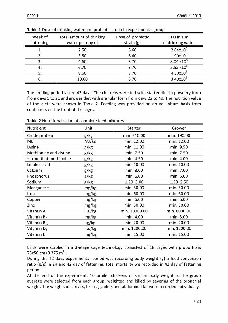
RFFCH Gödöllő, 2013
628
Table 1 Dose of drinking water and probiotic strain in experimental group
Week of fattening
Total amount of drinking water per day (l)
Dose of probiotic strain (g)
CFU in 1 ml of drinking water
1. 2.50 6.60 2.64x10⁶
2. 3.50 6.60 1.90x10⁶
3. 4.60 3.70 8.04 x10⁵
4. 6.70 3.70 5.52 x10⁵
5. 8.60 3.70 4.30x10⁵
6. 10.60 3.70 3.49x10⁵
The feeding period lasted 42 days. The chickens were fed with starter diet in powdery form from days 1 to 21 and grower diet with granular form from days 22 to 49. The nutrition value of the diets were shown in Table 2. Feeding was provided on an ad libitum basis from containers on the front of the cages. Table 2 Nutritional value of complete feed mixtures
Nutritient Unit Starter Grower
Crude protein g/kg min. 210.00 min. 190.00
ME MJ/kg min. 12.00 min. 12.00
Lysine g/kg min. 11.00 min. 9.50
Methionine and cistine g/kg min. 7.50 min. 7.50
– from that methionine g/kg min. 4.50 min. 4.00
Linoleic acid g/kg min. 10.00 min. 10.00
Calcium g/kg min. 8.00 min. 7.00
Phosphorus g/kg min. 6.00 min. 5.00
Sodium g/kg 1.20–3.00 1.20–2.50
Manganese mg/kg min. 50.00 min. 50.00
Iron mg/kg min. 60.00 min. 60.00
Copper mg/kg min. 6.00 min. 6.00
Zinc mg/kg min. 50.00 min. 50.00
Vitamin A i.u./kg min. 10000.00 min. 8000.00
Vitamin B2 mg/kg min. 4.00 min. 3.00
Vitamin B12 μg/kg min. 20.00 min. 20.00
Vitamin D3 i.u./kg min. 1200.00 min. 1200.00
Vitamin E mg/kg min. 15.00 min. 15.00
Birds were stabled in a 3-etage cage technology consisted of 18 cages with proportions 75x50 cm (0.375 m2). During the 42 days experimental period was recording body weight (g) a feed conversion ratio (g/g) in 24 and 42 day of fattening. total mortality we recorded in 42 day of fattening period. At the end of the experiment, 10 broiler chickens of similar body weight to the group average were selected from each group, weighted and killed by severing of the bronchial weight. The weights of carcass, breast, giblets and abdominal fat were recorded individually.
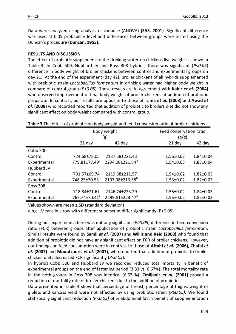
RFFCH Gödöllő, 2013
629
Data were analyzed using analysis of variance (ANOVA) (SAS, 2001). Significant difference was used at 0.05 probability level and differences between groups were tested using the Duncan’s procedure (Duncan, 1955). RESULTS AND DISCUSSION The effect of probiotic supplement to the drinking water on chickens live weight is shown in Table 3. In Cobb 500, Hubbard JV and Ross 308 hybrids, there was significant (P<0.05) difference in body weight of broiler chickens between control and experimental groups on day 21. At the end of the experiment (day 42), broiler chickens of all hybrids supplemented with prebiotic strain Lactobacillus fermentum in drinking water had higher body weight in compare of control group (P<0.05). These results are in agreement with Kabir et al. (2004) who observed improvement of final body weight of broiler chickens at addition of probiotic preparate. In contrast, our results are opposite to those of Lima et al. (2003) and Awad et al. (2008) who recorded reported that addition of probiotic to broilers diet did not show any significant effect on body weight compared with control group. Table 3 The effect of probiotic on body weight and feed conversion ratio of broiler chickens
Body weight (g)
Feed conservation ratio (g/g)
21 day 42 day 21 day 42 day
Cobb 500 Control 724.68±78.05 2137.58±221.43 1.56±0.02 1.84±0.04 Experimental 779.81±77.49a 2294.08±221,84a 1.54±0.03 1.83±0.04
Hubbard JV Control 701.57±69.74 2119.38±211.57 1.54±0.02 1.82±0.03 Experimental 748.25±70.53b 2197.98±213.58b 1.53±0.02 1.82±0.03
Ross 308 Control 718.84±71.67 2146.74±223.29 1.55±0.02 1.84±0.03 Experimental 765.74±70.41c 2299.81±223.47c 1.55±0.02 1.82±0.03
Values shown are mean ± SD (standard deviation) a,b,c Means in a row with different superscript differ significantly (P<0.05) During our experiment, there was not any significant (P≥0.05) difference in feed conversion ratio (FCR) between groups after application of probiotic strain Lactobacillus fermentum. Similar results were found by Samli et al. (2007) and Willis and Reid (2008) who found that addition of probiotic did not have any significant effect on FCR of broiler chickens. However, our findings on feed consumption were in contrast to those of Aftahi et al. (2006), Chafai et al. (2007) and Mountzouris et al. (2007), who reported that addition of probiotic to broiler chicken diets decreased FCR significantly (P<0.05). In hybrids Cobb 500 and Hubbard JV we recorded reduced total mortality in benefit of experimental groups on the end of fattening period (3.33 vs. 6.67%). The total mortality rate in the both groups in Ross 308 was identical (6.67 %). Cmiljanic et al. (2001) proved a reduction of mortality rate of broiler chickens due to the addition of probiotic. Data presented in Table 4 show that percentage of breast, percentage of thighs, weight of giblets and carcass yield were not affected by using probiotic strain (P≥0.05). We found
statistically significant reduction (P0.05) of % abdominal fat in benefit of supplementation
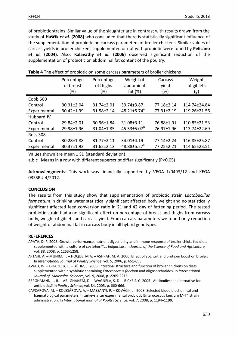
RFFCH Gödöllő, 2013
630
of probiotic strains. Similar value of the slaughter are in contrast with results drawn from the study of Haščík et al. (2008) who concluded that there is statistically significant influence of the supplementation of probiotic on carcass parameters of broiler chickens. Similar values of carcass yields in broiler chickens supplemented or not with probiotic were found by Pelicano et al. (2004). Also, Kalavathy et al. (2006) observed significant reduction of the supplementation of probiotic on abdominal fat content of the poultry. Table 4 The effect of probiotic on some carcass parameters of broiler chickens
Percentage of breast
(%)
Percentage of thighs
(%)
Weight of abdominal
fat (%)
Carcass yield (%)
Weight of giblets
(g)
Cobb 500 Control 30.31±2.04 31.74±2.01 33.74±3.87 77.18±2.14 114.74±24.84 Experimental 30.42±1.99 31.58±2.14 48.21±5.74a 77.31±2.19 119.26±21.56
Hubbard JV Control 29.84±2.01 30.96±1.84 31.08±3.11 76.88±1.91 110.85±21.53 Experimental 29.98±1.96 31.04±1.85 45.53±5.07b 76.97±1.96 113.74±22.69
Ross 308 Control 30.28±1.88 31.77±2.11 34.01±4.19 77.14±2.24 116.85±25.87 Experimental 30.37±1.92 31.62±2.13 48.88±5.27c 77.25±2.21 114.65±23.51
Values shown are mean ± SD (standard deviation) a,b,c Means in a row with different superscript differ significantly (P<0.05) Acknowledgments: This work was financially supported by VEGA 1/0493/12 and KEGA 035SPU-4/2012. CONCLUSION The results from this study show that supplementation of probiotic strain Lactobacillus fermentum in drinking water statistically significant affected body weight and no statistically significant affected feed conversion ratio in 21 and 42 day of fattening period. The tested probiotic strain had a no significant effect on percentage of breast and thighs from carcass body, weight of giblets and carcass yield. From carcass parameters we found only reduction of weight of abdominal fat in carcass body in all hybrid genotypes. REFERENCES APATA, D. F. 2008. Growth performance, nutrient digestibility and immune response of broiler chicks fed diets
supplemented with a culture of Lactobacillus bulgaricus. In Journal of the Science of Food and Agriculture, vol. 88, 2008, p. 1253-1258.
AFTAHI, A. – MUNIM, T. – HOQUE, M.A. – ASHRAF, M. A. 2006. Effect of yoghurt and protexin boost on broiler. In International Journal of Poultry Science, vol. 5, 2006, p. 651-655.
AWAD, W. – GHAREEB, K. – BÖHM, J. 2008. Intestinal structure and function of broiler chickens on diets supplemented with a synbiotic containing Enterococcus faecium and oligosaccharides. In International Journal of Molecular Sciences, vol. 9, 2008, p. 2205-2216.
BERGHMANN, L. R. – ABI-GHANEM, D. – WAGNELA, S. D. – RICKE S. C. 2005. Antibodies: an alternative for antibiotics? In Poultry Science, vol. 84, 2005, p. 660-666.
CAPCAROVA, M. – KOLESÁROVÁ, A. – MASSANYI, P. – KOVÁČIK, J. 2008. Selected blood biochemical and haematological parameters in turkeys after experimental probiotic Enterococcus faecium M-74 strain administration. In International Journal of Poultry Science, vol. 7, 2008, p. 1194–1199.
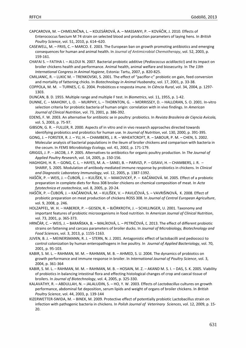
RFFCH Gödöllő, 2013
631
CAPCAROVA, M. – CHMELNIČNA, L. – KOLESÁROVÁ, A. – MASSANYI, P. – KOVÁČIK, J. 2010. Effects of Enterococcus faecium M 74 strain on selected blood and production parameters of laying hens. In British Poultry Science, vol. 51, 2010, p. 614–620.
CASEWELL, M. – FRIIS, C. – MARCO. E. 2003. The European ban on growth promoting antibiotics and emerging consequences for human and animal health. In Journal of Antimicrobial Chemotherapy, vol. 52, 2003, p. 159-161.
CHAFAI S. – FATIHA I. – ALLOUI N. 2007. Bacterial probiotic additive (Pediococcus acidilactici) and its impact on broiler chickens health and performance. Animal health, animal welfare and biosecurity. In The 13th International Congress in Animal Hygiene, Estonia: Tartu, 2007, p. 820-825.
CMILJANIC, R. – LUKIC M. – TRENKOVSKI, S. 2001. The effect of “paciflor-c” probiotic on gain, feed conversion and mortality of fattening chicks. In Biotechnology in Animal Husbandry, vol. 17, 2001, p. 33-38.
COPPOLA, M. M. – TURNES, C. G. 2004. Probióticos e resposta imune. In Ciência Rural, vol. 34, 2004, p. 1297-1303.
DUNCAN, B. D. 1955. Multiple range and multiple F test. In Biometrics, vol. 11, 1955, p. 1-42. DUNNE, C. – MAHONY, L. O. – MURPHY, L. – THORNTON, G. – MORRISSEY, D. – HALLORAN, S. O. 2001. In-vitro
selection criteria for probiotic bacteria of human origin: correlation with in vivo findings. In American Journal of Clinical Nutrition, vol. 73, 2001, p. 386-392.
EDENS, F. W. 2003. An alternative for antibiotic se in poultry: probiotics. In Revista Brasileira de Cięncia Avícola, vol. 5, 2003, p. 75-97.
GIBSON, G. R. – FULLER, R. 2000. Aspects of in vitro and in vivo research approaches directed towards identifying probiotics and probiotics for human use. In Journal of Nutrition, vol. 130, 2000, p. 391-395.
GONG, J. – FORSTER, R. J. – YU, H. – CHAMBERS, J. R. – WHEATCROFT, R. – SABOUR, P. M. – CHEN, S. 2002. Molecular analysis of bacterial populations in the ileum of broiler chickens and comparison with bacteria in the cecum. In FEMS Microbiology Ecology, vol. 41, 2002, p. 171-179.
GRIGGS, J. P. – JACOB, J. P. 2005. Alternatives to antibiotics for organic poultry production. In The Journal of Applied Poultry Research, vol. 14, 2005, p. 150-156.
HAGHIGHI, H. R. – GONG, C. L. – HAYES, M. A. – SANEI, B. – PARVIZI, P. – GISAVI, H. – CHAMBERS, J. R. – SHARIF, S. 2005. Modulation of antibody-mediated immune response by probiotics in chickens. In Clinical and Diagnostic Laboratory Immunology, vol. 12, 2005, p. 1387-1392.
HAŠČÍK, P. – WEIS, J. – ČUBOŇ, J. – KULÍŠEK, V. – MAKOVICKÝ, P. – KAČÁNIOVÁ. M. 2005. Effect of a probiotic preparation in complete diets for Ross 308 broiler chickens on chemical composition of meat. In Acta fytotechnica et zootechnica, vol. 8, 2005, p. 20-24.
HAŠČÍK, P. – ČUBOŇ, J. – KAČÁNIOVÁ, M. – KULÍŠEK, V. – PAVLIČOVÁ, S. – VAVRIŠINOVÁ, K. 2008. Effect of probiotic preparation on meat production of chickens ROSS 308. In Journal of Central European Agriculture, vol. 9, 2008, p. 246.
HOLZAPFEL, W. H. – HABERER, P. – GEISEN, R. – BJÖRKROTH, J. – SCHILLINGER, U. 2001. Taxonomy and important features of probiotic microorganisms in food nutrition. In American Journal of Clinical Nutrition, vol. 73, 2001, p. 365-373.
HRNČÁR, C. – WEIS, J. – BARAŇSKA, B. – MALÍKOVÁ, L. – PETRIČOVÁ, Ľ. 2013. The effect of different probiotic strains on fattening and carcass parameters of broiler ducks. In Journal of Microbiology, Biotechnology and Food Sciences, vol. 3, 2013, p. 1155-1163.
JUVEN, B. J. – MEINERSMANN, R. J. – STERN, N. J. 2001. Antagonistic effect of lactobacilli and pediococci to control colonization by human enteropathogens in live poultry. In Journal of Applied Bacteriology, vol. 70, 2001, p. 95-103.
KABIR, S. M. L. – RAHMAN, M. M. – RAHMAN, M. B. – AHMED, S. U. 2004. The dynamics of probiotics on growth performance and immune response in broiler. In International Journal of Poultry Science, vol. 3, 2004, p. 361-364
KABIR, S. M. L. – RAHMAN, M. M. – RAHMAN, M. B. – HOSAIN, M. Z. – AKAND M. S. I. – DAS, S. K. 2005. Viability of probiotics in balancing intestinal flora and effecting histological changes of crop and caecal tissue of broilers. In Journal of Biotechnology, vol. 4, 2005, p. 325-330.
KALAVATHY, R. – ABDULLAH, N. – JALALUDIN, S. – HO, Y. W. 2003. Effects of Lactobacillus cultures on growth performance, abdominal fat deposition, serum lipids and weight of organs of broiler chickens. In British Poultry Science, vol. 44, 2003, p. 139-144
KIZERWETTER-SWIDA, M. – BINEK, M. 2009. Protective effect of potentially probiotic Lactobacillus strain on infection with pathogenic bacteria in chickens. In Polish Journal of Veterinary Sciences, vol. 12, 2009, p. 15-20.
RFFCH Gödöllő, 2013
632
LIMA, A. C. F. – PIZAURO–JÚNIOR, J. M. – MACARI, M. – MALHEIROS, E. B. 2003. Efeito do uso de probiotic sobre o Desempenho e atividade de enzimas digestivas de frangos de corte. In Revista Brasileira de Zootecnia, vol 32, 2003, p. 200-207.
LIMA, E. T. – FILHO, R. L. A. – OKAMOTO, A. S. – JOSÉ C. – NOUJAIM, J. C. – BARROS, M. R. – ADALBERTO J. – CROCCI A. J. 2007. Evaluation in vitro of the antagonistic substances produced by Lactobacillus spp. isolated from chickens. In Canadian Journal of Veterinary Research, vol. 71, 2007, p. 103-107.
MALÍKOVÁ, L. – WEIS, J. – HRNČÁR, C. 2013. Effect of administration of probiotic preparation with strain of Lactobacillus fermentum on the weight gain of the young pigeons. In Journal of Microbiology, Biotechnology and Food Sciences, vol. 3, 2013, p. 1469-1476.
MESSAOUDI, D. F. – BERGER, C. N. – COCONNIER-POLTER, M. H. – MOAL, V. – SERVIN A. L. 2005. pH-, lactic acid-, and non-lactic acid-dependent activities of probiotic Lactobacilli against Salmonella enterica serovar Typhimurium. In Applied and Environmental Microbiology, vol. 71, 2005, p. 6008-6013.
MOUNTZOURIS, K.C. – TSIRTSIKOS, P. – KALAMARA, E. – NITSCH, S. – SCHATZMAYR, G. – FEGEROS, K. 2007. Evaluation of the efficacy of a probiotic containing Lactobacillus, Bifidobacterium, Enterococcus and Pediococcus strains in promoting broiler performance and modulating cecal microflora composition and metabolic activities. In Poultry Science, vol. 86, 2007, p. 309-317.
NAYEBPOR, M. – FARHOMAND, P. – HASHMI A. 2007. Effects of different levels of direct fed microbial (Primalac) on growth performance and humoral immune response in broiler chickens. In Journal of Animal and Veterinary Advances, vol. 6, 2007, p. 1308-1313.
PELICANO, E. R. L. – SOUZA, P. A. – SOUZA, H. B. A. – OBA, A. – NORKUS, E. A. – KODAWARA, L. M. – LIMA, T. M. A. 2004. Performance of broilers fed diets containing natural growth promoters. In Revista Brasileira de Cięncia Avícola, vol. 6, 2004, p. 231-236.
PATTERSON, J. A. – BURKHOLDER, K. M. 2003. Application of prebiotics and probiotics in poultry production. In Poultry Science, vol. 82, 2003, p. 627-631.
SAMLI, H. E. – SENKOYLU, N. – KOC, F. – KANTER, M. – AGMA, A. 2007. Effects of Enterococcus faecium and dried whey on broiler performance, gut histomorphology and intestinal microbiota. In Archives of Animal Nutrition, vol. 61, 2007, p. 42.
SAS, 2001. SAS User's Guide: Statistics version 6.12 Edn., SAS Institute, Inc., Cary, NC., USA. SCHEDLE, K. – HASLINGER, M. – LEITGEB, R. – BAUER, F. – ETTLE, T. – WINDISCH, W. 2006. Carcass and meat
quality of broiler chickens at different starving period before slaughter. In Veterinarija ir Zootechnika, vol. 35, 2006, p. 85-88.
SONGISEPP, E. – KALS, J. – KULLISAAR, T. – MÄNDAR, R. – HÜTT, P. – ZILMER, M. – MIKELSAAR, M. 2005. Evaluation of the functional efficacy of an antioxidative probiotic in healthy volunteers. In Nutrition Journal, vol. 4, 2005, p. 22-32.
TANNOCK, G. W. – TILSALA, A. – TIMISJÄRVI, S. – RODTONG, J. – MUNRO, K. – ALATOSSAVA, T. 1999. Identification of Lactobacillus isolates from the gastrointestinal tract, silage and yoghurt by 16S-23S rRNA gene intergenic spacer region sequence comparisons. In Applied and Environmental Microbiology., vol. 65, 1999, p. 4264-4267.
WEIS, J. – HRNČÁR, C. – MINDEK, S. 2008. Effect of probiotic preparates with different strain on meat production of broiler ducks. In Scientifical Papers. Animal Science and Biotechnologies, vol. 41, 2008, 717-720.
WEIS, J. – HRNČÁR, C. – PÁL, G. – BARAŇSKA, B. – BUJKO, J. – POLIČKA, M. – MALÍKOVÁ, L. 2010. Effect of probiotic strain Lactobacillus fermentum CCM 7158 supplement on performance and carcass characteristics of broiler chickens. In Acta fytotechnica et zootechnica, vol. 13, 2010, p. 96-98.
WILLIS, W. L. – REID, L. 2008. Investigating the effects of dietary probiotic feeding regimens on broiler chicken production and Campylobacter jejuni presence. In Poultry Science, vol. 87, 2008, p. 606-611.
YOON, C. – NA, C. S. – PARK, J. H. – HAN, S. K. – NAM, Y. M. – KWON, J. T. 2004. Effect of feeding multiple probiotics on performance and fecal noxious gas emission in broiler chicks. In Korean Journal of Poultry Science, vol. 3, 2004, p. 229-235.
Animal welfare, etológia és tartástechnológia
Animal welfare, ethology and housing systems
Volume 9 Issue 3
Különszám/Special Issue
Gödöllő
2013
RFFCH Gödöllő, 2013
633
ASSESSEMENT OF ACROLEIN-INDUCED CELLULAR DAMAGE IN THE YEAST SACCHAROMYCES CEREVISIAE CELLS USING MICROSCOPY TECHNIQUES
Renata ZADRAG-TECZA*, Magdalena KWOLEK-MIREK
University of Rzeszow, Faculty of Biology and Agriculture, Department of Biochemistry and Cell Biology, Zelwerowicza 4, 35-601 Rzeszow, Poland *Corresponding author: [email protected]
Abstract
Acrolein is well-known and commonly found a compound showing cytotoxic activity. This study was aimed at a comparison between numerous tests based on the technique of bright-field and fluorescence. We applied the non-fluorescent dye PhloxineB or the single or double staining procedure with specific fluorescent dyes like fluorescein diacetate (FDA), propidium iodide (PI), FUN-1, 4’,6-diamidyno-2-fenyloindol (DAPI), MitoTrackerGreen, Rhodamine-Phalloidin and immunofluorescence technique to assess the viability, metabolic activity, structural changes and oxidative protein damages in the yeast Saccharomyces cerevisiae cells exposed to acrolein. The comparison of the results showed that the microscopic methods are a simple and very useful research tool for testing the toxicity of chemicals at the cellular level. Keywords: yeast, acrolein, oxidative stress, fluorescence, microscopic methods Introduction
Fluorescence microscopy is an essential and very important technique used to assess viability and vitality of the cells, visualization of cellular organelles and localization of macromolecules damages. There are many fluorescent dyes specific for various intracellular structures e.g. DAPI to visualize nuclei, MitoTrackerGreen or RhodamineB to label mitochondria, Rhodamine-Phalloidin to visualize actin cytoskeleton, MDY-64 to label membrane of vacuole or to identify the metabolic activity of the cell e.g. FUN-1 or fluorescein diacetate (FDA). Moreover, the use of fluorescently labeled antibodies allows for the localization of the selected macromolecules. These methods can be especially usefull in toxicological studies.
Cytotoxic substances, which include a number of chemical compounds common occurring in the environment or food products show a multidirectional action in the cell. Disorders which result from their actions affect both the structural elements of the cell and the activities of numerous metabolic pathways. The cell response depends on the concentration and time of exposure of the toxic compounds and it may be: the cell cycle arrest, changes of the metabolic activity and/or intracellular structures of the cell and the cell death induction by necrosis or apoptosis. An example of this type of compound is acrolein, chemically classified to the unsaturated aldehydes. It is a common ingredient / cause of environmental pollution as a by-product the incomplete combustion of petrol or the basic fuel materials as carbon or wood. It is also compound of the tobacco smoke (Caffaro-Filho et al., 2010). This aldehyde is also found in many types of food undergoing heat treatment such as animal and vegetable fats (Abraham et al., 2011), but also in natural products as vegetables and fruits. It was reported that the toxicity of acrolein involves enhancement of cellular oxidative stress by depletion of glutathione content/level, generation of reactive oxygen species, macromolecules damages (mainly protein and DNA modification) and structural alteration on actin cytoskeleton and mitochondrial network (Kwolek-Mirek et al., 2009, Kwolek-Mirek and Bartosz, 2011).
RFFCH Gödöllő, 2013
634
In this paper we present various methods for studying the toxicity of acrolein based on the fluorescence microscopy, using a simple eukaryotic model of the yeast Saccharomyces cerevisiae. Material and methods Yeast strains
The following yeast strains were used: wild-type SP4 MATα leu1 arg4 (Bilinski et al., 1978),
and sod1 mutant, isogenic to SP4, MATα leu1 arg4 sod1::natMX (Koziol et al., 2005). Media, growth conditions and condition of incubation
Yeast was grown in a standard liquid medium (YPD; 1% yeast extract, 1% yeast bacto-peptone, 2% glucose) on the shaker (150 rpm/min), at a temperature 28°C.
Cells from exponential phase culture were centrifuged, washed twice, suspended to a final density of 108 cells/ml in 100 mM phosphate buffer, pH 7.0, containing 1 mM EDTA and 0.1% glucose, and incubated with shaking for 60 min with 0.4 mM allyl alcohol (precursor of acrolein in the cell) (Bilinski et al., 2005), at 28°C. Viability assays
For verification of budding cells, after incubation with allyl alcohol 5 µl of cell suspension were spotted on the plate with solid YPD medium. At the start of the experiment and after 24 h of culture. Cultures were grown at 28°C.
Viability of cells was (Minois et al., 2005). After incubation the cells were collected by centrifugation, washed twice and resuspended in phosphate-buffered saline (PBS). For image acquisition was used OLYMPUS BX-51 fluorescence microscope equipped with a DP-72 digital camera and analysis software Cell^D. Fluorescent dyes and antibodies
Fluorescein diacetate (FDA) and propidium iodide (PI) were from Sigma-Aldrich (Poznan, Poland), 4’,6-diamidyno-2-fenyloindol (DAPI), FUN-1, MitoTrackerGreen, Rhodamine-Phalloidin were from Molecular Probes (Eugene, OR, USA), primary polyclonal rabbit antibody specific to the 2,4-dinitrophenol-hydrazine (DNP) moiety of the proteins (ab6306) and goat anti-rabbit conjugated with Chromeo™ 546 secondary antibody (ab60317) were from Abcam (Cambridge, UK). Fluorescence microscopy
After incubation the cells were collected by centrifugation, washed twice and resuspended in appropriate medium depending on the staining procedure. Cell suspension contained about 1×10⁷ cell/ml were stained in appropriate conditions shown in Table 1 and according to the manufacturer’s protocols (Molecular Probes). For image acquisition was used OLYMPUS BX-51 fluorescence microscope equipped with a DP-72 digital camera and analysis software Cell^D.
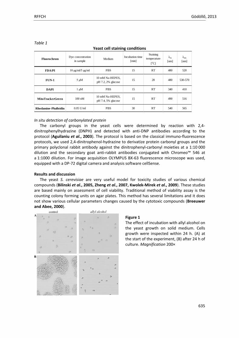
RFFCH Gödöllő, 2013
635
Table 1
Yeast cell staining conditions
FluorochromDye concentration
in sampleMedium
Incubation time
[min]
Staining
temperature
[oC]
λex
[nm]
λem
[nm]
FDA/PI 10 µg/ml/5 µg/ml PBS 15 RT 480 520
FUN-1 5 µM10 mM Na-HEPES,
pH 7.2, 2% glucose15 28 480 530-570
DAPI 1 µM PBS 15 RT 340 410
MitoTrackerGreen 100 nM10 mM Na-HEPES,
pH 7.4, 5% glucose15 RT 490 516
Rhodamine-Phalloidin 0.05 U/ml PBS 30 RT 540 565
In situ detection of carbonylated protein
The carbonyl groups in the yeast cells were determined by reaction with 2,4-dinitrophenylhydrazine (DNPH) and detected with anti-DNP antibodies according to the protocol (Aguilaniu et al., 2003). The protocol is based on the classical immuno-fluorescence protocols, we used 2,4-dinitrophenol-hydrazine to derivatize protein carbonyl groups and the primary polyclonal rabbit antibody against the dinitrophenyl-carbonyl moieties at a 1:10 000 dilution and the secondary goat anti-rabbit antibodies conjugated with Chromeo™ 546 at a 1:1000 dilution. For image acquisition OLYMPUS BX-63 fluorescence microscope was used, equipped with a DP-72 digital camera and analysis software cellSense. Results and discussion
The yeast S. cerevisiae are very useful model for toxicity studies of various chemical compounds (Bilinski et al., 2005, Zheng et al., 2007, Kwolek-Mirek et al., 2009). These studies are based mainly on assessment of cell viability. Traditional method of viability assay is the counting colony forming units on agar plates. This method has several limitations and it does not show various cellular parameters changes caused by the cytotoxic compounds (Breeuwer and Abee, 2000).
Figure 1 The effect of incubation with allyl alcohol on the yeast growth on solid medium. Cells growth were inspected within 24 h. (A) at the start of the experiment, (B) after 24 h of culture. Magnification 200×
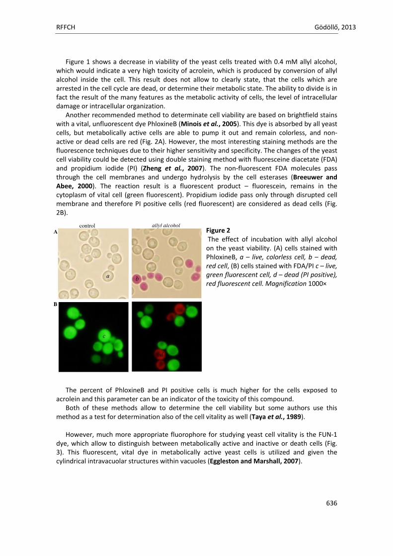
RFFCH Gödöllő, 2013
636
Figure 1 shows a decrease in viability of the yeast cells treated with 0.4 mM allyl alcohol, which would indicate a very high toxicity of acrolein, which is produced by conversion of allyl alcohol inside the cell. This result does not allow to clearly state, that the cells which are arrested in the cell cycle are dead, or determine their metabolic state. The ability to divide is in fact the result of the many features as the metabolic activity of cells, the level of intracellular damage or intracellular organization.
Another recommended method to determinate cell viability are based on brightfield stains with a vital, unfluorescent dye PhloxineB (Minois et al., 2005). This dye is absorbed by all yeast cells, but metabolically active cells are able to pump it out and remain colorless, and non-active or dead cells are red (Fig. 2A). However, the most interesting staining methods are the fluorescence techniques due to their higher sensitivity and specificity. The changes of the yeast cell viability could be detected using double staining method with fluoresceine diacetate (FDA) and propidium iodide (PI) (Zheng et al., 2007). The non-fluorescent FDA molecules pass through the cell membranes and undergo hydrolysis by the cell esterases (Breeuwer and Abee, 2000). The reaction result is a fluorescent product – fluorescein, remains in the cytoplasm of vital cell (green fluorescent). Propidium iodide pass only through disrupted cell membrane and therefore PI positive cells (red fluorescent) are considered as dead cells (Fig. 2B).
Figure 2 The effect of incubation with allyl alcohol on the yeast viability. (A) cells stained with PhloxineB, a – live, colorless cell, b – dead, red cell, (B) cells stained with FDA/PI c – live, green fluorescent cell, d – dead (PI positive), red fluorescent cell. Magnification 1000×
The percent of PhloxineB and PI positive cells is much higher for the cells exposed to
acrolein and this parameter can be an indicator of the toxicity of this compound. Both of these methods allow to determine the cell viability but some authors use this
method as a test for determination also of the cell vitality as well (Taya et al., 1989). However, much more appropriate fluorophore for studying yeast cell vitality is the FUN-1
dye, which allow to distinguish between metabolically active and inactive or death cells (Fig. 3). This fluorescent, vital dye in metabolically active yeast cells is utilized and given the cylindrical intravacuolar structures within vacuoles (Eggleston and Marshall, 2007).
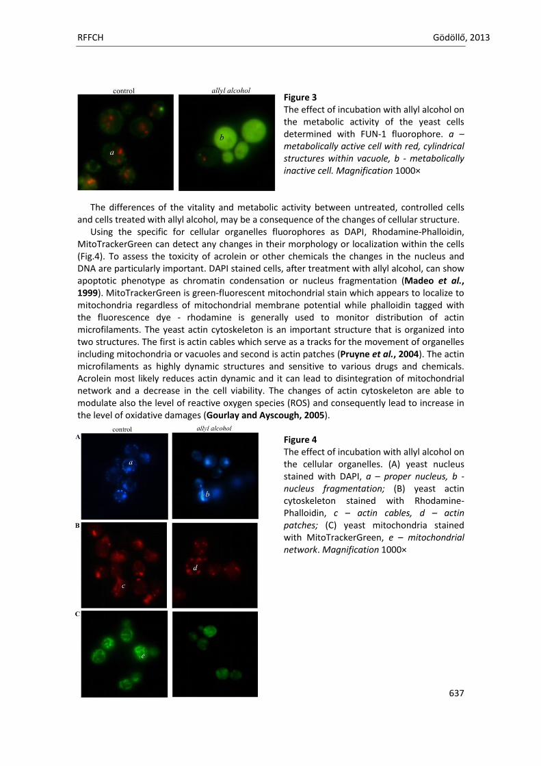
RFFCH Gödöllő, 2013
637
Figure 3 The effect of incubation with allyl alcohol on the metabolic activity of the yeast cells determined with FUN-1 fluorophore. a – metabolically active cell with red, cylindrical structures within vacuole, b - metabolically inactive cell. Magnification 1000×
The differences of the vitality and metabolic activity between untreated, controlled cells
and cells treated with allyl alcohol, may be a consequence of the changes of cellular structure. Using the specific for cellular organelles fluorophores as DAPI, Rhodamine-Phalloidin,
MitoTrackerGreen can detect any changes in their morphology or localization within the cells (Fig.4). To assess the toxicity of acrolein or other chemicals the changes in the nucleus and DNA are particularly important. DAPI stained cells, after treatment with allyl alcohol, can show apoptotic phenotype as chromatin condensation or nucleus fragmentation (Madeo et al., 1999). MitoTrackerGreen is green-fluorescent mitochondrial stain which appears to localize to mitochondria regardless of mitochondrial membrane potential while phalloidin tagged with the fluorescence dye - rhodamine is generally used to monitor distribution of actin microfilaments. The yeast actin cytoskeleton is an important structure that is organized into two structures. The first is actin cables which serve as a tracks for the movement of organelles including mitochondria or vacuoles and second is actin patches (Pruyne et al., 2004). The actin microfilaments as highly dynamic structures and sensitive to various drugs and chemicals. Acrolein most likely reduces actin dynamic and it can lead to disintegration of mitochondrial network and a decrease in the cell viability. The changes of actin cytoskeleton are able to modulate also the level of reactive oxygen species (ROS) and consequently lead to increase in the level of oxidative damages (Gourlay and Ayscough, 2005).
Figure 4 The effect of incubation with allyl alcohol on the cellular organelles. (A) yeast nucleus stained with DAPI, a – proper nucleus, b - nucleus fragmentation; (B) yeast actin cytoskeleton stained with Rhodamine-Phalloidin, c – actin cables, d – actin patches; (C) yeast mitochondria stained with MitoTrackerGreen, e – mitochondrial network. Magnification 1000×
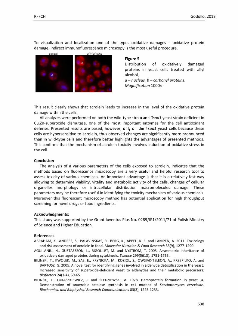
RFFCH Gödöllő, 2013
638
To visualization and localization one of the types oxidative damages – oxidative protein damage, indirect immunofluorescence microscopy is the most useful procedure.
Figure 5 Distribution of oxidatively damaged proteins in yeast cells treated with allyl alcohol, a – nucleus, b – carbonyl proteins. Magnification 1000×
This result clearly shows that acrolein leads to increase in the level of the oxidative protein damage within the cells.
All analyzes were performed on both the wild- sod1 yeast strain deficient in Cu,Zn-superoxide dismutase, one of the most important enzymes for the cell antioxidant defense. Presented results are based, however, sod1 yeast cells because these cells are hypersensitive to acrolein, thus observed changes are significantly more pronounced than in wild-type cells and therefore better highlights the advantages of presented methods. This confirms that the mechanism of acrolein toxicity involves induction of oxidative stress in the cell. Conclusion
The analysis of a various parameters of the cells exposed to acrolein, indicates that the methods based on fluorescence microscopy are a very useful and helpful research tool to assess toxicity of various chemicals. An important advantage is that it is a relatively fast way allowing to determine viability, vitality and metabolic activity of the cells, changes of cellular organelles morphology or intracellular distribution macromolecules damage. These parameters may be therefore useful in identifying the toxicity mechanism of various chemicals. Moreover this fluorescent microscopy method has potential application for high throughput screening for novel drugs or food ingredients. Acknowledgments: This study was supported by the Grant Iuventus Plus No. 0289/IP1/2011/71 of Polish Ministry of Science and Higher Education. References ABRAHAM, K., ANDRES, S., PALAVINSKAS, R., BERG, K., APPEL, K. E. and LAMPEN, A. 2011. Toxicology
and risk assessment of acrolein in food. Molecular Nutrition & Food Research 55(9), 1277-1290. AGUILANIU, H., GUSTAFSSON, L., RIGOULET, M. and NYSTROM, T. 2003. Asymmetric inheritance of
oxidatively damaged proteins during cytokinesis. Science 299(5613), 1751-1753. BILINSKI, T., KWOLEK, M., SAS, E., KRYNICKA, M., KOZIOL, S., OWSIAK-TELEON, A., KRZEPILKO, A. and
BARTOSZ, G. 2005. A novel test for identifying genes involved in aldehyde detoxification in the yeast. Increased sensitivity of superoxide-deficient yeast to aldehydes and their metabolic precursors. Biofactors 24(1-4), 59-65.
BILINSKI, T., LUKASZKIEWICZ, J. and SLEDZIEWSKI, A. 1978. Hemoprotein formation in yeast .4. Demonstration of anaerobic catalase synthesis in cz1 mutant of Saccharomyces cerevisiae. Biochemical and Biophysical Research Communications 83(3), 1225-1233.
RFFCH Gödöllő, 2013
639
BREEUWER, P. and ABEE, T. 2000. Assessment of viability of microorganisms employing fluorescence techniques. International Journal of Food Microbiology 55(1-3), 193-200.
CAFFARO-FILHO, R. A., WAGNER, R., UMBUZEIRO, G. A., GROSSMAN, M. J. and DURRANT, L. R. 2010. Identification of alpha-beta unsaturated aldehydes as sources of toxicity to activated sludge biomass in polyester manufacturing wastewater. Water Science and Technology 61(9), 2317-2324.
EGGLESTON, M. D. and MARSHALL, P. A. 2007. Saccharomyces cerevisiae samples stained with FUN-1 dye can be stored at-20 degrees C for later observation. Journal of Microscopy-Oxford 225(1), 100-103.
GOURLAY, C. W. and AYSCOUGH, K. R. 2005. A role for actin in aging and apoptosis. Biochemical Society Transactions 33), 1260-1264.
KOZIOL, S., ZAGULSKI, M., BILINSKI, T. A. and BARTOSZ, G. 2005. Antioxidants protect the yeast Saccharomyces cerevisiae against hypertonic stress. Free Radical Research 39(4), 365-371.
KWOLEK-MIREK, M. and BARTOSZ, G. 2011. Biochemical and cellular effects of acrolein toxicity in the yeast Saccharomyces cerevisiae, Xenobiotics Environmental exposure, toxicity and physiological response, Published by the University of Rzeszow, 271-281.
KWOLEK-MIREK, M., BEDNARSKA, S., BARTOSZ, G. and BILINSKI, T. 2009. Acrolein toxicity involves oxidative stress caused by glutathione depletion in the yeast Saccharomyces cerevisiae. Cell Biology and Toxicology 25(4), 363-378.
MADEO, F., FROHLICH, E., LIGR, M., GREY, M., SIGRIST, S. J., WOLF, D. H. and FROHLICH, K. U. 1999. Oxygen stress: A regulator of apoptosis in yeast. Journal of Cell Biology 145(4), 757-767.
MINOIS, N., FRAJNT, M., WILSON, C. and VAUPEL, J. W. 2005. Advances in measuring lifespan in the yeast Saccharomyces cerevisiae. Proceedings of the National Academy of Sciences of the United States of America 102(2), 402-406.
PRUYNE, D., LEGESSE-MILLER, A., GAO, L. N., DONG, Y. Q. and BRETSCHER, A. 2004. Mechanisms of polarized growth and organelle segregation in yeast. Annual Review of Cell and Developmental Biology 20), 559-591.
TAYA, M., YOSHIKAWA, M. and KOBAYASHI, T. 1989. Measurement of viable animal-cell concentration based on fluorometry. Journal of Chemical Engineering of Japan 22(1), 89-94.
ZHENG, K., PAN, J.-W., YE, L., FU, Y., PENG, H.-Z., WAN, B.-Y., GU, Q., BIAN, H.-W., HAN, N., WANG, J.-H., KANG, B., PAN, J.-H., SHAO, H.-H., WANG, W.-Z. and ZHU, M.-Y. 2007. Programmed cell death-involved aluminum toxicity in yeast alleviated by antiapoptotic members with decreased calcium signals. Plant Physiology 143(1), 38-49.
Animal welfare, etológia és tartástechnológia
Animal welfare, ethology and housing systems
Volume 9 Issue 3
Különszám/Special Issue
Gödöllő
2013
RFFCH Gödöllő, 2013
640
Acute Toxicity of Nickel Towards Freshwater Ciliate Paramecium bursaria
Patrycja Zagata & Magdalena Greczek-Stachura Institute of Biology, Pedagogical University, 30-084 Krakow Podchorążych 2
Heavy metals are common contaminants present in aquatic environment and biological systems. The ciliated protista are generally the most abundant group in aquatic ecosystems and they can be used as an effective biological indicators of some pollutants. Paramecium bursaria is an unicellular organism which is widely distributed in freshwater environment. P. bursaria cells harbor several hundred symbiotic algae in their cytoplasm. The main purpose of this study was to determine in laboratory tests the acute toxicity of nickel chloride (NiCl2) to Paramecium bursaria. The ciliates were incubated in solutions with 5x10⁻⁶ to 5x10⁻¹g/dm3 of NiCl2 at the temperature of 180C, in the light/dark conditions (12L/12D). Observations of cell shape and rate of cell division as well as the swimming behavior of paramecia were conducted after 24, 72 and 120 hours of incubation in the tested solutions and were compared to the control group. Nickel ions might be recognized as one of the most effective immobilizatory agents for cilates. After 10 minutes from the beginning of the incubation the velocity of swimming falls simultaneously the spiralization diminishes and the rotary movement round the long body axis become slower. After next few minutes ciliates becomes a short crawling and stop more frequently. In the immobilization phase cilia of the whole cell work but without performing an effective stroke. The next macronucleus becomes distinctly visible, the vacuoles enlarge and the cell is being deformed. Microscopic observations revealed that the lethal dose was 5x10⁻⁴g/dm3 and that in less constrained solutions the slowdown and characteristic movements like overturns were observed. The PEA measurements were carried on within 4 days, the first after a 24-hour incubation period in nickel solution. The measured parameter was Fv/Fm. Fv/Fm is a parameter widely used to indicate the maximum quantum efficiency of Photosystem II. These investigations revealed the highest fluorescence efficiency in culture incubating in the least concentration and the shortest time and the lowest one in a control sample, what can indicate the stimulating role of nickel to cells of Paramecium bursaria.
Animal welfare, etológia és tartástechnológia
Animal welfare, ethology and housing systems
Volume 9 Issue 3
Különszám/Special Issue
Gödöllő
2013
RFFCH Gödöllő, 2013
641
HAEMATOLOGICAL CHANGES IN RABBIT’S BLOOD AFTER TWO WEEKS EXPOSURE OF PATULIN.
Katarina Zbynovska*1, Jana Emrichova1, Anna Kalafova1, Peter Petruska1, Lubomir Ondruska2, Rastislav Jurcik2, Lubica Chrastinova2, Anton Kovacik1, Monika Schneidgenova1,Eva Tusimova1, Marcela Capcarova1
1Department of Animal Physiology, Faculty of Biotechnology and Food Sciences, Slovak University of Agriculture in Nitra, Tr. A. Hlinku 2, 949 76 Nitra, Slovak Republic 2Animal Production Research Centre Nitra, Hlohovecka 2, 949 01 Nitra, Slovak Republic
Abstract The aim of present study was to analyse haematological parameters in blood of rabbits after patulin administration during two weeks. Animals (adult female rabbits, body weight 4 ± 0.5 kg) were divided into two groups: control group (C) and experimental group (E). Animals in E group received intramuscular injection of patulin (10 µg.kg-1) 4 times. After 2 weeks of exposure the blood was collected and selected haematological parameters (total white blood cell count, lymphocytes count, medium size cell count, granulocytes count, red blood cell count, haemoglobin, haematocrit, mean corpuscular volume, mean corpuscular haemoglobin, mean corpuscular haemoglobin concentration, red cell distribution width, platelet count, platelet percentage, mean platelet volume and platelet distribution width) were analysed by Abacus Junior Vet (Diatron®, Vienna, Austria). Significant decrease (P < 0.05) of mean corpuscular haemoglobin concentration in E group in comparison with the C group was observed. In the case of hemoglobin concentration no significant change between both groups were found. Patulin had no effect on the other analysed haematological parameters. Keywords: patulin, haematology, rabbit Introduction Patulin, a genotoxic mycotoxin produced by several species of Aspergillus, Penicillium and Bysochlamys (Puel et al., 2010; Ozsoy et al., 2008) is the most common mycotoxin in apples and apple-derived products (Puel et al., 2010). It contaminates further fruits, such as grapes, oranges, pears, oranges, pears and peaches (Saxena et al., 2009; Ozsoy et al., 2008). The effects caused by this mycotoxin are based on several studies made during the past fifty years; these involve acute, chronic and cellular level effects (González-Osnaya et al., 2007). Cellular effects of patulin include formation of reactive oxygen species, cell cycle arrest, cytochrome c release from mitochondria, caspase-3 activation, PARP cleavage, ATF3 expression and subseyuent apoptosis (Kwon et al., 2012; Fribley et al., 2011; Ozsoy et al., 2008). Patulin may be neurotoxic, immunotoxic, immunosuppressive, genotoxic, teratogenic and carcinogenic (González-Osnaya et al., 2007). In vivo patulin caused severe damage in several organ systems like kidney, intestinal tissue (McKinley et al., 1982; Speijers et al., 1988) and immune system (Escoula et al., 1988), if applied at a range between 2.5 and 41 mg.kg-1 bw. Regarding carcinogenicity, the International Agency for Research on Cancer classified patulin in group C, since the evidence of carcinogenicity was considered limited in experimental animal (International Agency for Research on Cancer, 1986). Patulin has a strong affinity for sulfhydryl groups. Patulin adducts formed with cysteine are less toxic than the unmodified compound in acute toxicity, teratogenicity, and mutagenicity studies. Its affinity for SH-groups explains its inhibition of many enzymes (Puel et al., 2010). Toxic signs consistently reported in all studies were agitation, in some cases convulsions, dyspnea, pulmonary congestion, edema, and ulceration, hyperemia and distension of the gastro intestinal tract (WHO, 1998). Based on the provisional maximum tolerable daily intake (400 ng.kg-1 bw/day), several countries have set legislations for the maximum amount of patulin in apples products. In the Europen Unioin, the limit is
RFFCH Gödöllő, 2013
642
set to 50 µg.kg-1 in apple juice and cider, 25 µg.kg-1 in solid apple products (e.g. apple sauce) and 10 µg.kg-1 in products for infants and young children (Commission of the European Communities, 2003). The aim of present study was to analyse haematological parameters in blood of rabbits after patulin administration during two weeks. Materials and Methods Animals Adult female rabbits (n = 32), maternal albinotic line (crossbreed Newzealand white, Buskat rabbit, French silver) and paternal acromalictic line (crossbreed Nitra’s rabbit, Californian rabbit, Big light silver) were used in experiment. Rabbits were healthy and their condition was judged as good at the commencement of the experiment. Rabbits were healthy and their condition was judged as good at the commencement of the experiment. Water was available at any time from automatic drinking troughs. Groups of adult animals were balanced for age (150 days) and body weight (4 ± 0.5 kg) at the beginning of the experiment. Adult rabbits were fed diet of a 12.35 MJ.kg-1 of metabolizable diet (Table 1) composed of a pelleted concentrate. Animals were divided into two groups: control group (C) without addition of patulin and experimental group (E) with addition of patulin (10 µg.kg-1). Animlas from experimental group received patulin through intramuscular injection twice a week for two weeks. In this animal study, institutional and national guidelines for the care and use of animals were followed, and all experimental procedures involving animals were approved by ethical committee. Blood sampling and analyses Blood samples from vena auricularis were taken from all animals by macromethods after two weeks of patulin administration. In whole blood, selected haematological parameters [total white blood cell count (WBC), lymphocytes count (LYM), mediumsize cell count (MID), granulocytes count (GRA), red blood cell count (RBC), haemoglobin (HGB), haematocrit (HCT), mean corpuscular volume (MCV), mean corpuscular haemoglobin (MCH), mean corpuscular haemoglobin concentration (MCHC), red cell distribution width (RDWc), platelet count (PLT), platelet percentage (PCT), mean platelet volume (MPV) and platelet distribution width (PDWc)] were measured using haematology analyzer Abacus junior VET (Diatron®, Vienna, Austria). Statistical analyses The data used for statistical analyses represent means of values obtained in blood collection. To compare the results, t-test was applied to calculate basic statistic characteristics and to determine significant differences among the experimental and control groups. Statistical software SIGMA PLOT 12.0 (Jandel, Corte Madera, CA, USA) was used. Differences were compared for statistical significance at the level P< 0.05.
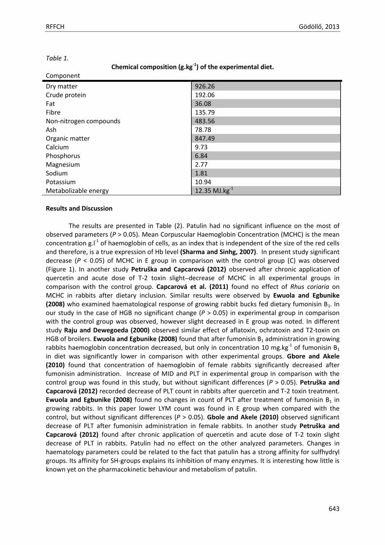
RFFCH Gödöllő, 2013
643
Table 1.
Chemical composition (g.kg-1) of the experimental diet. Component
Dry matter 926.26 Crude protein 192.06 Fat 36.08 Fibre 135.79 Non-nitrogen compounds 483.56 Ash 78.78 Organic matter 847.49 Calcium 9.73 Phosphorus 6.84 Magnesium 2.77 Sodium 1.81 Potassium 10.94 Metabolizable energy 12.35 MJ.kg-1
Results and Discussion The results are presented in Table (2). Patulin had no significant influence on the most of observed parameters (P > 0.05). Mean Corpuscular Haemoglobin Concentration (MCHC) is the mean concentration g.l-1 of haemoglobin of cells, as an index that is independent of the size of the red cells and therefore, is a true expression of Hb level (Sharma and Sinhg, 2007). In present study significant decrease (P < 0.05) of MCHC in E group in comparison with the control group (C) was observed (Figure 1). In another study Petruška and Capcarová (2012) observed after chronic application of quercetin and acute dose of T-2 toxin slight decrease of MCHC in all experimental groups in comparison with the control group. Capcarová et al. (2011) found no effect of Rhus coriaria on MCHC in rabbits after dietary inclusion. Similar results were observed by Ewuola and Egbunike (2008) who examined haematological response of growing rabbit bucks fed dietary fumonisin B1. In our study in the case of HGB no significant change (P > 0.05) in experimental group in comparison with the control group was observed, however slight decreased in E group was noted. In different study Raju and Dewegoeda (2000) observed similar effect of aflatoxin, ochratoxin and T2-toxin on HGB of broilers. Ewuola and Egbunike (2008) found that after fumonisin B1 administration in growing rabbits haemoglobin concentration decreased, but only in concentration 10 mg.kg-1 of fumonisin B1 in diet was significantly lower in comparison with other experimental groups. Gbore and Akele (2010) found that concentration of haemoglobin of female rabbits significantly decreased after fumonisin administration. Increase of MID and PLT in experimental group in comparison with the control group was found in this study, but without significant differences (P > 0.05). Petruška and Capcarová (2012) recorded decrease of PLT count in rabbits after quercetin and T-2 toxin treatment. Ewuola and Egbunike (2008) found no changes in count of PLT after treatment of fumonisin B1 in growing rabbits. In this paper lower LYM count was found in E group when compared with the control, but without significant differences (P > 0.05). Gbole and Akele (2010) observed significant decrease of PLT after fumonisin administration in female rabbits. In another study Petruška and Capcarová (2012) found after chronic application of quercetin and acute dose of T-2 toxin slight decrease of PLT in rabbits. Patulin had no effect on the other analyzed parameters. Changes in haematology parameters could be related to the fact that patulin has a strong affinity for sulfhydryl groups. Its affinity for SH-groups explains its inhibition of many enzymes. It is interesting how little is known yet on the pharmacokinetic behaviour and metabolism of patulin.
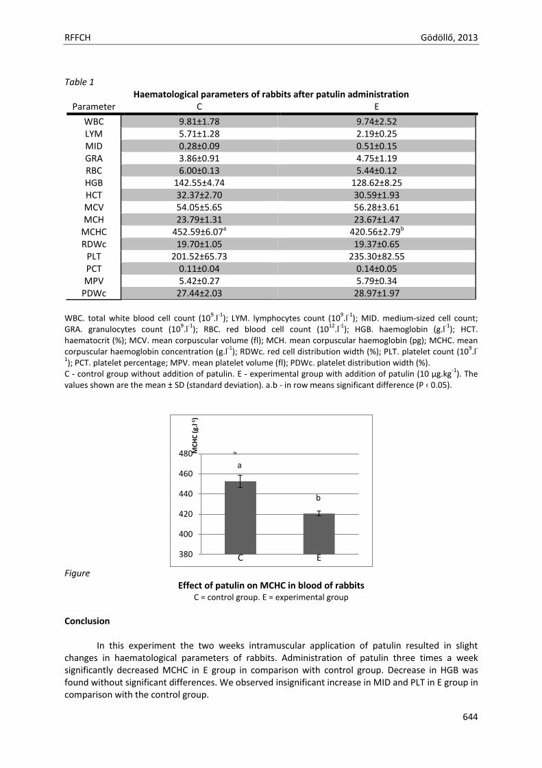
RFFCH Gödöllő, 2013
644
Table 1
Haematological parameters of rabbits after patulin administration Parameter C E
WBC 9.81±1.78 9.74±2.52 LYM 5.71±1.28 2.19±0.25 MID 0.28±0.09 0.51±0.15 GRA 3.86±0.91 4.75±1.19 RBC 6.00±0.13 5.44±0.12 HGB 142.55±4.74 128.62±8.25 HCT 32.37±2.70 30.59±1.93 MCV 54.05±5.65 56.28±3.61 MCH 23.79±1.31 23.67±1.47
MCHC 452.59±6.07a 420.56±2.79b
RDWc 19.70±1.05 19.37±0.65 PLT 201.52±65.73 235.30±82.55 PCT 0.11±0.04 0.14±0.05 MPV 5.42±0.27 5.79±0.34
PDWc 27.44±2.03 28.97±1.97
WBC. total white blood cell count (10
9.l
-1); LYM. lymphocytes count (10
9.l
-1); MID. medium-sized cell count;
GRA. granulocytes count (109.l
-1); RBC. red blood cell count (10
12.l
-1); HGB. haemoglobin (g.l
-1); HCT.
haematocrit (%); MCV. mean corpuscular volume (fl); MCH. mean corpuscular haemoglobin (pg); MCHC. mean corpuscular haemoglobin concentration (g.l
-1); RDWc. red cell distribution width (%); PLT. platelet count (10
9.l
-
1); PCT. platelet percentage; MPV. mean platelet volume (fl); PDWc. platelet distribution width (%).
C - control group without addition of patulin. E - experimental group with addition of patulin (10 µg.kg-1
). The values shown are the mean ± SD (standard deviation). a.b - in row means significant difference (P ‹ 0.05).
Figure
Effect of patulin on MCHC in blood of rabbits C = control group. E = experimental group
Conclusion
In this experiment the two weeks intramuscular application of patulin resulted in slight changes in haematological parameters of rabbits. Administration of patulin three times a week significantly decreased MCHC in E group in comparison with control group. Decrease in HGB was found without significant differences. We observed insignificant increase in MID and PLT in E group in comparison with the control group.
380
400
420
440
460
480 MC
HC
(g.
l-1)
C E
a
b
a
RFFCH Gödöllő, 2013
645
Further investigation with haematological response of animals will be worthy of further investigation. Acknowledgments This work was financially supported by VEGA scientific grant, 1/0084/12, and 1/0022/13. References Capcarová, M., Slamečka, J., Abbas, K., Kolesárová, A., Kalafová, A., Valent, M., Filipejová, T., Chrastinová, L.,
Ondruška, L., Massányi, P. (2011): Effects of dietary inclusion of Rhus coriaria on internal milieu of rabbits. Journal of Animal Physiology and Animal Nutrition, 96. 459-465.
Commission of thze European Communities. Commission Regulation (EC) No 1425/2003 of 11 August 2003 amending Regulation (EC) No 466/2001 as regards patulin 2003.
Escuola, L., Thomsen, M., Bourdiol, D., Pipy, B., Peuriere, S., Roubinet, F. (1988): Patulin immunotoxicology:effect on ohagocyte activation and the cellular and humoral immune system of mice and rabbits. International Journal of Immunopharmacology, 10. 983-989.
Ewuola, E.O., Egbunike, G.N. (2008): Haematological and serum biochemical response of growing rabbit bucks fed dietary fumonisin B1. African Journal of Biotechnology, 7. 4304-4309.
Fribley, A.M., Cruz, P.G., Miller, J.R., Callaghan, M.U., Cai, P., Narula, N., Neubig, R.R., Showalter, H.D., Larsen, S.D., Kirchhoff, P.D., Larsen, M.J., Burr, D.A., Schultz, P.J., Jacobs, R.R., Tamayo-Castillo, G., Ron, D., Sherman, D.H., Kaufman, R.J. (2011): Complementary cell-based high-throughput screens identify novel modulators of the unfolded protein response. Journal of Biomolecular Screening, 16. 825-835.
Gbore, F.A., Akele, O. (2010): Growth performance, haematology and serum biochemistry of female rabbits (Oryctolagus cuniculus) fed dietary fumonisin. Veterinarski Arhiv, 80. 431-443.
González-Osnaya, L., Soriano, J.M., Moltó, J.C., Mañes, J. (2007): Exposure to patulin from consumption of Apple-based products. Food Aditives and Contaminants, 24. 1268-1274.
IARC. (1986): Some Naturally Occurring and Synthetic Food Components, Furocoumarins and Ultraviolet Radiation. Monographs on the Evaluation of the Carcinogenic Risk of Chemicals to Humans. 40. 83–98.
Kwon, O., Soung, N.K., Thimmegowda, N.R., Jeong, S.J., Jang, J.H., Moon, D.O., Chung, J.K., Lee, K.S., Kwon, Y.T., Erikson, R.L., Ahn, J.S., Kim, B.Y. (2012): Patulin induces colorectal cancer cells apoptosis through EGR-1 dependent ATF3 up-regulation. Cell Signal, 24. 943-950.
McKinley, E.R., Carlton, W.W., Boon, G.D. (1982): Patulin mycotoxicosis in the rat: toxicology, pathology and clinical pathology. Food and Chemical Toxicology, 20. 289-300.
Ozsoy, N., Selmanoglu, G., Kockaya, E. A., Gul, N., Cebesoy, S. (2008): Effect of patulin on the interdigitating dendritic cells (IDCs) of rat thymus. Cell Biochemistry and Function, 26. 192-196.
Petruška, P., Capcarová, M. (2012): Effect of chronic application of quercetin and acute dose of T-2 toxin on haematological parameters of rabbits. Mendel Net, Internation students conference 2012 in Brno. 981-988.
Puel, O., Galtier, P., Oswald, I.P. (2010): Biosynthesis and toxicological effects of patulin. Toxins, 2. 613-631. Raju, M.V., Devegowda, G. (2000): Influence of esterified-glucomannan on performance and organ
morphology, serum biochemistry and haematology in broilers exposed to individual and combined mycotoxicosis (aflatoxin, ochratoxin and T-2 toxin). British Pultry Science, 41. 640-650.
Saxena, N., Ansari, K.M., Kumar, R., Dhawan, A., Dwivedi, P.D., Das, M. (2009): Patulin causes DNA damage leading to cell cycle arrest and apoptosis through modulation of Bax, p(53) and p(21/WAF1) proteins in skin of mice. Toxicology and Applied Pharmacology, 234. 192-201.
Sharma, G., Singh, S. (2007): Effect of indofil toxicity on MCHC of Channa punctatus. Journal of Environmental Research And Development, 1. 261-263.
Speijers, G.J., Franken, M.A., van Leeuwen, F.X. (1988): Subacute toxicity study of patulin in the rat:effects on the kidney and the gastrointestinal tract. Food and Chemical Toxicology, 26. 23-30.
WHO. Joint FAO/WHO Expert Committee on Food additives (JECFA), Position paper on patulin, 30th session, The Hague, The Netherlands, 9–13 March, 1998.
Animal welfare, etológia és tartástechnológia
Animal welfare, ethology and housing systems
Volume 9 Issue 3
Különszám/Special Issue
Gödöllő
2013
RFFCH Gödöllő, 2013
646
ENTEROTOXIGENIC POTENTIAL THE SAME BACTERIA OF STAPHYLOCOCCUS SPP. IN MASTITIS OF DAIRY COWS AND SHEEP
Zigo F1, Vasiľ M*1, Elečko J1, Farkašová Z1, Bulla J.2
1. Department of Animal Husbandry, University of Veterinary Medicine and Pharmacy, Komenskeho 73, 041 81 Kosice, Slovak Republic 2. Slovak University of Agriculture, A. Hlinku, Nitra, Slovak Republic
Abstract The aim of this work was to evaluate the occurrence of Staphylococcus spp. on the overall prevalence of mastitis in cows and sheep farms in eastern Slovakia, and determine incidence of enterotoxigenicity and other characteristics of bacterial isolates. Were tested presence of genes encoding production of enterotoxins (sea, seb, sec, sed, see) and the production individual types of staphylococcal enterotoxins (SEA, SEB, SEC, SED, SEE) in milk samples of cows and sheep. In a two-year experiment of testing 2916 individual samples of cow's and sheep's milk were recorded bacteria Staphylococcus spp. 18.41% (537). Using PCR methods was confirmed the presence of the gene (seg) encoding staphylococcal enterotoxins (SE) in 5.21% (28 cases). By using the Ridascreen® Set, (B), (C), (D), (E) was recorded in vitro production of SE in 4.28% (23 cases). Four species isolated from samples of cow's milk produced the following types of SE: S. aureus - SEC (4) and SEE (1), S. haemolyticus - SEA, S. chromogenes - SEC (2) and SED (1), S. simulans - SEA (1). The staphylococci isolated from samples of sheep's milk produce SE: S. aureus - SEA, SEB (1) and (2) SEC (3), S. intermedius, S. chromogenes - SEC-SEC (3), S. xylosus - SED (1). A common feature of the enterotoxigenic bacteria was the creation of a biofilm. Formation of hyaluronidase and lysins α, β and δ have been confirmed as positive for S. aureus and S. intermedius. Creation of lysine α has been confirmed for strains of S. chromogenes. The production of hyaluronidase in CPS was significant on significant level α = 0.001. Formation of hemolysines only on significant level α = 0.01. Other following properties were non significant.
In spite legislatively obligatory pasteurization of milk before further processing, a relatively high percentage of enterotoxigenic strains of staphylococci isolated from the milk of sheep and cows can pose a significant risk especially in the marketing of milk and milk products directly from the farm as organic products.
Keywords: mastitis; dairy cow; sheep; staphylococcal enterotoxins; genes
Introduction Bacteria of Staphylococcus spp. as the cause of a wide range of diseases in humans and animals are
well known, but in recent decades, especially coagulase-negative staphylococci (CNS) were subsequently diagnosed as the aetiological agent of a wide range of infectious processes and healthy complications (Bansal et al. 2004, Favre et al. 2005, Huang et al. 2006, Lalani et al. 2006). Common are also reported increases frequency of CNS as ruminant mastitis agents in many countries, with significant impact on the economy of milk production.
From samples of cows and sheep milk were isolated strains of bacteria, in which by ELISA and PCR methods were determined the production some of the types of staphylococcal enterotoxins (SE), and was confirmed the presence of the gene (seg) responsible for the production of SE (Fagundes et al. 2010).
Enterotoxigenic staphylococci activity is an important virulence factor and was mentioned in relation to their specific contribution to the pathogenesis of mastitis (da Silva et al. 2005) with very frequent

RFFCH Gödöllő, 2013
647
property is the ability to naturally acquire resistance to antibiotics (Orwin et al. 2001). Among
equally
important virulence factors of pathogens mammary gland include biofilm formation (Melchior et al. 2006).
Treatment of dairy milk for consumption ensures a standard of consumer protection to the public prior to the occurrence of staphylococcal enterotoxicosis. The free market, however, allows the consumption of milk in homes and other alternative sources without pasteurization. The solution for reducing the risk of enterotoxicosis in these cases is treatment by boiling milk.
The aim of this work was to evaluate the occurrence of Staphylococcus spp. on the overall prevalence of mastitis in cows and sheep farms in eastern Slovakia, and determine incidence of enterotoxigenicity and other characteristics of bacterial isolates.
Materials and methods
In the course of two years in the framework of the periodic complex examination were in herd of 120 dairy cows taken 968 samples, and in herd of 350 sheep exanimate 1948 individual milk samples. Clinical examination of udder, classification of mastitis and bacteriology examination were carried out according to Vasiľ (2004).
Taxonomy of staphylococci was used by set STAPHYtest 24 and evaluated by program TNW, version 7.0 ProAuto (Erba Lachema, Brno, ČR). Identified bacteria Staphylococcus spp. were examined on presence of genes coding their enterotoxigenic activity (sea, seb, sec, sed, see) by PCR method according to (Becker et al. 1998). On the separation of DNA was used QiAMP tissue kit (OIAGEM, Hilden, Germany). Within the PCR method were used reference strains for SEA, SAB, SEC, SED and SEE types of enterotoxins – (Bergdoll; CNCTC, Brno) and commercial oligonukleotids primers. Every strain with confirmed gene was examined for production of enterotoxins by Ridascreen ® Set A, B, C, D, E (R-Biopharm AG, Darmstadt, Germany).
In relation to forms of mastitis were in enterotoxigenic strains of staphylococci followed production of hemolysins α, β, δ, hyaluronidase and production of biofilm by methods according to Türkyilmaz and Kaya (2006), Boynukara et al. (2008), Krukowski et al. (2008).
On statistical evaluation was used Chi-quadrate test, was tested individual properties of enterotoxigenc CNS compare to enterotoxigenic CPS at significant level α = 0.001 (0.1%), and significant level α = 0.01, testing criteria G = 45.387; critical value df = 32.91.
Results and discussion
From 2916 examined milk samples were (24.24%) bacteriological positive while Staphylococcus spp. was found in 537 cases (18.41%). Summary of isolated staphylococci are described in Table 1.
Table 1 Total result of bacteriology examinations, prevalence of mastitis, percentage ration
of the Staphylococcus spp. isolated from individual milk samples of dairy cows and sheep
Individually milk samples Staphylococcus spp.
Sample examined positive find1 CPS CNS ∑ n n % n % n % n %
dairy cow 968 280 28.92 21 2.17 170 17.56 191 19.73
sheep 1948 427 19.73 38 1.95 308 15.81 346 17.76
summary 2916 707 24.24 59 2.02 478 16.39 537 18.41 1 prevalence of mastitis (percentage of positive cases from total number of examined cows and sheep); CPS – coagulase-positive staphylococci (S.aureus; S.intermedius); CNS – coagulase-negative staphylococci
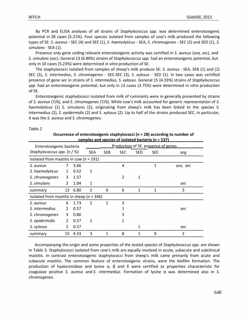
RFFCH Gödöllő, 2013
648
By PCR and ELISA analyses of all strains of Staphylococcus spp. was determined enterotoxigenic potential in 28 cases (5.21%). Four species isolated from samples of cow's milk produced the following types of SE: S. aureus - SEC (4) and SEE (1), S. haemolyticus - SEA, S. chromogenes - SEC (2) and SED (1), S. simulans - SEA (1).
Presence only gene coding relevant enterotoxigenic activity was certified in S. aureus (sea, sec), and S. simulans (sec). General 13 (6.80%) strains of Staphylococcus spp. had an enterotoxigenic potential, but only in 10 cases (5.23%) were determined in vitro production of SE.
The staphylococci isolated from samples of sheep's milk produce SE: S. aureus - SEA, SEB (1) and (2) SEC (3), S. intermedius, S. chromogenes - SEC-SEC (3), S. xylosus - SED (1). In two cases was certified presence of gene sec in strains of S. intermedius, S. xylosus. General 15 (4.33%) strains of Staphylococcus spp. had an enterotoxigenic potential, but only in 13 cases (3.75%) were determined in vitro production of SE.
Enterotoxigenic staphylococci isolated from milk of ruminants were in generally presented by strains of S. aureus (7/6), and S. chromogenes (7/6). While cow's milk accounted for generic representation of S. haemolyticus (1) S. simulanss (2), originating from sheep's milk has been linked to the species S. intermedius (2), S. epidermidis (2) and S. xylosus (2). Up to half of the strains produced SEC, in particular, it was the S. aureus and S. chromogenes.
Table 2
Occurrence of enterotoxigenic staphylococci (n = 28) according to number of samples and species of isolated bacteria (n = 537)
Enterotoxigenic bacteria Production of SE, presence of genes Staphylococcus spp. (n / %) SEA SEB SEC SED SEE seg
Isolated from mastitis in cow (n = 191)
S. aureus 7 3.66 4 1 sea, sec
S. haemolyticus 1 0.52 1
S. chromogenes 3 1.57 2 1
S. simulans 2 1.04 1 sec
summary 13 6.80 2 0 6 1 1 3
Isolated from mastitis in sheep (n = 346)
S. aureus 6 1.73 2 1 3
S. intermedius 2 0.57 1 sec
S. chromogenes 3 0.86 3
S. epidermidis 2 0.57 1 1
S. xylosus 2 0.57 1 sec
summary 15 4.33 3 1 8 1 0 2
Accompanying the origin and some properties of the tested species of Staphylococcus spp. are shown in Table 3. Staphylococci isolated from cow's milk are equally involved in acute, subacute and subclinical mastitis. In contrast enterotoxigenic staphylococci from sheep's milk came primarily from acute and subacute mastitis. The common feature of enterotoxigenic strains, were the biofilm formation. The production of hyaluronidase and lysine α, β and δ were certified as properties characteristic for coagulase positive S. aureus and S. intermedius. Formation of lysine α was determined also in S. chromogenes.
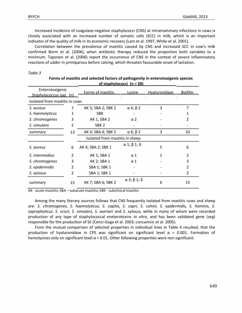
RFFCH Gödöllő, 2013
649
Increased incidence of coagulase-negative staphylococci (CNS) at intramammary infections in cows is closely associated with an increased number of somatic cells (SCC) in milk, which is an important indicator of the quality of milk in its economic recovery (Lam et al. 1997, White et al. 2001).
Correlation between the prevalence of mastitis caused by CNS and increased SCC in cow's milk confirmed Borm et al. (2006), when antibiotic therapy reduced the proportion both variables to a minimum. Taponen et al. (2006) report the occurrence of CNS in the context of severe inflammatory reactions of udder in primiparous before calving, which threaten favourable onset of lactation.
Table 3
Forms of mastitis and selected factors of pathogenity in enterotoxigenic species of staphylococci (n = 28)
Enterotoxigenic
Staphylococcus spp. (n) Forms of mastitis Lysine Hyaluronidase Biofilm
Isolated from mastitis in cows
S. aureus 7 AK 3; SBA 2; SBK 2 α 4; β 2 3 7
S. haemolyticus 1 SBK - - 1
S. chromogenes 3 AK 1, SBA 2 α 2 - 2
S. simulans 2 SBK 2 - - -
summary 13 AK 4; SBA 4; SBK 5 α 6; β 2 3 10
Isolated from mastitis in sheep
S. aureus
6
AK 4; SBA 2; SBK 1 α 1; β 1; δ
5
6
S. intermedius 2 AK 1; SBA 1 α 1 1 2
S. chromogenes 3 AK 2; SBA 1 α 1 - 3
S. epidermidis 2 SBA 1; SBK 1 - - 2
S. xylosus 2 SBA 1; SBK 1 - - 2
summary
15
AK 7; SBA 6; SBK 2 α 3; β 1; δ
6
15
AK - acute mastitis; SBA – subacute mastitis; SBK - subclinical mastitis
Among the many literary sources follows that CNS frequently isolated from mastitis cows and sheep
are: S. chromogenes, S. haemolyticus, S. capitis, S. capri, S. cohnii, S. epidermidis, S. hominis, S. saprophyticus. S. sciuri, S. simulans, S. warneri and S. xylosus, while in many of whom were recorded production of any type of staphylococcal eneterotoxins in vitro, and has been validated gene (seg) responsible for the production of SE (Cenci-Goga et al. 2003; Loncarevic et al. 2005).
From the mutual comparison of selected properties in individual lines in Table 4 resulted, that the production of hyaluronidase in CPS was significant on significant level α = 0.001. Formation of hemolysines only on significant level α = 0.01. Other following properties were non significant.
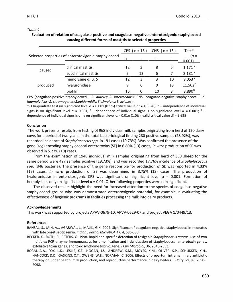
RFFCH Gödöllő, 2013
650
Table 4 Evaluation of relation of coagulase-positive and coagulase-negative enterotoxigenic staphylococci
causing different forms of mastitis to selected properties
CPS ( n = 15 ) CNS ( n = 13 ) Test* Selected properties of enterotoxigenic staphylococci (α =
+ - + - 0.001)
caused clinical mastitis 12 3 8 5 1.171 N
subclinical mastitis 3 12 6 7 2.181 N
hemolysine α, β, δ 12 3 3 10 9.053 X
produced hyaluronidase 9 6 0 13 11.502Z
biofilm 15 0 10 3 3.890N
CPS (coagulase-positive staphylococci – S. aureus; S. intermedius); CNS (coaguase-negative staphylococci – S. haemolyticus; S. chromogenes; S.epidermidis; S. simulans; S. xylosus); *- Chi-quadrate test (in significant level α = 0.001 (0.1%) critical value df = 10.828); N – independence of individual signs is on significant level α = 0.001; Z – dependence of individual signs is on significant level α = 0.001; X – dependence of individual signs is only on significant level α = 0.01n (1.0%), valid critical value df = 6.635
Conclusion The work presents results from testing of 968 individual milk samples originating from herd of 120 dairy
cows for a period of two years. In the total bacteriological finding 280 positive samples (28.92%), was recorded incidence of Staphylococcus spp. in 191 cases (19.73%). Was confirmed the presence of the gene (seg) encoding staphylococcal enterotoxins (SE) in 6.80% (13) cases, in vitro production of SE was observed in 5.23% (10) cases.
From the examination of 1948 individual milk samples originating from herd of 350 sheep for the same period were 427 samples positive (19.73%), and was recorded 17.76% incidence of Staphylococcus spp. (346 bacteria). The presence of the gene responsible for production of SE was reported in 4.33% (15) cases. In vitro production of SE was determined in 3.75% (13) cases. The production of hyaluronidase in enterotoxigenic CPS was significant on significant level α = 0.001. Formation of hemolysines only on significant level α = 0.01. Other following properties were non significant.
The observed results highlight the need for increased attention to the species of coagulase-negative staphylococci groups who was demonstrated enterotoxigenic potential, for example in evaluating the effectiveness of hygienic programs in facilities processing the milk into dairy products.
Acknowledgements This work was supported by projects APVV-0679-10, APVV-0629-07 and project VEGA 1/0449/13.
References BANSAL, S., JAIN, A.., AGARWAL, J., MALIK, G.K. 2004. Significance of coagulase negative staphylococci in neonates
with late onset septicaemia. Indian J Pathol Microbiol, 47, 4, 586-588. BECKER, K., ROTH, R., PETERS, G. 1998. Rapid and specific detection of toxigenic Staphylococcus aureus: use of two
multiplex PCR enzyme immunoassays for amplification and hybridization of staphylococcal enterotoxin genes, exfoliative toxin genes, and toxic syndrome toxin-1 gene. J Clin Microbiol, 36, 2548-2553.
BORM, A.A., FOX, L.K., LESLIE, K.E., HOGAN, J.S., ANDREW, S.M., MOYES, K.M., OLIVER, S.P., SCHUKKEN, Y.H., HANCOCK, D.D., GASKINS, C.T., OWENS, W.E., NORMAN, C. 2006. Effects of prepartum intramammary antibiotic therapy on udder health, milk production, and reproductive performance in dairy heifers. J Dairy Sci, 89, 2090- 2098.
RFFCH Gödöllő, 2013
651
BOYNUKARA, B., GULHAN, T., ALISARLI, M., GURTURK, K., SOLMAZ, H. 2008. Classical enterotoxigenic characteristics of Staphylococcus aureus strains isolated from bovine subclinical mastitis in Van. Turkey. Int J Food Microbiol, 125, 209-211.
CENCI-GOGA, B.T., KARAMA, M., ROSSITTO, P.V., MORGANTE, R.A., CULLOR, J.S. 2003. Research note enterotoxin production by Staphylococcus aureus isolated from mastitic cows. J Food Protect, 66, 9, 1693–1696.
DA SILVA, E.R., DO CARMO, L.S., DA SILVA, N. 2005. Detection of the enterotoxins A, B, and C genes in Staphylococcus aureus from goat and bovine mastitis in Brazilian dairy herds. Veterinary Microbiology, 106, 103–107.
FAGUNDES, H., BARCHESI, L., FILHO, A.N., FERREIRA, L.M., OLIVEIRA, C.A.F. 2010. Occurrence of Staphylococcus aureus in raw milk produced in dairy farms in São Paulo state, Brazil. Braz J Microbiol, 41, 376-380.
FAVRE, B., HUGONNET, S., CORREA, L., SAX, H., ROHNER, P., PITTET, D. 2005. Nosocomial bacteremia: clinical significance of a single blood culture positive for coagulase-negative staphylococci. Infect Control Hosp Epidemiol, 26, 8, 697-702.
HUANG, YHU-CH., WANG, Y.H., CHOU, YI-H., LIEN, R. 2006. Significance of coagulase-negative staphylococci isolated from a single blood culture from neonates in intensive care. Ann Trop Paediatr, 26(4), 311-318.
KRUKOWSKI, H., SZYMANKIEWICZ, M., LISOWSKI, A. 2008. Slime production by Staphylococcus aureus strains isolated from cases of bovine mastitis. Pol J Microbiol, 57, 3, 253-255.
LALANI, T., KANAFANI, Z.A., CHU, V.H., MOORE, L., COREY, G.R., PAPPAS, P., WOODS, C.W., CABELL, C.H., HOEN, B., SELTON-SUTY, C., DOCO-LECOMPTE, T., CHIROUZE, C., RAOULT, D., MIRO, J.M., MESTRES, C.A., OLAISON, L., EYKYN, S., ABRUTYN, E., FOWLER, V.G. 2006. Prosthetic valve endocarditis due to coagulase -negative staphylococci: findings from the International Collaboration on Endocarditis Merged Database. Eur J Clin Microbiol Infect Dis, 25, 6, 365-368.
LAM, T.J.G.M., SCHUKKEN, Y.H., VANVLIET, J.H., GROMMERS, F.J., TIELEN, M.J.M., BRAND, A. 1997. Effect of natural infection with minor pathogens on susceptibility to natural infection with major pathogens in the bovine mammary gland. Am J Vet Res, 58, 17-22.
LONCAREVIC, S., JØRGENSEN, H.J., LØVSETH, A., MATHISEN, T., RØRVIK, L.M. 2005. Diversity of Staphylococcus aureus enterotoxin types within single samples of raw milk and raw milk products. J Appl Microbiol, 98, 2, 344– 350.
MELCHIOR, M.B., VAARKAMP, H., FINK-GREMMELS, J. 2006. Biofilms: A role in recurrent mastitis Infections? Vet Journal, 171, 396-407.
ORWIN, P.M., LEUNG, D.Y.M., DONAHUE, H.L., NOVICK, R.P., SCHLIEVERT, P.M. 2001. Biochemical and biological properties of staphylococcal enterotoxin K. Infect Immun, 69, 360-366.
TAPONEN, S., SIMOJOKI, H., HAVERI, M., LARSEN, H.D., PYŐRÄLÄ, S. 2006. Clinical characteristics and persistence of bovine mastitis caused by different species of coagulase-negative staphylococci identified with API or AFLP. Vet Microbiol, 115, 199-207.
TURKYLMAZ, S., KAYA, O. 2006. Determination of some virulence factors in Staphylococcus spp. isolated from various clinical samples. Turkish Journal of Veterinary and Animal Science, 30, 1, 127-132.
VASIĽ, M. 2004. Inflammatory of udder in dairy cows. UVLF in Kosice, Study text on CD, 132 p. ISBN 89-8077-005-0. WHITE, L.J., SCHUKKEN, Y.H., LAM, L.J., MEDLEY, G.F., CHAPPELL, M.J. 2001. A multispecies model for the
transmission and control of mastitis in dairy cows. Epidemiol Infect, 127, 567-576.

Animal welfare, etológia és tartástechnológia
Animal welfare, ethology and housing systems
Volume 9 Issue 3
Különszám/Special Issue
Gödöllő
2013
RFFCH Gödöllő, 2013
652
LEAD AND COPPER CONTENT OF THE ROACH, WHITE BREAM AND PERCH ORGANS
ZYŚK Bartłomiej, BINKOWSKI Łukasz, GUZIK Marek, STAWARZ Robert, SCHIMSCHEINER Lucjan
Department of Zoology and Human Biology, Institute of Biology, Cracow Pedagogical University, ul. Podbrzezie 3; 30-054 Kraków
The aim of this study was to describe the place where elements like lead and copper has the highest level. Only females of three species of fish from Nida River were taken. After collected organs – (bones, gonads and heart muscle) elements by atomic absorption spectrophotometry (AAS) was determined in it. The highest level of lead and copper were detected in heart muscle of roach. But in case of the lowest level of lead and copper were detected in white bream gonads.
Keywords: white bream, roach, perch, AAS, lead, copper, fish organs
Introduction
These elements can get to an organism with food, by taking air or by a direct contact with skin. They have mainly an influence on the functionality of the organism they get to. The most harmful metals for living organisms and occurring in environment we are toxic elements such as cadmium, mercury, lead, or caesium. However, biogenic elements which are essential for the functionality of the organism such as zinc or copper, can become as toxic as xenobiotic elements if they are absorbed in too big amount.
Water organisms such as fish are particularly sensitive to water pollution. Since years scientists throughout the world have been doing some researches into the water pollution by different toxins and by some elements, too.
Copper is an essential element to live and it is presented in all organism tissues. . Copper is essential to the process of hemoglobin synthesis, as a coenzyme it falls in the composition of certain enzymes involved in redox processes. It is a component, inter alia, of ceruloplasmin (EC 1.16.3.1) – plasma protein which regulates metabolism and transport of iron, cytochrome c oxidase (EC 1.9.3.1) taking part in transport of electrons, tyrosinase (EC 1.14.18.1) involved in the process of melanin production, lysine oxidase (EC 1.4.3.13) which takes part, inter alia, in collagen fibers synthesis, dopamine beta – monooxygenase (EC 1.14.17.1) which takes part in the synthesis of peptide hormones, superoxide dismutase (EC 1.11.1.6) which takes part in the protection of aerobic organisms against cytotoxic activity of superoxide, and peptidyl α-amidating monooxygenase (PAM) (EC 1.14.17.3) taking part in the catalysis of incorporating an oxygen atom to the hydroxyl substrate (Mercer, 2001).
It is considered that the tissues of certain animals have a particular tendency to collect copper. The highest level of copper was found in invertebrates - in the blood of annelids, crustaceans and molluscs, where it is a component of hemocyanins (Kabata – Pendias, Pendias, 1999). In vertebrate animals, high copper content was found, inter alia, in the liver of birds (Eun – Young, et al., 1996), human milk (Dorea, 2000), muscles of dolphin (Wood, Van Vleet, 1996), kidneys of rat (Cui, Okayasu, 2008) and gills of fish (Romeo, et al., 1999).
However, lead is an element which gets to living organisms similarly as another xenobiotic element – cadmium by the digestive and respiratory track. Independently of the way of absorption lead gets to peripheral blood, where it is tied by erythrocyte membranes and it is transported together with
RFFCH Gödöllő, 2013
653
blood to different organs and tissues (Hoffman et al., 1985). The highest lead concentration in animals body occurs mainly in the liver, kidneys, brain and bones. Other organs like muscles, gonads, feathers or adipose tissue can also absorb easily lead, what was shown for example of small Australian birds – Zebra Finches by Dauwe et al. (2002). High concentration of this element was found also in human milk teeth, where the highest lead content was in the incisors (Karahalil, et al., 2007).
This metal has a strong influence on activity of many enzymes as acetylcholinesterase (EC 3.1.1.7) (Reddy, et al., 2007), which takes part in nervous impulses transmitting or glutamine synthetase (EC 1.4.1.14), which takes part in transforming glutamate into glutamine (Sierra, Tiffany-Castiglioni, 1991). It also impacts on blood and haematological system harmfully, it hinders the process of hemoglobin synthesis and it shortens the life of erythrocytes (Orłowski 2008). Lead is a strong cytoplasmic poison, it combines with groups of sulfhydryl enzymes and cellular proteins inactivating them. It disorders redox processes, nucleic acids synthesis (RNA and DNA), transformation of high-energy compounds as well as metabolism of some elements (Czerwionka-Szaflarska, Nowak, 1996).
In human’s point of view it is important how undesirable substances get to his body. The content of elements in various products eating by man was studied by many scientists throughout the world. The content of such metals for example as Pb, Ni, Cd or Cu was found among others things in honey and bees products (Formicki et.al. 2013), in milk (Abdou, K.A. and Korashy, E. 2001), or in cheese (Ekbal M.A. Ibrahim, 2004), as well as in fish muscles (Yilmaz, et al., 2007, Zyśk, 2013). Fish are an integral component of human’s diet so it is important to get to know the content of these elements which have important roles in life in these animal’s tissues. All the elements that got to the body wander through it with blood and get beside dorsal muscles also to the myocardium, gonads or bones. Three species of fish: roach (Rutilis rutilus L.), white bream (Blicca bjoerkna L.) and perch (Perca fluviatilis L.) were chosen to the study because they represent various ecological types and they are often caught by anglers in many countries. Roach was chosen as a pelagic species living in the water depths and it eats mainly plant food. White bream is a species living at the bottom of water body and it eats both plant food and animal food, whereas perch is a predatory species penetrating different parts of water.
Materials and methods
The study involved 10 females of three species of white bream (Blicca bjoerkna), roach (Rutilus rutilus) and perch (Perca fluviatilis). The fish were caught in Nida River in the south of Poland. From all caught alive fish were dissected samples (1g) of fresh weight. The samples were dissected from particular organs such as heart muscle (all), fragment of gonads, and bones (ribs). Taken organs were dried at 105 in order to obtain dry mass of organs. e t the material was mineralized in concentrated nitric acid ( O3) at the temperature of 0 until complete dissolution of tissues using VELP Scientifica DK 20 mineralizator. Next the samples were thinned with spectrally pure water to cubic capacity of 10 ml. The samples obtained this way were analysed for the content of lead and copper, which were assayed with atomic absorption spectrophotometry (AAS) in a Cole – Palmer, BUCK 200A apparatus. The results were subjected to statistical analysis using the STATISTICA software involving ANOVA tests and post hoc Tuckey analysis.
Results distribution was checked by Shapiro – Wilk’s test. Homogeneity of variance was checked by Levene’s test. Statistical significance was defined at P < 0,05.
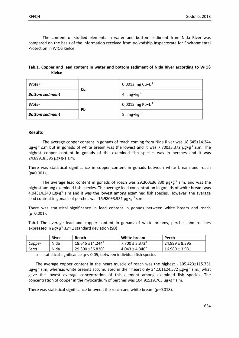
RFFCH Gödöllő, 2013
654
The content of studied elements in water and bottom sediment from Nida River was compared on the basis of the information received from Voivodship Inspectorate for Environmental Protection in WIOŚ Kielce.
Tab.1. Copper and lead content in water and bottom sediment of Nida River according to WIOŚ Kielce
Water Cu
0,0013 mg u•L-1
Bottom sediment 4 mg•kg-1
Water Pb
0,0015 mg Pb•L-1
Bottom sediment 8 mg•kg-1
Results
The average copper content in gonads of roach coming from Nida River was 18.645±14.244 µg•g-1 s.m but in gonads of white bream was the lowest and it was 7.700±3.372 µg•g-1 s.m. The highest copper content in gonads of the examined fish species was in perches and it was 24.8 ±8.3 5 µg•g-1 s.m.
There was statistical significance in copper content in gonads between white bream and roach (p=0.001).
The average lead content in gonads of roach was 2 .300±36.830 µg•g-1 s.m. and was the highest among examined fish species. The average lead concentration in gonads of white bream was 4.043±4.340 µg•g-1 s.m and it was the lowest among examined fish species. However, the average lead content in gonads of perches was 16. 80±3. 31 µg•g-1 s.m.
There was statistical significance in lead content in gonads between white bream and roach (p=0.001).
Tab.1 The average lead and copper content in gonads of white breams, perches and roaches expressed in µg•g-1 s.m.± standard deviation (SD)
River Roach White bream Perch
Copper Nida 18.645 ±14.244a 7.700 ± 3.372a 24.8 ± 8.3 5
Lead Nida 2 .300 ±36.830a 4.043 ± 4.340a 16. 80 ± 3. 31
a- statistical significance ,p < 0.05, between individual fish species
The average copper content in the heart muscle of roach was the highest - 105.423±115.751 µg•g-1 s.m, whereas white breams accumulated in their heart only 34.101±24.572 µg•g-1 s.m., what gave the lowest average concentration of this element among examined fish species. The concentration of copper in the myocardium of perches was 104. 15± .765 µg•g-1 s.m.
There was statistical significance between the roach and white bream (p=0.018).
RFFCH Gödöllő, 2013
655
The average lead content in the heart muscle of roach was 105.484±80.284 µg•g-1 s.m. and similarly like in case of copper was the highest in this species among examined fishes. However, the average concentration of lead in the myocardium of white bream was 58.281±108.76 µg•g-1 s.m., and in the myocardium of perch was 41.1 1±30.502 µg•g-1 s.m.
There was not statistical significance in lead content in the heart muscle between species (p=0.807).
Tab.2 The average lead and copper content in the heart muscle of white breams, perches and roaches expressed in µg•g-1 s.m.± standard deviation (SD)
River Roach White bream Perch
Copper Nida 105.432±115.751a 34.101±24.572a 104. 15± .765
Lead Nida 105.484±80.284 58.281±108.76 41.1 1±30.502 a- statistical significance p < 0.05, between individual fish species
The average copper content in bones of roach was 23.448±1 .633 µg•g-1 s.m., in white bream only .547±6.754 µg•g-1 s.m., while the highest average copper content was in bones of perch - 45.287±25.117 µg•g-1 s.m.
There was statistical significance in copper content in bones between white bream and roach (p=0.020), and between white bream and perch (p=0.026).
The highest average lead content was found in bones of roach - 70. 76±40. 2 µg•g-1 s.m., the lowest lead concentration was found in bones of white bream - 36.544±10.746 µg•g-1 s.m., and the average lead content was in bones of perches - 5 .816 ±10.646 µg•g-1 s.m.
There was statistical significance in lead content in bones between white bream and roach coming from the Nida (p=0.001).
Tab.3 The average lead and copper content in bones of white breams, perches and roaches expressed in µg•g-1 s.m.± standard deviation (SD)
River Roach White bream Perch
Copper Nida 23.448±1 .633a .547±6.754a,b 45.287±25.117b
Lead Nida 70. 76±40. 2 36.544±10.746 5 .816±10.646 a - statistical significance, p < 0.05, between individual fish species
b - statistical significance, p < 0.05, between individual fish species
Conclusion
Fish are a very specific group of vertebrate animals because they spend all their life in water. They are continually under the influence of various physical and chemical factors which occurs in water. Fish food are different water organisms, plants and animals which are under the influence of the same factors. Metals absorption to the body of fish takes place in two ways, the first one is to absorb them directly from the environment and the second to take them together with food (Sapota, 2008).
Taking into account the concentration of both studied elements in water and the bottom of water body of Nida River it was possible to expect the presence of these metals in examined fish tissues as well.
RFFCH Gödöllő, 2013
656
It was found that organs of all examined fish species – white bream, perch and roach contain as well copper as lead. However, the concentration of these elements in organs of particular fish species was different. Analyzing the results it is important to note high standard errors that were caused by a large result of single scattering group. This can prove about a big interspecific variation in the way of food intake or about presence of single individuals coming from Nida River.
The highest copper level among studied organs was found in the heart muscle of all examined fish species. The concentration of this element in the heart of roaches and perches was definitely higher than in the heart of white breams. Clearly higher copper content was also found in the bones of perches. Similar results of high copper accumulation in the bones were obtained by Moissenko i Kurdyavtseva (2001), studying organs of lavaret (Coregonus lavaretus) and brown trout (Salmo trutta morpha fario). High level of this metal in the heart muscle may be associated with high metabolic activity of this organ.
The lowest copper content was in gonads, wherein the lowest its level was found in white bream. However, copper concentration in animals gonads can have seasonal changes which depend on the biology of the species. This fact was found in common frog (Rana temporaria), between the period of winter torpor and the mating season of this species - that is the time spent in the water (Stawarz, 1998). According to the author copper content in ovaries of frogs rises in the mating season. Examined fishes were caught in the spring period where it is spawning season of these species. It can be inferred that copper accumulation in gonads of studied fishes was also higher in this period. Apart from that a big amount of substances as well as examined elements is washed out from fields and meadows directly to rivers where fish live during the spring thaw.
The highest lead content was in the heart muscle and bone tissue in all examined fish species. Average the highest lead level was in the heart of roach, whereas the lowest its concentration was in gonads of white bream. At the same time gonads were the place where the lowest was lead accumulation in all studied fish species.
The lead content in the bone tissue of fish was high. This can be attributed to the fact that this metal is continuously installing in the bone structure and its accumulation occurs throughout the life of the organism (Orłowski, 2008). High lead concentration in bones of 6 fish species from the Mediterranean was found by Khalifa et al. (2010), studying some elements content In their organs. Study of lead content, inter alia, in bones of amphibians was also conducted by Stawarz (1998). He found that lead accumulation in bones of common frog (Rana temporaria) was very high during the mating season and the period of winter torpor, that is the time spent in the water.
Lead is a metal that has a harmful influence on gonads, it impairs, inter alia, the secretion of testosterone in testicles, thus it causes disorders of spermatogenesis and steroidogenesis (Martynowicz, et al., 2005).
No great concentration of this metal in gonads of examined fish can provide about the processes occurring in the body, which protect gonads against the absorption of harmful substances such as lead to them. Kosior‘s et al. (2002) research confirms the low lead content in gonads of female fish living in the Southern Baltic Sea Lagoons.
The heart muscle of all examined by me fish species contained lead in a very large amount. Most likely it appears that the big amounts of lead in the heart muscle are caused by high metabolic activity of this organ. Lead is transported through the animals’ body with blood (Hoffman et al., 1985). The whole blood contained in the body together with all compounds contained therein,
RFFCH Gödöllő, 2013
657
passes through the heart, and some of these compounds like, for instance lead can be accumulated in cardiac tissue.
The conducted research also demonstrated that the species the least susceptible to accumulation of heavy metals in its organs is white bream, although, it takes the most amount of food from the bottom of water body. Perch and roach contained much more examined elements in their organs than white bream. Such high contents of examined elements that were marked in organs of roach can be explained by UV radiation, which penetrates easier in the high part of water, so in the place of roach living in the reservoirs. Formicki et al. (2006) wrote about such cases studying amphibian larvae. It is possible that physical and chemical factors or species-individual properties have also an influence on it but they were not taken into account. However, high concentration of examined chemical elements in perches can be ca mainly used by predation of this species.
References
Abdou, K.A., Korashy, E., 2001. Lead, Cadmium and management in milk and some milk products in upper Egypt. Assuit. Vet. Med. J., 45, 89: 336-348.
Cui, X., Okayasu, R., 2008. Arsenic accumulation, elimination, and interaction with copper, zinc and manganese in liver and kidney of rats. Food and Chemical Toxicology: 46, 12, 3646-3650.
Czerwionka – Szaflarska, M., owak, A., 1 6. Ołów – nowe spojrzenie na stary problem. Czy istnieje dziś zagrożenie ołowiem? Problemy Lekarskie: 35, 1-2, 65-69
Dauwe, T., Bervoets, L., Blust, R., Eens, M., 2002. Tissue Levels of Lead in Experimentally Exposed Zebra Finches (Taeniopygia guttata) with Particular Attention on the Use of Feathers as Biomonitors. Archives of Environmental Contamination and Toxicology: 42, 1, 88-92
Dorea, J.G., 2000. Iron and copper in human milk. Nutrition: 16, 3, 209-220. Ekbal, M.A., Ibrahim. 2004. Cadmium, Copper and Lead in Some Kinds of Cheeses. Benha Vet.Med.J., Vol.15,
No.2, Dec.2004, 55 Eun-Young, K., Ichihashi, H., Saeki, K., Atrashkevich, G., Tanabe, Sh., Tatsukawa, R., 1996. Metal accumulation in
tissues of seabirds from Chaun, Northeast Siberia, Rusia. Environmental Pollution: 92, 3, 247-252. Formicki, G., Stawarz, R., Zamachowski, W., 2006. Combined effects of lead ions and UV radiation on general
metabolism In common frog Rana temporaria Linnaeus, 1758 larvae. Ecological Chemistry and Engineering:13, 1-2, 23-27.
Formicki. G., Greń, A., Stawarz, R., Zyśk, B., Gał, A., 2013. Metal Content in Honey, Propolis, Wax and Bee Pollen and Implications for Metal Pollution Monitoring. Pol. J. Environ. Stud. Vol. 22. No. 1 (2013), 99-106
Hoffman, D.J., Franson, F.C., Pattee, O.H., Bunck, C.M., Murray, H.C., 1985. Biochemical and hematological effects of lead ingestion in nestling American kestrels (Falco sparverius). Comparative Biochemistry and Physiology: 80 C, 431-439.
Kabata-Pendias, A., Pendias, H., 1999. Biogeochemia pierwiastków śladowych. Wydawnictwo aukowe PW . Warszawa. 1-400.
Karahalil, B., Avkanat, B., Ertas, N., 2007. Dental lead levels in children from two different urban and suburban areas of Turkey. International Jurnal of Hygiene and Environmental Health: 210, 2, 107-112.
Khalifa, K.M., Hamil, A.M., Al-Houni, A.Q.A., Ackacha, M.A. 2010. Determination of Heavy Metals in Fish Species of the Mediterranean Sea (Libyan coastline) Using Atomic Absorption Spectrometry. Internatrional Journal of PharmTech Research: 2, 2,1350-1354
Kosior,M., Barska, I., Domagała-Wieloszewska, M., 2002. Heavy Metals, ΣDDT and ΣPCB in the Gonads of Pikeperch Females Spawning in Southern Baltic Sea Lagoons. Polish Journal of Environmental Studiem: 11, 2 (2002), 127-133
Martynowicz, ., Andrzejak, R., Mędraś, M., 2005. Wpływ ołowiu na funkcję gonad męskich. Medycyna Pracy: 56 (6), 495-500.
Mercer, J.F.B., 2001. The molecular basis of copper-transport diseases. Trends in Molecular Medicine: 7, 2, 64-69.
RFFCH Gödöllő, 2013
658
Moissenko, T.I., Kudryavtseva, L.P., 2001. Trace metal accumulation and fish pathologies in areas affected by mining and metallurgical enterprises in the Kola Region, Russia. Environmental Pollution: 114, 2, 285-297.
Orłowski, z., 2008. Metale. W Podstawy toksykologii. (red. Piotrowski J.), Wydawnictwo aukowo – Techniczne Warszawa: 6, 148-194.
Reddy, G.R., Devi, B.C., Chetty C.S., 2007. Developmental lead neurotoxicity: alterations in brain cholinergic system. Neurotoxicology: 28 (2), 402-407.
Roméo, M., Siau, Y., Sidoumou, Z., Gnassia-Barelli, M., 1999. Heavy metal distribution in different fish species from Mauritania coast. The Science of the Total Environment: 232, 3, 169-175.
Sapota, A., 2008. Drogi wchłaniania, metabolizm i wydalanie ksenobiotyków. W Podstawy toksykologii. (red. Piotrowski J.). Wydawnictwo Naukowo – Techniczne Warszawa 2008. 3, 63-86.
Sierra, E.M., Tiffany-Castiglioni, E., 1991. Reduction of glutamine synthetase activity in astroglia exposed in culture to low levels of inorganic lead. Toxicology: 65; 295-304.
Stawarz, R., 1 8. Analiza zawartości niektórych pierwiastków u żaby trawnej Rana temporaria L. w okresie odrętwienia zimowego i pory godowej. Maszynopis pracy doktorskiej. Zakład Zoologii Instytutu Biologii WSP w Krakowie.
Wood, C.M., Van Vleet, E.S., 1996. Copper, Cadmium and Zinc in Liver, Kidney and Muscle Tissues of Bottlenose Dolphins (Tursiops truncatus) Stranded in Florida. Marine Pollution Bulletin: 32, 12, 886-889.
Yilmaz, F., Özdemir, ., Demirak, A., Tuna, A.L. 2007. Heavy metal levels in two fish species Leuciscus cephalus and Lepomis gibbosus. Food Chemistry: 100, 2, 830-835.
Zyśk, B. 2013. The distribution of cadmium and copper in fish organs. The Journal of Microbiology, Biotechnology and Food Sciences. April – May 2013, vol. 2, no. 5, 2306-2309
Related Documents