PHYSIOLOGICAL RESEARCH • ISSN 0862-8408 (print) • ISSN 1802-9973 (online) 2016 Institute of Physiology of the Czech Academy of Sciences, Prague, Czech Republic Fax +420 241 062 164, e-mail: [email protected], www.biomed.cas.cz/physiolres Physiol. Res. 65: 833-842, 2016 Amelioration of Energy Metabolism by Melatonin in Skeletal Muscle of Rats With LPS Induced Endotoxemia E. OZKOK 1 , H. YORULMAZ 2 , G. ATES 3 , A. AKSU 4 , N. BALKIS 4 , Ö. ŞAHİN 5 , S. TAMER 3 1 Istanbul University, Institute of Experimental Medicine, Department of Neuroscience, Istanbul, Turkey, 2 Halic University, Medical Faculty, Istanbul, Turkey, 3 Istanbul University, Istanbul Medical Faculty, Department of Physiology, Istanbul, Turkey, 4 Department of Chemical Oceanography, Institute of Marine Sciences and Management, Istanbul University, Istanbul, Turkey, 5 Department of Pathology, Istanbul Faculty of Medicine, Istanbul University, Istanbul, Turkey Received December 21, 2015 Accepted June 21, 2016 Summary In the literature, few studies have investigated the effects of melatonin on energy metabolism in skeletal muscle in endotoxemia. We investigated the effects of melatonin on tissue structure, energy metabolism in skeletal muscle, and antioxidant level of rats with endotoxemia. We divided rats into 4 groups, control, lipopolysaccharide (LPS) (20 mg/kg, i.p., single dose), melatonin (10 mg/kg, i.p., three times), and melatonin + LPS. Melatonin was injected i.p. 30 min before and after the 2nd and 4th hours of LPS injection. Antioxidant status was determined by glutathione (GSH) measurement in the blood. Muscle tissue was stained using modified Gomori trichrome (MGT), succinic dehydrogenase (SDH), and cytochrome oxidase (COX) and histological scored. Also the sections were then stained with hematoxylin and eosin. The stained sections were visualized and photographed. Creatine, creatine phosphate, adenosine triphosphate (ATP), adenosine diphosphate (ADP), and adenosine monophosphate (AMP) levels were investigated using high performance liquid chromatography (HPLC) in muscle tissue. In the Melatonin + LPS group, blood GSH levels were increased compared with the LPS group (P<0.01). Melatonin reduced myopathic changes in the LPS group according to the histopathologic findings. In addition, ATP values were increased compared with the LPS group (P<0.05). Our findings showed melatonin treatment prevented muscle damage by increasing ATP and GSH levels in rats with LPS induced endotoxemia. Key words Energy • Lipopolysaccharide • Melatonin • Skeletal muscle Corresponding author E. Özkök, İstanbul University, Institute of Experimental Medicine, Department of Neuroscience, Vakif Gureba Cad, 34280 Istanbul, Turkey. E-mail: [email protected] Introduction Sepsis-induced organ failure is a major cause of death in critical care units and is characterized by a massive dysregulated inflammatory response and oxidative stress. Lipopolysaccharide (LPS) triggers the production of various inflammatory mediators involved in the development of sepsis, systemic inflammatory response syndrome (SIRS), and multiple organ failure (Kamisoglu et al. 2015). Skeletal tissue comprises 50-60 % of body cell mass and represents the largest organ affected by systemic inflammation. Muscle wasting is a serious complication of sepsis. Studies in patients with sepsis and experimental animals have provided evidence that myofibrillar proteins are particularly sensitive to the effects of sepsis (Wu et al. 2001). The ubiquitin- proteasome system has a major role in skeletal muscle proteolysis and is activated by several pathways, including proinflammatory cytokines, reactive oxygen species, and cyclooxygenases (Holecek et al. 2012). High-energy phosphorylated compounds and mitochondrial impairment have been reported in septic skeletal muscle (Giannesini et al. 2008). The effect of

Welcome message from author
This document is posted to help you gain knowledge. Please leave a comment to let me know what you think about it! Share it to your friends and learn new things together.
Transcript
PHYSIOLOGICAL RESEARCH • ISSN 0862-8408 (print) • ISSN 1802-9973 (online) 2016 Institute of Physiology of the Czech Academy of Sciences, Prague, Czech Republic Fax +420 241 062 164, e-mail: [email protected], www.biomed.cas.cz/physiolres
Physiol. Res. 65: 833-842, 2016
Amelioration of Energy Metabolism by Melatonin in Skeletal Muscle of Rats With LPS Induced Endotoxemia E. OZKOK1, H. YORULMAZ2, G. ATES3, A. AKSU4, N. BALKIS4, Ö. ŞAHİN5, S. TAMER3
1Istanbul University, Institute of Experimental Medicine, Department of Neuroscience, Istanbul, Turkey, 2Halic University, Medical Faculty, Istanbul, Turkey, 3Istanbul University, Istanbul Medical Faculty, Department of Physiology, Istanbul, Turkey, 4Department of Chemical Oceanography, Institute of Marine Sciences and Management, Istanbul University, Istanbul, Turkey, 5Department of Pathology, Istanbul Faculty of Medicine, Istanbul University, Istanbul, Turkey
Received December 21, 2015 Accepted June 21, 2016 Summary In the literature, few studies have investigated the effects of melatonin on energy metabolism in skeletal muscle in endotoxemia. We investigated the effects of melatonin on tissue structure, energy metabolism in skeletal muscle, and antioxidant level of rats with endotoxemia. We divided rats into 4 groups, control, lipopolysaccharide (LPS) (20 mg/kg, i.p., single dose), melatonin (10 mg/kg, i.p., three times), and melatonin + LPS. Melatonin was injected i.p. 30 min before and after the 2nd and 4th hours of LPS injection. Antioxidant status was determined by glutathione (GSH) measurement in the blood. Muscle tissue was stained using modified Gomori trichrome (MGT), succinic dehydrogenase (SDH), and cytochrome oxidase (COX) and histological scored. Also the sections were then stained with hematoxylin and eosin. The stained sections were visualized and photographed. Creatine, creatine phosphate, adenosine triphosphate (ATP), adenosine diphosphate (ADP), and adenosine monophosphate (AMP) levels were investigated using high performance liquid chromatography (HPLC) in muscle tissue. In the Melatonin + LPS group, blood GSH levels were increased compared with the LPS group (P<0.01). Melatonin reduced myopathic changes in the LPS group according to the histopathologic findings. In addition, ATP values were increased compared with the LPS group (P<0.05). Our findings showed melatonin treatment prevented muscle damage by increasing ATP and GSH levels in rats with LPS induced endotoxemia. Key words Energy • Lipopolysaccharide • Melatonin • Skeletal muscle
Corresponding author E. Özkök, İstanbul University, Institute of Experimental Medicine, Department of Neuroscience, Vakif Gureba Cad, 34280 Istanbul, Turkey. E-mail: [email protected] Introduction
Sepsis-induced organ failure is a major cause of death in critical care units and is characterized by a massive dysregulated inflammatory response and oxidative stress. Lipopolysaccharide (LPS) triggers the production of various inflammatory mediators involved in the development of sepsis, systemic inflammatory response syndrome (SIRS), and multiple organ failure (Kamisoglu et al. 2015).
Skeletal tissue comprises 50-60 % of body cell mass and represents the largest organ affected by systemic inflammation. Muscle wasting is a serious complication of sepsis. Studies in patients with sepsis and experimental animals have provided evidence that myofibrillar proteins are particularly sensitive to the effects of sepsis (Wu et al. 2001). The ubiquitin-proteasome system has a major role in skeletal muscle proteolysis and is activated by several pathways, including proinflammatory cytokines, reactive oxygen species, and cyclooxygenases (Holecek et al. 2012). High-energy phosphorylated compounds and mitochondrial impairment have been reported in septic skeletal muscle (Giannesini et al. 2008). The effect of
834 Ozkok et al. Vol. 65 sepsis on skeletal muscle mitochondria in rat models indicate decreased mitochondrial function, depletion of adenosine triphosphate (ATP) and adenosine 5'-diphosphate (ADP) stores, decreased mitochondrial protein synthesis, and damage to the mitochondrial transcription mechanism (Correa et al. 2015).
Melatonin is known to be an effective anti-inflammatory agent in various animal models of inflammation and sepsis. It has been demonstrated also to increase antioxidant defense and to exert protective effects on mitochondrial function (Reiter et al. 2008, Srinivasan et al. 2010).
Melatonin is mainly secreted from pineal gland is well known for being a potent free-radical scavenger, powerful antioxidant, and a regulator of mitochondrial bioenergetic function, especially ATP production via complexes I and IV (Paradis et al. 2015, Martin et al. 2002).
In a number of animal models of septic shock, as well as in patients with septic disease, melatonin reportedly exerted beneficial effects that reduced cellular damage and multiorgan failure (Sener et al. 2005, Kurcer et al. 2006).
It is now well documented that reactive oxygen species are involved in the pathogenesis of multiple organ failure following sepsis, which often leads to death (Heyland et al. 2006, Biolo et al. 2007).
Oxidative stress is the result of an imbalance between the production of reactive oxygen species (ROS) and their neutralization by antioxidants (Manzanares et al. 2009). Glutathione (GSH) is the main endogenous thiol antioxidant and plays an essential role in the protection against reactive oxygen species. GSH has a somewhat opposite effect in immunity; it is often regarded as anti-inflammatory because it inhibits the production and action of several inflammatory cytokines and chemokines (Biolo et al. 2007). Studies have shown that GSH is lower in sepsis (Biolo et al. 2007, Asci et al. 2015). Melatonin has also been shown to increase gene expression and enzyme activities of GSH and the mitochondrial GSH pool. In addition, melatonin has been shown to restore GSH homeostasis by stimulating mitochondrial gamma-glutamylcysteine synthase function under oxidative stress (Martin et al. 2000, Rodriguez et al. 2004).
Few studies have investigated the effects of melatonin on energy metabolism in skeletal tissue in sepsis models. In this study we aimed to investigate the effects of multidose melatonin treatment, which was
injected before and/or after endotoxemia on both energy metabolism and histologic changes in skeletal muscle and GSH level high-dose LPS-induced endotoxemia. Methods Experimental procedures
The study was approved by Istanbul University Local Ethics Committee for Animal Experiments. Male Wistar Albino rats that were 8-9 weeks old and weighed an average 200 – 250 g were housed in polypropylene cages with free access to food and drink, in a light/dark cycle of 12/12 h. Rats were randomly divided into 4 groups: the control group, LPS group, melatonin group, melatonin + LPS group, each comprised 8 animals.
The control group received no treatment. The LPS group received i.p. 20 mg/kg, as a single dose; LPS from Escherichia coli O127: B8 (Sigma-Aldrich, Product No: L5668) was dissolved in 1 ml of sterile saline solution. The melatonin group received a total of 30 mg/kg in 3 i.p. doses of 10 mg/kg at 2-hour intervals; melatonin (Sigma Aldrich, Product No: M5250) was dissolved in 2.5 % ml ethyl alcohol. The fourth group was injected with a dose of melatonin (10 mg/kg, i.p.) 30 min before the LPS administration. Two further doses of melatonin were administered 2 and 4 hours after LPS administration.
All rats from all 4 groups were sacrificed at the end of the 6th hour after the first injection using sodium pentothal anesthesia (i.p. 30 mg/kg) and gastrocnemius muscles were dissected from all animals. For this purpose, the gastrocnemius muscle was cut proximally, using the same line as the skin incision. The muscle samples were rapidly frozen by liquid nitrogen. Biochemical procedures
At the end of the experimental period, blood samples (2-3 ml) were collected from heart. Blood samples were centrifuged at 3000 rpm for 20 min. Serum concentrations of creatinine kinase, aspartate aminotransferase, alanine aminotransferase, glucose, and blood leucocyte were determined. Serum creatinine kinase, aspartate aminotransferase, alanine aminotransferase, glucose levels (Cobas autoanalyzer, DPC, Diagnostic products corporation, USA) and blood leucocytes (Beckman Coulter LH 780) were measured using the corresponding kit in an autoanalyzer.
2016 Melatonin on Skeletal Muscle With LPS 835
HPLC analysis of creatine, creatine phosphate, AMP, ADP, ATP Sample preparation
Muscle tissue samples weighing 200 mg were homogenized in 2 ml 0.42 M HClO4 using a homogenizer (Ultraturrax T25) for 30 sec. A 1.0 ml supernatant was taken for adjusting pH with 1.0 M K2HPO4 after centrifugation at 3000 rpm for 5 min. Measurement of creatine, creatine phosphate, and high-energy compounds
Creatine, creatine phosphate, and adenine nucleotides were evaluated with C18 column (5 µm, 250 mm x 4.6 mm, Nucleodur, USA) with isocratic elution using a KOH/KH2PO4 buffer (215 mM, pH 6.25), 3 mM tetrabutylammonium phosphate, and 5 % acetonitrile ion-paired reverse-phase chromatography using HPLC (Agilent 1100, USA) at 214 nm (Shellvold et al. 1986). Creatine, creatine phosphate, and adenine nucleotides were calculated from their external standard curves from different concentrations. Measurement of blood reduced GSH levels
GSH levels was determined with reduced GSH measurement in blood in accordance with the method of Buetler et al. Blood reduced GSH levels were measured spectrophotometrically using DTNB (5,5’dithio-bis-(2-nitro-benzoic acid) reagent (Buetler et al. 1963). After blood samples were hemolyzed with distilled water and precipitated proteins were removed by centrifugation, phosphate buffer and DTNB were added to the supernatant. The colored complex was measured at 412 nm at 5 min using a spectrophotometer (Shimadzu, Japan). Blood GSH concentrations were calculated using a GSH calibration curve. The results were expressed as mmol/l (Buetler et al. 1963). Histopathologic procedures
The gastrocnemius muscle was taken while the animals were under anesthesia. The muscle samples were rapidly frozen in isopentane cooled by liquid nitrogen. We took 8-μm-thick serial cross-sections and stained them with modified Gomori’s trichrome (MGT) method, succinic dehydrogenase (SDH), and cytochrome oxidase (COX) using standard protocols (Engel et al. 1963, Tanji 2008). The stained sections were visualized and photographed using a Nikon microscope (ECLIPSE 80i Nikon Corporation, JAPAN).
Both the intensity and the distribution of specific MGT, SDH and COX staining were scored. For each sample, a histological score (HSCORE) value was derived by summing the percentages of cells that stained at each intensity, multiplied by the weighted intensity of the staining [HSCORE = S Pi (i+1), where i is the intensity score (0-4) and Pi is the corresponding percentage of the cells]; staining was scored as 0= no staining, 1=weak staining, 2=middle staining, 3=dark staining, 4=very dark staining.
For hematoxylin and eosin (H&E), the muscle tissue samples were fixed in 10 % buffered formalin and embedded in parafin wax. Five-micrometer-thick sections were placed on polylysine-coated slides and stained with H&E. The slides were evaluated under a light microscopy (Olympus BX51; Olympus Corp., Tokyo, Japan) at 100 × magnification. Statistical analysis
Data are presented as mean values ± standard deviations. Groups of data were compared with an analysis of variance (ANOVA) followed by Tukey's multiple comparison tests. Values of P<0.05 were regarded as significant. Results Biochemical findings
Serum creatinine kinase, aspartate aminotransferase, alanine aminotransferase and blood leukocyte values were found increased in the LPS groups as compared with those of other groups (P<0.05).
In the LPS group, glucose levels were observed to be decreased compared with the control group (P<0.01). In the melatonin + LPS group; glucose levels were higher than in the LPS group (P<0.01) (Table 1). GSH findings
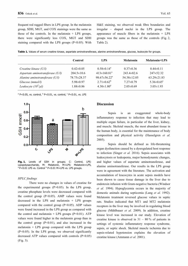
Blood GSH levels were found decreased in the LPS groups as compared with the control group, but blood GSH levels were found increased in the melatonin + LPS group compared with the LPS group (P<0.01) (Fig. 1). Histopathologic findings
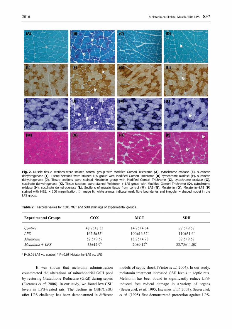
COX, MGT, and SDH staining was found normal in the control group. We saw no red fibers in the control group. In the LPS group, SDH and COX had significantly increased staining (P<0.01). There were
836 Ozkok et al. Vol. 65 frequent red ragged fibers in LPS group. In the melatonin group, SDH, MGT, and COX stainings were the same as those of the controls. In the melatonin + LPS groups, there were significantly less COX, MGT and SDH staining compared with the LPS groups (P<0.05). With
H&E staining, we observed weak fibre boundaries and irregular – shaped nuclei in the LPS group. The appearance of muscle fibers in the melatonin + LPS groups was the same as those of the controls (Fig. 2, Table 2).
Table 1. Values of serum creatine kinase, aspartate aminotransferase, alanine aminotransferase, glucose, leukocyte for groups.
Control LPS Melatonin Melatonin+LPS
Creatine kinase (U/l) 0.42±0.05 0.58±0.14a 0.37±0.36 0.44±0.11 Aspartate aminotransferase (U/l) 204.5±10.6 413±160.81a 243.4±82.6 247±52.32 Alanine aminotransferase (U/l) 78.75±28.37 98.67±56.22a 54.38±12.05 63.29±21.83 Glucose (mmol/l) 5.98±0.97 2.71±0.62b 7.27±0.79 5.36±0.65c Leukocyte (103/µl) 1.88±0.86 4.30±1.80a 2.05±0.69 3.05±1.93
a P<0.05, vs. control, b P<0.01, vs. control, c P<0.01, vs. LPS
Fig. 1. Levels of GSH in groups; C: Control, LPS: Lipopolysaccharide, M: Melatonin, M+LPS: Melatonin+LPS, a P<0.01 LPS vs. Control b P<0.01 M+LPS vs. LPS groups.
HPLC findings
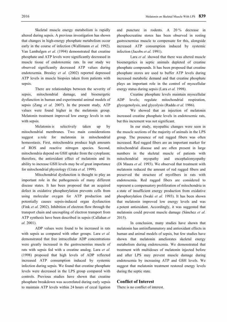
There were no changes in values of creatine for the experimental groups (P>0.05). In the LPS group, creatine phosphate levels were decreased compared with the control group (P<0.05). AMP values were found decreased in the LPS and melatonin + LPS groups compared with the control group (P<0.05). ADP values were found increased in the LPS group as compared with the control and melatonin + LPS groups (P<0.01). ATP values were found higher in the melatonin group than in the control group (P<0.01), and also increased in the melatonin + LPS group compared with the LPS group (P<0.05). In the LPS group, we observed significantly decreased ATP values compared with controls (P<0.05) (Fig. 3).
Discussion
Sepsis is an exaggerated whole-body inflammatory response to infection that may lead to multiple organ failure, in particular of the liver, kidney, and muscle. Skeletal muscle, the most abundant tissue in the human body, is essential for the maintenance of body composition and physical activity (Hasselgren et al. 2005).
Sepsis should be defined as life-threatening organ dysfunction caused by a dysregulated host response to infection (Singer et al. 2016). Sepsis associates with leukocytosis or leukopenia, major hemodynamic changes, and higher values of aspartate aminotransferase, and alanine aminotransferase. Our results in the LPS group were in agreement with the literature. The activation and accumulation of leucocytes in acute sepsis models have been shown to cause tissue damage in the liver due to endotoxin infusion with Gram-negative bacteria (Windsor et al. 1994). Hypoglycemia occurs in the majority of domestic animals during septicemia (Lang et al. 1992). Melatonin treatment reversed glucose values in septic rats. Studies indicated that MT1 and MT2 melatonin receptors in the liver may be involved in regulating blood glucose (Mühlbauer et al. 2009). In addition, creatine kinase level was increased in our study. Elevation of creatine kinase is observed in 31 – 80 % of patients in settings of systemic inflammatory response syndrome, sepsis, or septic shock. Skeletal muscle ischemia due to sepsis-related hypotension explains the elevation of creatine kinase (Ammann et al. 2001).
2016 Melatonin on Skeletal Muscle With LPS 837
Fig. 2. Muscle tissue sections were stained control group with Modified Gomori Trichrome (A), cytochrome oxidase (E), succinate dehydrogenase (I). Tissue sections were stained LPS group with Modified Gomori Trichrome (B) cytochrome oxidase (F), succinate dehydrogenase (J). Tissue sections were stained Melatonin group with Modified Gomori Trichrome (C), cytochrome oxidase (G), succinate dehydrogenase (K). Tissue sections were stained Melatonin + LPS group with Modified Gomori Trichrome (D), cytochrome oxidase (H), succinate dehydrogenase (L). Sections of muscle tissue from control (M), LPS (N), Melatonin (O), Melatonin+LPS (P) stained with H&E, × 100 magnification. In image N; white arrows indicate weak fibre boundaries and irregular – shaped nuclei in the LPS group.
Table 2. H-scores values for COX, MGT and SDH stainings of experimental groups.
Experimental Groups COX MGT SDH
Control 48.75±8.53 14.25±4.34 27.5±9.57 LPS 162.5±35a 100±16.32a 110±31.6a Melatonin 52.5±9.57 18.75±4.78 32.5±9.57 Melatonin + LPS 55±12.9b 20±9.12b 33.75±11.08b
a P<0.01 LPS vs. control, b P<0.05 Melatonin+LPS vs. LPS
It was shown that melatonin administration
counteracted the alterations of mitochondrial GSH pool by restoring Glutathione Reductase (GRd) during sepsis (Escames et al. 2006). In our study, we found low GSH levels in LPS-treated rats. The decline in GSH/GSSG after LPS challenge has been demonstrated in different
models of septic shock (Victor et al. 2004). In our study, melatonin treatment increased GSH levels in septic rats. Melatonin has been found to significantly reduce LPS-induced free radical damage in a variety of organs (Sewerynek et al. 1995, Escames et al. 2003). Sewerynek et al. (1995) first demonstrated protection against LPS-
838 Ozkok et al. Vol. 65 induced damage after melatonin administration. Melatonin’s direct antioxidant activity and its stimulatory effect on antioxidant enzyme activities may have clinical implications for the treatment of different pathologic conditions. It is suggested that melatonin has a potent glutathione peroxidase stimulating effect in response to the free-radical damage in severe sepsis. The evidence
that melatonin increases gene expression for glutathione peroxidase suggests a potential mechanism of action at the DNA level in different tissues (Tan et al. 1993, Antolin et al. 1996). A stimulatory effect of melatonin on brain glutathione peroxidase activity was also reported by Barlow-Walden et al. (1995).
Fig. 3. Levels of AMP (A), ADP (B), ATP (C), Creatine phosphate (D), and Creatine (E) in groups. C: Control, LPS: Lipopolysaccharide, M: Melatonin, M+LPS: Melatonin+LPS; a P<0.05 vs. Control, b P<0.01 vs. Control, c P<0.01 vs. LPS, d P<0.05 vs. LPS.
2016 Melatonin on Skeletal Muscle With LPS 839
Skeletal muscle energy metabolism is rapidly altered during sepsis. A previous investigation has shown that changes in high-energy phosphate metabolism occur early in the course of infection (Wallimann et al. 1992). Van Lambalgen et al. (1994) demonstrated that creatine phosphate and ATP levels were significantly decreased in muscle tissue of endotoxemic rats. In our study we observed significantly decreased ATP values during endotoxemia. Brealey et al. (2002) reported depressed ATP levels in muscle biopsies taken from patients with sepsis.
There are relationships between the severity of sepsis, mitochondrial damage, and bioenergetic dysfunction in human and experimental animal models of sepsis (Zang et al. 2007). In the present study, ATP values were found higher in the melatonin group. Melatonin treatment improved low energy levels in rats with sepsis.
Melatonin is selectively taken up by mitochondrial membranes. Two main considerations suggest a role for melatonin in mitochondrial homeostasis. First, mitochondria produce high amounts of ROS and reactive nitrogen species. Second, mitochondria depend on GSH uptake from the cytoplasm; therefore, the antioxidant effect of melatonin and its ability to increase GSH levels may be of great importance for mitochondrial physiology (Urata et al. 1999).
Mitochondrial dysfunction is thought to play an important role in the pathogenesis of many different disease states. It has been proposed that an acquired defect in oxidative phosphorylation prevents cells from using molecular oxygen for ATP production and potentially causes sepsis-induced organ dysfunction (Fink et al. 2002). Inhibition of electron flow through the transport chain and uncoupling of electron transport from ATP synthesis have been described in sepsis (Callahan et al. 2001).
ADP values were found to be increased in rats with sepsis as compared with other groups. Lara et al. demonstrated that free intracellular ADP concentrations were greatly increased in the gastrocnemius muscle of rats with sepsis fed with a creatine analog. Lara et al. (1998) proposed that high levels of ADP reflected increased ATP consumption induced by systemic infection during sepsis. We found that creatine phosphate levels were decreased in the LPS group compared with controls. Previous studies have shown that creatine phosphate breakdown was accerelated during early sepsis to maintain ATP levels within 24 hours of cecal ligation
and puncture in rodents. A 20 % decrease in phosphocreatine stores has been observed in resting gastrocnemius muscle to compensate for this, alongside increased ATP consumption induced by systemic infection (Jacobs et al. 1991).
Lara et al. showed that there was altered muscle bioenergetics in septic animals depleted of creatine phosphate compounds. It has been proposed that creatine phosphate stores are used to buffer ATP levels during increased metabolic demand and that creatine phosphate plays an important role in the control of myocellular energy status during sepsis (Lara et al. 1998).
Creatine phosphate levels maintain myocellular ADP levels; regulate mitochondrial respiration, glycogenolysis, and glycolysis (Radda et al. 1986).
We showed that an injection of melatonin increased creatine phosphate levels in endotoxemic rats, but this increment was not significant.
In our study, myopathic changes were seen in the muscle sections of the majority of animals in the LPS group. The presence of red ragged fibers was often increased. Red ragged fibers are an important marker for mitochondrial disease and are often present in large numbers in the skeletal muscle of patients with mitochondrial myopathy and encephalomyopathy (Di Maura et al. 1993). We observed that treatment with melatonin reduced the amount of red ragged fibers and preserved the structure of myofibers in rats with endotoxemia. Red ragged fibers are considered to represent a compensatory proliferation of mitochondria in a state of insufficient energy production from oxidative phosphorylation (Iwaki et al. 1993). It has been shown that melatonin improved low energy levels and was a potent antioxidant. Accordingly, it was suggested that melatonin could prevent muscle damage (Sánchez et al. 2015).
In conclusion, many studies have shown that melatonin has antiinflammatory and antioxidant effects in human and animal models of sepsis, but few studies have shown that melatonin ameliorates skeletal energy metabolism during endotoxemia. We demonstrated that treatment with multidoses of melatonin injected before and after LPS may prevent muscle damage during endotoxemia by increasing ATP and GSH levels. We suggest that melatonin treatment restored energy levels during the septic state. Conflict of Interest There is no conflict of interest.
840 Ozkok et al. Vol. 65 Acknowledgements Our study was granted from Istanbul University Research Projects (Project No: 42019). We would like also thank
Mr. David F. Chapman for editing of English and Ms. Hatice Taslı for preparing of muscle sections.
References AMMANN P, FEHR T, MINDER E, GUNTER C, BERTEL O: Elevation of troponin I in sepsis and septic shock.
Intensive Care Med 27: 965-969, 2001. ANTOLIN I, RODRIGUES C, SAINZ RM, MAYO JC, URIA H, KOTLER ML, RODRIGUEZ-COLUNGA MJ,
TOLIVIA D, MENENDEZ PEALEZ A: Neurohormone melatonin prevents cell damage: Effect on gene expression for antioxidative enzymes. FASEB J 10: 882-890, 1996.
ASCI A, SURMELI-ONAY O, ERKEKOGLU P, YIGIT S, YURDAKOK M, KOCER-GUMUSEL B: Oxidant and antioxidant status in neonatal proven and clinical sepsis in relation to selenium status. Pediatr Int 57: 1131-1137, 2015.
BALK RA: Severe sepsis and septic shock. Crit Care Clin 16: 179-192, 2000. BARLOW-WALDEN LR, REITER RJ, ABE M, PABLOS M, MENENDEZPELAEZ A, CHEN LD, POEGGELER B:
Melatonin stimulates brain glutathione peroxidase activity. Neurochem Int 26: 497-502, 1995. BIOLO G, ANTONIONE R, DE CICCO M: Glutathione metabolism in sepsis. Crit Care Med 35: 591-595, 2007. BREALEY D, BRAND M, HARGREAVES I, HEALES S, LAND J, SMOLENSKI R, DAVIES NA, COOPER CE,
SINGER M: Association between mitochondrial dysfunction and severity and outcome of septic shock. Lancet 360: 219-223, 2002.
BUETLER E, DURON O, KELLY BM: Improved method for the determination of blood glutathione. J Lab Clin Med 161: 882-888, 1963.
CALLAHAN LA, STOFAN D, SZWEDA L, NETHERY D, SUPINSKI GS: Free radicals alter maximal diaphragmatic oxygen consumption in endotoxin-induced sepsis. Free Radic Biol Med 30: 129-138, 2001.
CORREA TD, JAKOB SM, TAKALA J: Mitochondrial function in sepsis. Cri Care Hor 1: 31-41, 2015. CROUSER ED, JULIAN MW, BLAHO DV, PFEIFFER DR: Endotoxin-induced mitochondrial damage correlates with
impaired respiratory activity. Crit Care Med 30: 276-284, 2002. CRESPO E, MACIAS M, POZO D, ESCAMES G, MARTIN M, VIVES F, GUERRERO JM, ACUÑA-
CASTROVIEJO D: Melatonin inhibits expression of the inducible NO synthase II in liver and lung and prevents endotoxemia in lipopolysaccharide induced multiple organ dysfunction syndrome in rats. FASEB J 13: 1537‐1546, 1999.
CUZZOCREA S, REITER RJ: Pharmacological action of melatonin in shock, inflammation and ischemia/reperfusion injury. Eur J Pharmacol 426: 1-10, 2001.
DIMAURO S, MORAES C: Mitochondrial encephalomyopathies. Arch Neurol 50: 1197-208, 1993. ENGEL WK, CUNNINGHAM GG: Rapid examination of muscle tissue an improved trichrome method for fresh-
frozen biopsy sections. Neurology 13: 919-923, 1963. ESCAMES G, LOPEZ LC, TAPIAS V, UTRILLA P, REITER RJ, HITOS AB, LEÓN J, RODRÍGUEZ MI, ACUÑA-
CASTROVIEJO D: Melatonin counteracts inducible mitochondrial nitric oxide synthase-dependent mitochondrial dysfunction in skeletal muscle of septic mice. J Pineal Res 40: 71-78, 2006.
ESCAMES G, LEON J, MACIAS M, KHALDY H, ACUÑA-CASTROVIEJO D: Melatonin counteracts lipopolysaccharide‐induced expression and activity of mitochondrial nitric oxide synthase in rats. FASEB J 17: 932‐934, 2003.
FINK MP: Cytopathic hypoxia. Is oxygen use impaired in sepsis as a result of an acquired intrinsic derangement in cellular respiration? Crit Care Clin 18: 165-175, 2002.
GIANNESINI B, IZQUIERDO M, DALMASSO C, LE FUR Y, COZZONE PJ, VERLEYE M, LE GUERN ME, GILLARDIN JM, BENDAHAN D: Endotoxemia does not limit energy supply in exercising rat skeletal muscle. Muscle Nerve 37: 496-504, 2008.
2016 Melatonin on Skeletal Muscle With LPS 841
PARADIES G, PARADIES V, RUGGIERO FM, PETROSILLO G: Protective role of melatonin in mitochondrial dysfunction and related disorders. Arch Toxicol 89: 923-939, 2015.
HASSELGREN PO, FISCHER JE: Sepsis: stimulation of energy-dependent protein breakdown resulting in protein loss in skeletal muscle. World J Surg 122: 203-208, 1998.
HASSELGREN PO, MENCONI MJ, FAREED MU, YANG H, WEI W, EVENSON A: Novel aspects on the regulation of muscle wasting in sepsis. Int J Biochem Cell Biol 37: 2156-2168, 2005.
HEYLAND DK, DHALIWAL R, DAY A, MUSCEDERE J, DROVER J, SUCHNER U, COOK D, CANADIAN CRITICAL CARE TRIALS GROUP: REducing Deaths due to OXidative Stress (The REDOXS Study): rationale and study design for a randomized trial of glutamine and antioxidant supplementation in critically-ill patients. Proc Nutr Soc 65: 250-263, 2006.
HOLECEK M, SKOPEC F, SPRONGL L, PECKA M: Protein metabolism in specific tissues of endotoxin-treated rats: effect of nutritional status. Physiol Res 44: 399-406, 1995.
HOLECEK M: Muscle wasting in animal models of severe illness. Int J Exp Pathol 93: 157-171, 2012. IWAKI T, IWAKI A, GOLDMAN JE: Alpha B-crystallin in oxidative muscle fibers and its accumulation in ragged-red
fibers: a comparative immunohistochemical and histochemical study in human skeletal muscle. Acta Neuropathol 85: 475-480, 1993.
JACOBS DO, KOBAYASHI T, IMAGIRE J, GRANT C, KESSELLY B, WILMORE DW: Sepsis alters skeletal muscle energetics and membrane function. Surgery 110: 318-325, 325-326, 1991.
KAMISOGLU K, HAIMOVICH B, CALVANO SE, COYLE SM, CORBETT SA, LANGLEY RJ, KINGSMORE SF, ANDROULAKIS IP: Human metabolic response to systemic inflammation: assessment of the concordance between experimental endotoxemia and clinical cases of sepsis/SIRS. Crit Care 19: 71, 2015.
KURCER Z, IRAZ M, KELESYILMAZ N, KILIC N, OLMEZ E: Beneficial effects of melatonin on diaphragmatic contractility and fatigability in Escherichia coli endotoxemic rats. Arzneimittelforschung 56: 90‐95, 2006.
LANG CH, BAGBY GJ, DOBRESCU C, OTTLAKAN A, SPITZER JJ: Sepsis- and endotoxin-induced increase in organ glucose uptake in leukocyte-depleted rats. Am J Physiol 263: R1324-R1332, 1992.
LARA TM, WONG MS, ROUNDS J, ROBINSON MK, WILMORE DW, JACOBS DO: Skeletal muscle phosphocreatine depletion depresses myocellular energy status during sepsis. Arch Surg 133: 1316-1321, 1998.
MANZANARES W, BIESTRO A, GALUSSO F, TORRE MH, MAÑAY N, PITTINI G, FACCHIN G, HARDY G: Serum selenium and glutathione peroxidase-3 activity: biomarkers of systemic inflammation in the critically ill. Intensive Care Med 35: 882-889, 2009.
MARTIN M, MACIAS M, ESCAMES G, LEÓN J, ACUÑA-CASTROVIEJO D: Melatonin but not vitamins C and E maintains glutathione homeostasis in t-butyl hydroperoxide-induced mitochondrial oxidative stress. FASEB J 14: 1677-1679, 2000.
MARTIN M, MACIAS M, LEÓN J, ESCAMES G, KHALDY H, ACUÑA-CASTROVIEJO D: Melatonin increases the activity of the oxidative phosphorylation enzymes and the production of ATP in rat brain and liver mitochondria. Int J Biochem Cell Biol 34: 348-357, 2002.
MUHLBAUER E, GROSS E, LABUCAY K, WOLGAST S, PESCHKE E: Loss of melatonin signalling and its impact on circadian rhythms in mouse organs regulating blood glucose. Eur J Pharmacol 606: 61-71, 2009.
PARADIES G, PARADIES V, RUGGIERO FM, PETROSILLO G: Protective role of melatonin in mitochondrial dysfunction and related disorders. Arch Toxicol 89: 923-939, 2015.
RADDA GK: The use of NMR spectroscopy for the understanding of disease. Science 8: 640-645, 1986. REITER RJ, PAREDES SD, KORKMAZ A, JOU MJ, TAN DX: Melatonin combats molecular terrorism at the
mitochondrial level. Interdiscip Toxicol 1: 137-149, 2008. RODRIGUEZ C, MAYO JC, SAINZ RM, ANTOLIN I, HERRERA F, MARTIN V, REITER RJ: Regulation of
antioxidant enzymes: a significant role for melatonin. J Pineal Res 36: 1-9, 2004. SACCANI A, SACCANI S, ORLANDO S, SIRONI M, BERNASCONI S, GHEZZI P, MANTOVANIA, SICA A:
Redox regulation of chemokine receptor expression. Proc Natl Acad Sci U S A 97: 2761-2766, 2000. SÁNCHEZ A, CALPENA AC, CLARES B: Evaluating the oxidative stress in inflammation: Role of Melatonin. Int J
Mol Sci 16: 16981-17004, 2015.
842 Ozkok et al. Vol. 65 SENER G, TOKLU H, KAPUCU C, ERCAN F, ERKANLI G, KACMAZ A, TILKI M, YEGEN BC: Melatonin
protects against oxidative organ injury in a rat model of sepsis. Surg Today 35: 52‐59, 2005. SEWERYNEK E, MELCHIORRI D, CHEN LD, REITER RJ: Melatonin reduces both basal and bacterial
lipopolysaccharide-induced lipid peroxidation. Free Radic Biol Med 19: 903-909, 1995. SELLEVOLD OF, JYNGE P, AARSTAD K: High performance liquid chromatography: a rapid isocratic method for
determination of creatine compounds and adenine nucleotides in the myocardial tissues. J Mol Cell Cardiol 18: 517-527, 1986.
SIES H, SHAROV VS, KLOTZ LO, BRIVIBA K: Glutathione peroxidase protects against peroxynitrite-mediated oxidations. A new function for selenoproteins as peroxynitrite reductase. J Biol Chem 272: 27812-27817, 1997.
SINGER M, DEUTSCHMAN CS, SEYMOUR CW, SHANKAR-HARI M, ANNANE D, BAUER M, BELLOMO R, BERNARD GR, CHICHE JD, COOPERSMITH CM, HOTCHKISS RS, LEVY MM, MARSHALL JC, MARTIN GS, OPAL SM, ET AL.: The Third International Consensus Definitions for Sepsis and Septic Shock (Sepsis-3). JAMA 315: 801-810, 2016.
SRINIVASAN V, PANDI-PERUMAL SR, SPENCE DW, KATO H, CARDINALI DP: Melatonin in septicshock: some recent concepts. J Crit Care 25: e1-6, 2010.
TAN DX, CHEN LD, POEGGELER B, MANCHESTER LC, REITER RJ: Melatonin: A potent, endogenous hydroxyl radical scavenger. Endocrine J 1: 57-60, 1993.
TANJI K, BONILLA E: Light microscopic methods to visualize mitochondria on tissue sections. Methods 46: 274-280, 2008.
TAYLOR DE, PIANTADOSI CA: Oxidative metabolism in sepsis and sepsis syndrome. J Crit Care 10: 122-135, 1995.
TRUMBECKAITE S, POALKA JR, NEUHOF C, ZIERZ S, GELLERICH FN: Different sensitivity of rabbit heart and skeletal muscle to endotoxin-induced impairment of mitochondrial function. Eur J Biochem 268: 1422-1429, 2001.
URATA Y, HONMA S, GOTO S, TODOROKI S, UEDA T, IRIDA T, CHO S, HONMA K, KONDO T: Melatonin induces gamma-glutamylcysteine synthetase mediated by activator protein-1 in human vascular endothelial cells. Free Rad Biol Med 27: 838-847, 1999.
VICTOR VM, DE LA FUENTE M: Immune cells redox state from mice with endotoxin-induced oxidative stress. Involvement of NF-kappaB. Free Radic Res 37: 19-27, 2003.
VILLA P, SACCANI A, SICA A, GHEZZI P: Glutathione protects mice from lethal sepsis by limiting inflammation and potentiating host defense. J Infect Dis 15: 1115-1120, 2002.
WALLIMANN T, WYSS M, BRDICZKA D, NICOLAY K, EPPENBERGER HM: Intracellular compartmentation, structure and function of creatine kinase isoenzymes in tissues with high and fluctuating energy demands: the 'phosphocreatine circuit' for cellular energy homeostasis. Biochem J 1: 21-40, 1992.
WINDSOR AC, MULLEN PG, WALSH CJ, FISHER BJ, BLOCHER CR, JESMOK G, FOWLER AA 3RD, SUGERMAN HJ: Delayed tumor necrosis factor alpha blockade attenuates pulmonary dysfunction and metabolic acidosis associated with experimental gram-negative sepsis. Arch Surg 129: 80-89, 1994.
WU LL, TANG C, LIU MS: Altered phosphorylation and calcium sensitivity of cardiac myofibrillar proteins during sepsis. Am J Physiol Regul Integr Comp Physiol 281: R408-R416, 2001.
VAN LAMBALGEN AA, VAN KRAATS AA, MULDER MF, TEERLINK T, VAN DEN BOS GC: High-energy phosphates in heart, liver, kidney, and skeletal muscle of endotoxemic rats. Am J Physiol 266: 1581-1587, 1994.
ZANG Q, MAASS DL, TSAI SJ, HORTON JW: Cardiac mitochondrial damage and inflammation responses in sepsis. Surg Infect (Larchmt) 8: 41-54, 2007.
Related Documents