Alveolar Distraction Osteogenesis for Dental Implant Preparation: An Update Luis G. Vega, DDS a, *, Arturo Bilbao, MD, PhD b,c Current standards in implant dentistry are intended to provide natural prosthetic restorations with the finest esthetic and functional outcomes. Several parameters have been suggested to achieve gold-standard results: adequate bone height, width, and anteroposterior projection; adequate soft tissue quantity and quality; preser- vation of buccal sulcus; and adequate papillae and gingival contour. 1 The preservation and reconstruction of the alveolar bone and surrounding soft tissues for the placement of dental implants has become fundamental in the contemporary practice of oral and maxillofacial surgery. As described elsewhere in this issue, multiple techniques have been used for these purposes. Since its introduction in 1996, 2 alveolar distrac- tion osteogenesis has been considered a viable technique for reconstruction of alveolar bone before implant placement. In 2004, the Oral and Maxillofacial Clinics of North America published an article on alveolar distraction osteogenesis. Batal and Cottrell 3 comprehensively reviewed the history, biologic principles, devices, clinical appli- cations, and surgical techniques in alveolar distraction osteogenesis, and readers are referred to this text for the basic concepts on alveolar distraction. This article discusses newer research and provides clinical advice on the practice of alveolar distraction osteogenesis. BIOLOGY OF ALVEOLAR DISTRACTION OSTEOGENESIS Alveolar distraction osteogenesis uses biologic principles described in the orthopedic literature. 4,5 After performing an alveolar bone osteotomy, a distractor device is placed in the transport segment, which remains fully vascularized via its periosteum. Subsequently, the bony segment is subjected to gradual traction that separates it from the basal bone; this traction activates tissue growth and regeneration, forming a distraction callus that progressively matures into bone. The resultant bone mass and shape depends on the vector of distraction, mechanical forces, and the blood supply. Several biologic processes occur during and after distraction. In recent years, several publica- tions have reported specifically on the biology of human alveolar distraction osteogenesis. 6–11 Chiapasco and colleagues 8 reported that, after 12 weeks of consolidation, the percentage of mineralized bone that formed in the distracted a Division of Oral & Maxillofacial Surgery, Department of Surgery, Health Science Center at Jacksonville, University of Florida, 653-1 West 8th Street, Jacksonville, FL 32209, USA b Santiago de Compostela University Hospital, Travesı ´a da Choupana s/n, 15706 Santiago de Compostela, La Corun ˜ a, Spain c Private Practice, La Rosaleda Hospital Policlı ´nico, Santiago Leo ´ n de Caracas, 1, 15701 Santiago de Composte- la, La Corun ˜ a, Spain * Corresponding author. E-mail address: [email protected] KEYWORDS Alveolar distraction Alveolar reconstruction Dental implants Bone grafting Oral Maxillofacial Surg Clin N Am 22 (2010) 369–385 doi:10.1016/j.coms.2010.04.004 1042-3699/10/$ – see front matter ª 2010 Elsevier Inc. All rights reserved. oralmaxsurgery.theclinics.com

Welcome message from author
This document is posted to help you gain knowledge. Please leave a comment to let me know what you think about it! Share it to your friends and learn new things together.
Transcript
Alveolar DistractionOsteogenesis forDental ImplantPreparation:An Update
Luis G. Vega, DDSa,*, Arturo Bilbao, MD, PhDb,cKEYWORDS
� Alveolar distraction � Alveolar reconstruction� Dental implants � Bone grafting
om
Current standards in implant dentistry areintended to provide natural prosthetic restorationswith the finest esthetic and functional outcomes.Several parameters have been suggested toachieve gold-standard results: adequate boneheight, width, and anteroposterior projection;adequate soft tissue quantity and quality; preser-vation of buccal sulcus; and adequate papillaeand gingival contour.1 The preservation andreconstruction of the alveolar bone andsurrounding soft tissues for the placement ofdental implants has become fundamental in thecontemporary practice of oral and maxillofacialsurgery. As described elsewhere in this issue,multiple techniques have been used for thesepurposes.
Since its introduction in 1996,2 alveolar distrac-tion osteogenesis has been considered a viabletechnique for reconstruction of alveolar bonebefore implant placement. In 2004, the Oral andMaxillofacial Clinics of North America publishedan article on alveolar distraction osteogenesis.Batal and Cottrell3 comprehensively reviewed thehistory, biologic principles, devices, clinical appli-cations, and surgical techniques in alveolardistraction osteogenesis, and readers are referredto this text for the basic concepts on alveolar
a Division of Oral & Maxillofacial Surgery, DepartmentUniversity of Florida, 653-1 West 8th Street, Jacksonvilleb Santiago de Compostela University Hospital, Travesıa dCoruna, Spainc Private Practice, La Rosaleda Hospital Policlınico, Santiala, La Coruna, Spain* Corresponding author.E-mail address: [email protected]
Oral Maxillofacial Surg Clin N Am 22 (2010) 369–385doi:10.1016/j.coms.2010.04.0041042-3699/10/$ – see front matter ª 2010 Elsevier Inc. All
distraction. This article discusses newer researchand provides clinical advice on the practice ofalveolar distraction osteogenesis.
BIOLOGY OF ALVEOLAR DISTRACTIONOSTEOGENESIS
Alveolar distraction osteogenesis uses biologicprinciples described in the orthopedic literature.4,5
After performing an alveolar bone osteotomy,a distractor device is placed in the transportsegment, which remains fully vascularized via itsperiosteum. Subsequently, the bony segment issubjected to gradual traction that separates itfrom the basal bone; this traction activates tissuegrowth and regeneration, forming a distractioncallus that progressively matures into bone. Theresultant bone mass and shape depends on thevector of distraction, mechanical forces, and theblood supply.
Several biologic processes occur during andafter distraction. In recent years, several publica-tions have reported specifically on the biology ofhuman alveolar distraction osteogenesis.6–11
Chiapasco and colleagues8 reported that, after12 weeks of consolidation, the percentage ofmineralized bone that formed in the distracted
of Surgery, Health Science Center at Jacksonville,, FL 32209, USAa Choupana s/n, 15706 Santiago de Compostela, La
go Leon de Caracas, 1, 15701 Santiago de Composte-
rights reserved. oral
max
surg
ery.
thec
lini
cs.c
Vega & Bilbao370
region ranged from 21.6% to 57.8%. The newlyformed bone was oriented perpendicular to the os-teotomy cut and consisted of woven bone rein-forced by parallel-fibered bone. Turker andcolleagues10 reported similar histologic findingsafter 12 weeks of consolidation. They also corre-lated those findings with panoramic radiographs,dental computed tomography (CT) scans, andbone density analysis. Panoramic radiographs atthe end of distraction showed radiolucent gaps;after 12 weeks the distraction gaps appeared tobe mostly radio-opaque with some radiolucentareas, and after 1 year the appearances were thesame as the preexisting bone. Dental CT scanstaken 12 weeks after distraction confirmed theincrease of alveolar heights and filling of thedistraction chamber; after 1 year the CT scansshowed formation of bone that appeared similarto preexisting bone. Bone density analysis fromthe dental CT showed that the newly formedbone after 12 weeks of consolidation was denserthan medullary bone. Iizuka and colleagues6 foundthat a bidirectional alveolar distractor formed high-density new bone with complex architecture. Thenew bone was oriented in several different layers.They concluded that the favorable bone regenera-tion was achieved as a result of the combination ofslow distraction and gradual anterior angulation.
Consolo and colleagues7 compared the use oftraditional alveolar distraction with an intermittentloading alveolar distraction. After reaching thedistraction goal, the individual started an activa-tion-deactivation protocol for 8 weeks during theconsolidation phase. The histologic results at 6, 8,and 12 weeks of consolidation showed evidence ofearly bone formation with superior structure quality.
Adequate blood supply is crucial for the devel-opment, remodeling, and regeneration of bone.Amir and colleagues9 found a positive correlationbetween blood vessel volume and bone volumedensity in newly formed bone after alveolar distrac-tion. This finding supports the concept that vascu-larity is necessary for the formation of new bone.
Lindeboom and colleagues11 reported on thevascular density changes in oral mucosa afteralveolar distraction. They showed that the mainincrease of vascularity was during the activationphase. The vessel density during consolidationwas comparable with preoperative levels.
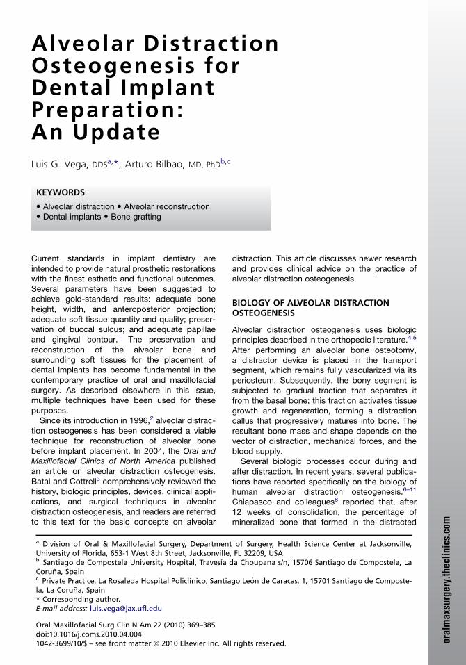
Fig. 1. General alveolar distraction timeline.
DISTRACTION PROTOCOL
After almost 15 years of widespread use, there isstill controversy regarding the best protocol. Asnew devices and applications have been designed,different distraction protocols have been testedand established. However, the original clinical
phases of distraction remain the same: osteotomy,latency, distraction, and consolidation (Fig. 1).
Osteotomy
Osteotomy has traditionally been performed withrotary burs, different kinds of saws, and osteo-tomes. Piezosurgery for alveolar distractionosteotomies has also been reported in the litera-ture.12,13 By comparing piezoelectric with conven-tional osteotomies for alveolar distraction,Gonzalez-Garcıa and colleagues13 found that thesurgical difficulty and the incidence of intraopera-tive complications were significantly lower in thepiezoelectric group. Their results showed that thepostdistraction alveolar morphology was worsein the piezoelectric group. They theorize that thepiezoelectric osteotomies will create a wider initialgap that may favor the appearance of granulationtissue without good osteogenic potential.
Latency Period
Latency period is defined as time from surgery tothe beginning of distraction. In an alveolar distrac-tion systematic review from 1996 to 2006, themost common latency period was 7 days (66%of the cases reviewed) to allow for healing of themucoperiosteum and reduce the risk of wound

Box 1General applications for alveolar distractionosteogenesis
� Moderate to severe vertical alveolar bonedefects� Segmental deficiencies of the alveolar ridge� Narrow alveolar ridges� Adjuvant to other bone graft techniques� Gradual vertical movement of ankylosed
teeth� Gradual vertical movement of an osseointe-
grated implant together with thesurrounding alveolar bone
Alveolar Distraction Osteogenesis: An Update 371
dehiscence. Extended latency periods of morethan 15 days were applied to ensure completerevascularization of the transport segment incases in which the mucoperiosteal pedicle is smallor endangered.14
Distraction Period
The distraction period encompasses the timebetween initial activation and end of the activationof the distractor device. The amount of distractionrequired is generally based on the amount of tissuenecessary to fulfill the implant and dental rehabili-tation goals. Several studies have focused on theamount of alveolar distraction relapse, and theirrecommendation is to overcorrect by 20% to25%.15,16 Apart from the amount of distractionneeded, the distraction rate and rhythm are ofparamount importance during this period.
Distraction rateThe daily amount of bone to be distracted isknown as distraction rate. Saulacic andcolleagues14 reported in a systematic review thatthe mean distraction rate was 0.71 (�0.27) mm.They also noted a lower distraction rate of 0.4 to0.5 mm in cases in which distractor implants andhorizontal distraction were used. According toAmir and colleagues,17 a distraction rate of 0.5mm per day results in faster osteogenesis thana distraction rate of 1 mm in elderly patients.
Distraction rhythmDistraction rhythm is the number of distractionactivations per day. According to Saulacic andcolleagues,14 the rhythm in alveolar distractionhas tended to be chosen empirically, in partbecause of a lack of experimental findings. Theyreviewed 209 distractions in 197 patients, andfound that the rhythm of distraction rangedbetween 1 (62%), 2 (35%), and 4 times daily (3%).
Consolidation Period
This is the period that allows for maturation andcorticalization of the regenerated bone. Accordingto Amir and colleagues,9 a minimum of 10 weeks isrequired for new bone to bridge a 10 mm alveolardistraction gap. It has been suggested that thepoorly mineralized bone tissue found after 10weeks of consolidation will start an adaptiveresponse that would increase the bone matrixmineralization with placement of dental implants.18
A systematic review found that the mean consoli-dation period was 12.22 (�5.58) weeks. A differ-ence was noted in the consolidation period whendifferent distractor devices were used. The meanconsolidation period on intraosseous distractorswas 8.82 (�2.67) weeks, 11.44 (�2.55) weeks for
the extraosseous distractors, and 18.02 (�3.50)weeks before prosthetic treatment started indistraction implants.
ALVEOLAR DISTRACTION DEVICES
Novel alveolar distraction designs are constantlybeing developed for research and clinicalpurposes. As a general rule they have been classi-fied as intraosseous and extraosseous, dependingon the placement in relation to the bone. In a studycomparing clinical outcomes of intraosseous andextraosseous alveolar distractors, Uckan andcolleagues,19 found no significant statistical differ-ence despite the higher complication rate andlower implant success in the intraosseous group.Devices can also be categorized as unidirectionaland multidirectional, depending on the directionof the movement. Initial alveolar distractor designsallowed for only a unidirectional movement,making correct positioning of the device andvector control most important. Recent publica-tions have shown the clinical value of multidirectional alveolar distraction devices.6,20,21 Aretrospective study comparing outcomes of unidi-rectional and bidirectional distractor devices,Schleier and colleagues21 found no significantstatistical differences in the bone gain or implantsuccess. Moreover, several cases with unidirec-tional distraction had to be bone grafted at thetime of implant placement. They concluded thatthis difference was caused by the precise controlof the distraction process in the bidirectionaldistraction group.
INDICATIONS FOR ALVEOLAR DISTRACTIONOSTEOGENESIS
Several clinical indications for alveolar distractionosteogenesis have been reported in the literature(Box 1).22–24 Alveolar reconstruction in preparationfor dental implant placement continues to be the

Vega & Bilbao372
most common indication (Fig. 2). Reconstructionefforts have used alveolar distraction as a definitiveprocedure to establish the ideal alveolar ridge oras an adjunctive procedure used to gain bone aspart of a larger reconstruction plan. Overall, alve-olar distraction offers several advantagescompared with other augmentation techniques(Boxes 2 and 3).25
Alveolar distraction has traditionally been usedfor vertical augmentation of the alveolar ridge,but horizontal26–28 and segmental alveolar distrac-tion29,30 have also been described. The main indi-cation for alveolar distraction is to manage the
Fig. 2. Alveolar reconstruction using alveolar distraction iposttraumatic vertical defect. (B) Osteotomy. (C) Alveolar d(E) Panoramic radiograph after the end of the distraction.graph at the end of the treatment.
vertical defects in the anterior maxilla andmandible. Posterior maxillary defects are best ad-dressed with traditional techniques such as sinuslift or bone grafts. Vertical defects of the posteriormandible can be treated with alveolar distractionbut, if the defect also has a horizontal component,a more traditional approach with an onlaybone graft or guided tissue regeneration isrecommended.31,32
To facilitate the evaluation and treatment ofvertical alveolar defects, Jensen and Block33
proposed a classification system in which theydefined a class I defect as a mild alveolar vertical
n preparation for dental implants. (A) Right maxillaryistractor in place. (D) Vector control using a prosthesis.(F) Placement of dental implants. (G) Panoramic radio-

Box 2Advantages of alveolar distractionosteogenesis for preparation for implantplacement
� Simple technique� Simultaneous augmentation of bone and soft
tissues� Less resorption than traditional bone grafts� Transport segment can include teeth or
implants, facilitating the correction ofocclusal or prosthetic defects� Elimination of donor-site morbidity� Shorter treatment times compared with tradi-
tional bone grafting techniques� Allows the implementation of complemen-
tary techniques when results are not optimal
Alveolar Distraction Osteogenesis: An Update 373
deficiency with up to 5 mm that ideally can betreated by a sandwich osteotomy or more tradi-tional bone graft techniques, although distractioncan be considered when there are prostheticconcerns in the esthetic zone. Class II defectsconsist of a moderate vertical loss of 6 to 10 mmthat ideally will benefit from alveolar distraction.Class III defects are severe vertical losses greaterthan 10 mm. Treatment of these defects dependson the available bone stock. If sufficient boneexists, distraction can be carried out first, anddefinitive alveolar bone form and position can beperformed with a bone graft. If the amount ofbone is not sufficient for distraction, boneaugmentation is carried out first followed bydistraction. Vertical defects that involve adjacentteeth with significant bone loss are designated asclass IV. In these cases, by extracting the affecteddentition, the defect will be converted into a classII or III defect, making the problem more predict-able and easier to resolve.
PLANNING FOR ALVEOLAR DISTRACTIONOSTEOGENESIS
Clinical examination will establish a preliminaryidea of the patient prosthetic needs, occlusion,
Box 3Disadvantages of alveolar distractionosteogenesis for preparation for implantplacement
� Patient acceptance and compliance� Requires careful vector control� Interference with occlusion might require the
construction of protective appliances� High device cost
and the size and shape of the alveolar defect.Maxillary and mandibular models with a diag-nostic wax-up will allow corroboration of the clin-ical findings. They can also be used to fabricatea surgical splint that could be use for vectorcontrol as well as temporary restoration. Modelsalso play an important role in planning for thedistraction vector, allowing preadaptation of thedevice, decreasing surgical time, and identifyingpossible device interferences with opposingdentition. In more complex cases, sterolitho-graphic models are a good option for treatmentplanning.
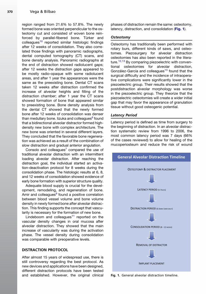
Radiographic examination with plain films orCT scan is useful for alveolar defect assessmentbut it also allows for planning the length andheight of the osteotomy. Of great importance isthe amount of bone stock and its relationshipwith the inferior alveolar nerve, inferior borderof the mandible, nasal floor, and maxillary sinus.These factors could limit device placement or thedistraction procedure. Added considerationshould be given to the prophylactic plating ofthe mandible in which the remaining basal boneis scarce, to prevent fracture and retention ofcompromised teeth adjacent to the distractorprocedure and to help with vector control. Newertechnologies, such as computer-assistedsurgical planning, are also being applied to alve-olar distraction (Fig. 3).34
ALVEOLAR DISTRACTION: SURGICAL ADVICE
During the different phases of alveolar distraction,there are a series of considerations that cancontribute to a successful outcome. Allocatingsufficient time for surgical planning is probablythe single most important element in alveolardistraction.
Incision
� Special consideration should be given tothe location of the incision, because itwill affect the quality of the soft tissuethat will be augmented at the end oftreatment� Use sound surgical principles that will guar-
antee proper blood supply to the mucosaand bone� Careful and conservative dissection will
maintain the vascularity of the transportsegment, decreasing excessive resorptionand avoiding damage to adjacentstructures.
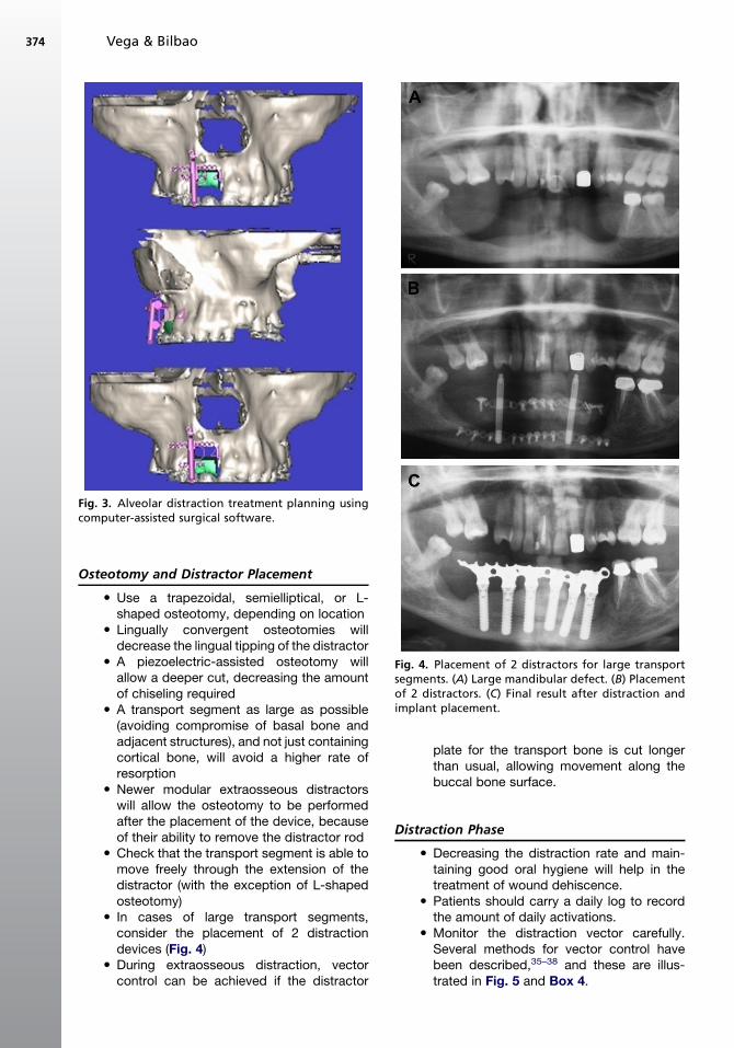
Fig. 3. Alveolar distraction treatment planning usingcomputer-assisted surgical software.
Vega & Bilbao374
Osteotomy and Distractor Placement
Fig. 4. Placement of 2 distractors for large transportsegments. (A) Large mandibular defect. (B) Placementof 2 distractors. (C) Final result after distraction andimplant placement.
� Use a trapezoidal, semielliptical, or L-shaped osteotomy, depending on location� Lingually convergent osteotomies will
decrease the lingual tipping of the distractor� A piezoelectric-assisted osteotomy will
allow a deeper cut, decreasing the amountof chiseling required� A transport segment as large as possible
(avoiding compromise of basal bone andadjacent structures), and not just containingcortical bone, will avoid a higher rate ofresorption� Newer modular extraosseous distractors
will allow the osteotomy to be performedafter the placement of the device, becauseof their ability to remove the distractor rod� Check that the transport segment is able to
move freely through the extension of thedistractor (with the exception of L-shapedosteotomy)� In cases of large transport segments,
consider the placement of 2 distractiondevices (Fig. 4)� During extraosseous distraction, vector
control can be achieved if the distractor
plate for the transport bone is cut longerthan usual, allowing movement along thebuccal bone surface.
Distraction Phase
� Decreasing the distraction rate and main-taining good oral hygiene will help in thetreatment of wound dehiscence.� Patients should carry a daily log to record
the amount of daily activations.� Monitor the distraction vector carefully.
Several methods for vector control havebeen described,35–38 and these are illus-trated in Fig. 5 and Box 4.
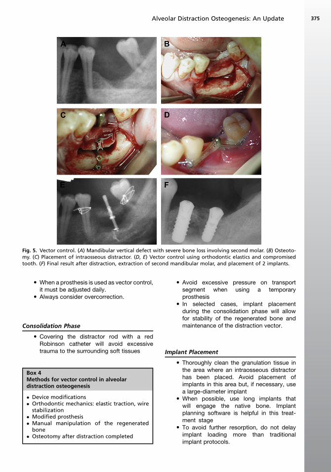
Fig. 5. Vector control. (A) Mandibular vertical defect with severe bone loss involving second molar. (B) Osteoto-my. (C) Placement of intraosseous distractor. (D, E) Vector control using orthodontic elastics and compromisedtooth. (F) Final result after distraction, extraction of second mandibular molar, and placement of 2 implants.
BoMdis
��
��
�
Alveolar Distraction Osteogenesis: An Update 375
� When a prosthesis is used as vector control,it must be adjusted daily.� Always consider overcorrection.
Consolidation Phase
� Covering the distractor rod with a redRobinson catheter will avoid excessivetrauma to the surrounding soft tissues
x 4ethods for vector control in alveolartraction osteogenesis
Device modificationsOrthodontic mechanics: elastic traction, wirestabilizationModified prosthesisManual manipulation of the regeneratedboneOsteotomy after distraction completed
� Avoid excessive pressure on transportsegment when using a temporaryprosthesis� In selected cases, implant placement
during the consolidation phase will allowfor stability of the regenerated bone andmaintenance of the distraction vector.
Implant Placement
� Thoroughly clean the granulation tissue inthe area where an intraosseous distractorhas been placed. Avoid placement ofimplants in this area but, if necessary, usea large-diameter implant� When possible, use long implants that
will engage the native bone. Implantplanning software is helpful in this treat-ment stage� To avoid further resorption, do not delay
implant loading more than traditionalimplant protocols.
Vega & Bilbao376
CLINICAL OUTCOMES IN ALVEOLARDISTRACTION OSTEOGENESISVertical Bone Gain
Data of 181 patients from a recent review byChiapasco and colleagues39 showed that theamount of bone gain after distraction osteogene-sis had a range of 3 to 20 mm. Saulacic andcolleagues14 reported in their systematic reviewthe mean bone gain obtained by different typesof distractors: distraction implants, 5.02 (�0.09)mm; intraosseous distractors, 7.86 (�0.36) mm;and extraosseous distractors, 9.31 (�0.45) mm.A clinical assessment of 40 patients subjectedto an extraosseous distraction showed that thebone augmentation average was 9.5 mm inheight, showing a 92.5% success rate.40 Kannoand colleagues16 reported comparable resultson bone gain using extraosseous distractors;they also noted that, during the consolidationperiod, there was 15% to 25% bone heightreduction. These findings are similar to theprevious reports in the literature that recommend20% to 25% overcorrection in vertical alveolardistraction.15,41,42 Perdijk and colleagues43
pointed out the influence of vector of distractionon vertical gain. They studied 34 cases of alveolardistraction on atrophic mandible in which nearlyall patients had lingual tipping of the segment bya mean of 12�. This finding meant that, in thosecases, only 87% of maximum vertical bone gaincould be achieved.
Alveolar Distraction Compared withConventional Bone Grafting Techniques
In 2004, Chiapasco and colleagues44 comparedalveolar distraction osteogenesis with guidedbone regeneration on vertically deficient alveolarridges. This prospective study evaluated parame-ters such as bone resorption of the regenerated
Table 1Comparison of augmentation techniques on edentul
Technique Success
Guided bone regeneration 60–100
Onlay bone grafts 92–100
Split ridge 98–100
Alveolar distraction 96.7–10
Microvascular flaps 87.5
Data from Chiapasco M, Zaniboni M, Boisco M. Augmentationridges with oral implants. Clin Oral Implants Res 2006;17(Supp
ridges before and after implant placement,peri-implant bone loss at 1, 2, and 3 years afterprosthetic loading of the implant, and successrates of implants. The results suggested thatalveolar distraction might offer more predictablelong-term results for bone gain maintenance andperi-implant bone resorption. Furthermore,implant success rates were significantly higher inthe alveolar distraction group. Chiapasco andcolleagues45 also compared alveolar osteogene-sis with autogenous onlay bone grafts using thesimilar parameters. This study found that boneresorption before implant placement was signifi-cantly higher in the autogenous onlay bone graftgroup. For implant success, no difference wasencountered between the groups. Uckan andcolleagues46 also compared alveolar distractionwith autogenous onlay graft using complicationand implant survival rates. Their results showeda higher complication rate with the alveolardistraction (66.8% vs 33.8%). But they alsoreported that those complications were minorand easier to treat than those of the autogenousonlay graft. Again, implant survival rates weresimilar between the groups (91.4% alveolardistraction vs 93.7% autogenous onlay graft). Ina prospective study comparing alveolar distractionwith inlay bone grafting in the posterior mandible,Bianchi and colleagues47 showed that, althoughthe mean bone gain with alveolar distraction wassignificantly better (10 mm vs 5.8 mm), the compli-cation rate was significantly higher in the alveolardistraction group (60%) than in the inlay bone graftgroup (14.3%).
Two literature reviews of bone augmentationprocedures on edentulous ridges for dentalimplants concluded that it is difficult to demon-strate that one surgical procedure offered betteroutcomes than another because of the poor meth-odological quality of the articles published(Table 1).39,48 Their recommendation is to give
ous ridges for dental implant placement
Rate (%) Implant Survival (%)
92–100
60–100
91–97.3
0 90.4–100
88.2
procedures for the rehabilitation of deficient edentulousl 2):136–59.

Alveolar Distraction Osteogenesis: An Update 377
priority to those procedures that are simpler, lessinvasive, involve less risk of complications, andreach their goals in the shortest time.
Alveolar Distraction on a PreviouslyReconstructed Site
Reconstruction of severe maxillary and mandib-ular defects for dental implants after trauma ortumor ablation is often a difficult task. Case reportsin the literature describe the use of alveolardistraction as adjuvant to enhance sites previouslyreconstructed with iliac bone grafts,49,50 scapulafree flaps,51,52 and fibular free flaps.53,54
In a retrospective study, Kunkel and colleagues50
reported on 4 patients who underwent iliac crestbone graft for mandibular reconstruction aftertumor ablation and later alveolar distraction withan intraosseous device. The vertical gain rangewas from 5 to 9 mm and, of the 12 implants placed,1 failed and 1 had critical bone loss after 40 monthsof follow-up. Hirota and colleagues51,52 describedthe use alveolar distraction to enhance the mandib-ular reconstruction carried out with free scapulaflaps in 2 patients. They reported vertical gains of9 and 10 mm and placement of 9 implants witha 100% success rate after 2 years of follow-up. In2009, Lizio and colleagues54 used alveolar dis-traction to increase the vertical bone height on6 patients after reconstruction with free fibula flaps.Themeanverticalbonegain was14mm(12–15mm).They placed 35 implants, 4 of which failed during thefollow-up period, bringing the cumulative implantsurvival to 89%. They also reported 1 case withfracture of the remaining basal fibula duringconsolidation.
Success of Dental Implantsin Distracted Bone
Prosthetic rehabilitation facilitated by the place-ment of dental implants is the ultimate goal ofalveolar distraction. Controversy still remainsregarding the best time for implant placement.
A prospective multicenter study reported theoutcomes of 138 implants placed in distractedbone after 2 or 3 months of consolidation. Aftera mean follow-up of 34 months after prostheticloading, the success rate was 94.2% with a cumu-lative survival rate of 100%. No statistically signif-icant differences were found between the differentcenters.55
Using 92 distractor implants on 46 patients withseverely atrophic mandibles, Raghoebar andcolleagues56 reported a survival rate of 97% aftera minimum of 62 months of follow-up. In a retro-spective study, Elo and colleagues57 compared
the implant success rates in distracted bone withautogenous bone-grafted sites. They placed 184implants on 65 patients reconstructed with autog-enous bone, with an implant success rate of 97%.The distraction group contained 56 implants on 17patients and a success rate of 98%. Again, nostatistical difference was noted between groups.
A systematic review on alveolar distractionanalyzed a total of 469 implants placed indistracted bone. The mean osseointegrationperiod was 4.59 (�1.34) months. The overallsurvival rate was 97%. They reported 14 implantfailures, 10 of them before loading. The meanfollow-up was 14.19 (�11.03) months. This anal-ysis also found no significant difference in implantfailure rate associated with location, indicationfor distraction, latency period, and daily rate andrhythm. The mean augmentation ratesapproached a statistically significant difference:rate on successful implants was 6.79 (�2.51) mmand 8.40 (�2.31) mm on failed implants. A signifi-cant difference was encountered in the relation-ship between implant failures and distractionimplants. Consolidation period also showed signif-icant differences; failed implants were placed after8.10 (�2.51) weeks, compared with 12.43 (�5.62)weeks for successful implants. Peri-implant bonelevel was reported for 301 implants. Stable peri-implant bone level was maintained in 285 (95%)of the implants.14 Recent studies reported peri-implant bone loss values of 0.89 to 1.9 mm/y inareas of alveolar bone distraction.42,58
Immediate Loading of Implantson Distracted Bone
In 2004, Degidi and colleagues59 presented a caseof immediate loading of implants placed indistracted bone. Although this practice has notbeen popular, a study was carried out comparingdata from radiofrequency analysis on implantsplaced in native bone and distracted bone. Evenwhen the results were significantly inferior forimplants placed in distracted bone, the investiga-tors concluded that the values obtained suggestthe possibility of immediate loading with outcomessimilar to those of implants in native bone.60
Adjunctive Techniques to Improve theOutcomes of Alveolar Distraction
Research is being conducted on ways to improvethe outcome of alveolar distraction. Robiony andcolleagues61 reported on their long-term experi-ence with 12 patients after alveolar distractionand a combination of autologous bone graft withplatelet-rich plasma on severely atrophic mandi-bles. After performing an osteotomy, the distractor
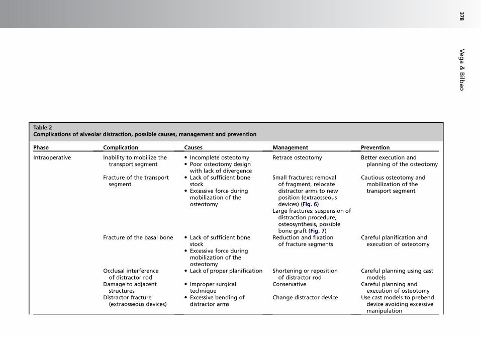
Table 2Complications of alveolar distraction, possible causes, management and prevention
Phase Complication Causes Management Prevention
Intraoperative Inability to mobilize thetransport segment
� Incomplete osteotomy� Poor osteotomy design
with lack of divergence
Retrace osteotomy Better execution andplanning of the osteotomy
Fracture of the transportsegment
� Lack of sufficient bonestock
� Excessive force duringmobilization of theosteotomy
Small fractures: removalof fragment, relocatedistractor arms to newposition (extraosseousdevices) (Fig. 6)
Large fractures: suspension ofdistraction procedure,osteosynthesis, possiblebone graft (Fig. 7)
Cautious osteotomy andmobilization of thetransport segment
Fracture of the basal bone � Lack of sufficient bonestock
� Excessive force duringmobilization of theosteotomy
Reduction and fixationof fracture segments
Careful planification andexecution of osteotomy
Occlusal interferenceof distractor rod
� Lack of proper planification Shortening or repositionof distractor rod
Careful planning using castmodels
Damage to adjacentstructures
� Improper surgicaltechnique
Conservative Careful planning andexecution of osteotomy
Distractor fracture(extraosseous devices)
� Excessive bending ofdistractor arms
Change distractor device Use cast models to prebenddevice avoiding excessivemanipulation
Veg
a&
Bilb
ao
378
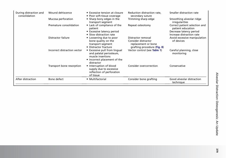
During distraction andconsolidation
Wound dehiscence � Excessive tension at closure� Poor soft-tissue coverage
Reduction distraction rate,seconda suture
Smaller distraction rate
Mucosa perforation � Sharp bony edges in thetransport segment
Trimming arp edge Smoothing alveolar ridgeirregularities
Premature consolidation � Lack of compliance of thepatient
� Excessive latency period� Slow distraction rate
Repeat ost otomy Correct patient selection andpatient education
Decrease latency periodIncrease distraction rate
Distractor failure � Loosening due to poorbone quality on thetransport segment
� Distractor fracture
Distractor movalConsider d tractor
replacem nt or bonegrafting rocedure (Fig. 8)
Avoid excessive manipulationof devices
Incorrect distraction vector � Excessive pull from lingualand palatal periosteum,muscle insertions
� Incorrect placement of thedistractor
Vector con rol (see Table 1) Careful planning, closemonitoring
Transport bone resorption � Interruption of bloodsupply due to excessivereflection of perforationof tissue
Consider o ercorrection Conservative
After distraction Bone defect � Multifactorial Consider b ne grafting Good alveolar distractiontechnique
Alve
ola
rD
istractio
nO
steo
gen
esis:
An
Up
date
379
rysh
e
reisept
v
o
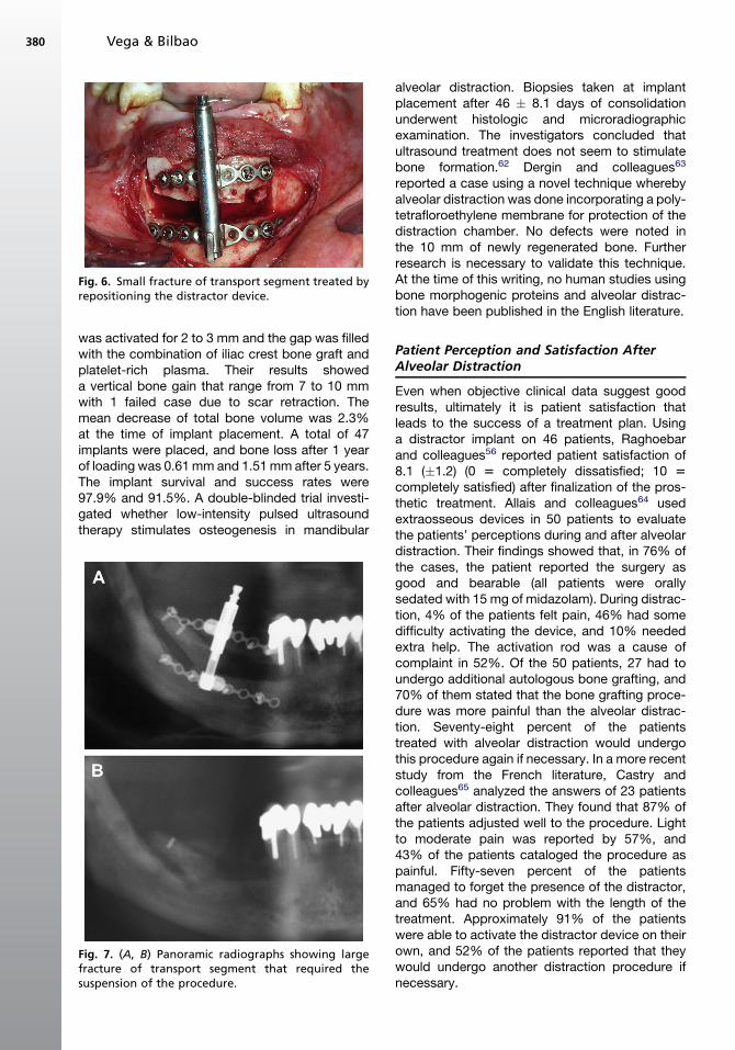
Fig. 6. Small fracture of transport segment treated byrepositioning the distractor device.
Vega & Bilbao380
was activated for 2 to 3 mm and the gap was filledwith the combination of iliac crest bone graft andplatelet-rich plasma. Their results showeda vertical bone gain that range from 7 to 10 mmwith 1 failed case due to scar retraction. Themean decrease of total bone volume was 2.3%at the time of implant placement. A total of 47implants were placed, and bone loss after 1 yearof loading was 0.61 mm and 1.51 mm after 5 years.The implant survival and success rates were97.9% and 91.5%. A double-blinded trial investi-gated whether low-intensity pulsed ultrasoundtherapy stimulates osteogenesis in mandibular
Fig. 7. (A, B) Panoramic radiographs showing largefracture of transport segment that required thesuspension of the procedure.
alveolar distraction. Biopsies taken at implantplacement after 46 � 8.1 days of consolidationunderwent histologic and microradiographicexamination. The investigators concluded thatultrasound treatment does not seem to stimulatebone formation.62 Dergin and colleagues63
reported a case using a novel technique wherebyalveolar distraction was done incorporating a poly-tetrafloroethylene membrane for protection of thedistraction chamber. No defects were noted inthe 10 mm of newly regenerated bone. Furtherresearch is necessary to validate this technique.At the time of this writing, no human studies usingbone morphogenic proteins and alveolar distrac-tion have been published in the English literature.
Patient Perception and Satisfaction AfterAlveolar Distraction
Even when objective clinical data suggest goodresults, ultimately it is patient satisfaction thatleads to the success of a treatment plan. Usinga distractor implant on 46 patients, Raghoebarand colleagues56 reported patient satisfaction of8.1 (�1.2) (0 5 completely dissatisfied; 10 5completely satisfied) after finalization of the pros-thetic treatment. Allais and colleagues64 usedextraosseous devices in 50 patients to evaluatethe patients’ perceptions during and after alveolardistraction. Their findings showed that, in 76% ofthe cases, the patient reported the surgery asgood and bearable (all patients were orallysedated with 15 mg of midazolam). During distrac-tion, 4% of the patients felt pain, 46% had somedifficulty activating the device, and 10% neededextra help. The activation rod was a cause ofcomplaint in 52%. Of the 50 patients, 27 had toundergo additional autologous bone grafting, and70% of them stated that the bone grafting proce-dure was more painful than the alveolar distrac-tion. Seventy-eight percent of the patientstreated with alveolar distraction would undergothis procedure again if necessary. In a more recentstudy from the French literature, Castry andcolleagues65 analyzed the answers of 23 patientsafter alveolar distraction. They found that 87% ofthe patients adjusted well to the procedure. Lightto moderate pain was reported by 57%, and43% of the patients cataloged the procedure aspainful. Fifty-seven percent of the patientsmanaged to forget the presence of the distractor,and 65% had no problem with the length of thetreatment. Approximately 91% of the patientswere able to activate the distractor device on theirown, and 52% of the patients reported that theywould undergo another distraction procedure ifnecessary.
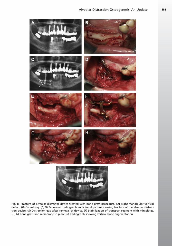
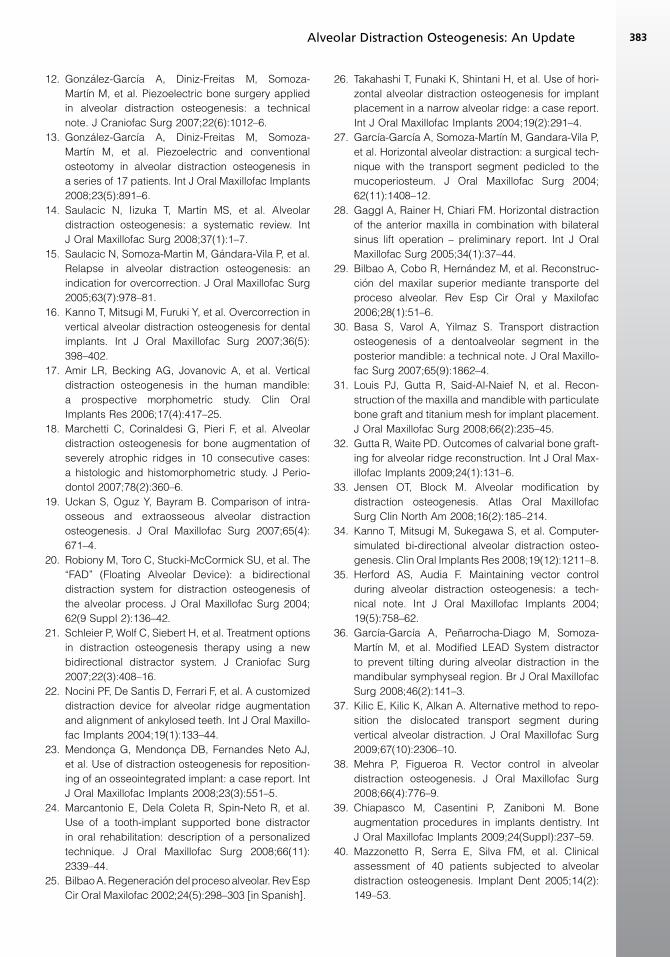
Fig. 8. Fracture of alveolar distractor device treated with bone graft procedure. (A) Right mandibular verticaldefect. (B) Osteotomy. (C, D) Panoramic radiograph and clinical picture showing fracture of the alveolar distrac-tion device. (E) Distraction gap after removal of device. (F) Stabilization of transport segment with miniplates.(G, H) Bone graft and membrane in place. (I) Radiograph showing vertical bone augmentation.
Alveolar Distraction Osteogenesis: An Update 381

Vega & Bilbao382
Complication Rates of Alveolar Distraction
Despite almost 15 years of clinical practice,growing popularity, and newer technologies, alve-olar distraction continues to be a challengingprocedure. Alveolar distraction complicationshave conventionally been classified according tothe distraction phases in which they occurred:intraoperative, during distraction, during consoli-dation, and after distraction. They can also beclassified as minor complications or major compli-cations that are more difficult to manage and couldjeopardize the distraction procedure. In addition tothe common complications of any surgical proce-dure, such as excessive bleeding, hematoma,infection, and paresthesia, there is a set of specificcomplications for alveolar distraction. Thesecomplications, the possible causes, management,and prevention are listed in Table 2.
In more recent years, several articles have focuson the complications of alveolar distraction. Thesestudies have reported a wide array of complicationrates, ranging from 36% to 100%.41,42,66–70 Ina comprehensive review of the literature from1996 to 2008, Saulacic and colleagues71 studiedthe complication rate of alveolar distraction. Theirresults showed an overall complication rate of30%. The most common complication was insuffi-cient bone formation after the consolidation period(8%), followed by regression of distractiondistance (7%), and problems related to the device(6%). Intraoperative complications includebleeding from the floor of the mouth (4%) andtemporary paresthesia (4%). During the distractionperiod, wound dehiscence was found on 1% of thepatients. Pain was reported in 1% of the patients,as well as mild soft-tissue resistance. Vector devi-ation was found in 2%. More severe complicationswere found during the consolidation period,including a mandibular fracture rate of 2% andproblems related to the device in 6% of the cases.They also found that insufficient bone formationand evidence of complications were significantlyrelated to the type of distractor and augmentationrates greater than 0.5 mm daily. The investigatorsconcluded that, although complications in alveolardistraction are frequent, they rarely cause severeproblems. They suggested that most of thecomplications could be related to lack of experi-ence and the learning process.
SUMMARY
Alveolar distraction is a technique in constantevolution. A review of the literature within thepast 14 years reveal that there are clear indicationsfor its use, with outcomes similar to and
sometimes even more predictable than traditionalbone grafting techniques in preparation for implantplacement. Although complications exist withalveolar distraction, it seems that most are minorand easy to manage. Appropriate patient selectionand a better understanding of the technique areparamount in successful bone regeneration withalveolar distraction osteogenesis.
REFERENCES
1. Guerrero C, Lopez P, Figueroa F, et al. Three-dimen-
sional alveolar distraction osteogenesis. In: Bell W,
Guerrero C, editors. Distraction osteogenesis of
the facial skeleton. 1st edition. Hamilton (Canada):
BC Decker; 2007. p. 457–93.
2. Chin M, Toth BA. Distraction osteogenesis in maxillo-
facial surgery using internal devices: review of five
cases. J Oral Maxillofac Surg 1996;54(1):45–53.
3. Batal HS, Cottrell DA. Alveolar distraction osteogen-
esis for implant site development. Oral Maxillofac
Surg Clin North Am 2004;16(1):91–109.
4. Ilizarov GA. The tension-stress effect on the genesis
and growth of tissues. Part I. The influence of
stability of fixation and soft-tissue preservation. Clin
Orthop Relat Res 1989;238:249–81.
5. Ilizarov GA. The tension-stress effect on the genesis
and growth of tissues: Part II. The influence of the
rate and frequency of distraction. Clin Orthop Relat
Res 1989;239:263–85.
6. Iizuka T, Hallermann W, Seto I, et al. Bi-directional
distraction osteogenesis of the alveolar bone using
an extraosseous device. Clin Oral Implants Res
2005;16(6):700–7.
7. Consolo U, Bertoldi C, Zaffe D. Intermittent loading
improves results in mandibular alveolar distraction
osteogenesis. Clin Oral Implants Res 2006;17(2):
179–87.
8. Chiapasco M, Biglioli F, Autelitano L, et al. Clinical
outcome of dental implants placed in fibula-free
flaps used for the reconstruction of maxillo-
mandibular defects following ablation for tumors
or osteoradionecrosis. Clin Oral Implants Res
2006;17(2):220–8.
9. Amir LR, Becking AG, Jovanovic A, et al. Formation
of new bone during vertical distraction osteogenesis
of the human mandible is related to the presence of
blood vessels. Clin Oral Implants Res 2006;17(4):
410–6.
10. Turker N, Basa S, Vural G. Evaluation of osseous
regeneration in alveolar distraction osteogenesis
with histological and radiological aspects. J Oral
Maxillofac Surg 2007;65(4):608–14.
11. Lindeboom JA, Mathura KR, Milstein DMJ, et al.
Microvascular soft tissue changes in alveolar
distraction osteogenesis. Oral Surg Oral Med Oral
Pathol Oral Radiol Endod 2008;106(3):350–5.
Alveolar Distraction Osteogenesis: An Update 383
12. Gonzalez-Garcıa A, Diniz-Freitas M, Somoza-
Martın M, et al. Piezoelectric bone surgery applied
in alveolar distraction osteogenesis: a technical
note. J Craniofac Surg 2007;22(6):1012–6.
13. Gonzalez-Garcıa A, Diniz-Freitas M, Somoza-
Martın M, et al. Piezoelectric and conventional
osteotomy in alveolar distraction osteogenesis in
a series of 17 patients. Int J Oral Maxillofac Implants
2008;23(5):891–6.
14. Saulacic N, Iizuka T, Martin MS, et al. Alveolar
distraction osteogenesis: a systematic review. Int
J Oral Maxillofac Surg 2008;37(1):1–7.
15. Saulacic N, Somoza-Martin M, Gandara-Vila P, et al.
Relapse in alveolar distraction osteogenesis: an
indication for overcorrection. J Oral Maxillofac Surg
2005;63(7):978–81.
16. Kanno T, Mitsugi M, Furuki Y, et al. Overcorrection in
vertical alveolar distraction osteogenesis for dental
implants. Int J Oral Maxillofac Surg 2007;36(5):
398–402.
17. Amir LR, Becking AG, Jovanovic A, et al. Vertical
distraction osteogenesis in the human mandible:
a prospective morphometric study. Clin Oral
Implants Res 2006;17(4):417–25.
18. Marchetti C, Corinaldesi G, Pieri F, et al. Alveolar
distraction osteogenesis for bone augmentation of
severely atrophic ridges in 10 consecutive cases:
a histologic and histomorphometric study. J Perio-
dontol 2007;78(2):360–6.
19. Uckan S, Oguz Y, Bayram B. Comparison of intra-
osseous and extraosseous alveolar distraction
osteogenesis. J Oral Maxillofac Surg 2007;65(4):
671–4.
20. Robiony M, Toro C, Stucki-McCormick SU, et al. The
‘‘FAD’’ (Floating Alveolar Device): a bidirectional
distraction system for distraction osteogenesis of
the alveolar process. J Oral Maxillofac Surg 2004;
62(9 Suppl 2):136–42.
21. Schleier P, Wolf C, Siebert H, et al. Treatment options
in distraction osteogenesis therapy using a new
bidirectional distractor system. J Craniofac Surg
2007;22(3):408–16.
22. Nocini PF, De Santis D, Ferrari F, et al. A customized
distraction device for alveolar ridge augmentation
and alignment of ankylosed teeth. Int J Oral Maxillo-
fac Implants 2004;19(1):133–44.
23. Mendonca G, Mendonca DB, Fernandes Neto AJ,
et al. Use of distraction osteogenesis for reposition-
ing of an osseointegrated implant: a case report. Int
J Oral Maxillofac Implants 2008;23(3):551–5.
24. Marcantonio E, Dela Coleta R, Spin-Neto R, et al.
Use of a tooth-implant supported bone distractor
in oral rehabilitation: description of a personalized
technique. J Oral Maxillofac Surg 2008;66(11):
2339–44.
25. Bilbao A. Regeneracion del proceso alveolar. Rev Esp
Cir Oral Maxilofac 2002;24(5):298–303 [in Spanish].
26. Takahashi T, Funaki K, Shintani H, et al. Use of hori-
zontal alveolar distraction osteogenesis for implant
placement in a narrow alveolar ridge: a case report.
Int J Oral Maxillofac Implants 2004;19(2):291–4.
27. Garcıa-Garcıa A, Somoza-Martın M, Gandara-Vila P,
et al. Horizontal alveolar distraction: a surgical tech-
nique with the transport segment pedicled to the
mucoperiosteum. J Oral Maxillofac Surg 2004;
62(11):1408–12.
28. Gaggl A, Rainer H, Chiari FM. Horizontal distraction
of the anterior maxilla in combination with bilateral
sinus lift operation – preliminary report. Int J Oral
Maxillofac Surg 2005;34(1):37–44.
29. Bilbao A, Cobo R, Hernandez M, et al. Reconstruc-
cion del maxilar superior mediante transporte del
proceso alveolar. Rev Esp Cir Oral y Maxilofac
2006;28(1):51–6.
30. Basa S, Varol A, Yilmaz S. Transport distraction
osteogenesis of a dentoalveolar segment in the
posterior mandible: a technical note. J Oral Maxillo-
fac Surg 2007;65(9):1862–4.
31. Louis PJ, Gutta R, Said-Al-Naief N, et al. Recon-
struction of the maxilla and mandible with particulate
bone graft and titanium mesh for implant placement.
J Oral Maxillofac Surg 2008;66(2):235–45.
32. Gutta R, Waite PD. Outcomes of calvarial bone graft-
ing for alveolar ridge reconstruction. Int J Oral Max-
illofac Implants 2009;24(1):131–6.
33. Jensen OT, Block M. Alveolar modification by
distraction osteogenesis. Atlas Oral Maxillofac
Surg Clin North Am 2008;16(2):185–214.
34. Kanno T, Mitsugi M, Sukegawa S, et al. Computer-
simulated bi-directional alveolar distraction osteo-
genesis. Clin Oral Implants Res 2008;19(12):1211–8.
35. Herford AS, Audia F. Maintaining vector control
during alveolar distraction osteogenesis: a tech-
nical note. Int J Oral Maxillofac Implants 2004;
19(5):758–62.
36. Garcıa-Garcıa A, Penarrocha-Diago M, Somoza-
Martın M, et al. Modified LEAD System distractor
to prevent tilting during alveolar distraction in the
mandibular symphyseal region. Br J Oral Maxillofac
Surg 2008;46(2):141–3.
37. Kilic E, Kilic K, Alkan A. Alternative method to repo-
sition the dislocated transport segment during
vertical alveolar distraction. J Oral Maxillofac Surg
2009;67(10):2306–10.
38. Mehra P, Figueroa R. Vector control in alveolar
distraction osteogenesis. J Oral Maxillofac Surg
2008;66(4):776–9.
39. Chiapasco M, Casentini P, Zaniboni M. Bone
augmentation procedures in implants dentistry. Int
J Oral Maxillofac Implants 2009;24(Suppl):237–59.
40. Mazzonetto R, Serra E, Silva FM, et al. Clinical
assessment of 40 patients subjected to alveolar
distraction osteogenesis. Implant Dent 2005;14(2):
149–53.
Vega & Bilbao384
41. Wolvius EB, Scholtemeijer M, Weijland M, et al.
Complications and relapse in alveolar distraction
osteogenesis in partially dentulous patients. Int
J Oral Maxillofac Surg 2007;36(8):700–5.
42. Ettl T, Gerlach T, Schusselbauer T, et al. Bone
resorption and complications in alveolar distraction
osteogenesis. Clin Oral Investig 2009. DOI:10.1007/
s00784-009-0340-y.
43. Perdijk FB, Meijer GJ, van Strijen PJ, et al. Effect of
extraosseous devices designed for vertical distrac-
tion of extreme resorbed mandibles on backward
rotation of upper bone segments. Br J Oral Maxillo-
fac Surg 2009;47(1):31–6.
44. Chiapasco M, Romeo E, Casentini P, et al. Alveolar
distraction osteogenesis vs. vertical guided bone
regeneration for the correction of vertically deficient
edentulous ridges: a 1–3-year prospective study on
humans. Clin Oral Implants Res 2004;15(1):82–95.
45. Chiapasco M, Zaniboni M, Rimondini L. Autogenous
onlay bone grafts vs. alveolar distraction osteogene-
sis for the correction of vertically deficient edentu-
lous ridges: a 2–4-year prospective study on
humans. Clin Oral Implants Res 2007;18(4):432–40.
46. Uckan S, Veziroglu F, Dayangac E. Alveolar distrac-
tion osteogenesis versus autogenous onlay bone
grafting for alveolar ridge augmentation: technique,
complications, and implant survival rates. Oral
Surg Oral Med Oral Pathol Oral Radiol Endod
2008;106(4):511–5.
47. Bianchi A, Felice P, Lizio G, et al. Alveolar distraction
osteogenesis versus inlay bone grafting in posterior
mandibular atrophy: a prospective study. Oral Surg
Oral Med Oral Pathol Oral Radiol Endod 2008;
105(3):282–92.
48. Chiapasco M, Zaniboni M, Boisco M. Augmentation
procedures for the rehabilitation of deficient edentu-
lous ridges with oral implants. Clin Oral Implants Res
2006;17(Suppl 2):136–59.
49. Alkan A, Basx B, Inal S. Alveolar distraction osteogen-
esis of bone graft reconstructed mandible. Oral
Surg Oral Med Oral Pathol Oral Radiol Endod
2005;100(3):e39–42.
50. Kunkel M, Wahlmann U, Reichert TE, et al. Recon-
struction of mandibular defects following tumor abla-
tion by vertical distraction osteogenesis using
intraosseous distraction devices. Clin Oral Implants
Res 2005;16(1):89–97.
51. Hirota M, Mizuki N, Iwai T, et al. Vertical distraction of
a free vascularized osteocutaneous scapular flap in
the reconstructed mandible for implant therapy. Int
J Oral Maxillofac Surg 2008;37(5):481–3.
52. Hirota M, Matsui Y, Mizuki N, et al. Management
considerations in reconstruction of postablative
defects of the mandible: vertical distraction of
a scapular bone flap and removable lip support:
a case report. Oral Surg Oral Med Oral Pathol Oral
Radiol Endod 2008;106(6):e6–9.
53. Levin L, Carrasco L, Kazemi A, et al. Enhancement
of the fibula free flap by alveolar distraction for
dental implant restoration: report of a case. Facial
Plast Surg 2003;19(1):87–94.
54. Lizio G, Corinaldesi G, Pieri F, et al. Problems with
dental implants that were placed on vertically
distracted fibular free flaps after resection: a report of
sixcases. Br J Oral Maxillofac Surg2009;47(6):455–60.
55. Chiapasco M, Consolo U, Bianchi A, et al. Alveolar
distraction osteogenesis for the correction of verti-
cally deficient edentulous ridges: a multicenter
prospective study on humans. Int J Oral Maxillofac
Implants 2004;19(3):399–407.
56. Raghoebar GM, Stellingsma K, Meijer HJ, et al.
Vertical distraction of the severely resorbed edentu-
lous mandible: an assessment of treatment outcome.
Int J Oral Maxillofac Implants 2008;23(2):299–307.
57. Elo JA, Herford AS, Boyne PJ. Implant success in
distracted bone versus autogenous bone-grafted
sites. J Oral Implantol 2009;35(4):181–4.
58. Polo WC, de Araujo NS, Lima YB, et al. Peri-
implant bone loss around posterior mandible
dental implants placed after distraction osteogene-
sis: preliminary findings. J Periodontol 2007;78(2):
204–8.
59. Degidi M, Pieri F, Marchetti C, et al. Immediate
loading of dental implants placed in distracted
bone: a case report. Int J Oral Maxillofac Implants
2004;19(3):448–54.
60. Bilbao A, Oliveira MH, Varela-Centelles PI, et al.
Assessment of dental implant stability in osseo-
distraction-generated bone: a resonance freq-
uency analysis. Clin Oral Implants Res 2009;
20(8):772–7.
61. Robiony M, Zorzan E, Polini F, et al. Osteogenesis
distraction and platelet-rich plasma: combined use
in restoration of severe atrophic mandible. Long-
term results. Clin Oral Implants Res 2008;19(11):
1202–10.
62. Schortinghuis J, Bronckers AL, Gravendeel J, et al.
The effect of ultrasound on osteogenesis in the verti-
cally distracted edentulous mandible: a double-
blind trial. Int J Oral Maxillofac Surg 2008;37(11):
1014–21.
63. Dergin G, Gurler G, Guvercin M, et al. Vertical alve-
olar bone distraction with polytetrafloroethylene
membrane for implant application: a technical
note. J Oral Maxillofac Surg 2007;65(5):1050–4.
64. Allais M, Maurette PE, Mazzonetto R, et al. Patient’s
perception of the events during and after osteogenic
alveolar distraction. Med Oral Patol Oral Cir Bucal
2007;12(3):E225–8.
65. Castry G, Ella B, Emparanza A, et al. Impact psy-
chologique de la distraction alveolaire mandibulaire
[Psychological impact of alveolar mandibular
distraction]. Rev Stomatol Chir Maxillofac 2009;
110(5):251–5 [in French].

Alveolar Distraction Osteogenesis: An Update 385
66. Enislidis G, Fock N, Ewers R. Distraction osteogen-
esis with subperiosteal devices in edentulous
mandibles. Br J Oral Maxillofac Surg 2005;43(5):
399–403.
67. Mazzonetto R, Allais M, Maurette PE, et al. A retro-
spective study of the potential complications during
alveolar distraction osteogenesis in 55 patients. Int
J Oral Maxillofac Surg 2007;36(1):6–10.
68. Saulacic N, Somosa Martın M, de Los Angeles Leon
Camacho M, et al. Complications in alveolar distrac-
tion osteogenesis: a clinical investigation. J Oral
Maxillofac Surg 2007;65(2):267–74.
69. Perdijk FB, Meijer GJ, Strijen PJ, et al. Complications
in alveolar distraction osteogenesis of the atrophic
mandible. Int J Oral Maxillofac Surg 2007;36(10):
916–21.
70. Gunbay T, Koyuncu BO, Akay MC, et al. Results and
complications of alveolar distraction osteogenesis to
enhance vertical bone height. Oral Surg Oral Med
Oral Pathol Oral Radiol Endod 2008;105(5):e7–13.
71. Saulacic N, Zix J, Iizuka T. Complication rates and
associated factors in alveolar distraction osteogene-
sis: a comprehensive review. Int J Oral Maxillofac
Surg 2009;38(3):210–7.
Related Documents