Adipose Tissue Digital Laboratory It’s best to view this in Slide Show mode, especially for the quizzes. This module will take approximately 30 minutes to complete.

Adipose Tissue Digital Laboratory It’s best to view this in Slide Show mode, especially for the quizzes. This module will take approximately 30 minutes.
Dec 16, 2015
Welcome message from author
This document is posted to help you gain knowledge. Please leave a comment to let me know what you think about it! Share it to your friends and learn new things together.
Transcript

Adipose TissueDigital Laboratory
It’s best to view this in Slide Show mode, especially for the quizzes.
This module will take approximately 30 minutes to complete.
After completing this exercise, you should be able to: • Distinguish, at the light microscope level, each of the following components of adipose tissue:
• Adipose tissue• Unilocular (WAT)• Multilocular (BAT and developing WAT)
• Immunocytochemistry to differentiate white adipose tissue (WAT) and brown adipose tissue (BAT) in humans
• Describe the distribution of white adipose tissue (WAT) and brown adipose tissue (BAT) in humans

White adipose tissue (WAT) and brown adipose tissue (BAT) are distributed differently in the body, are histologically distinct and serve very different functions.
WAT is also termed unilocular adipose tissue because each cell contains a single large droplet of fat, which displaces other organelles to the cell periphery. In contrast, BAT cells contain many small fat droplets (thus, also termed multilocular fat)
ADIPOSE TISSUE
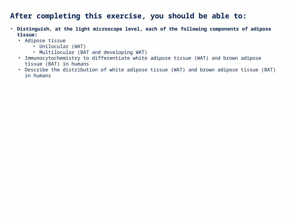
A section of adipose tissue, showing the large, unilocular adipocytes (an example of one cell is in shaded circle) – sometimes described as “signet rings”, because the large lipid droplet displaces the nucleus (green arrow) and remainder of the cytoplasm (red arrow) to the edge of the cell. The clear areas in the section are truly empty, because the triglycerides in the fat droplet are soluble in the organic solvents used during processing of the section, and are therefore removed from the section.
Many small blood vessels are also evident (blue arrows) – typical of WAT, which is richly vascularized. Sympathetic nerve endings (not visible in H&E) are also present.
UNILOCULAR ADIPOSE TISSUE (WAT)
There are thin layers of loose connective tissue between the cells here as well (e.g. a larger region is outlined). Because the cytoplasm of the adipocytes and the collagen fibers of the connective tissue are both eosinophilic, it’s often difficult to tell the exact border between the two.
http://pathology.mc.duke.edu/research/Histo_course/whitefat.jpg
Lipid droplet

Link to SL 034Be able to identify:
• Unilocular adipose tissue
Video of aged thymus showing unilocular adipose tissue – SL34
UNILOCULAR ADIPOSE TISSUE (WAT)

This transmission EM shows a single unilocular adipose cell. One large lipid droplet (Ld) is evident, although the EM shows that “unilocular” cells also contain smaller droplets that may be in the process of coalescing with the large droplet. The droplet squeezes the cytoplasm (arrows) and nucleus (N, cropped at bottom) to the periphery of the cell, resulting in the so-called “signet ring” morphology.
All cellular organelles (Golgi, rER, mitochondria) are present, although not abundant. A collagen fiber (Cf) and fibroblast (Fb) surrounding the cell are also present.
UNILOCULAR ADIPOSE TISSUE (WAT)

This scanning EM shows a cluster of unilocular adipocytes – which appear as spheres. Each spherical unilocular cell is almost entirely composed of a large, single droplet of fat, which is liquid at body temperature.
Reticular fibers, which look like small threads, enmesh the adipocytes.
UNILOCULAR ADIPOSE TISSUE (WAT)
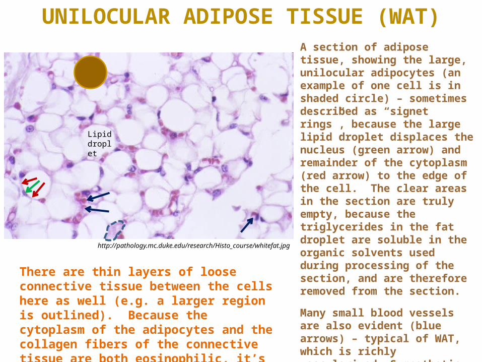
http://pathology.mc.duke.edu/research/Histo_course/brownfat.jpg
In contrast to the unilocular WAT cells, multilocular adipocytes contain many small fat droplets (these are the clear areas). Two cells have been outlined, each containing 10-20 visible droplets. Note that, in contrast to WAT, nuclei of BAT cells are round or oval and often centrally located (green arrows).
Many capillaries and small vessels are evident.
MULTILOCULAR ADIPOSE TISSUE (BAT)
The nuclei that you see around the periphery of the adipocytes (e.g. red arrow) likely belong to fibroblasts of the connective tissue between the adipose cells, or endothelial cells of the blood vessels.

Link to SL 012Be able to identify:
• multilocular adipose tissue
Video of axilla showing multilocular adipose tissue – SL12
MULTILOCULAR ADIPOSE TISSUE (BAT)
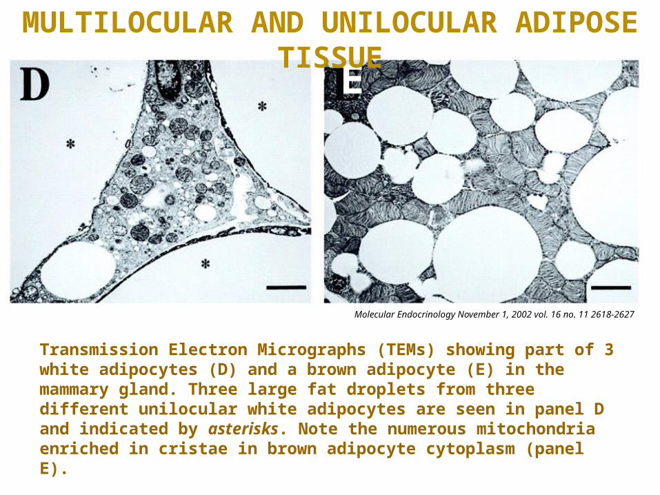
Transmission Electron Micrographs (TEMs) showing part of 3 white adipocytes (D) and a brown adipocyte (E) in the mammary gland. Three large fat droplets from three different unilocular white adipocytes are seen in panel D and indicated by asterisks. Note the numerous mitochondria enriched in cristae in brown adipocyte cytoplasm (panel E).
Molecular Endocrinology November 1, 2002 vol. 16 no. 11 2618-2627
E
MULTILOCULAR AND UNILOCULAR ADIPOSE TISSUE

There is one caveat to the categorization of adipose tissue: Developing white fat cells are multilocular!As you can see in the drawing on the left, both types of adipose cells begin as multilocular. The lipid droplets in BAT remain separate, while those in WAT coalesce to form one large droplet in mature cells.
MULTILOCULAR AND UNILOCULAR ADIPOSE TISSUE
Thus, when looking at slides, it’s probably best to use the terms “unilocular adipose” and “multilocular adipose”. If you say “white adipose” or “brown adipose”, we’ll presume you mean “unilocular” and “multilocular”, respectively. You may qualify your statement by saying “either brown fat or developing white fat” if the tissue is multilocular.

Here’s an example of developing white fat. You can clearly see that this tissue contains both unilocular and multilocular cells. Eventually, this entire tissue will consist of unilocular adipose tissue.
As mentioned above, from a practical standpoint, you would call the area outlined in blue unilocular adipose, and the area outlined in green multilocular adipose.
MULTILOCULAR AND UNILOCULAR ADIPOSE TISSUE

UCP-1
BAT WAT
As mentioned on the previous slides, when you see multilocular adipose stained with H&E, you cannot definitely say whether it is brown fat or developing white fat. However, you can distinguish the two with special staining procedures such as histochemistry. In this example, both tissues were stained for uncoupling protein (UCP-1), which shows up as brown in this preparation.
Uncoupling protein (UCP-1) is found in mitochondria of BAT cells but not WAT cells (it is not found in developing white fat either). This protein dissipates the proton gradient before it can be used to provide the energy for oxidative phosphorylation, resulting in production of heat instead of ATP.
MULTILOCULAR AND UNILOCULAR ADIPOSE TISSUE

WAT BAT
Both WAT and BAT are highly vascularized; BAT especially so.
Here, vessels are visualized by injection of an opaque dye
The vasculature conveys fatty acids to WAT for storage as triglycerides and, during mobilization, distributes the hydrolyzed fatty acids and glycerol to other tissues.
In BAT, the vasculature distributes the heat generated during arousal from hibernation in animals, or from cold- or diet-induced thermogenesis in man.

The next set of slides is a quiz for this module. You should review the structures covered in this module, and try to visualize each of these in light and electron micrographs. • Distinguish, at the light microscope level, each of the following components of adipose tissue:
• Adipose tissue• Unilocular (WAT)• Multilocular (BAT and developing WAT)
• Immunocytochemistry to differentiate white adipose tissue (WAT) and brown adipose tissue (BAT) in humans
• Describe the distribution of white adipose tissue (WAT) and brown adipose tissue (BAT) in humans

Self check:Identify the indicated tissues
Upper panel =
Lower panel =
Which tissue has more blood vessels?
FINAL QUIZ
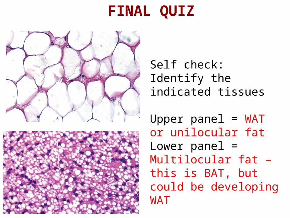
Self check:Identify the indicated tissues
Upper panel = WAT or unilocular fatLower panel = Multilocular fat – this is BAT, but could be developing WAT
Which tissue has more blood vessels? BAT
FINAL QUIZ
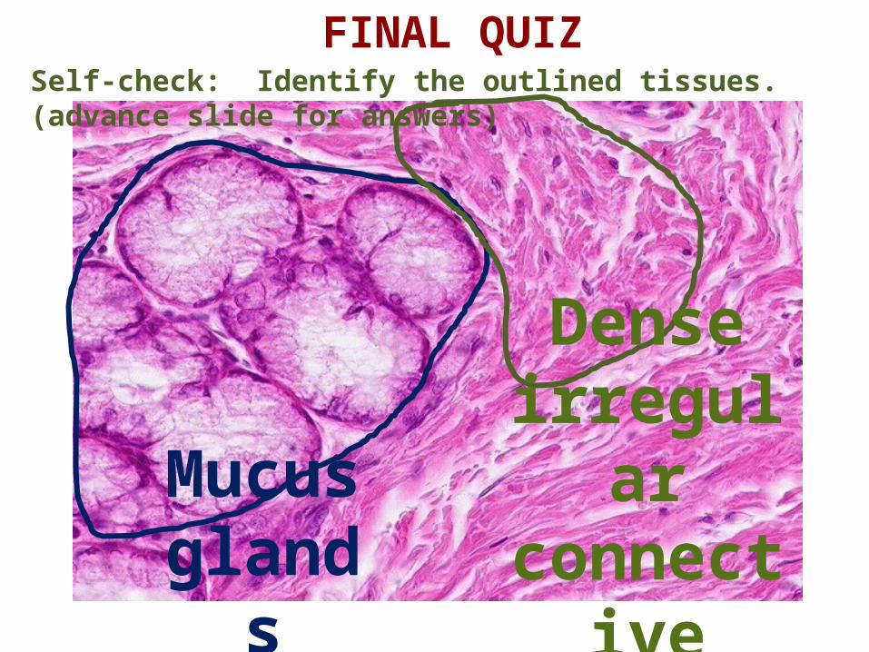
FINAL QUIZSelf-check: Identify the outlined tissues. (advance slide for answers)
Mucus glands
Dense irregular
connective tissue
Copright © 2011 Nephron.Permission is granted to copy, distribute and/or modify this image under the terms of theGNU Free Documentation License Version 1.2 ( http://www.gnu.org/copyleft/fdl.html )
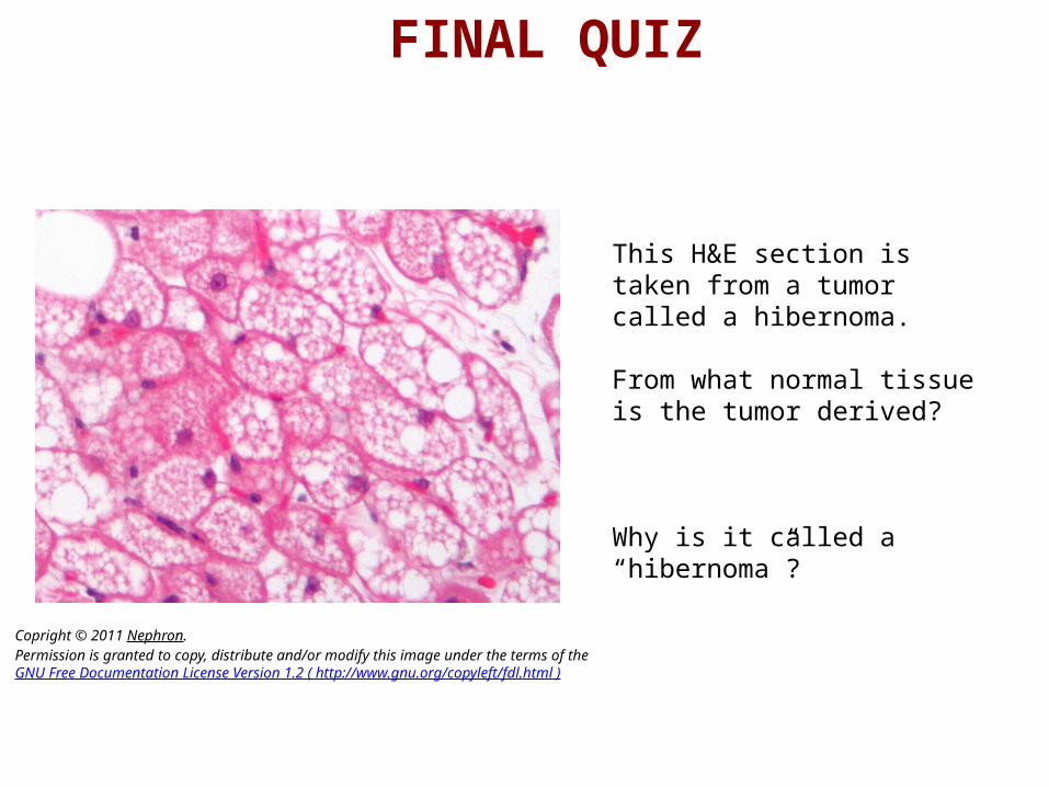
This H&E section is taken from a tumor called a hibernoma.
From what normal tissue is the tumor derived?
Why is it called a “hibernoma”?
FINAL QUIZ
Copright © 2011 Nephron.Permission is granted to copy, distribute and/or modify this image under the terms of theGNU Free Documentation License Version 1.2 ( http://www.gnu.org/copyleft/fdl.html )
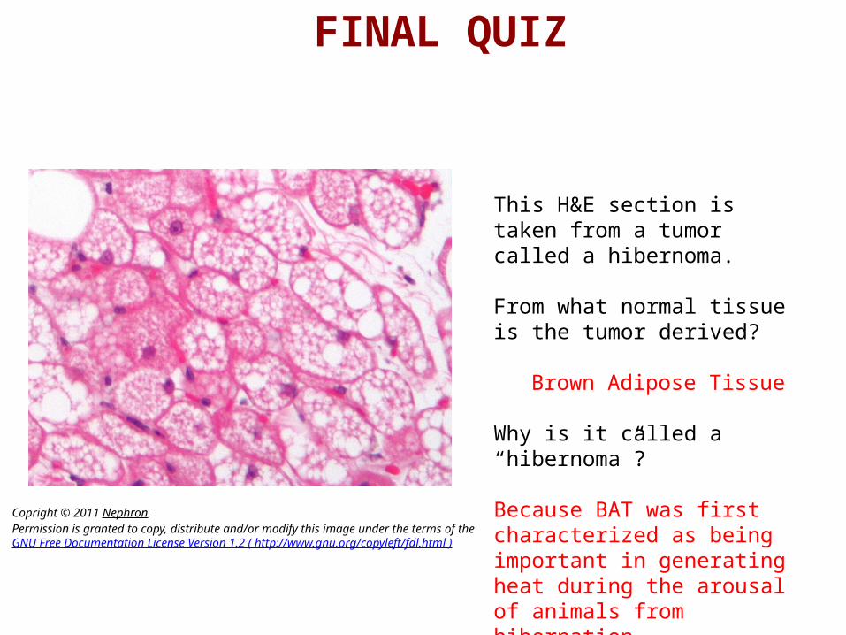
This H&E section is taken from a tumor called a hibernoma.
From what normal tissue is the tumor derived?
Brown Adipose Tissue
Why is it called a “hibernoma”?
Because BAT was first characterized as being important in generating heat during the arousal of animals from hibernation.
FINAL QUIZ

FINAL QUIZSelf-check: Identify the outlined tissues. (advance slide for answers)
Unilocular adipose
Mucus glands

• Self check: Identify the tissues indicated at left
• 1 =
• 2 =
1
2
FINAL QUIZ
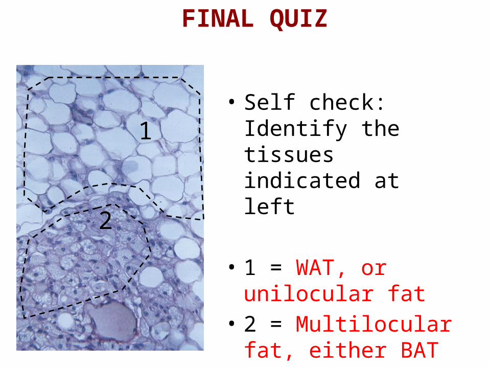
• Self check: Identify the tissues indicated at left
• 1 = WAT, or unilocular fat
• 2 = Multilocular fat, either BAT or developing WAT
1
2
FINAL QUIZ
• Self check:• How could you
determine whether the indicated tissue is BAT or developing WAT?
FINAL QUIZ
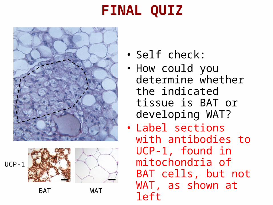
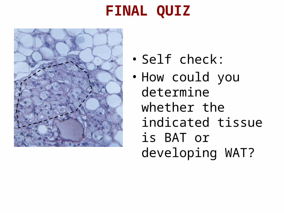
• Self check:• How could you determine
whether the indicated tissue is BAT or developing WAT?
• Label sections with antibodies to UCP-1, found in mitochondria of BAT cells, but not WAT, as shown at leftUCP-1
BAT WAT
FINAL QUIZ
Related Documents