1212 Current Drug Targets, 2009, 10, 1212-1226 1389-4501/09 $55.00+.00 © 2009 Bentham Science Publishers Ltd. Activated Protein C and Acute Kidney Injury: Selective Targeting of PAR-1 Akanksha Gupta 1 , Mark D. Williams 2 , William L. Macias 2 , Bruce A. Molitoris 3 and Brian W. Grinnell *,1 1 Biotechnology Discovery Research; 2 Acute Care Medical, Lilly Research Laboratories, Indianapolis, IN; 3 Division of Nephrology, Department of Medicine, Indiana University School of Medicine, Indianapolis, IN, USA Abstract: Protein C is a plasma serine protease that when activated plays a central role in modulating the function of the vascular endothelium and its interface with the innate immune system. Activated protein C (APC) has a dual mechanism of action via the feedback inhibition of thrombin generation, and as an agonist of protease activated receptor-1 (PAR-1). Through different cofactor interactions, this dual mechanism of antithrombotic and cytoprotective activity results in the ability of APC to modulate endothelial dysfunction by blocking cytokine signaling, functional cell adhesion expression, vascular permeability, apoptosis, and modulating leukocyte migration and adhesion. Deficiency in protein C, which occurs during systemic inflammatory activation, is highly associated with organ dysfunction. APC has shown efficacy in a number of preclinical models of thrombosis and ischemia, and the recombinant human APC drotrecogin alfa (activated), reduces mortality in patients with high-risk severe sepsis. The ability of APC to suppress pro-inflammatory pathways and enhance cellular survival suggests that APC plays a key role in the adaptive response to protect the vessel wall from insult and to enhance endothelial, cellular, and organ survival. The focus of this review will be to summarize the emerging data suggesting the potential therapeutic benefit of APC and related members of the pathway in the prevention and treatment of acute kidney injury. Key Words: Activated Protein C, PAR-1, Acute kidney injury, thrombin, apoptosis, inflammation, thrombomodulin, EPCR. INTRODUCTION Acute kidney injury (AKI) leading to renal failure is a common complication in the ICU with a prevalence varying from 30-50% [1]. In patients undergoing cardiac surgery, up to 30% develop AKI [2], and in patients with severe sepsis or shock, the reported incidence ranges from 20-50% [3-5]. AKI during sepsis is an independent risk factor for death [6- 11], with an associated mortality rate as high as 75% compared to 45% in ICU patients with AKI not from sepsis [12, 13]. Despite advances in supportive care, coupled with the lack of proven pharmacological intervention, mortality rates for AKI patients have remained either largely unchanged [14] or slightly reduced [15, 16]. The pathophysiology of AKI involves a complex inter- play between various processes, including tubular and endothelial cell injury and inflammation. In particular, microvascular dysfunction following injury to the kidney appears to play a key role in initiating and extending tubular injury, and is associated with significant inflammatory activation that contributes to the extension of renal dysfunc- tion (reviewed in [17]). In patients with sepsis, damage to the endothelium resulting in alterations in renal blood flow, soluble mediator-induced injury (e.g., from circulating cyto- kines, endothelins, and reactive oxygen species), increased vascular permeability, leukocyte adherence and loss of nor- mal endothelial function, has been well documented [17-19] *Address correspondence to this author at the Biotechnology Discovery Research, Lilly Research Laboratories, IN 46285-0444, USA; Tel: 317-276- 2293; E-mail: [email protected] and the resulting hemodynamic alterations with microvas- cular impairment contribute to impaired renal function [7]. Multiple approaches have been taken in the attempt to identify key pathways for therapeutic intervention, ranging from the modulation of apoptosis, vascular tone, inflamma- tion and cellular repair (reviewed in [20]). Recent preclinical studies have explored the role of the protein C pathway in the pathophysiology of AKI and the role of activated protein C (APC) as a therapeutic approach. While APC has tradi- tionally been considered an anti-thrombotic agent, recent data have demonstrated that APC plays an important cyto- protective role at the endothelial/leukocyte interface via direct agonism of protease activated receptor-1 (PAR-1) (reviewed in [21]). A human recombinant APC, drotrecogin alfa (activated), has been shown to be efficacious in the treatment of patients with severe sepsis at high risk of death [22]. In this review, we will provide an overview the biology of human activated protein C and its mechanistic role in the pathogenesis of AKI. In particular, we will focus on its unique ability to modulate multiple pathways of relevance to renal dysfunction, making it an attractive agent for the potential treatment of AKI. HUMAN PROTEIN C Protein C is a member of the vitamin K-dependent family of blood coagulation proteases, which circulates as an inactive zymogen and becomes activated under conditions of thrombotic and inflammatory stress. The structure of human protein C has been reviewed in detail [23, 24]. The protein is a multi domain serine protease containing a -carboxy gluta-

Welcome message from author
This document is posted to help you gain knowledge. Please leave a comment to let me know what you think about it! Share it to your friends and learn new things together.
Transcript

1212 Current Drug Targets, 2009, 10, 1212-1226
1389-4501/09 $55.00+.00 © 2009 Bentham Science Publishers Ltd.
Activated Protein C and Acute Kidney Injury: Selective Targeting of PAR-1
Akanksha Gupta1, Mark D. Williams
2, William L. Macias
2, Bruce A. Molitoris
3 and
Brian W. Grinnell*,1
1Biotechnology Discovery Research;
2Acute Care Medical, Lilly Research Laboratories, Indianapolis, IN;
3Division of
Nephrology, Department of Medicine, Indiana University School of Medicine, Indianapolis, IN, USA
Abstract: Protein C is a plasma serine protease that when activated plays a central role in modulating the function of the
vascular endothelium and its interface with the innate immune system. Activated protein C (APC) has a dual mechanism
of action via the feedback inhibition of thrombin generation, and as an agonist of protease activated receptor-1 (PAR-1).
Through different cofactor interactions, this dual mechanism of antithrombotic and cytoprotective activity results in the
ability of APC to modulate endothelial dysfunction by blocking cytokine signaling, functional cell adhesion expression,
vascular permeability, apoptosis, and modulating leukocyte migration and adhesion. Deficiency in protein C, which
occurs during systemic inflammatory activation, is highly associated with organ dysfunction. APC has shown efficacy in a
number of preclinical models of thrombosis and ischemia, and the recombinant human APC drotrecogin alfa (activated),
reduces mortality in patients with high-risk severe sepsis. The ability of APC to suppress pro-inflammatory pathways and
enhance cellular survival suggests that APC plays a key role in the adaptive response to protect the vessel wall from insult
and to enhance endothelial, cellular, and organ survival. The focus of this review will be to summarize the emerging data
suggesting the potential therapeutic benefit of APC and related members of the pathway in the prevention and treatment
of acute kidney injury.
Key Words: Activated Protein C, PAR-1, Acute kidney injury, thrombin, apoptosis, inflammation, thrombomodulin, EPCR.
INTRODUCTION
Acute kidney injury (AKI) leading to renal failure is a common complication in the ICU with a prevalence varying from 30-50% [1]. In patients undergoing cardiac surgery, up to 30% develop AKI [2], and in patients with severe sepsis or shock, the reported incidence ranges from 20-50% [3-5]. AKI during sepsis is an independent risk factor for death [6-11], with an associated mortality rate as high as 75% compared to 45% in ICU patients with AKI not from sepsis [12, 13]. Despite advances in supportive care, coupled with the lack of proven pharmacological intervention, mortality rates for AKI patients have remained either largely unchanged [14] or slightly reduced [15, 16].
The pathophysiology of AKI involves a complex inter-play between various processes, including tubular and endothelial cell injury and inflammation. In particular, microvascular dysfunction following injury to the kidney appears to play a key role in initiating and extending tubular injury, and is associated with significant inflammatory activation that contributes to the extension of renal dysfunc-tion (reviewed in [17]). In patients with sepsis, damage to the endothelium resulting in alterations in renal blood flow, soluble mediator-induced injury (e.g., from circulating cyto-kines, endothelins, and reactive oxygen species), increased vascular permeability, leukocyte adherence and loss of nor-mal endothelial function, has been well documented [17-19]
*Address correspondence to this author at the Biotechnology Discovery
Research, Lilly Research Laboratories, IN 46285-0444, USA; Tel: 317-276-
2293; E-mail: [email protected]
and the resulting hemodynamic alterations with microvas-cular impairment contribute to impaired renal function [7].
Multiple approaches have been taken in the attempt to identify key pathways for therapeutic intervention, ranging from the modulation of apoptosis, vascular tone, inflamma-tion and cellular repair (reviewed in [20]). Recent preclinical studies have explored the role of the protein C pathway in the pathophysiology of AKI and the role of activated protein C (APC) as a therapeutic approach. While APC has tradi-tionally been considered an anti-thrombotic agent, recent data have demonstrated that APC plays an important cyto-protective role at the endothelial/leukocyte interface via direct agonism of protease activated receptor-1 (PAR-1) (reviewed in [21]). A human recombinant APC, drotrecogin alfa (activated), has been shown to be efficacious in the treatment of patients with severe sepsis at high risk of death [22]. In this review, we will provide an overview the biology of human activated protein C and its mechanistic role in the pathogenesis of AKI. In particular, we will focus on its unique ability to modulate multiple pathways of relevance to renal dysfunction, making it an attractive agent for the potential treatment of AKI.
HUMAN PROTEIN C
Protein C is a member of the vitamin K-dependent family of blood coagulation proteases, which circulates as an inactive zymogen and becomes activated under conditions of thrombotic and inflammatory stress. The structure of human protein C has been reviewed in detail [23, 24]. The protein is a multi domain serine protease containing a -carboxy gluta-
Activated Protein C and Acute Kidney Injury Current Drug Targets, 2009, Vol. 10, No. 12 1213
mate domain (GLA), two EGF-like structural domains and a serine protease. The molecule contains multiple post-translational modifications and the complete -carboxylation of the light-chain, -hydroxylation of Asp 71, glycosylation and correct pro-peptide processing all affect the functional activity as reviewed previously [23, 24].
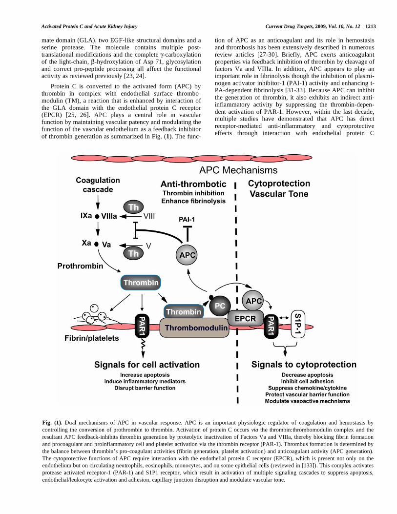
Protein C is converted to the activated form (APC) by thrombin in complex with endothelial surface thrombo-modulin (TM), a reaction that is enhanced by interaction of the GLA domain with the endothelial protein C receptor (EPCR) [25, 26]. APC plays a central role in vascular function by maintaining vascular patency and modulating the function of the vascular endothelium as a feedback inhibitor of thrombin generation as summarized in Fig. (1). The func-
tion of APC as an anticoagulant and its role in hemostasis and thrombosis has been extensively described in numerous review articles [27-30]. Briefly, APC exerts anticoagulant properties via feedback inhibition of thrombin by cleavage of factors Va and VIIIa. In addition, APC appears to play an important role in fibrinolysis though the inhibition of plasmi-nogen activator inhibitor-1 (PAI-1) activity and enhancing t-PA-dependent fibrinolysis [31-33]. Because APC can inhibit the generation of thrombin, it also exhibits an indirect anti-inflammatory activity by suppressing the thrombin-depen-dent activation of PAR-1. However, within the last decade, multiple studies have demonstrated that APC has direct receptor-mediated anti-inflammatory and cytoprotective effects through interaction with endothelial protein C
Fig. (1). Dual mechanisms of APC in vascular response. APC is an important physiologic regulator of coagulation and hemostasis by
controlling the conversion of prothrombin to thrombin. Activation of protein C occurs via the thrombin:thrombomodulin complex and the
resultant APC feedback-inhibits thrombin generation by proteolytic inactivation of Factors Va and VIIIa, thereby blocking fibrin formation
and procoagulant and proinflammatory cell and platelet activation via the thrombin receptor (PAR-1). Thrombus formation is determined by
the balance between thrombin’s pro-coagulant activities (fibrin generation, platelet activation) and anticoagulant activity (APC generation).
The cytoprotective functions of APC require interaction with the endothelial protein C receptor (EPCR), which is present not only on the
endothelium but on circulating neutrophils, eosinophils, monocytes, and on some epithelial cells (reviewed in [133]). This complex activates
protease activated receptor-1 (PAR-1) and S1P1 receptor, which result in activation of multiple signaling cascades to suppress apoptosis,
endothelial/leukocyte activation and adhesion, capillary junction disruption and modulate vascular tone.
1214 Current Drug Targets, 2009, Vol. 10, No. 12 Gupta et al.
receptor (EPCR) [34-36], and this complex exhibits direct cellular effects via differential signaling through PAR-1 (reviewed in [21]), and in some settings via cross-talk with the S1P1 receptor [37-40]. Various mechanistic studies have demonstrated that the direct modulation of PAR-1 by APC results in cytoprotection by several mechanisms. Via PAR-1 signaling, APC has been shown to suppress endothelial and leukocyte apoptosis [34, 41, 42], leukocyte adhesion and rolling [21, 42-44], inhibit inflammatory activation [40], modulate vascular tone [45, 46], and suppress endothelial barrier disruption both in cell culture [38, 47] and in vivo [37, 45]. Thus, APC plays a fundamental role in a coordi-nated system for controlling thrombosis, limiting inflamma-tory responses and potentially decreasing endothelial cell apoptosis and vascular maintenance in response to inflam-matory and ischemic injury. The interplay of these mechanisms with regard to AKI pathophysiology will be highlighted in additional detail below.
ROLE OF PROTEIN C IN RENAL PHYSIOLOGY
Preclinical Studies
While many soluble factors change during the inflam-matory response, there is substantial evidence that a reduc-tion in plasma levels of the PC is prognostic for sepsis and sepsis severity (reviewed in [48]). Studies have also suggested that PC deficiency may appear before the onset of defined clinical parameters of severe sepsis or septic shock [49]. Moreover, a retrospective evaluation of the PROWESS severe sepsis clinical trial [22] indicated that severe PC defi-ciency was associated with early death resulting from refrac-tory shock and multiple organ failure [50]. While these early studies provided an overall association of PC deficiency and outcome, the link to the pathophysiology of specific organ dysfunction, such as occurs in the kidney, remained unclear. Using both genetic and acquired PC deficiency models, preclinical studies have begun to elucidate the importance of the protein C pathway in conferring resistance to inflamma-tory insult to the kidney
Using heterozygote PC deficient mice treated with E. coli endotoxin, Levi and colleagues [51] observed more severe disseminated intravascular coagulation, liver and kidney injury, and higher mortality when compared to animals with normal PC. Histologic evaluation of the kidneys of these heterozygote mice showed more extensive fibrin deposition compared to wild-type controls. Similarly, using a cecal ligation and puncture (CLP) model of sepsis Ganopolsky and Castellino [52] showed that heterozygote PC deficient mice developed significantly more systemic hypotension, and reduced renal function as measured by significantly increased levels of serum BUN and creatinine at 24-hours post CLP. These investigators also utilized genetic enginee-ring to produce PC deficient mice of varying degrees (e.g., 1%,3%, 5%, 18% and 50% of wild-type Protein C levels) [53]. They demonstrated that endotoxin challenge in these mice resulted in higher mortality in the more severely PC deficient mice, with increased organ injury, including increased apoptosis of the kidney.
Similar to clinical observation in septic patients, low PC levels were highly predictive of early death in a rat model of
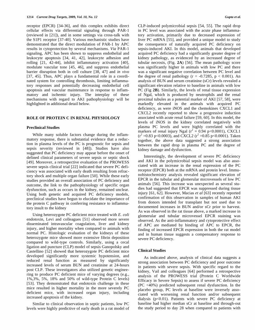
CLP-induced polymicrobial sepsis [54, 55]. The rapid drop in PC level was associated with the acute phase inflamma-tory activation, primarily due to decreased expression of liver PC mRNA [55], and provided a unique model to study the consequence of naturally acquired PC deficiency on sepsis-induced AKI. In this model, animals that developed acquired PC deficiency had a significantly greater degree of kidney pathology, as evidenced by an increased degree of tubular necrosis, (Fig. 2A) [56]. The mean pathology score was significantly higher in animals with low PC and there was a significant negative correlation between PC level and the degree of renal pathology (r = -0.7285, p < 0.001). An analysis of BUN and serum creatinine (sCr) levels revealed a significant elevation relative to baseline in animals with low PC (Fig 2B). Similarly, the levels of renal tissue expression of Ngal, which is produced by neutrophils and the renal proximal tubules as a potential marker of AKI [57, 58], were markedly elevated in the animals with acquired PC deficiency, as were iNOS and the chemokines CXCL1 and CXCL2 recently reported to show a progressive induction associated with acute renal failure [59, 60]. In this model, the levels of iNOS in the kidney correlated negatively with plasma PC levels and were highly correlated with the markers of renal injury Ngal (r
2 = 0.94 p<0.0001), CXCL1
(r2 =0.83 p<0.0003), and CXCL2 (r
2 =0.85 p<0.0001). Taken
together, the above data suggested a strong association between the rapid drop in plasma PC and the degree of kidney damage and dysfunction.
Interestingly, the development of severe PC deficiency and AKI in the polymicrobial sepsis model was also asso-ciated with an increase in the renal expression of the PC receptor (EPCR) both at the mRNA and protein level. Immu-nohistochemistry analysis revealed significant elevation of EPCR in the tubular and glomerular microvessels of low PC animals [56]. This increase was unexpected as several stu-dies had suggested that EPCR was suppressed during tissue injury [61, 62]. However, Macias et al [63] recently reported confirmation of this observation in samples of human AKI from donors intended for transplant but not used due to documented increases in BUN and/or sCr prior to harvest. As was observed in the rat tissue above, a marked increase in glomerular and tubular microvessel EPCR staining was observed. As the anti-inflammatory and cytoprotective effect of APC are mediated by binding of APC to EPCR, the finding of increased EPCR expression in both the rat model and in human tissue suggests a compensatory response to severe PC deficiency.
Clinical Studies
As indicated above, analysis of clinical data suggests a strong association between PC deficiency and poor outcome in patients with severe sepsis. With specific regard to the kidney, Vail and colleagues [64] performed a retrospective analysis of the PROWESS trial (Protein C Worldwide Efficacy in Severe Sepsis) to assess if severe PC deficiency (PC <40%) predicted subsequent renal dysfunction. In the placebo group, PC levels at baseline were inversely asso-ciated with worsening renal function and/or subsequent dialysis (p<0.01). Patients with severe PC deficiency at baseline had higher median sCr at baseline and through-out the study period to day 28 when compared to patients with
Activated Protein C and Acute Kidney Injury Current Drug Targets, 2009, Vol. 10, No. 12 1215
baseline PC >40%, 1.2 mg/dl vs. 0.90 mg/dl, respectively (p 0.0001). Moreover, PC deficiency at baseline was asso-ciated with significantly decreased improvement in subse-quent sCr change compared to those with PC >40% at baseline.
While much of the clinical association of PC with outcome has been in the context of sepsis, recent studies have begun to elucidate the important role PC plays in non-septic patients. In a surgical ICU study, low PC levels were observed in approximately 50% of the patients, and found to be associated with organ dysfunction and to predict outcome independent of the presence of sepsis [65]. Studies in trauma patients found that low PC levels were significantly associa-ted with increased mortality and blood transfusion require-ments. Of relevance to this discussion low PC levels were associated with increased AKI [66].
While the cause and effect relationship of low PC and clinical outcome has not been proven, the data from both genetic and acquired PC deficiency models, supported by the clinical associations, suggest that low endogenous PC levels during systemic inflammatory response may be pathophysio-logically related to renal dysfunction by reducing the ability to modulate not only coagulopathic dysfunction, but by dampening the cascading inflammatory activation in the kidney following injury The low level of PC likely com-promises the ability to naturally generate APC, which results in a reduction in the natural protective mechanism of the vasculature to inflammatory and ischemic injury.
STUDIES OF ACTIVATED PROTEIN C IN AKI
Model studies have provided direct evidence for a protective effect of APC in treatment of AKI and direct support for the contention that low PC is pathophysiolo-gically linked to worsened renal function. Krishnamurti and colleagues [67] first observed in endotoxin-treated rabbits that bovine-derived APC, co-administered with human tissue-type plasminogen activator (t-PA), markedly inhibited fibrin deposition in the kidney. PAI-1 activity was markedly inhibited by APC in this model, suggesting that modulation of fibrinolysis by APC might provide benefit in the renal vasculature. The potential therapeutic effect of APC was also examined in a model of ischemia-reperfusion (I/R) injury [68]. In these studies, multiple renal parameters including renal blood flow, renal microvascular permeability, histopa-thology, myeloperoxidase activity and serum levels of fibrin degradation products were assessed. The administration of human plasma-derived APC markedly reduced I/R-induced changes in all parameters. In contrast, the use of active-site inhibited APC had no effect, nor did heparin or active-site-blocked factor Xa, suggesting that anticoagulation alone might not be sufficient for protection. Induction of leuko-penia prior to the I/R injury also markedly reduced the extent of injury and the authors suggested APC-induced improve-ment in renal function might result in part from inhibition of neutrophil-mediated injury.
Several studies in models of sepsis have demonstrated significant efficacy and mechanistic insight into APC as a
Fig. (2). Analysis of renal pathology and renal injury markers as a function of acquired PC deficiency during polymicrobial sepsis. Animals
were analyzed 22 hrs after the induction of sepsis with cecal ligation and puncture (A) H & E staining of kidney section from normal and low
PC animals showing significant evidence of acute tubular necrosis in animals with acquired PC deficiency. (B) Summary of changes in
various measures of renal dysfunction and inflammation. Serum creatinine (sCr), blood urea nitrogen (BUN) neutrophil gelatinase associated
lipocalin (Ngal). Data are mean +/- SEM, n= 20 to 25. Adapted from reference [56].
1216 Current Drug Targets, 2009, Vol. 10, No. 12 Gupta et al.
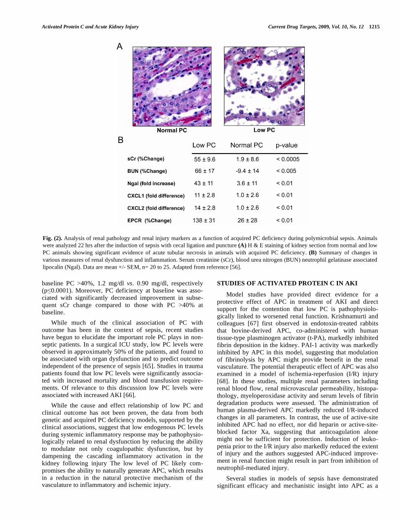
potential therapy for AKI. The effect of APC on the kidney was studied in a model of E. coli lipopolysaccharide (LPS) using intravital 2-photon microscopy to quantify functional defects on the renal vasculature [45]. As shown in Fig. (3), administration of LPS resulted in rapid injury to the kidney as measured by obstructed peritubular capillary flow, leuko-cyte adhesion and rolling, and defects in tubular function by 3h post-endotoxemia. Following treatment with APC, the deficit in renal blood flow was restored in these endotoxemic rats. APC treatment also significantly reduced the total number of static and rolling leukocytes in the renal micro-vasculature. The accumulation of leukocytes is well demons-trated during AKI in both humans and model systems [69, 70], and activated leukocytes can initiate an inflammatory cascade that leads to endothelial cell injury and disruption of the endothelial permeability barrier thus contributing to renal injury [71]. The authors also examined whether or not APC could protect endothelial barrier disruption in vivo, as determined by leakage of low and high molecular weight dextrans, and showed that the pronounced induction of microvascular leak induced by LPS was largely eliminated by APC treatment in this model. In addition, APC treatment improved tubular function as shown in Fig. (3), where the endocytic uptake by proximal tubules was reduced by LPS but maintained with APC treatment. Suppression of various inflammatory mediators including renal IL-6, IL-18, iNOS mRNA and NOx levels was also observed upon APC treatment. Overall, these data demonstrate that in acute endotoxemia, APC can protect the renal microvasculature from injury by improving capillary blood flow, glomerular
filtration and proximal tubular function, and reducing endothelial permeability and inflammation.
In a model of endotoxin-induced AKI, bolus APC treatment completely restored renal function as measured by improvement in renal flow (P=0.001) [45] and reduction in serum BUN levels to control levels. In a similar rat endotoxemia model, APC infusion significantly restored kidney function compared with standard fluid resuscitation [72]. Studies by Xu et al [73], also provide evidence of APC-induced renal protection where they have shown that LPS-treated mice injected with an antibody that blocked binding of APC to the endothelium had higher BUN and sCr levels, as well as much higher IL-6 levels. In addition, antibody blockade of endogenous APC induced
significant
vascular leakage in low-dose LPS-challenged mice in the organ beds, including the kidney, as measured by Evans blue extravasation [37].
Renoprotective effects of APC in experimental models of sepsis-induced AKI have also been demonstrated. In a rat CLP model, the effect of APC on renal function was examined in a delayed treatment starting 10 hrs after the induction of polymicrobial sepsis [56]. By 22 hrs post-sepsis, the brief infusion of APC significantly reduced plasma BUN and renal pathology. The effect of APC was most pronounced in those animals with elevated BUN at the time infusion was started, and these animals had significantly reduced PC levels (42 +/- 12 % vs. 99 +/- 8% of normal in animals without elevation in BUN) confirming that the low PC animals are at greater risk of early renal dysfunction and
Fig. (3). Effect of APC on renal blood flow, leukocyte adhesion and tubular function. Mean blood cell velocity per vessel was measured by
modified RBC line-scan method [134]. Leukocyte effects were determined by intravital microscopy 3h after treatment. The effect of APC
treatment on proximal tubule function as determined by the uptake of a low molecular weight dextran (red) by the proximal tubules. The
images were taken at timed intervals post-injection of the dextran to obtain a qualitative assessment. Adapted from references [45, 135].
Activated Protein C and Acute Kidney Injury Current Drug Targets, 2009, Vol. 10, No. 12 1217
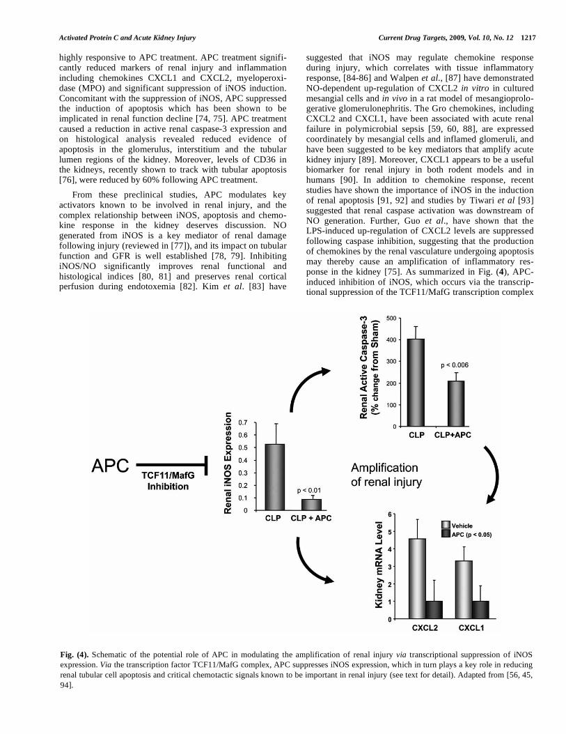
highly responsive to APC treatment. APC treatment signifi-cantly reduced markers of renal injury and inflammation including chemokines CXCL1 and CXCL2, myeloperoxi-dase (MPO) and significant suppression of iNOS induction. Concomitant with the suppression of iNOS, APC suppressed the induction of apoptosis which has been shown to be implicated in renal function decline [74, 75]. APC treatment caused a reduction in active renal caspase-3 expression and on histological analysis revealed reduced evidence of apoptosis in the glomerulus, interstitium and the tubular lumen regions of the kidney. Moreover, levels of CD36 in the kidneys, recently shown to track with tubular apoptosis [76], were reduced by 60% following APC treatment.
From these preclinical studies, APC modulates key activators known to be involved in renal injury, and the complex relationship between iNOS, apoptosis and chemo-kine response in the kidney deserves discussion. NO generated from iNOS is a key mediator of renal damage following injury (reviewed in [77]), and its impact on tubular function and GFR is well established [78, 79]. Inhibiting iNOS/NO significantly improves renal functional and histological indices [80, 81] and preserves renal cortical perfusion during endotoxemia [82]. Kim et al. [83] have
suggested that iNOS may regulate chemokine response during injury, which correlates with tissue inflammatory response, [84-86] and Walpen et al., [87] have demonstrated NO-dependent up-regulation of CXCL2 in vitro in cultured mesangial cells and in vivo in a rat model of mesangioprolo-gerative glomerulonephritis. The Gro chemokines, including CXCL2 and CXCL1, have been associated with acute renal failure in polymicrobial sepsis [59, 60, 88], are expressed coordinately by mesangial cells and inflamed glomeruli, and have been suggested to be key mediators that amplify acute kidney injury [89]. Moreover, CXCL1 appears to be a useful biomarker for renal injury in both rodent models and in humans [90]. In addition to chemokine response, recent studies have shown the importance of iNOS in the induction of renal apoptosis [91, 92] and studies by Tiwari et al [93] suggested that renal caspase activation was downstream of NO generation. Further, Guo et al., have shown that the LPS-induced up-regulation of CXCL2 levels are suppressed following caspase inhibition, suggesting that the production of chemokines by the renal vasculature undergoing apoptosis may thereby cause an amplification of inflammatory res-ponse in the kidney [75]. As summarized in Fig. (4), APC-induced inhibition of iNOS, which occurs via the transcrip-tional suppression of the TCF11/MafG transcription complex
Fig. (4). Schematic of the potential role of APC in modulating the amplification of renal injury via transcriptional suppression of iNOS
expression. Via the transcription factor TCF11/MafG complex, APC suppresses iNOS expression, which in turn plays a key role in reducing
renal tubular cell apoptosis and critical chemotactic signals known to be important in renal injury (see text for detail). Adapted from [56, 45,
94].
1218 Current Drug Targets, 2009, Vol. 10, No. 12 Gupta et al.
[94], may be central to the protective mechanism by inhibiting both the release of chemokines and the induction of apoptosis, thereby suppressing the amplification of renal injury well described in the literature.
VASO-MODULATION AND RENAL PROTECTION
BY APC
APC can elicit modulation of the hypotensive response in both model systems and in humans. In a study of human endotoxin challenge, human recombinant APC (drotrecogin alfa - activated) prevented the hypotensive response [95] and data from the PROWESS registration trial suggested that patients on drotrecogin alfa (activated) treatment had reduced requirement for vasopressor support, higher mean arterial pressure and fewer treated deaths from shock [96]. In animal models of endotoxemia, APC blocked hypotension [97, 98] and this effect was associated with suppression of the vasoactive peptide adrenomedullin (ADM) [46]. Moreover, suppression of ADM by drotrecogin alfa (activated) was strongly associated with improved mean arte-rial pressures in human endotoxin challenge [46]. In light of these significant effects on systemic hypotension, the ques-tion arises whether the effect of APC on the kidney might be secondary to improvement in systemic hemodynamics. Several lines of evidence would suggest this is not the case. Using an endotoxemia model where animals showed a profound drop in MAP, volume expansion by infusion resulted in an increase in MAP of 26 mmHg, similar to the effect of APC, but did not significantly alter tubular dysfunc-tion or leukocyte margination [45]. Likewise, induction of hypotension by administration of NO donor, sodium nitroprusside, did not result in compromised renal blood flow [99] consistent with reports that renal blood flow can be maintained in the presence of hypotension, attributable to persistence of autoregulation in the kidney [100-103]. Moreover, using variants that separate functions of APC (discussed in detail below), renal blood flow, pathology and BUN improvements occurred in the absence of changes in systemic MAP, and dose-response data separated the anti-hypotensive effect from effects on renal microvascular function [99]. Outside of these studies with APC, Lopez et al. [102] have reported that inhibiting hypotension using pharmacological iNOS inhibitors was unable to prevent sepsis-induced organ injury, and studies with thrombin inhibition using heparin infusion in rats, showed improved organ microvascular blood flow independent of blood pressure changes [104]. In the patient setting, measures of cardiovascular dysfunction were not predictors of sepsis-associated AKI [105]. These data support the contention that the protective effects of APC are not secondary to prevention of sepsis-induced hypotension.
A hallmark of AKI is enhanced vasoconstriction of the renal microvasculature. APC appears to have a unique ability to suppress systemic hypotensive response, while also suppressing the vasoconstrictive response in the kidney as observed by in vivo imaging. A possible mechanism may come from the ability of APC to modulate the local renin-angiotensin system (RAS), particularly ACE1, ACE2 and angiotensinogen. Numerous studies suggest that abnormal activation of the RAS system in the kidney, leading to excess
production of AngII, results in tubulointerstitial injury and subsequent loss of renal function [106]. As a counter-regulatory measure to peripheral vasodilation, subsequent RAS activation culminates to maintain hemodynamic stability, inducing renal vasoconstriction that in turn further contributes to the pathogenesis of sepsis-induced AKI [107]. Using two-photon imaging, Gupta, Molitoris and colleagues demonstrated enhanced renal vasoconstriction following LPS challenge in rats that was associated with elevated renal AngII levels, angiotensinogen and ACE1 but diminished ACE2 expression [45]. Treatment with APC resulted in a suppression of the local synthesis of AngII in the kidney by up-regulating ACE2 and inhibiting angiotensinogen and ACE1. With regard to ACE2, similar observations were made by Ye et al., where they suggested that increased ACE2 expression coupled with low ACE activity may be renoprotective in diabetes [108]. As AngII has been shown to induce neutrophil accumulation in vivo [109], reduction of renal AngII levels by APC may also have contributed to its ability to suppress leukocyte adhesion and endothelial activation, which in both humans and animals may contribute to renal vasoconstriction [110, 111].
APC DIFFERENTIALLY TARGETS PAR-1-
DEPENDENT PATHWAYS IN THE KIDNEY
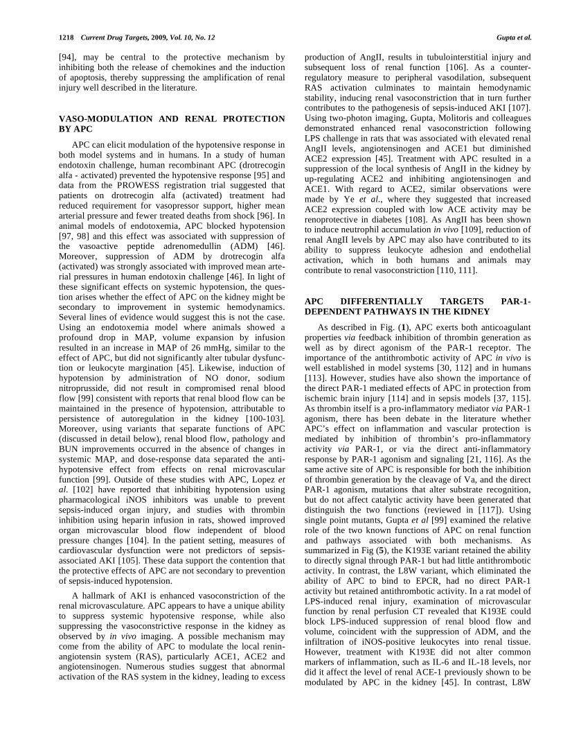
As described in Fig. (1), APC exerts both anticoagulant properties via feedback inhibition of thrombin generation as well as by direct agonism of the PAR-1 receptor. The importance of the antithrombotic activity of APC in vivo is well established in model systems [30, 112] and in humans [113]. However, studies have also shown the importance of the direct PAR-1 mediated effects of APC in protection from ischemic brain injury [114] and in sepsis models [37, 115]. As thrombin itself is a pro-inflammatory mediator via PAR-1 agonism, there has been debate in the literature whether APC’s effect on inflammation and vascular protection is mediated by inhibition of thrombin’s pro-inflammatory activity via PAR-1, or via the direct anti-inflammatory response by PAR-1 agonism and signaling [21, 116]. As the same active site of APC is responsible for both the inhibition of thrombin generation by the cleavage of Va, and the direct PAR-1 agonism, mutations that alter substrate recognition, but do not affect catalytic activity have been generated that distinguish the two functions (reviewed in [117]). Using single point mutants, Gupta et al [99] examined the relative role of the two known functions of APC on renal function and pathways associated with both mechanisms. As summarized in Fig (5), the K193E variant retained the ability to directly signal through PAR-1 but had little antithrombotic activity. In contrast, the L8W variant, which eliminated the ability of APC to bind to EPCR, had no direct PAR-1 activity but retained antithrombotic activity. In a rat model of LPS-induced renal injury, examination of microvascular function by renal perfusion CT revealed that K193E could block LPS-induced suppression of renal blood flow and volume, coincident with the suppression of ADM, and the infiltration of iNOS-positive leukocytes into renal tissue. However, treatment with K193E did not alter common markers of inflammation, such as IL-6 and IL-18 levels, nor did it affect the level of renal ACE-1 previously shown to be modulated by APC in the kidney [45]. In contrast, L8W
Activated Protein C and Acute Kidney Injury Current Drug Targets, 2009, Vol. 10, No. 12 1219
suppressed ACE-1, IL-6, thrombospondin (TSP) and IL-18 induction, i.e., markers previously shown to be activated by thrombin agonism via PAR-1. Both variants effectively reduced active caspase-3, renal pathology and blood urea nitrogen. While K193E could completely block the reduction in mean arterial pressure induced by LPS, L8W had no effect consistent with studies by Isobe et al. [118] showing that inhibition of thrombin generation by active-site-inhibited Factor Xa could not alter LPS-induced hypotension in an rat endotoxin model. Thus protection of the kidney could occur either by APC’s distinct effects as a PAR-1 agonist (K193E), or by suppression of thrombin-mediated PAR-1 agonism by inhibition of thrombin generation (L8W). Although control of hypotensive shock was strictly via APC agonism of PAR-1, which may explain preferential effect of signaling variants on mortality in shock models [119], both functions of APC may play independent, and possibly the redundant roles that function independently depending on the disease context. For example, a variant with little anticoagulant activity may be preferred in AKI in the post-surgical setting, but in sepsis-induced AKI where DIC is present a variant with a balance of antithrombotic and PAR-agonism may be ideal. The results also suggested an important role for differential PAR-1 signaling in attenuating renal injury.
INSIGHTS FROM CLINCAL STUDIES OF
DROTRECOGIN ALFA (ACTIVATED)
The pre-clinical data strongly support a role for the protein C pathway in the pathophysiology of AKI. While prospective human studies specifically investigating drotre-cogin alfa (activated) (human recombinant APC) as a ther-apy for AKI are lacking, studies in both healthy subjects administered an endotoxin challenge (either intravenous or endobronchial) and retrospective analyses from clinical studies with drotrecogin alfa (activated) demonstrate many of the effects of APC observed in animal models AKI.
As reviewed above, drotrecogin alfa (activated) can pre-vent hypotension in healthy subjects administered an endo-toxin challenge [95], and significantly improve hypotension in severe sepsis [96]. In healthy subjects administered endo-toxin endobronchially, the administration of drotrecogin alfa (activated) significantly reduced leukocyte accumulation in the airspaces [120], and neutrophils recovered from the bronchoalveolar lavage fluid (BALF) of drotrecogin alfa (activated) treated subjects demonstrated decreased chemo-taxis ex vivo. Proteomic analysis has been performed on a subset of 243 PROWESS patients, which has provided additional data on the anti-inflammatory activity of drotre-cogin alfa (activated) [121]. For this study, 116 drotrecogin
Fig. (5). APC variants that separate the two functions of APC both modulate LPS-induced AKI by distinct pathways converging in
differential PAR-1 signaling. Substitution of a Glu for Arg at position 193 in the 37 loop (substrate recognition region) of the protease
domain of APC (K193E) resulted in a variant with little anticoagulant activity but high ability to directly agonize PAR-1. A Typ for Leu
substitution at position 8 in the GLA domain (L8W), which blocked binding to EPCR (required for PAR-1 signaling by APC), retained the
ability to block the generation of thrombin, but with no measurable activity as a PAR-1 agonist. APC protected the renal function by either
direct PAR-1 agonism (K193E), or by suppression of thrombin-mediated PAR-1 agonism through inhibition of thrombin generation (L8W).
Redundant mechanisms appear to allow APC to improve vascular function, as both variants improve renal blood flow (RBF) and volume
(RBV) and function by modulating distinct pathways, both centered on altering differential signaling by different PAR-1 activators. Adapted
from [99].
1220 Current Drug Targets, 2009, Vol. 10, No. 12 Gupta et al.
alfa (activated) and 127 placebo-treated with baseline APACHE II scores > 25 and serum samples obtained at baseline and 24 hrs from start of study drug were compared across 143 proteins (included cytokines, chemokines, soluble cytokine receptors, receptor antagonists, adhesion molecules, and hormones). Drotrecogin alfa (activated) treatment was predominantly associated with a change in chemokine response. Compared to placebo, RANTES, P-selectin, platelet factor 4 (PF-4) and hemofiltrate CC chemokine 4 (HCC4) levels were significantly lower in the drotrecogin alfa (activated) group following infusion. With regard to changes from baseline, the chemokines TARC, GRO-beta and GRO-gamma also exhibited a significant decrease with drotrecogin alfa (activated) treatment. Analyses of variance demonstrated similar findings: levels of RANTES, P-selectin, PF-4 and E-selectin (endothelial leukocyte adhesion molecule-1) were significantly lower (p<0.05) in drotrecogin alfa (activated) than placebo-treated patients. Overall, these clinical data show that the effects of APC on systemic hemodynamics, leukocyte activation and chemokine res-ponse observed in animals are also observed in humans administered a therapeutic dose of drotrecogin alfa (activated).
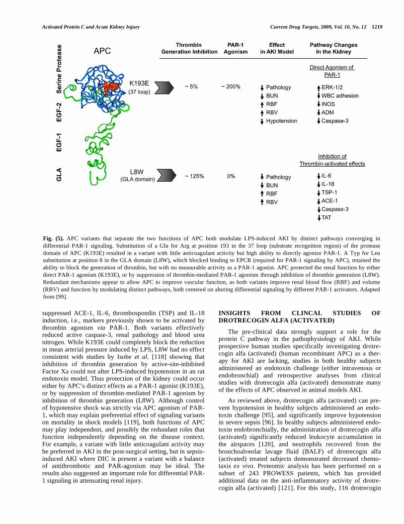
Exploratory analyses of the PROWESS dataset confirm that renal dysfunction is associated with a marked increase in mortality for placebo-treated patients regardless of whether
dysfunction was classified by oliguria alone, renal SOFA score, calculated creatinine clearance or APACHE II score classification for acute renal failure (Table 1). Interestingly, a larger treatment effect, both by absolute and relative benefit, was observed with drotrecogin alfa (activated) therapy in patients that had moderate renal insufficiency (Table 1). Less effect was observed in the population of patients with a renal SOFA score of “0” or with a calculated creatinine clearance of 50 mL/minute. However, concluding that the treatment effect associated with drotrecogin alfa (activated) might vary by baseline renal function assessment, other than that related to severity of illness, would require prospective validation.
With regard to PC deficiency, a retrospective analysis of the PROWESS trial sought to determine whether or not therapy with drotrecogin alfa (activated) improves renal function in severe sepsis patients with Protein C deficiency [64]. The authors analyzed serum creatinine changes on days 1-28 stratified by severe PC deficiency and drotrecogin alfa (activated) vs. placebo using ANOVA on the ranks and last observation carried forward. Therapy with drotrecogin alfa (activated) was associated with improved renal function compared to placebo in patients with severe PC deficiency, Fig (6A). In patients with concomitant baseline renal insufficiency (creatinine of 2.0-3.4 mg/dl), median creatinine changes from baseline at day 28 were -0.81 mg/dl and 0
Table 1. 28-day All-Cause Mortality by Baseline Renal Function Assessment in Prowess
Population Drotrecogin alfa (activated)
% 28-day mortality (N)
Placebo
% 28-day mortality (N)
Relative Risk
Reduction
All Patients1 24.7%(850) 30.8% (840) 19.8%
Renal Dysfunction2
Yes 32.5%(357) 40.5% (353) 19.8%
No 19.1%(493) 23.8% (487) 19.7%
Renal SOFA Score
0 19.8%(323) 21.4%(323) 7.5%
1 21.3%(235) 31.5% (241) 32.4%
2 33.0%(188) 43.2% (176) 23.6%
3 31.5%(73) 38.5% (52) 18.2%
4 33.3%(30) 37.8% (45) 11.9%
Baseline Creatinine Clearance (mL/minute) 3
50 18.4%(478) 19.4% (484) 5.2%
20 to <50 = 31.7%(278) 46.8% (278) 32.3%
<20 41.8%(67) 46.0% (63) 9.1%
APACHE II Classification for Acute Renal Failure
Yes 38.3% (180) 50.5% (192) 24.2%
No 21.0% (668) 25.0% (644) 16.0%
1Statistical comparison is provided only for the primary analysis (P=0.005). Mortality rates for subgroups defined by baseline assessment of renal dysfunction are provided for descriptive purposes only. 2Renal dysfunction was defined as urine output <0.5 mL/kg/hour despite adequate fluid resuscitation. Adequate fluid resuscitation was defined as either a) the administration of an
intravenous fluid bolus (>=500 mL crystalloid solution, >=20 g albumin, or >=200 mL other colloid) over a 30 minute or less interval; b) pulmonary artery wedge pressure >=12 mm Hg; or c) central venous pressure >=8 mm Hg. 3Creatinine clearance was calculated by the Cockcroft-Gault equation. “N” = number of patients within treatment group. (Data on file, Eli Lilly and Company; clinical report for study F1K-MC-EVAD. Table adapted from [63]).
Activated Protein C and Acute Kidney Injury Current Drug Targets, 2009, Vol. 10, No. 12 1221
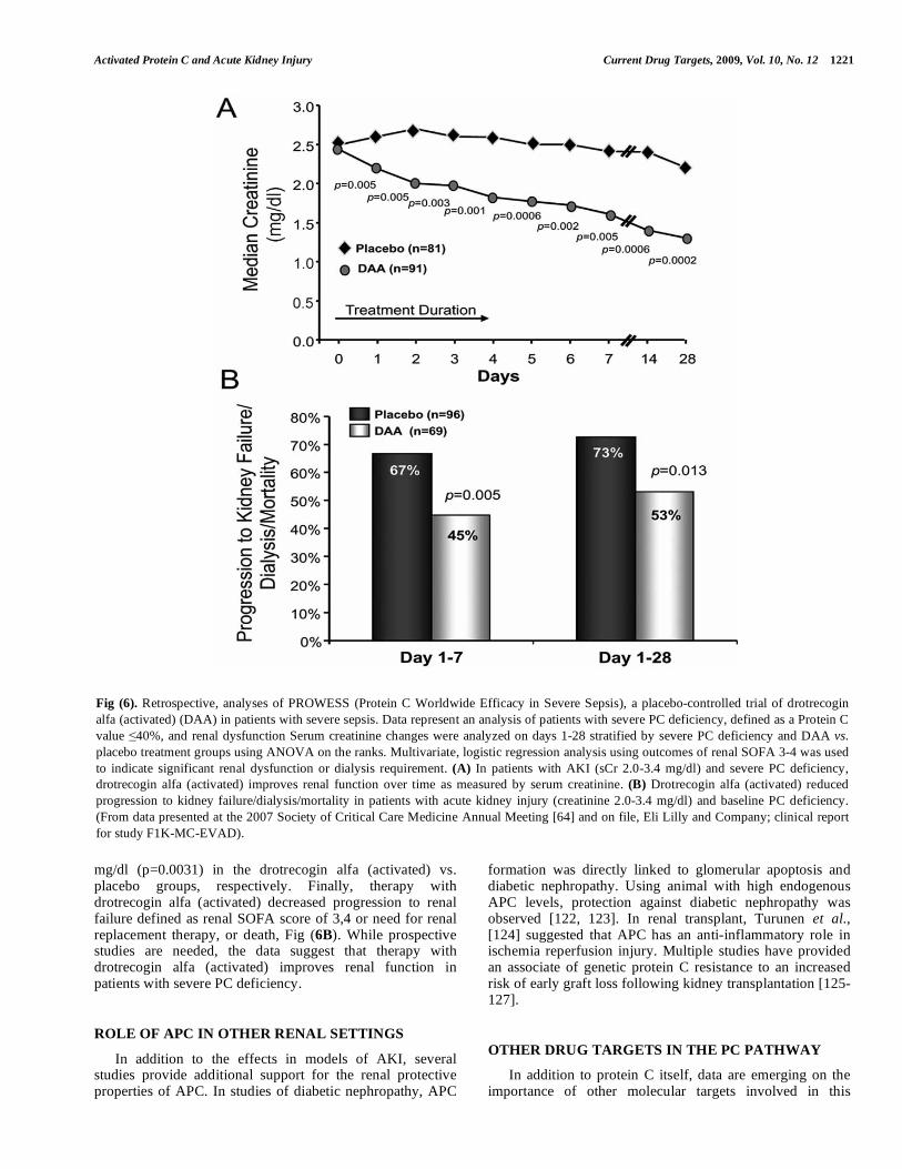
mg/dl (p=0.0031) in the drotrecogin alfa (activated) vs. placebo groups, respectively. Finally, therapy with drotrecogin alfa (activated) decreased progression to renal failure defined as renal SOFA score of 3,4 or need for renal replacement therapy, or death, Fig (6B). While prospective studies are needed, the data suggest that therapy with drotrecogin alfa (activated) improves renal function in patients with severe PC deficiency.
ROLE OF APC IN OTHER RENAL SETTINGS
In addition to the effects in models of AKI, several studies provide additional support for the renal protective properties of APC. In studies of diabetic nephropathy, APC
formation was directly linked to glomerular apoptosis and diabetic nephropathy. Using animal with high endogenous APC levels, protection against diabetic nephropathy was observed [122, 123]. In renal transplant, Turunen et al., [124] suggested that APC has an anti-inflammatory role in ischemia reperfusion injury. Multiple studies have provided an associate of genetic protein C resistance to an increased risk of early graft loss following kidney transplantation [125-127].
OTHER DRUG TARGETS IN THE PC PATHWAY
In addition to protein C itself, data are emerging on the importance of other molecular targets involved in this
Fig (6). Retrospective, analyses of PROWESS (Protein C Worldwide Efficacy in Severe Sepsis), a placebo-controlled trial of drotrecogin
alfa (activated) (DAA) in patients with severe sepsis. Data represent an analysis of patients with severe PC deficiency, defined as a Protein C
value 40%, and renal dysfunction Serum creatinine changes were analyzed on days 1-28 stratified by severe PC deficiency and DAA vs.
placebo treatment groups using ANOVA on the ranks. Multivariate, logistic regression analysis using outcomes of renal SOFA 3-4 was used
to indicate significant renal dysfunction or dialysis requirement. (A) In patients with AKI (sCr 2.0-3.4 mg/dl) and severe PC deficiency,
drotrecogin alfa (activated) improves renal function over time as measured by serum creatinine. (B) Drotrecogin alfa (activated) reduced
progression to kidney failure/dialysis/mortality in patients with acute kidney injury (creatinine 2.0-3.4 mg/dl) and baseline PC deficiency.
(From data presented at the 2007 Society of Critical Care Medicine Annual Meeting [64] and on file, Eli Lilly and Company; clinical report
for study F1K-MC-EVAD).
1222 Current Drug Targets, 2009, Vol. 10, No. 12 Gupta et al.
pathway in the kidney. As indicated above, thrombomodulin plays a key role in the activation of protein C and soluble forms of the molecule can mimic functions of the transmem-brane form. Ikeguchi et al. [128] evaluated the effects of recombinant human soluble thrombomodulin in a lethal model of thrombotic glomerulonephritis induced in rats by administration of lipopolysaccharide and rabbit anti-rat glo-merular basement membrane antibody. The sTM attenuated leukocyte/neutrophil infiltration in the glomerulus, progre-ssion of the disease and mortality. In a model of ischemia/ reperfusion, recombinant sTM directly injected into the kidney decreased BUN and sCr levels and prevented a reduction in cortical blood flow, tubular damage and macro-phage/neutrophil infiltration [129]. Sharfuddin et al. [130] recently described the protective effect of sTM in a hypoperfusion model of ischemic kidney injury. In this rat model, infrarenal aortic blood flow was markedly reduced by partial suprarenal aortic clamp of rats, resulting in acute tubular necrosis, markedly reduced flow and increased WBC adhesion in the microvasculature, and defects in endothelial permeability. Administration of recombinant sTM in this model reduced both ischemia-reperfusion-induced renal dysfunction, tubular histologic injury scores, microvascular endothelial leukocyte rolling and attachment, and significan-tly improved microvascular erythrocyte flow rates. In geneti-cally engineered animals with a defect in TM expression, more renal injury was observed following the induction of diabetic nephropathy [122]. With regard to PAR-1, a recent study by Sevastos et al., [131] have shown that PAR-1
-/-
mice were protected from renal failure and had reduced mortality, tubular injury, neutrophil accumulation, and lower levels of the chemokines suggesting that thrombin-mediated
PAR-1 agonism plays an important role in renal IR, and is consistent with the protective effect of APC L8W described in Fig. (5). While the focus of this review has been on therapeutic proteins and extracellular modulation, as our understanding of the signaling involved in the protection by this pathway through differential agonism of PAR1, and the interrelationship with the sphingosine receptor pathways [37, 38, 40], key intracellular mediators will likely emerge as important targets for future drug discovery.
SUMMARY
The development of AKI is associated with a substantial morbidity and mortality, and recent studies suggest that episodes of AKI may accelerate progression of renal disease [132]. The pathophysiology of AKI is complex and as previ-ously reviewed, clinical trial design is equally complex [63]. While there have been a number of promising approaches targeting various pathophysiological pathways for the treatment and prevention of AKI, [20], there has been little clinical success toward this high unmet medical need. As we have reviewed, APC appears to be a unique multi-pathway modulator of vascular physiology with potential for the treatment of AKI clinically. Acquired Protein C deficiency is common in severe sepsis, the surgical ICU and in trauma patients, and is associated with a higher incidence of AKI and mortality, potentially as a result of decreased generation of endogenous APC. In model systems, PC deficiency is associated with the development of AKI and can be reversed by APC treatment. The administration of APC appears to improve renal perfusion, decrease leukocyte-endothelial interactions, down-regulate iNOS and NO production and
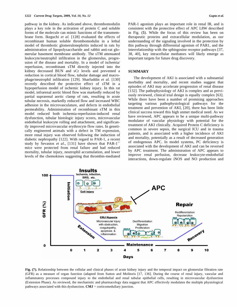
Fig. (7). Relationship between the cellular and clinical phases of acute kidney injury and the temporal impact on glomerular filtration rate
(GFR) as a measure of organ function (adapted from Sutton and Molitoris [17, 136]. During the course of renal injury, vascular and
inflammatory processes compound injury to the endothelial and renal tubular epithelial cells, resulting in microvascular dysfunction
(Extension Phase). As reviewed, the mechanistic and pharmacology data suggest that APC effectively modulates the multiple physiological
pathways associated with this dysfunction. CMJ = corticomedullary junction.
Activated Protein C and Acute Kidney Injury Current Drug Targets, 2009, Vol. 10, No. 12 1223
improve renal function in a variety of animal models of AKI. As summarized in Fig (7), the extension phase of renal injury results from the complex interplay of endothelial, leukocyte and tubular function. APC appears to have the ability to effectively modulate this phase of injury allowing significant recovery of function. Based on the preclinical and retrospective clinical analyses, the protein C pathway appears to be an excellent area to target for the development of therapeutics to prevent and treat AKI. In particular, the emerging understandings of the mechanisms for the protective effect(s) of APC suggest that differential modulation of PAR-1 signaling may be a key target for future drug discovery.
ACKNOWLEDGEMENTS
We thank Dave Berg, Mark Richardson, Bruce Gerlitz, Ruben Sandoval, Silvia Campos and George Rhodes, who were instrumental in generating published data reviewed here. We also thank Dave Nelson for statistical analysis used in the published studies cited on retrospective analysis of PROWESS. Dr. Molitoris’ work was supported by National Institutes of Health Grants DK79312 and DK069408, a VA Merit Review and a research grant from Lilly Research Laboratories.
DISCLOSURES
The authors disclose that AG, WLM, MDW and BWG are employed by Lilly Research Laboratories, a division of Eli Lilly and Co who produces recombinant APC for the treatment of severe sepsis. BAM is a consultant for Lilly Research Laboratories in AKI.
REFERENCES
[1] Langenberg C, Bellomo R, May C, Wan L, Egi M, Morgera S. Renal blood flow in sepsis. Crit Care 2005; 9: R363-74.
[2] Rosner M, Okusa M. Acute kidney injury associated with cardiac surgery. Clin J Am Soc Nephrol 2006; 1: 19-32.
[3] Rangel-Frausto MS, Pittet D, Costigan M, Hwang T, Davis CS, Wenzel RP. The natural history of the systemic inflammatory
response syndrome (SIRS). A prospective study JAMA 1995; 273: 117-23.
[4] Riedemann NC, Guo RF, Ward PA. The enigma of sepsis. J Clin Invest 2003; 112: 460-67.
[5] Vincent J, Bota D, De Backer D. Epidemiology and outcome in renal failure. Int J Artif Organs 2004; 27: 1013-8.
[6] Chertow GM, Burdick E, Honour M, Bonventre JV, Bates DW. Acute kidney injury, mortality, length of stay, and costs in
hospitalized patients. J Am Soc Nephrol 2005; 16: 3365-70. [7] Ronco C, Kellum JA, Bellomo R, House AA. Potential
interventions in sepsis-related acute kidney injury. Clin J Am Soc Nephrol 2008; 3: 531-44.
[8] Schwilk B, Wiedeck H, Stein B, Reinelt H, Treiber H, Bothner U. Epidemiology of acute renal failure and outcome of
haemodiafiltration in intensive care. Intens Care Med 1997; 23: 1204-11.
[9] Ely EW, Laterre PF, Angus DC, Helterbrand JD, Levy H, Dhainaut JF, et al. Drotrecogin alfa (activated) administration across
clinically important subgroups of patients with severe sepsis. Crit Care Med 2003; 31: 12-19.
[10] Levy MM, Macias WL, Vincent JL, Russell JA, Silva E, Trzaskoma B, et al. Early changes in organ function predict
eventual survival in severe sepsis. Crit Care Med 2005; 33: 2194-201.
[11] Ympa YP, Sakr Y, Reinhart K, Vincent JL. Has mortality from
acute renal failure decreased? A systematic review of the literature. Am J Med 2005; 118: 827-32.
[12] Schrier RW, Wang W. Acute renal failure and sepsis. N Engl J Med 2004; 351: 159-69.
[13] Schrier RW, Wang W, Poole B, Mitra A. Acute renal failure: definitions, diagnosis, pathogenesis, and therapy. J Clin Invest
2004; 114: 5-14. [14] Ympa Y, Sakr Y, Reinhart K, Vincent J-L. Has mortality from
acute renal failure decreased? A systematic review of the literature. Am J Med 2005; 118: 827-32.
[15] Xue J, Daniels F, Star R, Kimmel P, Eggers P, Molitoris B, et al. Ncidence and mortality of acute renal failure in Medicare
beneficiaries, 1992 to 2001. J Am Soc Nephrol 2006; 17: 1135-42. [16] Waikar S, Curhan G, Wald R, McCarthy E, Chertow G. Declining
mortality in patients with acute renal failure, 1988 to 2002. J Am Soc Nephrol 2006; 17: 1143-50.
[17] Molitoris B, Sutton T. Endothelial injury and dysfunction: role in the extention phase of acute renal failure. Kidney Int 2004; 66:
496-9. [18] Mutunga M, Fulton B, Bullock R, Batchelor A, Gascoigne A,
Gillespie JI, et al. Circulating endothelial cells in patients with septic shock. Am J Respir Crit Care Med 2001; 163: 195-200.
[19] Klenzak J, Himmelfarb J. Sepsis and the kidney. Crit Care Clin 2005; 21: 211-22.
[20] Jo S, Rosner M, Okusa M. Pharmacologic treatment of acute kidney injury: Why drugs haven't worked and what is on the
horizon. Clin J Am Soc Nephrol 2007; 2: 356-65. [21] Mosnier L, Zlokovic B, Griffin J. The cytoprotective protein C
pathway. Blood 2007; 109: 3161-72. [22] Bernard G, Vincent J, Laterre P, LaRosa S, Dhainaut J, Lopez-
Rodriguez A, et al. Efficacy and safety of recombinant human activated protein C for severe sepsis. New Engl J Med 2001; 344:
699-705. [23] Grinnell B, Yan S, Macias W. Activated Protein C. In: McGrath B,
Walsh G, eds. Directory of Therapeutic Enzymes. CRC Press: Boca Raton-London-New York-Washington, D. C. 2006; pp. 69-95.
[24] O'Brien L, Gupta A, Grinnell B. Activated protein C and sepsis Front Biosci 2006; 11: 676-98.
[25] Esmon CT, Xu J, Gu JM, Qu D, Laszik Z, Ferrell G, et al. Endothelial protein C receptor. Thromb Haemost 1999; 82: 251-8.
[26] Preston RJ, Villegas-Mendez A, Sun YH, Hermida J, Simioni P, Philippou H, et al. Selective modulation of protein C affinity for
EPCR and phospholipids by Gla domain mutation. FEBS J 2005; 272: 97-108.
[27] Castellino FJ. Gene targeting in hemostasis. Protein C Front Biosci 2001; 6: D807-19.
[28] Esmon CT, Ding W, Yasuhiro K, Gu JM, Ferrell G, Regan LM, et al. The protein C pathway: new insights Thromb Haemost 1997;
78: 70-4. [29] Esmon CT, Moore K, Comp PC, Taylor FB, D'Angelo A, Vigano-
D'Angelo S, et al. Functions of the protein C anticoagulant pathway: modulation in disease states. In: Suttie JW Ed. Current
Advances in Vitamin K Research. Elsevier Science Publishing Co.: New York 1988; pp. 85-94.
[30] Grinnell B. Joyce DE. Recombinant human activated protein C: A system modulator of vascular function for treatment of severe
sepsis. Crit Care Med 2001; 29: S53-61. [31] Bajzar L, Nesheim M, Tracy PB. The profibrinolytic effect of
activated Protein C in clots formed from plasma is TAFI-dependent. Blood 1996; 88: 2093-100.
[32] Jackson CV, Shetler TJ, Bailey BD, Gertlitz BE, Berg DT, Grinnell BW. Recombinant human activated protein C (LY203638) induced
reperfusion in a canine model of occlusive coronary artery thrombosis. Circulation 1998; 98: I454.
[33] Esmon CT, Gu JM, Xu J, Qu D, Stearns-Kurosawa DJ, Kurosawa S. Regulation and functions of the protein C anticoagulant
pathway. Haematologica 1999; 84: 363-8. [34] Joyce DE, Gelbert L, Ciaccia A, Dehoff B, Grinnell BW. Gene
expression profile of antithrombotic Protein C defines new mechanisms modulating inflammation and apoptosis. J Biol Chem
2001; 276: 11199-203. [35] Mosnier LO, Griffin JH. Inhibition of staurosporine-induced
apoptosis of endothelial cells by activated protein C requires protease activated receptor-1 and endothelial cell protein C
receptor. Biochem J 2003; 373: 65-70.
1224 Current Drug Targets, 2009, Vol. 10, No. 12 Gupta et al.
[36] Riewald M, Petrovan R, Donner A, Mueller B, Ruf W. Activation
of endothelial cell protease activated receptor 1 by the protein C pathway. Science 2002; 296: 1880-2.
[37] Niessen F, Furlan-Freguia C, Fernandez JA, Mosnier LO, Castellino FJ, Weiler H, et al. Endogenous EPCR/aPC-PAR1
signaling prevents inflammation-induced vascular leakage and lethality. Blood 2009; 113: 2859-66.
[38] Feistritzer C, Riewald M. Endothelial barrier protection by activated protein C through PAR1-dependent sphingosine 1-
phosphate receptor-1 crossactivation. Blood 2005; 105: 3178-84. [39] Finigan JH, Dudek SM, Singleton PA, Chiang ET, Jacobson JR,
Camp SM, et al. Activated protein C mediates novel lung endothelial barrier enhancement: role of sphingosine 1-phosphate
receptor transactivation. J Biol Chem 2005; 280: 17286-93. [40] O'Brien L, Richardson M, Mehrbod S, Berg D, Gerlitz B, Gupta A,
et al. Activated protein C decreases tumor necrosis factor related apoptosis-inducing ligand by an EPCR- independent mechanism
involving Egr-1/Erk-1/2 activation. Arterioscler Thromb Vasc Biol 2007; 27: 2634-41.
[41] Mosnier LO, Griffin JH. Inhibition of staurosporine-induced apoptosis of endothelial cells by activated protein C requires
protease-activated receptor-1 and endothelial cell protein C receptor. Biochem J 2003; 373: 65-70.
[42] Joyce DE, Nelson DR, Grinnell BW. Leukocyte and endothelial cell interactions in sepsis: relevance of the protein C pathway. Crit
Care Med 2004; 32: S280-6. [43] Hoffmann JN, Vollmar B, Laschke MW, Inthorn D, Fertmann J,
Schildberg FW, et al. Microhemodynamic and cellular mechanisms of activated protein C action during endotoxemia. [see comment].
Crit Care Med 2004; 32: 1011-7. [44] Joyce DE, Gelbert L, Ciaccia A, DeHoff B, Grinnell BW. Gene
expression profile of antithrombotic protein c defines new mechanisms modulating inflammation and apoptosis. J Biol Chem
2001; 276: 11199-203. [45] Gupta A, Rhodes G, Berg D, Gerlitz B, Molitoris B, Grinnell B.
Activated protein C ameliorates LPS-induced acute kidney injury and down-regulates renal iNOS and angiotensin 2. Am J Physiol
Renal Physiol 2007; 293: F245 - 54. [46] Gupta A, Berg D, Gerlitz G, Richardson M, Galbreath E, Syed S, et
al. Activated protein C suppresses adrenomedullin and ameliorates LPS-induced hypotension. Shock 2007; 28: 468-76.
[47] Finigan JH, Dudek SM, Singleton PA, Chiang ET, Jacobson JR, Camp SM, et al. Activated protein C mediates novel lung
endothelial barrier enhancement: role of sphingosine 1-phosphate receptor transactivation. J Biol Chem 2005; 280: 17286-93.
[48] Fisher CJ, Yan SB. Protein C levels as a prognostic indicator of outcome in sepsis and related diseases. Crit Care Med 2000; 28:
S49-56. [49] Mesters R, Helterbrand H, Utterback BG, Yan SB, Chao B,
Fernandez JA. Prognostic value of protein C levels in Neutropenic patients at high risk of severe septic complications. Crit Care Med
2000; 28: 2209-16. [50] Macias WL, Nelson DR. Severes protein C deficiency predicts
early death in severe sepsis. Crit Care Med 2004; 32: S223-8. [51] Levy MM, Fink MP, Marshall JC, Abraham E, Angus D, Cook D,
et al. 2001 SCCM/ESICM/ACCP/ATS/SIS International Sepsis Definitions Conference. Crit Care Med 2003; 31: 1250-6.
[52] Ganopolsky JG, Castellino FJ. A protein C deficiency exacerbates inflammatory and hypotensive responses in mice during
polymicrobial sepsis in a cecal ligation and puncture model. Am J Pathol 2004; 165: 1433-46.
[53] Lay AJ, Donahue D, Tsai MJ, Castellino FJ. Acute inflammation is exacerbated in mice genetically predisposed to a severe protein C
deficiency. Blood 2007; 109: 1984-91. [54] Heuer JG, Sharma GR, Gerlitz B, Zhang T, Bailey DL, Ding C, et
al. Evaluation of protein C and other biomarkers as predictors of mortality in a rat cecal ligation and puncture model of sepsis [see
comment]. Crit Care Med 2004; 32: 1570-8. [55] Berg D, Gerlitz B, Sharma G, Richardson M, Stephens E, Grubbs
R, et al. FoxA2 Involvement in Suppression of Protein C, an Outcome Predictor in experimental Sepsis. Clin Vaccine Immunol
2006; 13: 426-32. [56] Gupta A, Berg D, Gerlitz B, Sharma G, Syed S, Richardson M, et
al. Role of protein C in renal dysfunction after polymicrobial sepsis. J Am Soc Nephrol 2007; 18: 860-7.
[57] Wagener G, Jan M, Kim M, Mori K, Barasch J, Sladen R, et al.
Association between increases in urinary neutrophil gelatinase-associated lipocalin and acute renal dysfunction after adult cardiac
surgery. Anesthesiology 2006; 105: 485-91. [58] Trachtman H, Christen E, Cnaan A, Patrick J, Mai V, Mishra J, et
al. Urinary neutrophil gelatinase-associated lipocalcin in D+HUS: a novel marker of renal injury. Pediatr Nephrol 2006; 21: 989-94.
[59] Maier S, Emmanuilidis K, Entleutner M, Zantl N, Werner M, Pfeffer K et al. Massive chemokine transcription in acute renal
failure due to polymicrobial sepsis. Shock 2000; 14: 187-92. [60] Hertting O, Khalil A, Jaremko G, Chromek M, Li Y, Bakhiet M, et
al. Enhanced chemokine response in experimental acute Escherichia coli pyelonephritis in IL-1beta-deficient mice. Clin
Exp Immunol 2003; 131: 225-33. [61] Dahlback B, Villoutreix B. The anticoagulant protein C pathway.
FEBS Lett 2005; 579: 3310-6. [62] Esmon CT. The protein C pathway. Chest 2003; 124: 26S-32S.
[63] Macias W, Gupta A, Vail G, Grinnell B. Activated protein C therapy and sepsis-associated acute kidney injury. In: Ronco C,
Bellomo R, Kelum J, eds. Critical Care Nephrology. Saunders Elsevier: Philidelphia. 2009: pp. 1717-22.
[64] Vail GM, Wang D, Macias WL, Nelson DR, Williams MD. In patients with severe sepsis, renal dysfunction is predicted by severe
protein C deficiency which is improved by drotrecogin alfa (activated) therapy. Crit Care Med 2006; 34: A101.
[65] Brunkhorst F, Sakr Y, Hagel S, Reinhart K. Protein C concentrations correlate with organ dysfunction and predict
outcome independent of the presence of sepsis. Anesthesiology 2007; 107: 15-23.
[66] Brohi K, Cohen M, Ganter M, Matthay M, Mackersie R , Pittet J. Acute traumatic coagulopathy: initiated by hypoperfusion:
modulated through the protein C pathway? Ann Surg 2007; 245: 812-8.
[67] Krishnamurti C, Young GD, Barr CF, Colleton CA, Alving BM. Enhancement of tissue plasminogen activator-induced fibrinolysis
by activated protein C in endotoxin-treated rabbits. J Lab Clin Med 1991; 118: 523-30.
[68] Mizutani A, Okajima K, Uchiba M, Noguchi T. Activated protein C reduces ischemia/reperfusion-induced renal injury in rats by
inhibiting leukocyte activation. Blood 2000; 95: 3781-7. [69] Hellberg PO, Kallskog OT, Ojteg G, Wolgast M. Peritubular
capillary permeability and intravascular RBC aggregation after ischemia: effects of neutrophils. Am J Physiol 1990; 258: F1018-
25. [70] Solez K, Kramer EC, Fox JA, Heptinstall RH. Medullary plasma
flow and intravascular leukocyte accumulation in acute renal failure. Kidney Int 1974; 6: 24-37.
[71] Kelly KJ, Molitoris BA. Acute renal failure in the new millennium: time to consider combination therapy. Semin Nephrol 2000; 20: 4-
19. [72] Almac E, Johannes T, Mik E, Legrand M, Unertl K, Ince C.
Activated protein C restores kidney function in endotoxin-induced acute renal failure in the rat. Critical Care 2009; 13(Suppl 1): P356.
[73] Xu J, Ji Y, Zhang X, Drake M, Esmon CT. Endogenous activated protein C signaling is critical to protection of mice from
lipopolysaccaride induced septic shock. J Thromb Haemost 2009; 7: 851-6.
[74] Daemen MA, de Vries B, Buurman WA. Apoptosis and inflammation in renal reperfusion injury Transplantation 2002; 73:
1693-700. [75] Guo R, Wang Y, Minto AW, Quigg RJ, Cunningham PN. Acute
renal failure in endotoxemia is dependent on caspase activation. J Am Soc Nephrol 2004; 15: 3093-102.
[76] Susztak K, Ciccone E, McCue P, Sharma K, Bottinger E. Multiple metabolic hits converge on CD36 as novel mediator of tubular
epithelial apoptosis in diabetic nephropathy. PLoS Med 2005; 2: e45.
[77] Goligorsky M, Brodsky S, Noiri E. Nitric oxide in acute renal failure: NOS versus NOS. Kidney Int 2002; 61: 855-61.
[78] Millar CG, Thiemermann C. Intrarenal haemodynamics and renal dysfunction in endotoxaemia: effects of nitric oxide synthase
inhibition. Br J Pharmacol 1997; 121: 1824-30. [79] Sutton TA, Kelly KJ, Mang HE, Plotkin Z, Sandoval RM, Dagher
PC. Minocycline reduces renal microvascular leakage in a rat model of ischemic renal injury. Am J Physiol Renal Physiol 2005;
288: F91-7.
Activated Protein C and Acute Kidney Injury Current Drug Targets, 2009, Vol. 10, No. 12 1225
[80] Zahmatkesh M, Kadkhodaee M, Arab H, Shams S. Effects of co-
administration of an iNOS inhibitor with a broad-spectrum reactive species scavenger in rat renal ischemia/reperfusion injury. Nephron
Exp Nephrol 2006; 103: e119-25. [81] Guan Z, Gobe G, Willgoss D, Endre Z. Renal endothelial
dysfunction and impaired autoregulation after ischemia-reperfusion injury result from excess nitric oxide. Am J Physiol Renal Physiol
2006; 291: F619-28. [82] Tiwari MM, Brock RW, Megyesi JK, Kaushal GP, Mayeux PR.
Disruption of renal peritubular blood flow in lipopolysaccharide-induced renal failure: role of nitric oxide and caspases. Am J
Physiol Renal Physiol 2005; 289: F1324-32. [83] Kim JY, Kim D, Lee EM, Choi I, Park CG, Kim KS, et al.
Inducible nitric oxide synthase inhibitors prolonged the survival of skin xenografts through selective down-regulation of pro-
inflammatory cytokine and CC-chemokine expressions. Transpl Immunol 2003; 12: 63-72.
[84] Dugo L, Marzocco S, Mazzon E, Di Paola R, Genovese T, Caputi AP, et al. Effects of GW274150, a novel and selective inhibitor of
iNOS activity, in acute lung inflammation. Br J Pharmacol 2004; 141: 979-87.
[85] Toga H, Tobe T, Ueda Y, Yang GH, Osanai K, Ishigaki M, et al. Inducible nitric oxide synthase expression and nuclear factor-
kappaB activation in alveolar type II cells in lung injury. Exp Lung Res 2001; 27: 485-504.
[86] Chen LW, Hwang B, Chang WJ, Wang JS, Chen JS, Hsu CM. Inducible nitric oxide synthase inhibitor reverses exacerbating
effects of hypertonic saline on lung injury in burn. Shock 2004; 22: 472-7.
[87] Walpen S, Beck KF, Schaefer L, Raslik I, Eberhardt W, Schaefer RM, et al. J Nitric oxide induces MIP-2 transcription in rat renal
mesangial cells and in a rat model of glomerulonephritis. FASEB J 2001; 15: 571-3.
[88] Miura M, Fu X, Zhang QW, Remick DG, Fairchild RL. Neutralization of Gro alpha and macrophage inflammatory protein-
2 attenuates renal ischemia/reperfusion injury. Am J Pathol 2001; 159: 2137-45.
[89] Wu X, Dolecki G, Lefkowith J. GRO chemokines: a transduction, integration, and amplification mechanism in acute renal inflam-
mation. Am J Physiol 1995; 269: F248-56. [90] Molls R, Savransky V, Liu M, Bevans S, Mehta T, Tuder R, et al.
Keratinocyte-derived chemokine is an early biomarker of ischemic acute kidney injury. Am J Physiol Renal Physiol 2006; 290: F1187-
93. [91] Vinas JL, Sola A, Genesca M, Alfaro V, Pi F, Hotter G. NO and
NOS isoforms in the development of apoptosis in renal ischemia/reperfusion. Free Radic Biol Med 2006; 40: 992-1003.
[92] Du C, Guan Q, Diao H, Yin Z, Jevnikar AM. Nitric oxide induces apoptosis in renal tubular epithelial cells through activation of
caspase-8. Am J Physiol Renal Physiol 2006; 290: F1044-54. [93] Tiwari MM, Brock RW, Megyesi JK, Kaushal GP, Mayeux PR.
Disruption of renal peritubular blood flow in lipopolysaccharide-induced renal failure: role of nitric oxide and caspases. Am J
Physiol Renal Physiol 2005; 289: F1324-32. [94] Berg D, Gupta A, Richardson M, O'Brien L, Calnek D, Grinnell B.
Negative regulation of inducible nitric-oxide synthase expression mediated through transforming growth factor-beta-dependent
modulation of transcription factor TCF11. J Biol Chem 2007; 282: 36837-44.
[95] Kalil A, Coyle SM, Um JY, Larosa SP, Turlo MA, Calvano SE. Effects of drotrecogin alfa (activated) in human endotoxemia.
Shock 2004; 21: 222-9. [96] Vincent JL, Angus DC, Artigas A, Kalil A, Basson BR, Jamal HH,
et al. Effects of drotrecogin alfa (activated) on organ dysfunction in the PROWESS trial. Crit Care Med 2003; 31: 834-40.
[97] Wang Z, Su F, Rogiers P, Vincent J. Beneficial effects of recombinant human activated protein C in a ewe model of septic
shock. Crit Care Med 2007; 35: 2594-600. [98] Isobe H, Okajima K, Uchiba M, Mizutani A, Harada N, Nagasaki
A, et al. Activated protein C prevents endotoxin-induced hypotension in rats by inhibiting excessive production of nitric
oxide. Circulation 2001; 104: 1171-5. [99] Gupta A, Gerlitz B, Richardson MA, Bull C, Berg DT, Syed S, et
al. Distinct functions of activated protein C differentially attenuate acute kidney injury. J Am Soc Nephrol 2009; 20: 267-77.
[100] Hegarty NJ, Young LS, Kirwan CN, O'Neill AJ, Bouchier-Hayes
DM, Sweeney P, et al. Nitric oxide in unilateral ureteral obstruction: effect on regional renal blood flow. Kidney Int 2001;
59: 1059-65. [101] Leighton KM, Bruce C, MacLeod BA. Sodium nitroprusside-
induced hypotension and renal blood flow. Can Anaesth Soc J 1977; 24: 637-40.
[102] Lopez A, Lorente JA, Steingrub J, Bakker J, McLuckie A, Willatts S, et al. Multiple-center, randomized, placebo-controlled, double-
blind study of the nitric oxide synthase inhibitor 546C88: effect on survival in patients with septic shock. Crit Care Med 2004; 32: 21-
30. [103] Wang HH, Liu LM, Katz RL. A comparison of the cardiovascular
effects of sodium nitroprusside and trimethaphan. Anesthesiology 1977; 46: 40-48.
[104] Weber J, Angstwurm K, Rosenkranz T, Lindauer U, Freyer D, Bürger W, et al. Heparin inhibits leukocyte rolling in pial vessels
and attenuates inflammatory changes in a rat model of experimental bacterial meningitis. J Cereb Blood Flow Metab
1997: 17:1221-9. [105] Chawla LS, Seneff MG, Nelson DR, Williams M, Levy H, Kimmel
PL, et al. Elevated plasma concentrations of IL-6 and elevated APACHE II score predict acute kidney injury in patients with
severe sepsis. Clin J Am Soc Nephrol 2007; 2: 22-30. [106] Mezzano SA, Ruiz-Ortega M, Egido J. Angiotensin II and renal
fibrosis. Hypertension 2001; 38: 635-8. [107] Cumming AD, Driedger AA, McDonald JW, Lindsay RM, Solez
K, Linton AL. Vasoactive hormones in the renal response to systemic sepsis. Am J Kidney Dis 1988; 11: 23-32.
[108] Ye M, Wysocki J, Naaz P, Salabat MR, LaPointe MS, Batlle D. Increased ACE 2 and decreased ACE protein in renal tubules from
diabetic mice: a renoprotective combination? Hypertension 2004; 43: 1120-5.
[109] Nabah YN, Mateo T, Estelles R, Mata M, Zagorski J, Sarau H, et al. Angiotensin II induces neutrophil accumulation in vivo through
generation and release of CXC chemokines. Circulation 2004; 110: 3581-6.
[110] Pleiner J, Heere-Ress E, Langenberger H, Sieder AE, Bayerle-Eder M, Mittermayer F, et al. Adrenoceptor hyporeactivity is
responsible for Escherichia coli endotoxin-induced acute vascular dysfunction in humans. Arterioscler Thromb Vasc Biol 2002; 22:
95-100. [111] Chauhan SD, Seggara G, Vo PA, Macallister RJ, Hobbs AJ,
Ahluwalia A. Protection against lipopolysaccharide-induced endothelial dysfunction in resistance and conduit vasculature of
iNOS knockout mice. FASEB J 2003; 17: 773-5. [112] Choi G, Hofstra JJ, Roelofs JJ, Florquin S, Bresser P, Levi M, et al.
Recombinant human activated protein C inhibits local and systemic activation of coagulation without influencing inflammation during
Pseudomonas aeruginosa pneumonia in rats. Crit Care Med 2007; 35: 1362-8.
[113] Dahlback B. Blood coagulation. Lancet 2000; 355: 1627-32. [114] Cheng T, Liu D, Griffin J, Fernandez J, Castellino F, Rosen E, et
al. Activated protein C blocks p53-mediated apoptosis in ischemic human brain endothelium and is neuroprotective. Nat Med 2003; 9:
338-42. [115] Kerschen E, Fernandez J, Cooley B, Yang X, Sood R, Mosnier L,
et al. Endotoxemia and sepsis mortality reduction by non-anticoagulant activated protein. C J Exp Med 2007; 204: 2439-48.
[116] Esmon CT. Is APC activation of endothelial cell PAR1 important in severe sepsis? J Thromb Haemost 2005; 3: 1910-1.
[117] Weiler HA, Ruf WB. Activated protein C in sepsis: the promise of nonanticoagulant activated protein C. Curr Opin Hematol 2008;
15:487-93. [118] Isobe H, Okajima K, Uchiba M, Mizutani A, Harada N, Nagasaki
A, et al. Activated protein C prevents endotoxin-induced hypotension in rats by inhibiting excessive production of nitric
oxide. Circulation 2001; 104: 1171-5. [119] Mosnier L, Zampolli A, Kerschen E, Schuepbach R, Banerjee Y,
Fernández J, et al. Hyperantithrombotic, noncytoprotective Glu149Ala-activated protein C mutant. Blood 2009; 113: 5970-8.
[120] Nick JA, Coldren CD, Geraci MW, Poch KR, Fouty BW, O'Brien J, et al. Recombinant human activated protein C reduces human
endotoxin-induced pulmonary inflammation via inhibition of neutrophil chemotaxis. Blood 2004; 104: 3878-85.

1226 Current Drug Targets, 2009, Vol. 10, No. 12 Gupta et al.
[121] Williams M, Grinnell B, Kingsmore S, Nelson D.
Pharmacodynamic markers for drotrecogin alfa (activated) from proteomic analysis. ESCIM Annual Meeting 2008; 0081.
[122] Isermann B, Vinnikov IA, Madhusudhan T, Herzog S, Kashif M, Blautzik J, et al. Activated protein C protects against diabetic
nephropathy by inhibiting endothelial and podocyte apoptosis. Nat Med 2007; 13: 1349-58.
[123] Gilbert RE, Marsden PA. Activated protein C and diabetic nephropathy. N Engl J Med 2008; 358: 1628-30.
[124] Turunen AJ, Fernandez JA, Lindgren L, Salmela KT, Kyllonen LE, Makisalo H, et al. Activated protein C reduces graft neutrophil
activation in clinical renal transplantation. Am J Transplant 2005; 5: 2204-12.
[125] Ekberg H, Svensson PJ, Simanaitis M, Dahlback B. Factor V R506Q mutation (activated protein C resistance) is an additional
risk factor for early renal graft loss associated with acute vascular rejection. Transplantation 2000; 69: 1577-81.
[126] Hocher B, Slowinski T, Hauser I, Vetter B, Fritsche L, Bachert D, et al. Association of factor V Leiden mutation with delayed graft
function, acute rejection episodes and long-term graft dysfunction in kidney transplant recipients. Thromb Haemost 2002; 87: 194-8.
[127] Wuthrich RP, Cicvara-Muzar S, Booy C, Maly FE. Heterozygosity for the factor V Leiden (G1691A) mutation predisposes renal
transplant recipients to thrombotic complications and graft loss. Transplantation 2001; 72: 549-50.
[128] Ikeguchi H, Maruyama S, Morita Y, Fujita Y, Kato T, Natori Y, et al. Effects of human soluble thrombomodulin on experimental
glomerulonephritis. Kidney Int 2002; 61: 490-501.
[129] Ozaki T, Anas C, Maruyama S, Yamamoto T, Yasuda K, Morita Y,
et al. Intrarenal administration of recombinant human soluble thrombomodulin ameliorates ischaemic acute renal failure. Nephrol
Dial Transplant 2008; 23: 110-9. [130] Sharfuddin AA, Sandoval RM, Berg DT, McDougal GE, Campos
SB, Phillips CL, et al. Soluble thrombomodulin protects ischemic kidneys. J Am Soc Nephrol 2009; 20: 524-34.
[131] Sevastos J, Kennedy S, Davis D, Sam M, Peake P, Charlesworth J, et al. Tissue factor deficiency and PAR-1 deficiency are protective
against renal ischemia reperfusion injury. Blood 2007 109: 577-83. [132] Ishani A, Xue JL, Himmelfarb J, Eggers PW, Kimmel PL,
Molitoris BA, et al. Acute kidney injury increases risk of ESRD among elderly. J Am Soc Nephrol 2009; 20: 223-8.
[133] Macias WL, Yan SB, Williams MD, Um SL, Sandusky GE, Ballard DW, et al. New insights into the protein C pathway:
potential implications for the biological activities of drotrecogin alfa (activated). Crit Care 2005; 9(Suppl 4): S38-45.
[134] Molitoris BA, Sandoval RM. Intravital multiphoton microscopy of dynamic renal processes. Am J Physiol Renal Physiol 2005; 288:
F1084-9. [135] Grinnell B, Gupta A, Sandoval R, Molitoris B. Multi-photon
microscopy: Linking drug mechanism to disease physiology Am. Pharm. Review 2007; 10: 68-73.
[136] Sutton T, Fisher C, Molitoris B. Microvascular endothelial injury and dysfunction during ischemic acute renal failure. Kidney Int
2002; 62: 1539-49.
Received: July 25, 2009 Revised: August 08, 2009 Accepted: August 08, 2009
Related Documents