ن م ح ر ل ه ا ل ل م ا س ب م ي ح ر ل اMaxillary molars Dr:3mmar

Welcome message from author
This document is posted to help you gain knowledge. Please leave a comment to let me know what you think about it! Share it to your friends and learn new things together.
Transcript
الرحمن الله بسمالرحيم
Maxillary molars
Dr:3mmar
Surface anatomy of permanent
teeth

This is happening only in Egypt


General Features:
1.Twelve in number.
2 .Largest and strongest teeth.
3 .Have no deciduous predeccesors.
4 .Lower 1st molars are formed from 5 lobes.
5 .Second and some third molars may have only 4 lobes.
6 .The 1st molar is the most developed molar.
7 .The 3rd molar exhibits the most variable morphology.
8 .Molars are multirooted teeth (2-3 roots).
9 .Used in griding food.
10 .They support and maintain the vertical dimension of the face.
11 .They have wide occlusal surface.
12 .They are the most posteriorly situated teeth in the mouth (distal to the 2nd premolar).
13 .They are multicuspid teeth.
14 .Cervical margin is much less curved than in the anterior teeth.
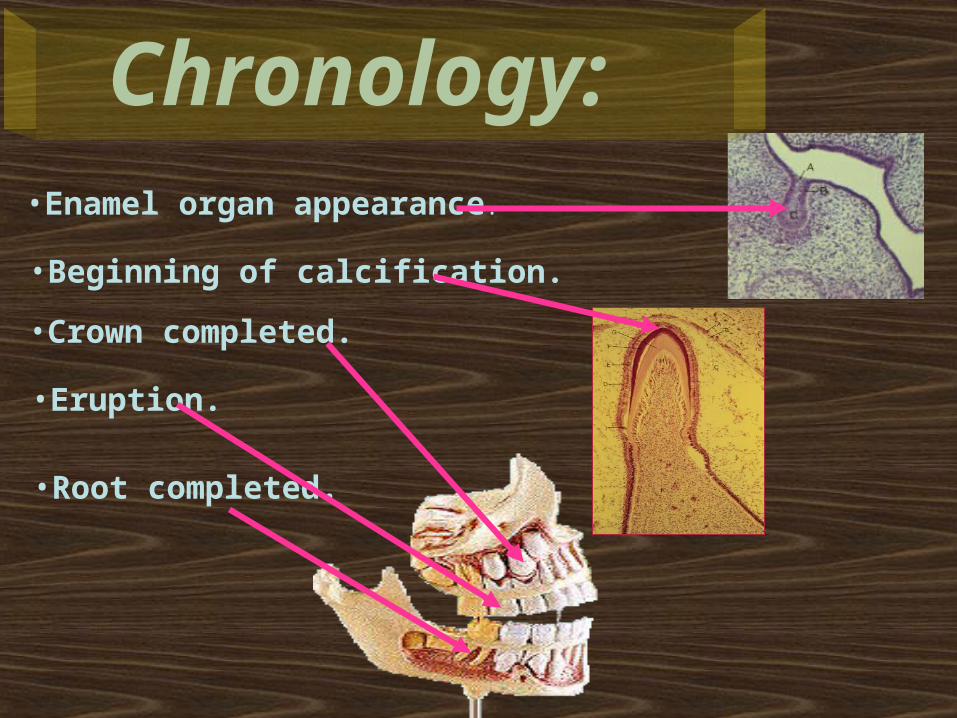
•Enamel organ appearance.
•Beginning of calcification.
•Crown completed.
•Eruption.
•Root completed.
Chronology:
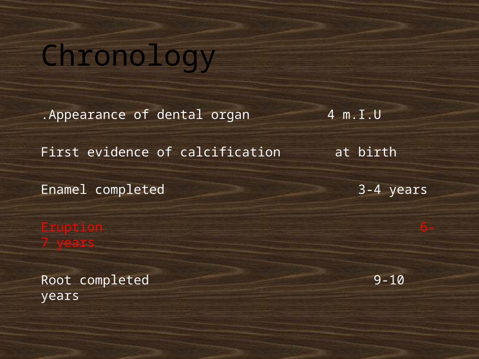
Chronology
Appearance of dental organ 4 m.I.U.
First evidence of calcification at birth
Enamel completed 3-4 years
Eruption 6-7 years
Root completed 9-10 years
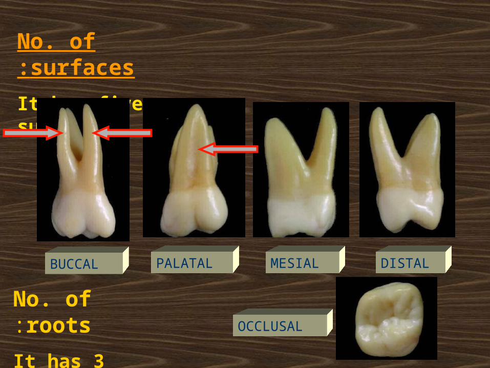
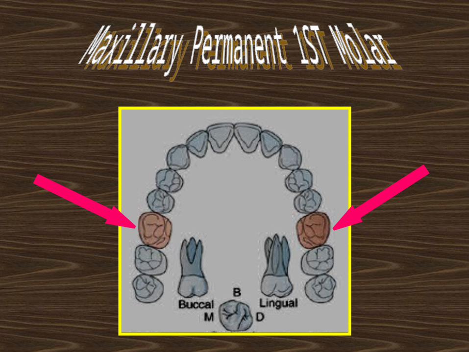
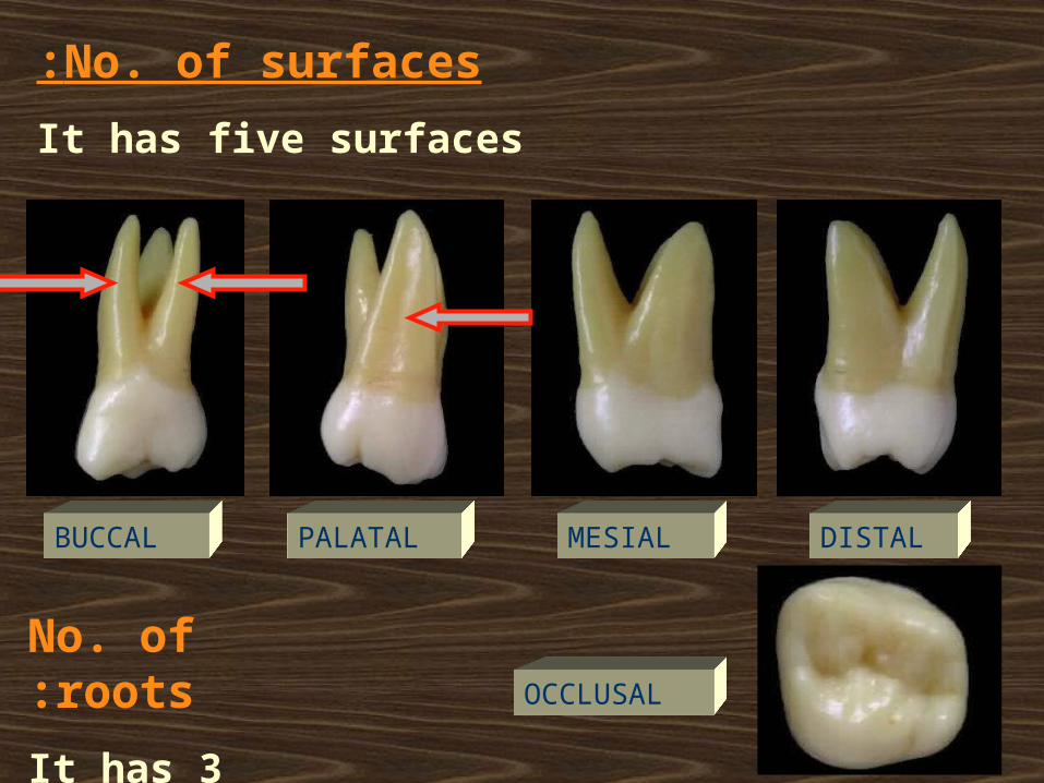
No. of surfaces:
It has five surfaces
No. of roots:
It has 3 roots
BUCCAL PALATAL MESIAL DISTAL
OCCLUSAL
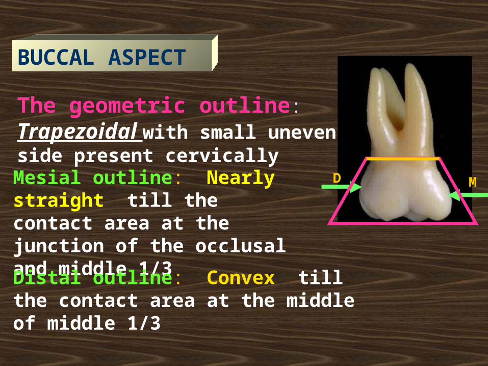
BUCCAL ASPECT
The geometric outline: Trapezoidal with small uneven side present cervically
DMesial outline: Nearly straight till the contact area at the junction of the occlusal and middle 1/3
Distal outline: Convex till the contact area at the middle of middle 1/3
M
D M
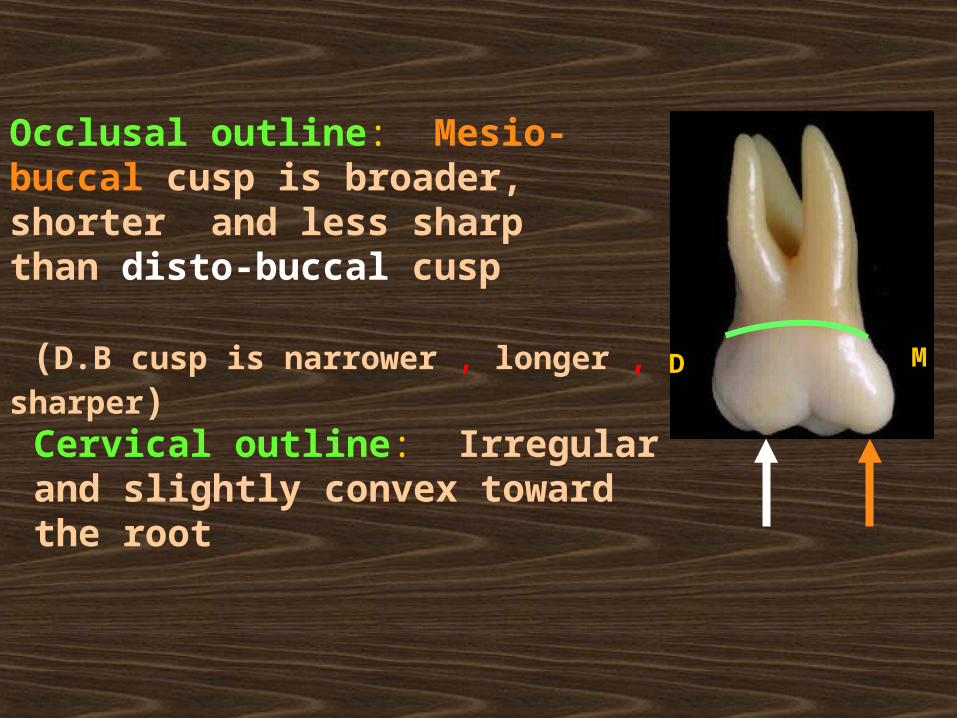
Occlusal outline: Mesio-buccal cusp is broader, shorter and less sharp than disto-buccal cusp
(D.B cusp is narrower , longer , sharper)
Cervical outline: Irregular and slightly convex toward the root
D M
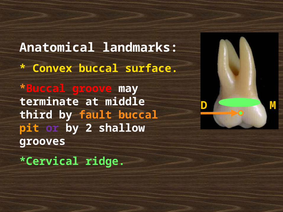
Anatomical landmarks:
* Convex buccal surface.
*Buccal groove may terminate at middle third by fault buccal pit or by 2 shallow grooves
*Cervical ridge.
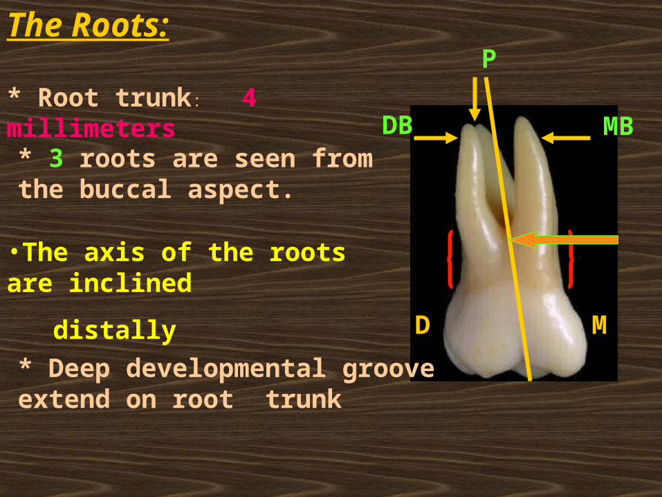
The Roots:
* Root trunk: 4 millimeters
* 3 roots are seen from the buccal aspect.
P
DB MB
•The axis of the roots are inclined
distallyD M
* Deep developmental groove extend on root trunk
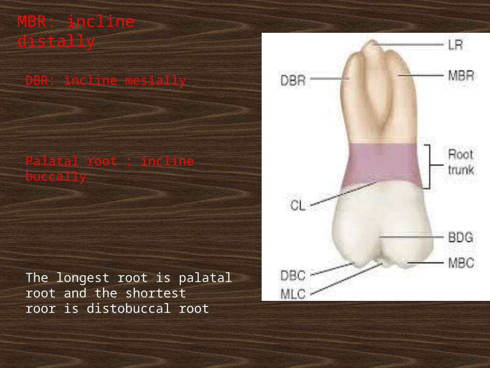
MBR: incline distally
DBR: incline mesially
Palatal root : incline buccally
The longest root is palatal root and the shortest roor is distobuccal root
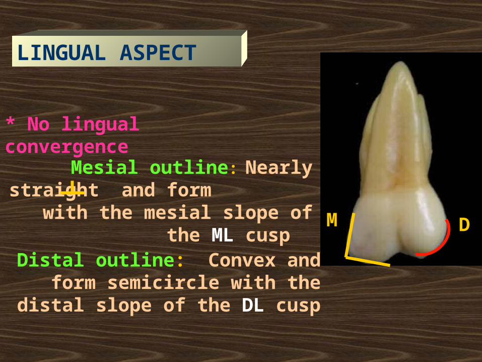
* No lingual convergence
Mesial outline: Nearly straight and form with the mesial slope of the
ML cusp
Distal outline: Convex and form semicircle with the distal slope of the
DL cusp
DM
LINGUAL ASPECT
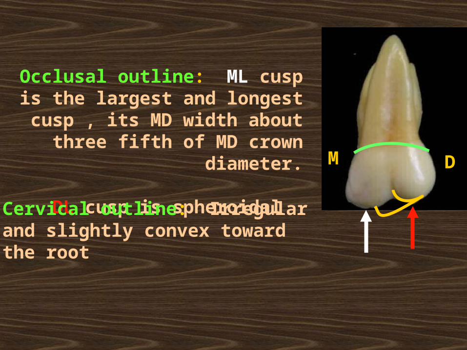
Occlusal outline: ML cusp is the largest and longest cusp , its MD
width about three fifth of MD crown diameter.
DL cusp is spheroidal
Cervical outline: Irregular and slightly convex toward the root
DM
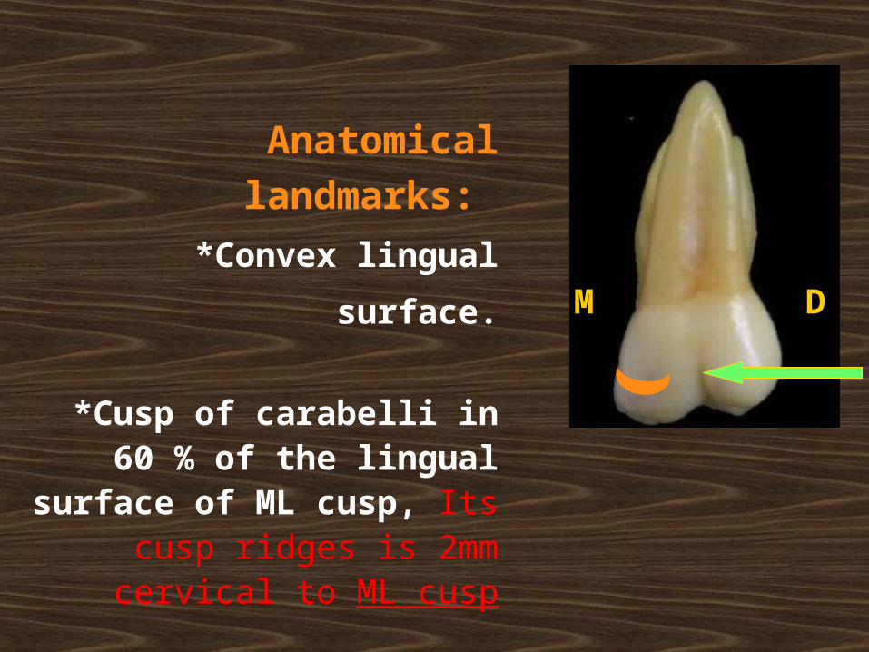
Anatomical landmarks:
*Convex lingual surface.
*Cusp of carabelli in 60 % of the lingual surface of ML cusp, Its
cusp ridges is 2mm cervical to ML cusp
*Lingual developmental groove.
DM
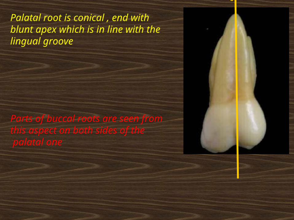
Palatal root is conical , end with blunt apex which is in line with the lingual groove
Parts of buccal roots are seen from this aspect on both sides of the palatal one
LB
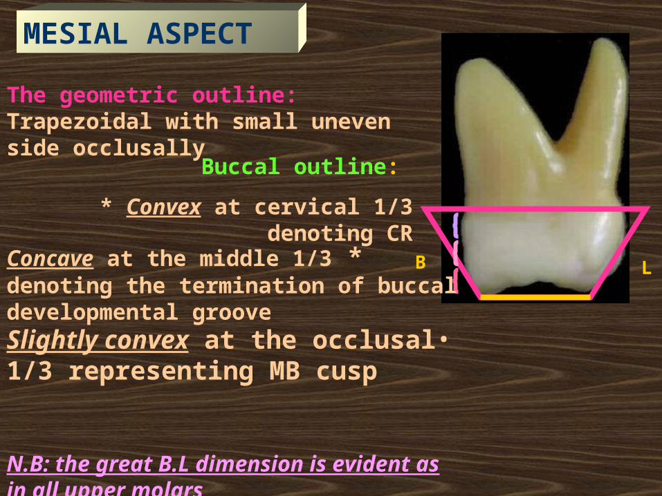
The geometric outline: Trapezoidal with small uneven side occlusally
Buccal outline:
* Convex at cervical 1/3 denoting CR
*Concave at the middle 1/3 denoting the termination of buccal developmental groove
•Slightly convex at the occlusal 1/3 representing MB cusp
N.B: the great B.L dimension is evident as in all upper molars
MESIAL ASPECT
LB
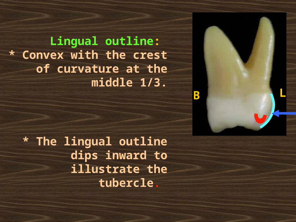
Lingual outline: * Convex with the crest of
curvature at the middle 1/3.
* The lingual outline dips inward to illustrate the tubercle.
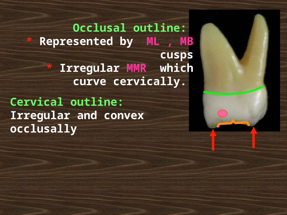
Occlusal outline: * Represented by ML , MB cusps
* Irregular MMR which curve cervically.
Cervical outline:Irregular and convex occlusally
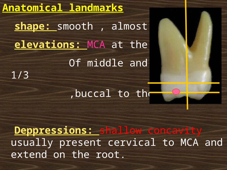
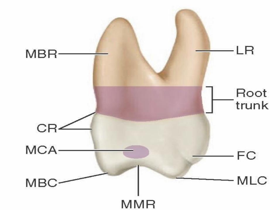
Anatomical landmarks
shape: smooth , almost flat
elevations: MCA at the junction
Of middle and occlusal 1/3
,buccal to the center.
Deppressions: shallow concavity usually present cervical to MCA and extend on the root.
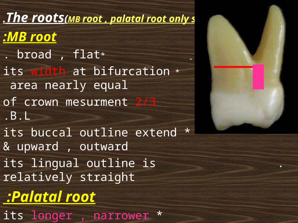
The roots(MB root , palatal root only seen).
MB root: *broad , flat.
*its width at bifurcation area nearly equal 2/3 of crown mesurment B.L.
* its buccal outline extend upward , outward&
. its lingual outline is relatively straight
Palatal root : * its longer , narrower than MB root &
its banana-shaped. * has blunt apex.
DB root: hidden.
The root trunk is aboout 3mm (the shortest root trunk)
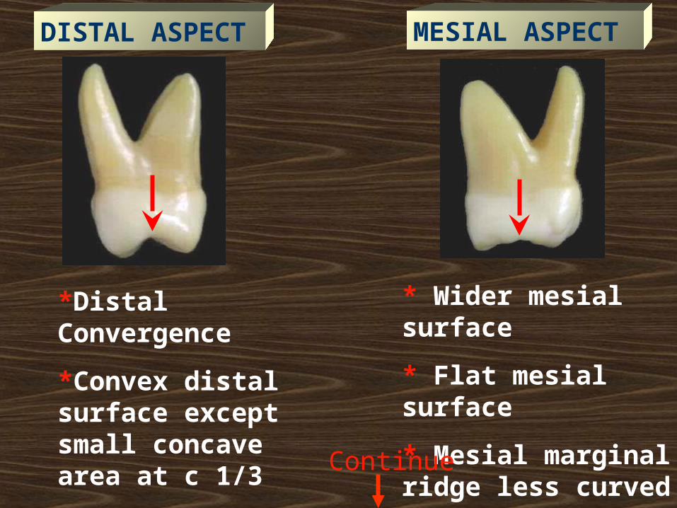
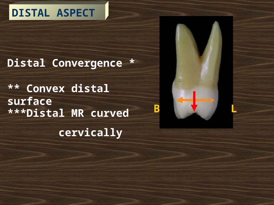
*Distal Convergence
*Convex distal surface except small concave area at c 1/3
*Distal MR curved cervically
* Wider mesial surface
* Flat mesial surface
* Mesial marginal ridge less curved
DISTAL ASPECT MESIAL ASPECT
Continue
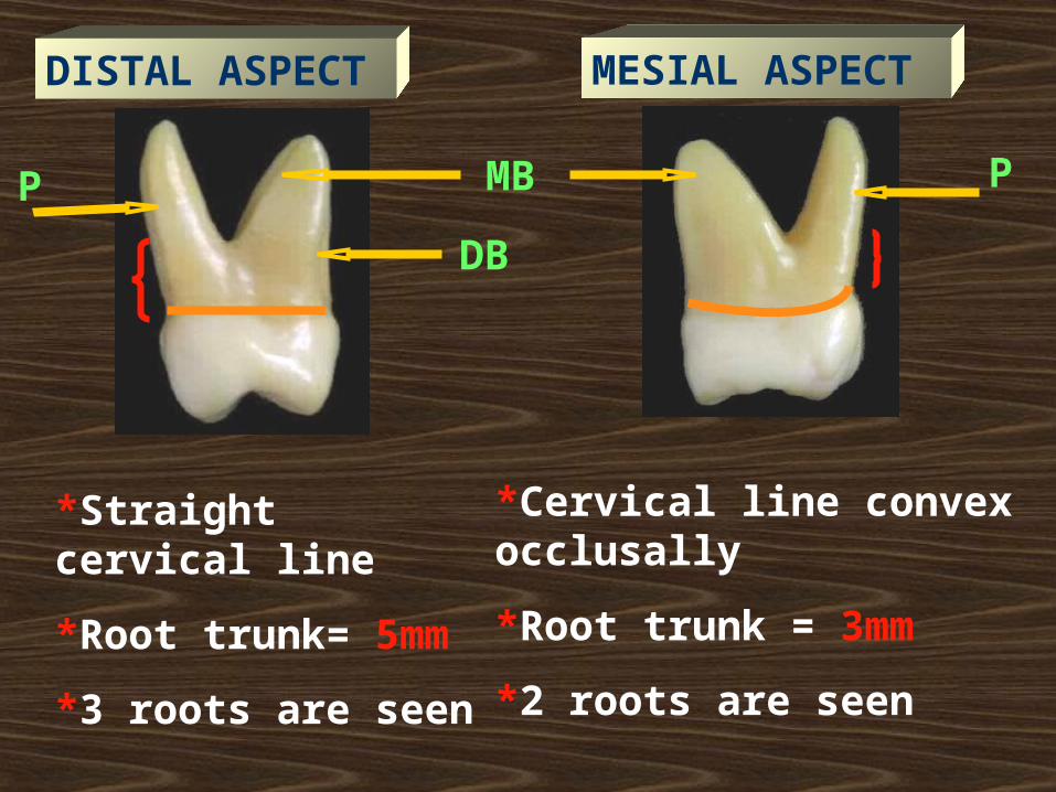
*Straight cervical line
*Root trunk= 5mm
*3 roots are seen
*Cervical line convex occlusally
*Root trunk = 3mm
*2 roots are seen
DB
MBP P
DISTAL ASPECT MESIAL ASPECT
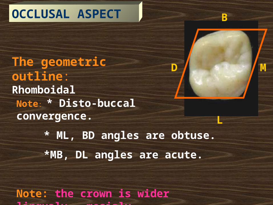
The geometric outline: Rhomboidal
MD
B
L
Note: * Disto-buccal convergence.
* ML, BD angles are obtuse.
*MB, DL angles are acute.
Note: the crown is wider lingualy ,mesialy
OCCLUSAL ASPECT
MD
B
L
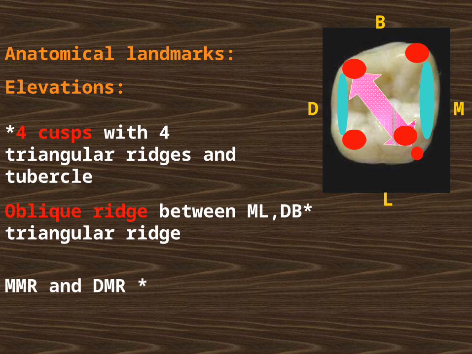
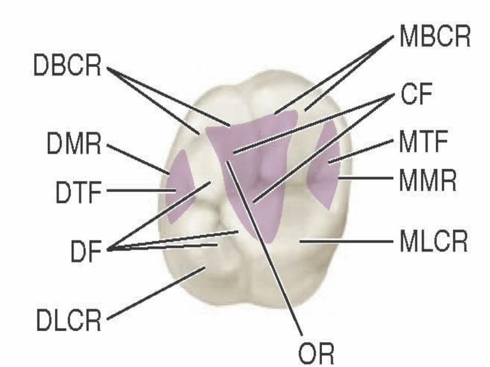
Anatomical landmarks:
Elevations:
*Oblique ridge between ML,DB triangular ridge
*4 cusps with 4 triangular ridges and tubercle
*MMR and DMR
MD
B
L
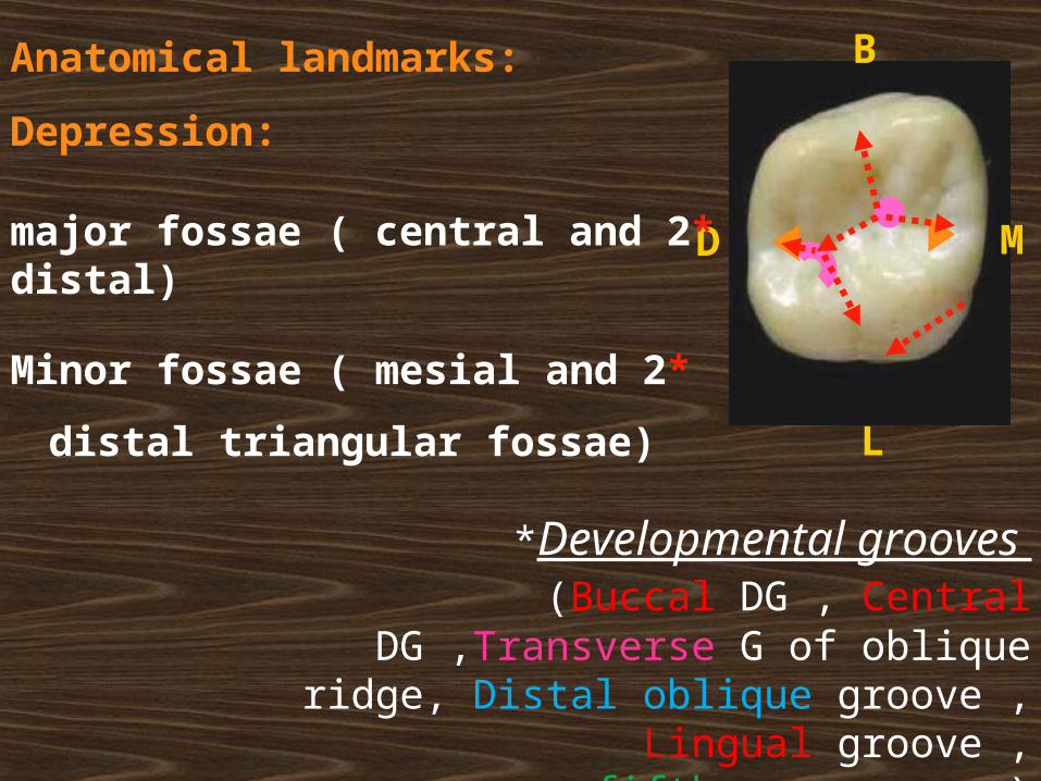
Anatomical landmarks:
Depression:
*2 major fossae ( central and distal)
*2 Minor fossae ( mesial and distal
triangular fossae)
*Developmental grooves (Buccal DG , Central DG ,Transverse G of oblique
ridge, Distal oblique groove , Lingual groove , fifth cusp groove)
Note:
The maxillary 1st molar is the
only molar that is wider lingually than buccally.
The mesiolingual cusp is the largest cusp, followed by the
rounded mesiobuccal, the sharp distobuccal, the small
distolingual, and the tubercle of
Carabelli.
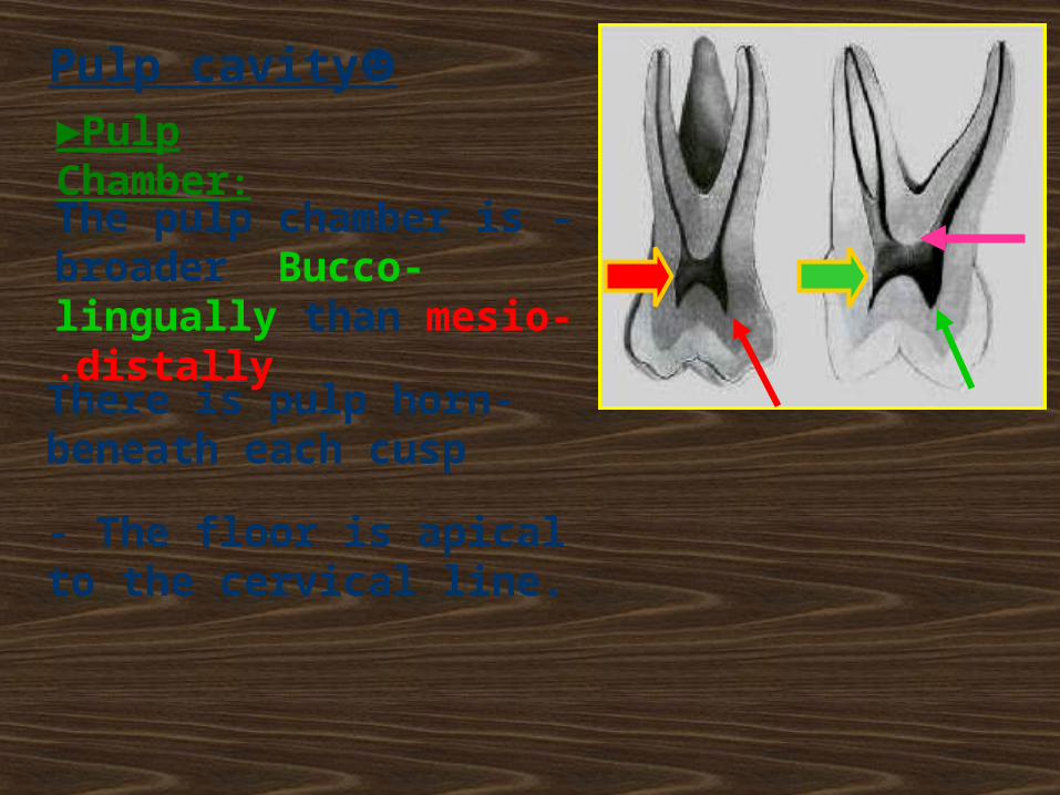
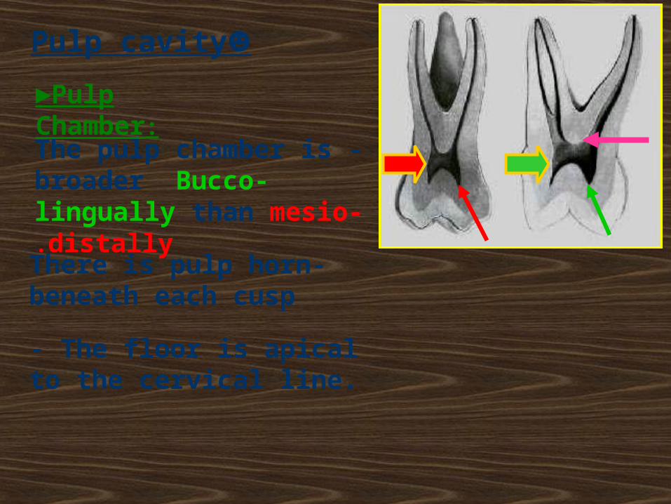
-The pulp chamber is broader Bucco-lingually than mesio-distally.
- The floor is apical to the cervical line.
-There is pulp horn beneath each cusp
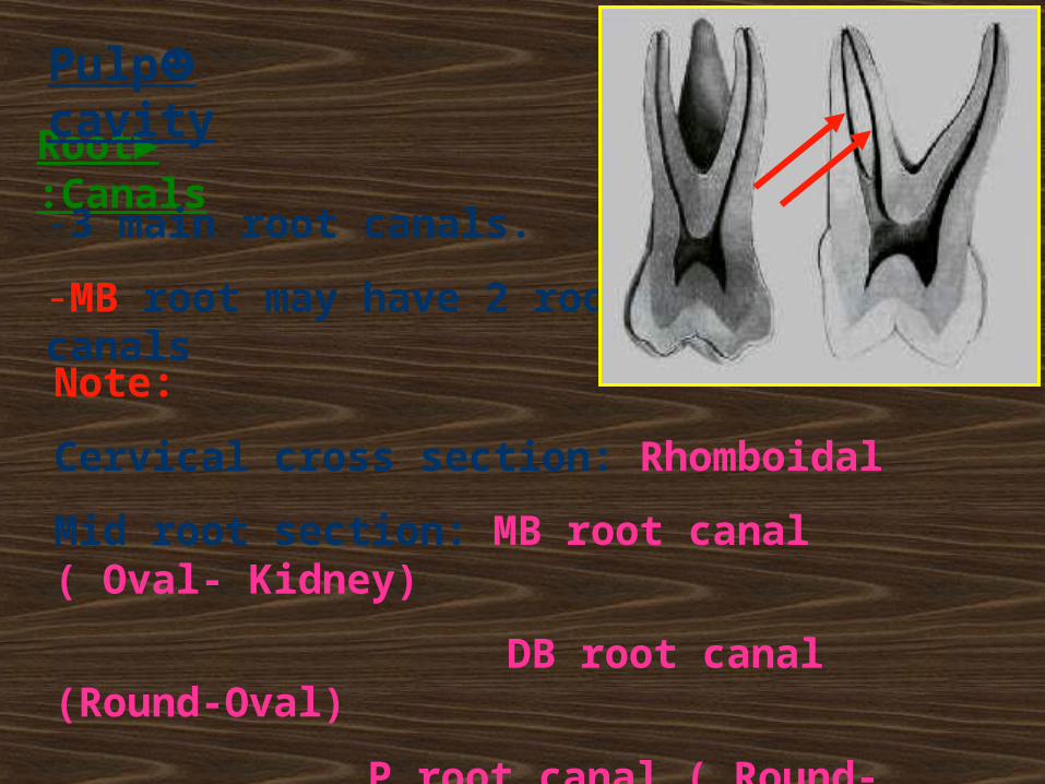
►Pulp Chamber:
☻Pulp cavity
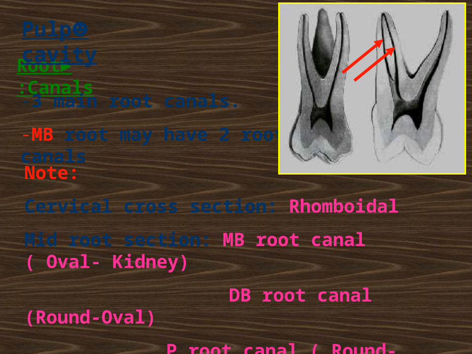
►Root Canals:
-3 main root canals.
-MB root may have 2 root canals
☻Pulp cavity
Note:
Cervical cross section: Rhomboidal
Mid root section: MB root canal ( Oval- Kidney)
DB root canal (Round-Oval)
P root canal ( Round- Oval)
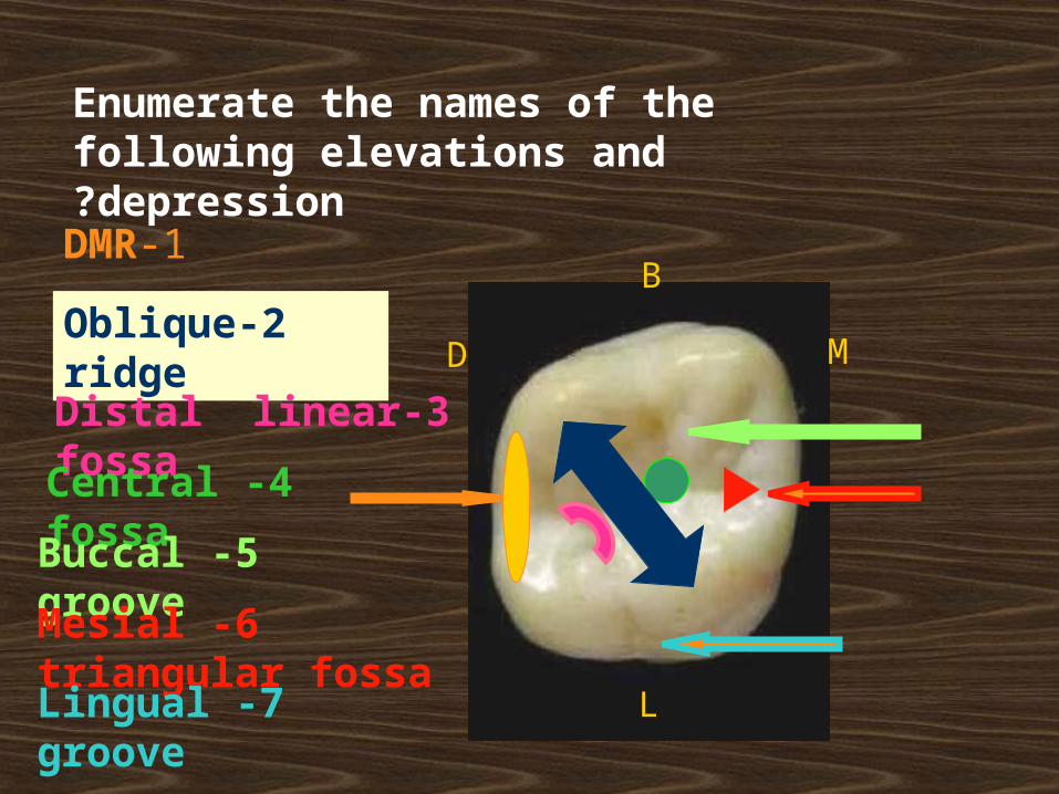
Enumerate the names of the following elevations and depression?
1-DMR
2-Oblique ridge
3-Distal linear fossa
4 -Central fossa
5 -Buccal groove
6 -Mesial triangular fossa
7 -Lingual groove
D M
L
B

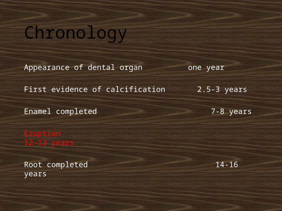
Chronology
Appearance of dental organ one year
First evidence of calcification 2.5-3 years
Enamel completed 7-8 years
Eruption 12-13 years
Root completed 14-16 years
No. of surfaces:
It has five surfaces
No. of roots:
It has 3 roots
BUCCAL PALATAL MESIAL DISTAL
OCCLUSAL
76
MMD D
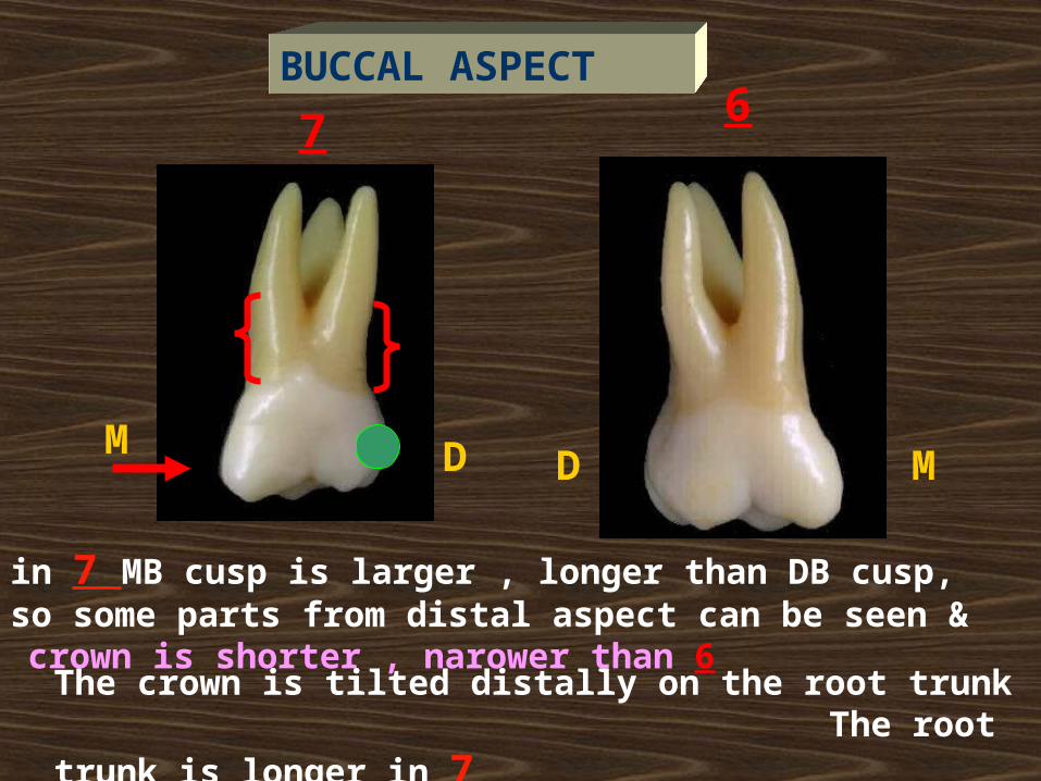
in 7 MB cusp is larger , longer than DB cusp, so some parts from distal aspect can be seen & crown is shorter , narower than 6
The crown is tilted distally on the root trunk The root trunk is longer in 7
BUCCAL ASPECT
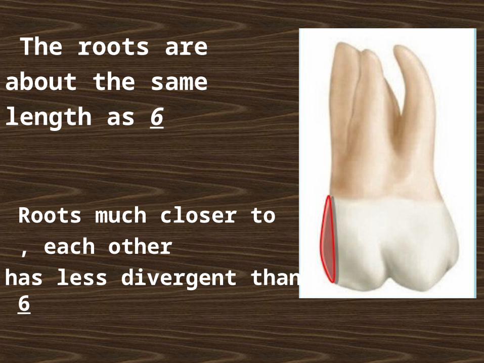
The roots are
about the same length as 6
Roots much closer to
each other ,
has less divergent than 6
7 6
MD D
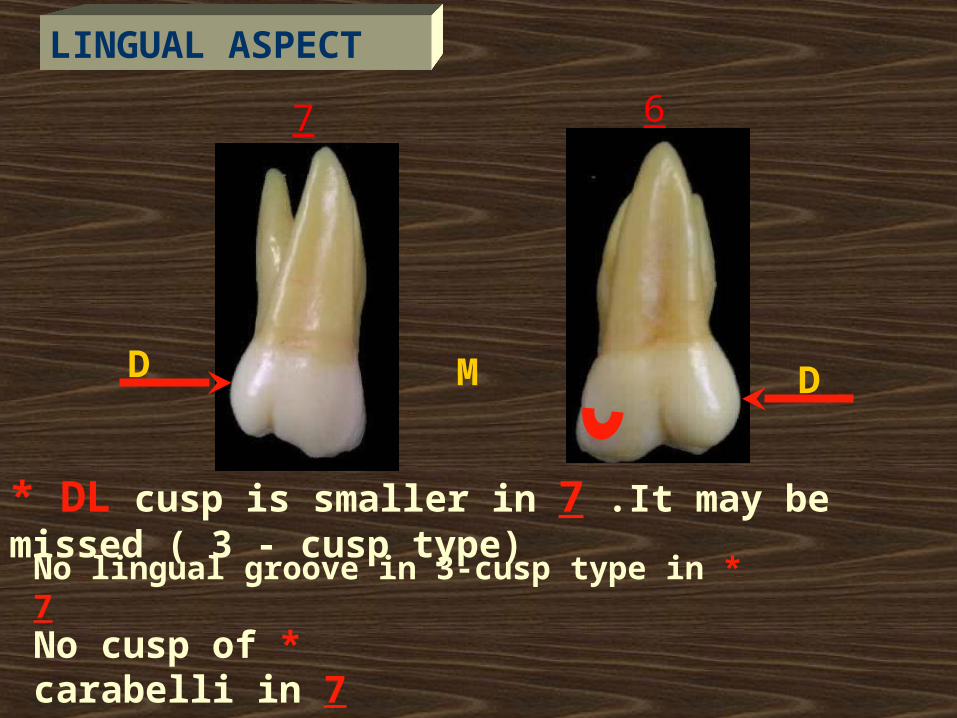
* DL cusp is smaller in 7 .It may be missed ( 3 - cusp type)
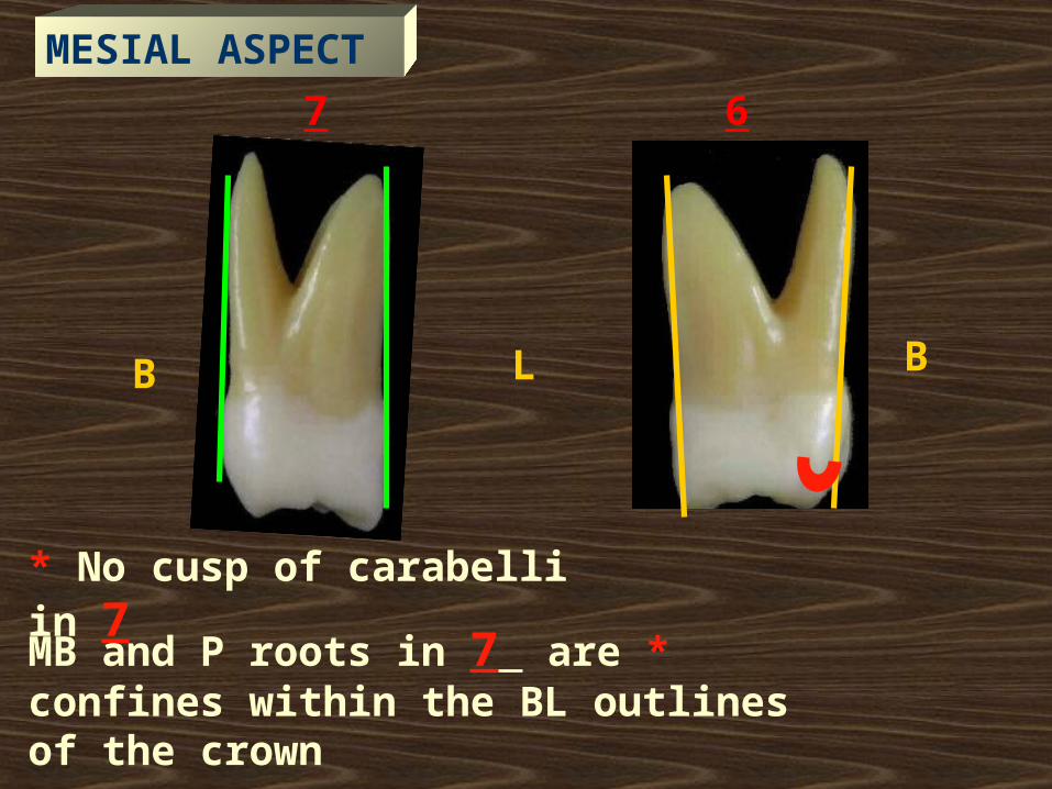
* No cusp of carabelli in 7
LINGUAL ASPECT
* No lingual groove in 3-cusp type in 7
7 6
LB B
* No cusp of carabelli in 7
*MB and P roots in 7 are confines within the BL outlines of the crown
MESIAL ASPECT
***Distal MR curved
cervically
*Distal Convergence
** Convex distal surface
B L
DISTAL ASPECT
MD
B
L
B
M
L
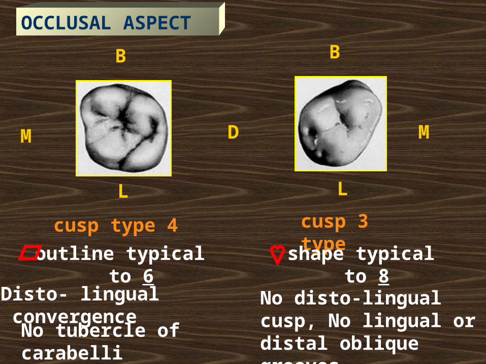
4 cusp type 3 cusp type
outline typical to 6 shape typical to 8
Disto- lingual convergence
No tubercle of carabelli
No disto-lingual cusp, No lingual or distal oblique grooves
OCCLUSAL ASPECT
-The pulp chamber is broader Bucco-lingually than mesio-distally.
- The floor is apical to the cervical line.
-There is pulp horn beneath each cusp
►Pulp Chamber:
☻Pulp cavity
►Root Canals:
-3 main root canals.
-MB root may have 2 root canals
☻Pulp cavity
Note:
Cervical cross section: Rhomboidal
Mid root section: MB root canal ( Oval- Kidney)
DB root canal (Round-Oval)
P root canal ( Round- Oval)

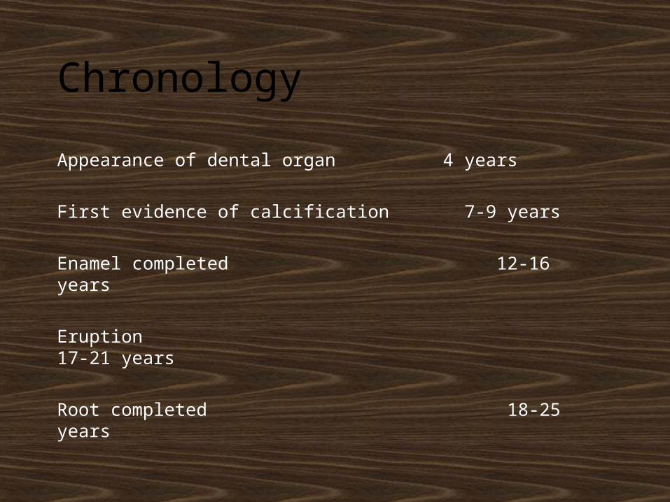
Chronology
Appearance of dental organ 4 years
First evidence of calcification 7-9 years
Enamel completed 12-16 years
Eruption 17-21 years
Root completed 18-25 years
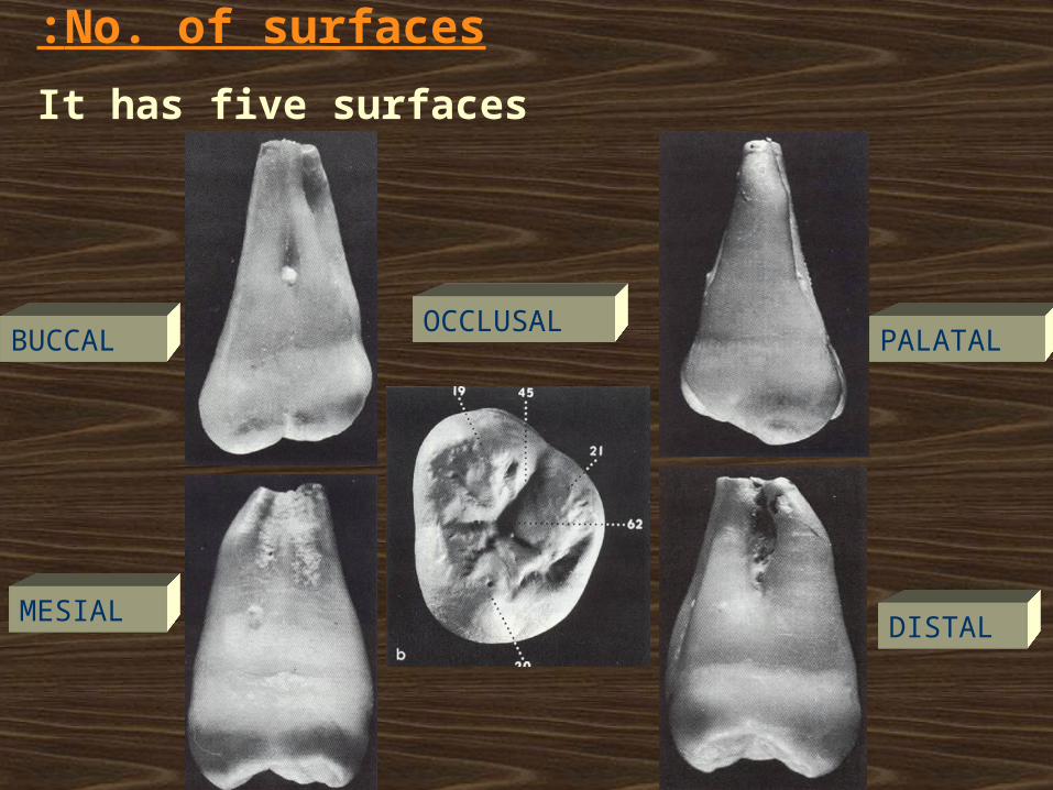
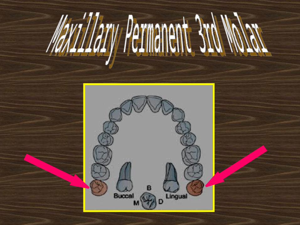
No. of surfaces:
It has five surfaces
BUCCAL PALATAL
MESIAL DISTAL
OCCLUSAL
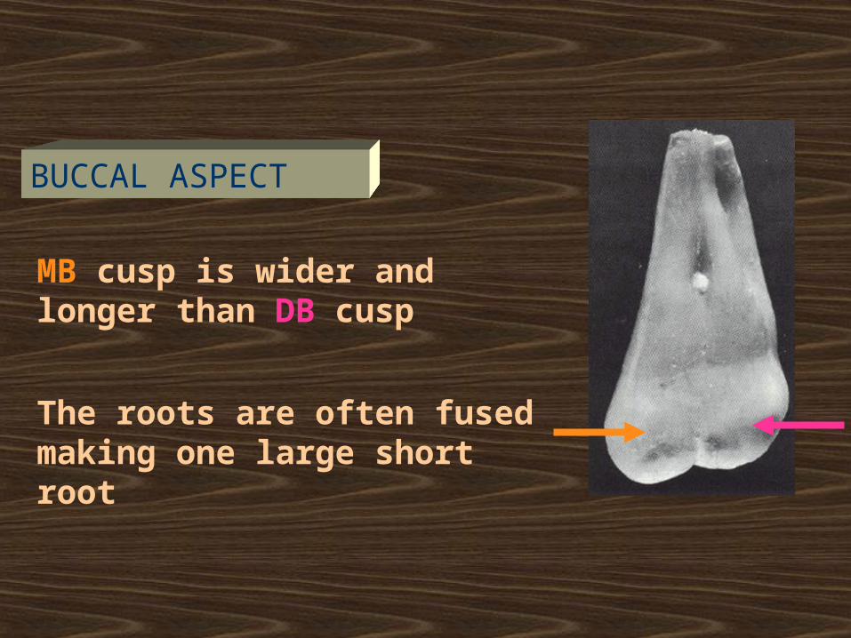
MB cusp is wider and longer than DB cusp
The roots are often fused making one large short root
BUCCAL ASPECT
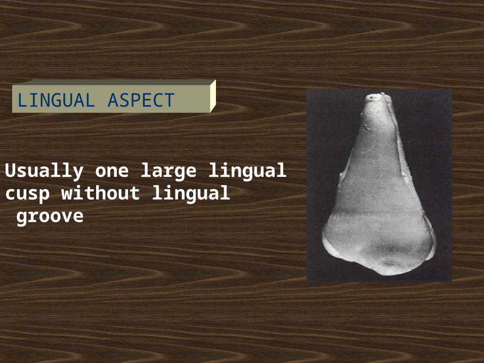
Usually one large lingual cusp without lingual groove
LINGUAL ASPECT
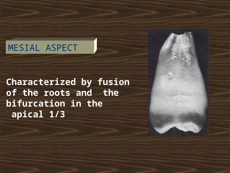
Characterized by fusion of the roots and the bifurcation in the apical 1/3
MESIAL ASPECT
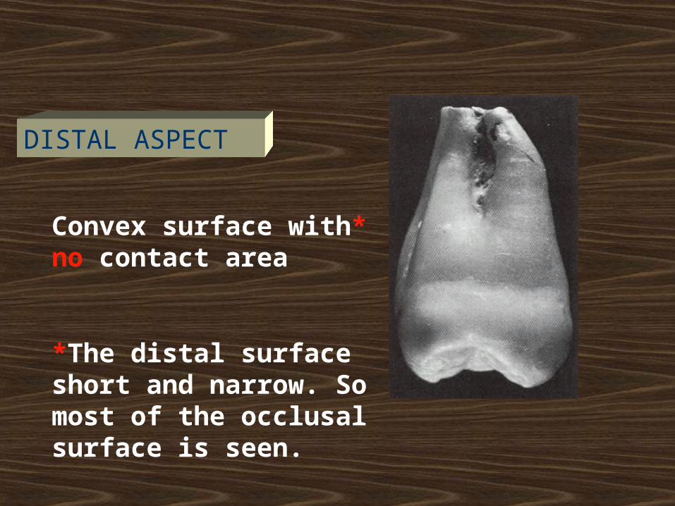
*Convex surface with no contact area
*The distal surface short and narrow. So most of the occlusal surface is seen.
DISTAL ASPECT
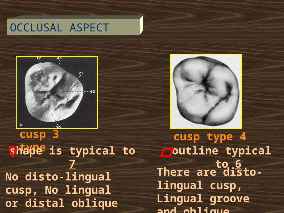
3 cusp type
shape is typical to 7
No disto-lingual cusp, No lingual or distal oblique grooves
4 cusp type outline typical to 6
There are disto- lingual cusp, Lingual groove and oblique ridge.
OCCLUSAL ASPECT
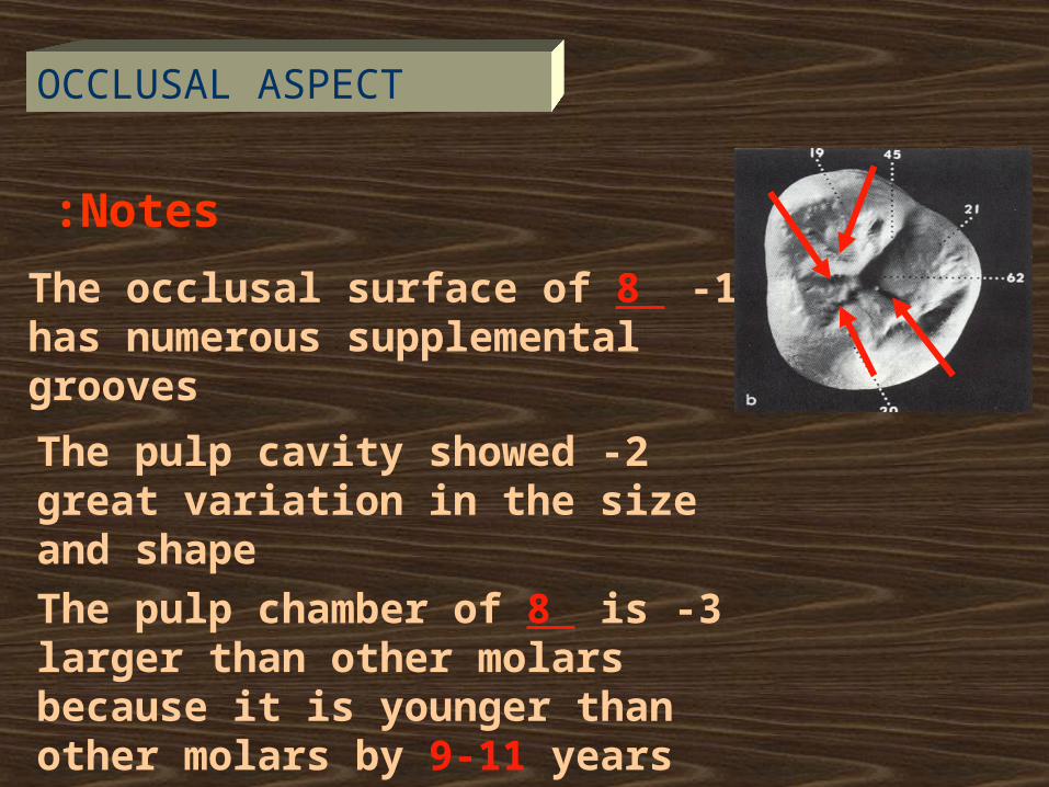
Notes:
1 -The occlusal surface of 8 has numerous supplemental grooves
2 -The pulp cavity showed great variation in the size and shape
3 -The pulp chamber of 8 is larger than other molars because it is younger than other molars by 9-11 years
OCCLUSAL ASPECT

THANK YOU

ANY QUESTION ?
Related Documents