1 Tuberculosis—History, Epidemiology, and Pathogenesis Thomas J. Kishen, Nazim Mughal Egyptian medical treatise, Ebers papyrus (1550 BC), described pulmonary tuberculosis with cervical lymphadenopathy and recommended surgical lancing of the abscess followed by appli- cation of a mixture of acacia seyal, peas, fruits, animal and insect blood, honey, and salt. 1,2 Tuberculosis has also been mentioned in ancient Indian religious texts and termed yaksma (wasting disease) in the Rigveda (1500 BC) and balasa in the Atharvaveda. The Sushruta Samhita (600 BC) recommended treatment with breast milk, various meats, alcohol, and rest, whereas the Yajurveda advised that patients with tuberculosis should reside at higher altitudes. 1,2 Hippocrates (Greek physician, 460–370 BC) described fever, cough with thick sputum, loss of thirst and appetite culminating in delirium, and death as the characteristic features of tuberculosis in his book “Of the Epidemics.” Hippocrates attempted to correct kyphotic deformities due to tuberculosis of the spine with manual pressure, traction, and mechanical appliances, but failed. 1,2 Moreover, Hippocrates believed that the disease was hereditary in nature, while Aristotle thought the disease to be contagious. René Laennec (French physician, 1781–1826), who invented the stethoscope, highlighted the utility of pulmonary auscultation in diagnosing tuberculosis. He died at the age of 45, after con- tracting tuberculosis from contagious patients and infected bodies. Jean Villemin (French phy- sician, 1827–1892) proved the contagious nature of the disease by inoculating tuberculous material from humans into laboratory rabbits, which then became infected. Percivall Pott (British surgeon, 1714–1788) described the features of spinal tuberculosis as ‘‘That kind of palsy of lower limbs, which is frequently found to accompany a curvature of the spine,’’ and Introduction Tuberculosis has been known to infect mankind since the early civilizations. The discovery of the causative organism (Mycobacterium tuber- culosis) and subsequent introduction of anti- tuberculous antibiotics helped to control the disease to some extent. However, urbanization with poverty and pollution, large-scale migra- tions, emergence of human immunodeficiency virus (HIV), and drug-resistant tuberculosis has caused a resurgence of the infection. Nearly 10 million people continue to be infected with tuberculosis every year. Although spinal tuberculosis forms a small proportion of the entire load of tuberculosis infection, it can lead to devastating sequelae including spinal deformities and paraplegia. This chapter discusses the historical evolu- tion of the treatment of tuberculosis along with the epidemiology of the disease and pathogenesis of spinal tuberculosis. History Tuberculosis (TB), one of the first infectious diseases known to mankind, has been vari- ously referred to as consumption, phthisis, white plague, scrofula (tuberculous cervical lymphadenopathy), and Pott’s disease (spinal tuberculosis). Morphological and molecular evidence of tuberculous infection was found in 9,000-year-old Neolithic human remains recovered in Atlit Yam (Israel) and Heidelberg (Germany). The disease has also been found in ancient Egyptian mummies dated between 3000 and 2400 BC, with evidence of hospitals for tuberculosis being around as early as 1500 BC. The mummy of the Egyptian priest Nesperehen, discovered in 1881, revealed evidence of spinal tuberculosis with a psoas abscesses. The ancient

Welcome message from author
This document is posted to help you gain knowledge. Please leave a comment to let me know what you think about it! Share it to your friends and learn new things together.
Transcript
1 Tuberculosis—History, Epidemiology, and PathogenesisThomas J. Kishen, Nazim Mughal
Egyptian medical treatise, Ebers papyrus (1550 BC), described pulmonary tuberculosis with cervical lymphadenopathy and recommended surgical lancing of the abscess followed by application of a mixture of acacia seyal, peas, fruits, animal and insect blood, honey, and salt.1,2
Tuberculosis has also been mentioned in anci ent Indian religious texts and termed yaksma (wasting disease) in the Rigveda (1500 BC) and balasa in the Atharvaveda. The Sushruta Samhita (600 BC) recom mended treatment with breast milk, various meats, alcohol, and rest, whereas the Yajurveda advised that patients with tuber culosis should reside at higher altitudes.1,2
Hippocrates (Greek physician, 460–370 BC) described fever, cough with thick sputum, loss of thirst and appetite culminating in delirium, and death as the characteristic features of tuber culosis in his book “Of the Epidemics.” Hippocrates attempted to correct kyphotic defor mities due to tuberculosis of the spine with manual pressure, traction, and mechanical appli ances, but failed.1,2 Moreover, Hippocrates believed that the disease was hereditary in nature, while Aristotle thought the disease to be contagious.
René Laennec (French physician, 1781–1826), who invented the stethoscope, highlighted the utility of pulmonary auscultation in diagnosing tuberculosis. He died at the age of 45, after contracting tuberculosis from contagious patients and infected bodies. Jean Villemin (French physician, 1827–1892) proved the contagious nature of the disease by inoculating tuberculous material from humans into laboratory rabbits, which then became infected. Percivall Pott (British surgeon, 1714–1788) described the fea tures of spinal tuberculosis as ‘‘That kind of palsy of lower limbs, which is frequently found to accompany a curvature of the spine,’’ and
Introduction
Tuberculosis has been known to infect mankind since the early civilizations. The discovery of the causative organism (Mycobacterium tuberculosis) and subsequent introduction of antitu berculous antibiotics helped to control the disease to some extent. However, urbanization with poverty and pollution, largescale migrations, emergence of human immunodeficiency virus (HIV), and drugresistant tuberculosis has caused a resurgence of the infection. Nearly 10 million people continue to be infected with tuberculosis every year. Although spinal tuberculosis forms a small proportion of the entire load of tuberculosis infection, it can lead to devastating sequelae including spinal deformities and paraplegia.
This chapter discusses the historical evolution of the treatment of tuberculosis along with the epidemiology of the disease and pathogenesis of spinal tuberculosis.
History
Tuberculosis (TB), one of the first infectious diseases known to mankind, has been variously referred to as consumption, phthisis, white plague, scrofula (tuberculous cervical lymphad enopathy), and Pott’s disease (spinal tuberculosis). Morphological and molecular evidence of tuberculous infection was found in 9,000yearold Neolithic human remains recovered in Atlit Yam (Israel) and Heidelberg (Germany). The disease has also been found in ancient Egyptian mummies dated between 3000 and 2400 BC, with evidence of hospitals for tuberculosis being around as early as 1500 BC. The mummy of the Egyptian priest Nesperehen, discovered in 1881, revealed evidence of spinal tuberculosis with a psoas abscesses. The ancient
Spinal Tuberculosis CHAPTER 1 Tuberculosis—History, Epidemiology, and Pathogenesis4
spinal tuberculosis came to be known as “Pott’s disease.”
However, it was Robert Koch (Prussian physician, 1843–1910) who discovered the causative organism (Mycobacterium tuberculosis) using staining methods applied to the sputum of patients with tuberculosis. He identified, isolated, and cultivated the bacillus in animal serum and was able to reproduce the disease in laboratory animals by inoculating them with the bacillus. Koch presented his research at the Physiological Society of Berlin on 24th March 1882 in a lecture entitled Über Tuberculose. March 24th is now commemorated as World Tuberculosis Day. He also went on to demonstrate that Mycobacterium was the cause of all forms of tuberculosis. Although tuberculin, a purified protein derivative of the bacteria developed by Koch in 1890, proved to be an ineffective means of treatment, it was subsequently utilized to perform an intradermal diagnostic test for tuberculosis by Charles Mantoux in 1908.
In the 19th century and first half of the 20th century, before antibiotics were discovered, tuberculosis sanatoria were popular for the treat ment of tuberculosis. These sanatoria were usually located at higher altitudes and pro vided patients with rest, clean air, and a nutritious diet. Gradually, sanatoria became estab lished in cities and low altitudes also. However, the disease continued to take a huge toll on the afflicted patients. The natural history of skeletal tuberculosis without chemotherapy passed through three stages.2 In the “stage of onset” lasting from 1 month to 1 year, the localized disease led to a warm tender swelling and minimal destruction. Over the next 1 to 3 years (stage of destruc tion), disease progression resulted in vertebral destruction, deformity, subluxation, contractures, and abscess formation. Rupture of the abscesses led to ulcers and sinuses and subsequently secondary bacterial infection. With superimposed bacterial infection, the immune system was compromised leading to cachexia, disseminated tuberculosis (miliary tuberculosis and tuberculous meningitis), and death in nearly onethird of the patients. The survivors entered the “stage of repair and ankylosis” with improvement in general
condition, resorption of abscesses, healing of sinuses, and bony healing in kyphosis. Plaster cast immobilization or plaster beds for 2 to 3 years were used to achieve the healed stage in the least disabling position.2 Only 30 to 44% of the patients who developed tuberculosis were able to return to work while the remaining either died (30–50%) or were paralyzed and crippled.3 In the preantibiotic era, surgery aimed at the infected focus (predominantly anterior) was associated with poor outcomes, and Albee and Hibbs described posterior spinal fusion for tuberculous infection (away from the focus of infection). The aim was to shorten the period of immobilization in a plaster cast/bed by providing internal stabilization. However, this surgery did not change the course of the infection.2
The Bacillus Calmette–Guérin (BCG) vaccine, developed by Albert Calmette and Camille Guérin in 1906, from attenuated bovinestrain tuberculosis and first used on humans in 1921, gained general acceptance only in the second half of the 20th century. The modern era in the treatment of tuberculosis began with the discovery of streptomycin (1944), followed by isoniazid (1952) and rifampicin (1970). In the period between 1950 and 1960, major anterior excisional surgery was advocated in the belief that drugs are unable to reach the site of a tuberculous lesion (Tuli 1964). Although, radiotagged isoniazid4 was shown to penetrate the infected focus, it was the international multicentric controlled clinical trials conducted by the Medical Research Council of Great Britain (MRC) that laid the foundation for the current management of uncomplicated spinal tuberculosis (without paraplegia). The conclusions from the various MRC trial centers were as follows: (1) The standard drugs were potent for florid spinal tuberculosis in children and bed rest was not necessary (Masan, Korea MRC 1973a), (2) plaster jackets offer no benefit (Pusan, Korea 1973b), (3) the addition of anterior debridement did not enhance the efficacy of ambulatory chemotherapy alone (Bulawayo MRC 1974a), (4) radical anterior debride ment and strut grafting (Hong Kong procedure) allowed early achievement of a “favorable status” with reduced tendency for deformity pro gression (Hong Kong MRC 1974b), (5) ambu latory chemotherapy for 6 or 9 months
Spinal Tuberculosis CHAPTER 1 Tuberculosis—History, Epidemiology, and Pathogenesis 5
(rifampicin and isoniazid only) was sufficient and radical anterior debridement and strut grafting did not enhance efficacy of the shortcourse chemotherapy regimen (Madras MRC).5
The introduction of antibiotics along with improved standards of living, better nutrition and hygiene contributed largely to the reduction in the burden of tuberculosis infection in affluent countries. However, in the developing world, the disease has persisted due to poverty, mal nutrition, overcrowding, pollution, and the emergence of HIV and multidrugresistant tuberculosis.
Epidemiology
The World Health Organization (WHO) Global Tuberculosis Report 20176 presents the following salient facts:
•• TB is the ninth leading cause of death worldwide and the leading cause from a single infectious agent, ranking above HIV/AIDS.6
•• About 10.4 million people (90% adults; 65% male; 10% people living with HIV) were newly diagnosed to have TB worldwide in 2016, with 2.8 million cases being reported from India alone.6
•• Globally, children (aged < 15 years) accounted for 6.9% of the new TB cases notified in 2016.6
•• The maximum number of incident (new) cases in 2016 occurred in the Southeast
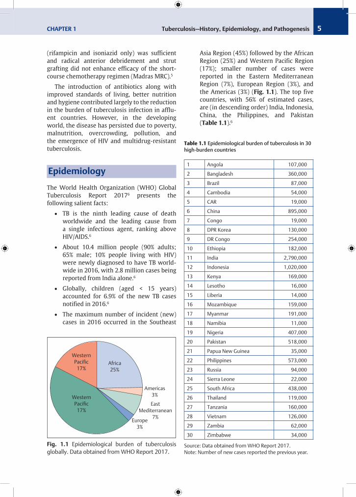
Asia Region (45%) followed by the African Region (25%) and Western Pacific Region (17%); smaller number of cases were reported in the Eastern Mediterranean Region (7%), European Region (3%), and the Americas (3%) (Fig. 1.1). The top five countries, with 56% of estimated cases, are (in descending order) India, Indonesia, China, the Philippines, and Pakistan (Table 1.1).6
Western Pacific
17%
East Mediterranean
7%
Western Pacific
17%
Africa 25%
Americas 3%
Europe 3%
Fig. 1.1 Epidemiological burden of tuberculosis globally. Data obtained from WHO Report 2017.
Table 1.1 Epidemiologicalburdenoftuberculosisin30high-burden countries
1 Angola 107,000
2 Bangladesh 360,000
3 Brazil 87,000
4 Cambodia 54,000
5 CAR 19,000
6 China 895,000
7 Congo 19,000
8 DPR Korea 130,000
9 DR Congo 254,000
10 Ethiopia 182,000
11 India 2,790,000
12 Indonesia 1,020,000
13 Kenya 169,000
14 Lesotho 16,000
15 Liberia 14,000
16 Mozambique 159,000
17 Myanmar 191,000
18 Namibia 11,000
19 Nigeria 407,000
20 Pakistan 518,000
21 Papua New Guinea 35,000
22 Philippines 573,000
23 Russia 94,000
24 Sierra Leone 22,000
25 South Africa 438,000
26 Thailand 119,000
27 Tanzania 160,000
28 Vietnam 126,000
29 Zambia 62,000
30 Zimbabwe 34,000
Source: Data obtained from WHO Report 2017.Note: Number of new cases reported the previous year.
Spinal Tuberculosis CHAPTER 1 Tuberculosis—History, Epidemiology, and Pathogenesis6
•• In 2016, there were 600,000 new cases with resistance to rifampicin (RRTB), of which 490,000 (81.6%) had multidrugresistant TB (MDRTB). Almost half (47%) of these cases were in India, China, and the Russian Federation. There were about 240,000 deaths from MDR/RRTB in 2016. An estimated 4.1% of new cases and 19% of previously treated cases had MDR/RRTB. The treatment success rate for extremely drugresistant TB (XDRTB) (2014 cohort) was 30%.6
•• The incidence of MDRTB varies widely in India, ranging from 1.1% in Bangalore and New Delhi, 2% in Ernakulam, 2.4% in Gujrat, and 24% in Mumbai.7
•• An estimated 10% of the newly diagnosed TB cases in 2016 were among people living with HIV. The proportion of TB cases coinfected with HIV was highest in countries in the African Region, exceeding 50% in parts of southern Africa. The risk of developing TB in people living with HIV was 21 times higher than the rest of the population. The treatment success rate for HIVassociated TB (2015 cohort) was 78%.6
•• In 2016, there were an estimated 1.3 million TB deaths among HIVnegative people (down from 1.7 million in 2000) and an additional 374, 000 deaths among HIVpositive people.6
•• Globally, the TB mortality rate is falling at about 3% per year. TB incidence is falling at about 2% per year, and 16% of TB cases die from the disease.6
•• Zoonotic TB, predominantly caused by Mycobacterium bovis, belongs to the M. tuberculosis complex. In humans, there were an estimated 147,000 new cases of zoonotic TB and 1,200 deaths due to the disease in 2016.6
•• Globalization and immigration has resulted in the spread of tuberculosis around the world. The WHO report on Tuberculosis Surveillance in Europe (2017) shows that immigrants (foreignborn individuals) constitute 30% of all TB cases.8 Between 1991 and 1999, 57% of the reported cases of tuberculosis in Denmark
were among foreignborn immigrants.9 The majority of cases between 2000 and 2011 in both the United States (56%) and the United Kingdom (64%) were also among foreignborn persons,10 and foreignborn children and adolescents accounted for 31% of 18,659 reported TB cases in persons younger than age 18 years from 1994 to 2007 in United States.11
Mycobacterium Tuberculosis—The Bacillus
Tuberculosis is an infectious disease caused by the bacillus M. tuberculosis. M. tuberculosis is an obligate aerobe, acidfast, nonsporing and nonmotile, rodshaped bacillus measuring about 3 mm in length and 0.5 mm in width. It is metabolically catalase and phenylasepositive without adhesion pili or fimbriae in the cell wall surface. The outer cell wall consists of lipids and proteins while the inner wall consists of peptidoglycan (PG), arabinogalactan (AG), and mycolic acid (MA). The mycolic acid containing cell wall, a distinguishing feature of organisms belonging to the genus Mycobacterium, resists staining with acid alcohol when stained with aniline dyes, leading to the term acidfast bacilli. Mycobacteria are typically slow growing, requiring 20 to 24 hours of generation time in artificial media, and 2 to 6 weeks for detectable growth in culture media. M. tuberculosis appears as brown, granular colonies when cultured in Löwenstein–Jensen medium. The load of Mycobacterium in pulmonary lesions is high (107–109), whereas in osteoarticular tissue, it is much lower (< 105). Mycobacterium does not produce adhesion molecules known as biofilm (glycocalyx) around metallic implants thereby rendering these implants inert and safe to use without affecting the efficacy of antibiotic therapy in the presence of tuberculous infection.
Tuberculosis infection is most commonly acquired by inhaling respiratory droplets (< 5 μm in size) aerosolized by coughing from an indi vidual with pulmonary tuberculosis. About 25% of those exposed to TB become infected, and only 10 to 18% of infected individuals develop active tuberculosis disease
Spinal Tuberculosis CHAPTER 1 Tuberculosis—History, Epidemiology, and Pathogenesis 7
within 5 years.12 The disease may be “primary” or reactivation of a previous latent infection. A number of host and environmental factors determine who acquires the infection and who develops progression of the disease. If the host immune response is adequate, the infection may heal although the Mycobacterium may persist in a dormant/latent state to cause reactivation at a later stage. However, if the immune response is inadequate, the primary infection progresses to primary disease. Various factors play a role in the progression of the infection to disease. These include poverty, illiteracy, malnutrition, close contact with a person with tuberculosis, alcoholism, narcotic abuse, diabetes mellitus and immunosuppressive treatment, HIV infection, chronic peritoneal dialysis, silicosis, health care workers, previous tuberculous infection, and vitamin D receptor gene polymorphism. Malnourishment and diabetes cause defects in the humoral and cellular immune responses. Humoral defects include deficits in C3 and C4 complement proteins and alteration in antibody production. Cellular deficits include alterations in cytokine signaling through tumor necrosis factor (TNF), interleukins, insulin growth factor, and direct effects on Tcells. Tcell–mediated cellular immunity plays a role in mycobacterial infection. Numerous host genes play an important role in susceptibility/resistance to tuberculosis infection.
Pathogenesis of Tuberculosis
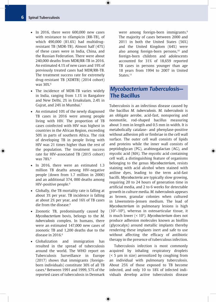
Pulmonary tuberculosis forms 75% of the burden of infection with extrapulmonary tuberculosis contributing to the remaining 25%. Bone and joint tuberculosis forms 15 to 20% of the burden of extrapulmonary tuberculosis of which spinal tuberculosis accounts for 50% of the disease (Fig. 1.2). M. tuberculosis lodges itself in tissues with highoxygen content like the upper lobe of the lung and metaphysis of bones. The reticuloendothelial system responds by accumulation of polymorphonuclear cells, which are subsequently replaced by phagocytic mononuclear cells (macrophages and mono cytes). M. tuberculosis enters the macrophages passively through phagocytosis as it cannot actively enter cells by its own motility. Phagocytosis and breakdown of the bacilli occur
Pulmonary TB 75%
Extra-pulomonary TB
15%
Both 10%
Pleural 18%
Lymphatics 42%
Musculoskeletal 11%
Genitourinary 5%
Meningeal 6%
Peritoneal 6%Others
12%
All other bones and
joints 50%
Spine 50%
Anatomical distribution of Tuberculosis
Distribution of Extrapulmonary Tuberculosis
Distribution of Musculoskeletal Tuberculosis
Fig. 1.2 Anatomical distribution of tuberculosis.
with subsequent dispersal of its lipid content throughout the cytoplasm of the mononuclear cells transforming them into epithelioid cells. Phagosomes (vesicles formed around an engulfed particle/bacterium in a phagocyte) fuse with lysosomes allowing the lysosomal enzymes to destroy the bacteria that come in contact. However, mycobacteriumproduced sulfa tide can inhibit the phagolysosomal fusion process thereby allowing some engulfed mycobacteria to escape into the cytoplasm of the macrophage where they multiply and enter
Spinal Tuberculosis CHAPTER 1 Tuberculosis—History, Epidemiology, and Pathogenesis8
the bloodstream and spread to other parts of the body. Tubercle bacilli are resistant to killing by macrophages and can be found alive within the macrophages leading to chronic and latent infections. Mycobacteria survive inside the granulomas (composed of host defense cells such as neutrophils, macrophages, Tcells, Bcells, dendrite cells, fibroblast, and matrix com ponent) by production of sulfatides, cord factors, and wax D, which are the main cell wall components.
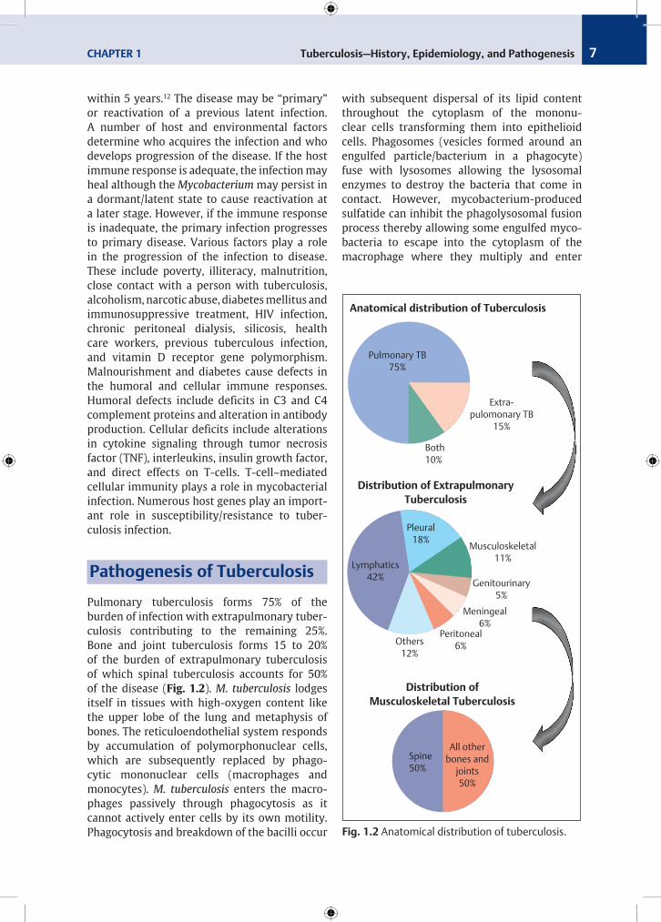
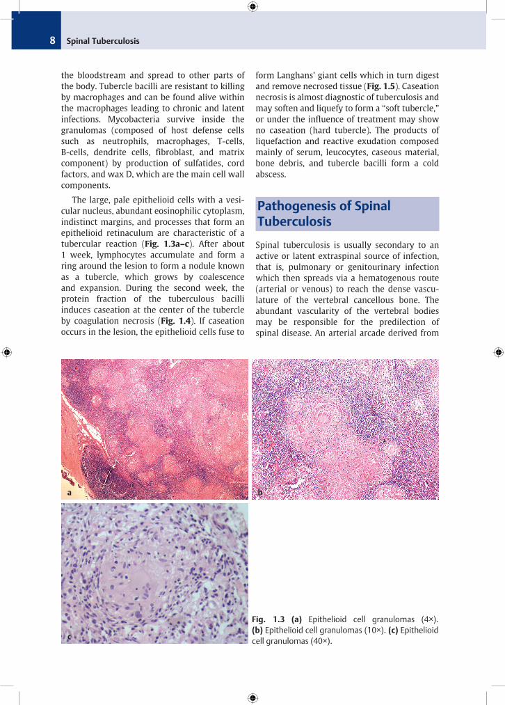
The large, pale epithelioid cells with a vesicular nucleus, abundant eosinophilic cytoplasm, indistinct margins, and processes that form an epithelioid retinaculum are characteristic of a tubercular reaction (Fig. 1.3a–c). After about 1 week, lymphocytes accumulate and form a ring around the lesion to form a nodule known as a tubercle, which grows by coalescence and expansion. During the second week, the protein fraction of the tuberculous bacilli induces caseation at the center of the tubercle by coagulation necrosis (Fig. 1.4). If caseation occurs in the lesion, the epithelioid cells fuse to
form Langhans’ giant cells which in turn digest and remove necrosed tissue (Fig. 1.5). Caseation necrosis is almost diagnostic of tuberculosis and may soften and liquefy to form a “soft tubercle,” or under the influence of treatment may show no caseation (hard tubercle). The products of liquefaction and reactive exudation composed mainly of serum, leucocytes, caseous material, bone debris, and tubercle bacilli form a cold abscess.
Pathogenesis of Spinal Tuberculosis
Spinal tuberculosis is usually secondary to an active or latent extraspinal source of infection, that is, pulmonary or genitourinary infection which then spreads via a hematogenous route (arterial or venous) to reach the dense vasculature of the vertebral cancellous bone. The abundant vascularity of the vertebral bodies may be responsible for the predilection of spinal disease. An arterial arcade derived from
Fig. 1.3 (a) Epithelioid cell granulomas (4×). (b) Epithelioid cell granulomas (10×). (c) Epithelioid cell granulomas (40×).
b
c
a
Spinal Tuberculosis CHAPTER 1 Tuberculosis—History, Epidemiology, and Pathogenesis 9
the ant erior and posterior spinal arteries forms a vascular plexus in the subchondral region of each vertebrae resulting in the infection localizing in the paradiscal regions. Simultaneous involve ment of the paradiscal regions of adjacent vertebrae with its common blood supply lends credence to a hematogenous route being a mode for spread of infection.13–15 Strenuous activities like coughing can cause the infection to spread to the spine via the valveless Batson’s paravertebral venous plexus from the abdominal or thoracic cavity leading to central vertebral body lesions and noncontiguous ver tebral
tuberculosis. Based on its location, tuber cular involvement of the vertebra can be classi fied as paradiscal, central, anterior, appendiceal, indeterminate, and nonosseous type.
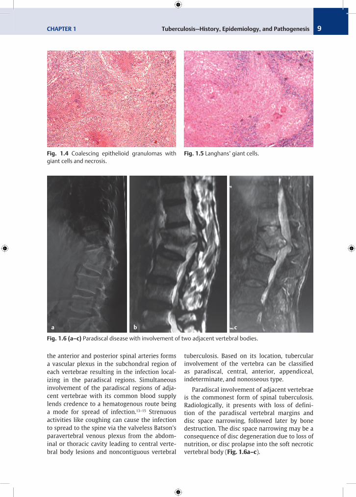
Paradiscal involvement of adjacent vertebrae is the commonest form of spinal tuberculosis. Radiologically, it presents with loss of definition of the paradiscal vertebral margins and disc space narrowing, followed later by bone destruction. The disc space narrowing may be a consequ ence of disc degeneration due to loss of nutrition, or disc prolapse into the soft necrotic vertebral body (Fig. 1.6a–c).
Fig. 1.4 Coalescing epithelioid granulomas with giant cells and necrosis.
Fig. 1.5 Langhans’ giant cells.
Fig. 1.6 (a–c) Paradiscal disease with involvement of two adjacent vertebral bodies.
b ca
Spinal Tuberculosis CHAPTER 1 Tuberculosis—History, Epidemiology, and Pathogenesis10
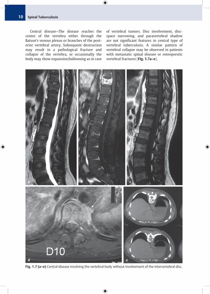
Central disease—The disease reaches the center of the vertebra either through the Batson’s venous plexus or branches of the posterior vertebral artery. Subsequent destruction may result in a pathological fracture and collapse of the vertebra, or occasionally the body may show expansion/ballooning as in case
of verte bral tumors. Disc involvement, discspace narrowing, and paravertebral shadow are not significant features in central type of vertebral tuberculosis. A similar pattern of vertebral collapse may be observed in patients with metastatic spinal disease or osteoporotic vertebral fractures (Fig. 1.7a–e).
Fig. 1.7 (a–e) Central disease involving the vertebral body without involvement of the intervertebral disc.
b c
d
a
e
Spinal Tuberculosis CHAPTER 1 Tuberculosis—History, Epidemiology, and Pathogenesis 11
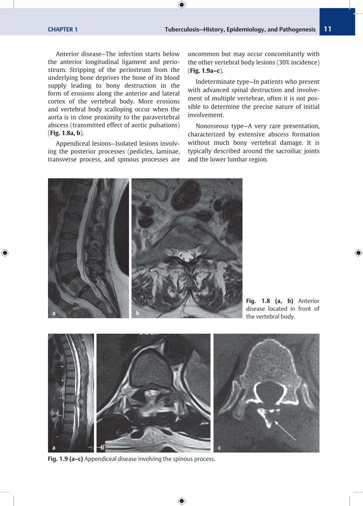
Anterior disease—The infection starts below the anterior longitudinal ligament and periosteum. Stripping of the periosteum from the underlying bone deprives the bone of its blood supply leading to bony destruction in the form of erosions along the anterior and lateral cortex of the vertebral body. More erosions and vertebral body scalloping occur when the aorta is in close proximity to the paravertebral abscess (transmitted effect of aortic pulsations) (Fig. 1.8a, b).
Appendiceal lesions—Isolated lesions involving the posterior processes (pedicles, laminae, transverse process, and spinous processes are
uncommon but may occur concomitantly with the other vertebral body lesions (30% incidence) (Fig. 1.9a–c).
Indeterminate type—In patients who present with advanced spinal destruction and involvement of multiple vertebrae, often it is not possible to determine the precise nature of initial involvement.
Nonosseous type—A very rare presentation, characterized by extensive abscess formation without much bony vertebral damage. It is typically described around the sacroiliac joints and the lower lumbar region.
Fig. 1.8 (a, b) Anterior disease located in front of the vertebral body.
Fig. 1.9 (a–c) Appendiceal disease involving the spinous process.
b
b
a
a c
Spinal Tuberculosis CHAPTER 1 Tuberculosis—History, Epidemiology, and Pathogenesis12
Spinal Deformity Secondary to Tuberculosis
The average number of vertebrae involved vary from 2.5 to 3.8,13,16,17 with 7 to 16% presenting with noncontiguous spinal lesions13,18 and 12% incidence of concomitant nonosteoarticular (pulmonary/visceral/glandular) disease.13 The infection causes increased local osseous blood flow, osteoporosis, and lytic bone destruction leading to compression, collapse, and deformation of the bone. Additionally, arterial occlusion secondary to thromboembolic phenomenon, endarteritis, and periarteritis causes ischemic necrosis that subsequently results in collapse of the vertebra. Ischemic changes can also form bony or cartilaginous sequestra. The inter vertebral disc being relatively avascular in adults is not primarily involved but may be involved in children because of the vascularity. Distortion of the spinal column can lead to progres sive deformity and eventually to neural compression and paraplegia. Reduction in the height of the disc space and wedging of the vertebral body can change the alignment of the spine. Anterior vertebral body wedging involving one to two vertebrae produces a small prominence of the spinous process (knuckle kyphosis), and involvement of three or more vertebral bodies produces an angular kyphosis.13 The kyphosis is progressive in the active stage of the disease due to progressive vertebral body destruction. In children, the deformity may progress even after the infection has healed.19 The preexist ing thoracic kyphosis leads to increased kyphosis with thoracic spine involvement, but the lordotic lumbar spine with larger verte bra provides some protection against the development of a kyphotic deformity. The mean increase in kyphotic deformity in conservatively treated patients is 15 degrees and 3 to 5% develop a deformity of greater than 60 degrees.20,21 Vertebrae within the arms of a kyphotic segment can show increased longitudinal growth (tall vertebra) when the disease has occurred during childhood.22 Lateral vertebral wedg ing can lead to a scoliotic spinal deformity.
Neurological Complications Secondary to Spinal Tuberculosis
Paraplegia secondary to tuberculosis has been classified into two groups by Griffiths et al.23
Group A—Early-Onset Paraplegia or Paraplegia Associated with Active Disease Earlyonset paraplegia occurs during the active phase of the disease (within 2 years of the onset) is caused by mechanical compression, instab ility and inflam matory edema, granulation tissue/abscess, or rarely spinal cord infarction. Intraspinal abscess or granulation tissue can cause extra dural cord compression leading to paraplegia. Vertebral destruction leading to kyphosis, ver tebral dislocation, and vertebral body seque stration can also narrow the spinal canal and cause instability leading to neurological deficits. Inflammatory spinal cord edema due to venous stasis or toxins from tubercular inflam mation is an early cause of neurological deficits. Infarction of the spinal cord can occur secondary to endar teritis, periarteritis, or spinal artery thromb osis caused by inflammation and pre sents a poor prognosis for complete neuro logical recovery.24
Group B—Late-Onset Paraplegia or Paraplegia Associated with Healed Disease The lateonset paraplegia, occurring after 2 years, is usually caused by mechanical compression from tubercular debris, sequestrated bone or disc, internal gibbus, or severe kyphotic deformity. Occasionally, thick granulation tissue around the dura can contract and undergo cicatrization leading to recurrence of paraplegia in longstanding disease. The prognosis is less favorable even after surgical decompression.24
Spinal Tuberculosis CHAPTER 1 Tuberculosis—History, Epidemiology, and Pathogenesis 13
Conclusion
Tuberculosis, a disease known for centuries, continues to exact a huge toll being the ninth leading cause of death worldwide with 10 million newly diagnosed cases every year. Tuberculosis in HIV patients, drugresistant tuberculosis, and spread of tuberculosis by immi grant populations continue to pose a challenge. Although spinal tuberculosis forms a small pro portion of the burden of tuberculosis, it leads to devastating sequelae like spinal deformity and paraplegia. The pathogenesis of tuberculosis infection and the host response is better understood, and newer imaging modalities and laboratorybased tests have allowed for easier diagnosis.
Key Points
•• Nearly 10 million new cases of tuberculosis are diagnosed every year.
•• HIV, drugresistant tuberculosis, and immi gration have led to the continued spread of tuberculosis.
•• Mycobacterium tuberculosis is the causative organism, and caseation necrosis is almost diagnostic of tuberculosis.
•• Based on its location, tubercular involvement of the vertebra can be classified as paradiscal, central, anterior, appendiceal, indeterminate, and nonosseous type.
•• Neurological deficits/paraplegia may occur during the active stage of the disease or during the healed stage due to vary ing mechanisms.
References 1. Bick KM. Classics in Orthopaedics. Philadelphia,
PA: JB Lippincott Co.; 1976 2. Tuli SM. Historical aspects of Pott’s disease
(spinal tuberculosis) management. Eur Spine J 2013;22(Suppl 4):529–538
3. Dobson J. Tuberculosis of the spine; an analysis of the results of conservative treatment and of
the factors influencing the prognosis. J Bone Joint Surg Br 1951;33B(4):517–531
4. Barclay WR, Ebert RH, Le Roy GV, Manthei RW, Roth LJ. Distribution and excretion of radioactive isoniazid in tuberculous patients. J Am Med Assoc 1953;151(16):1384–1388
5. Reetha AMSS, Parthasarathy R, Somasundaram PR, Prabhakar R. Fiveyear findings of a comparison of ambulatory short course chemotherapy with radical surgery plus chemotherapy for tuberculosis of the spine in Madras. Indian J Orthop 1994;28:7–13
6. Global Tuberculosis Report 2017: World Health Organisation. Available from: apps.who.int/iris/bitstream/hamdle/10665/259366/ 9789241565516eng.pdf
7. Sharma SK, Kaushik G, Jha B, et al. Prevalence of multidrugresistant tuberculosis among newly diagnosed cases of sputumpositive pulmonary tuberculosis. Indian J Med Res 2011;133:308–311
8. Tuberculosis surveillance and monitoring in Europe 2017. European Centre for Disease Prevention and Control/WHO Regional Office for Europe. Available from: https://ecdc.europa.eu/sites/portal/files/media/en/publications/Publications/ecdctuberculosissurveillancemonitoringEurope2017.pdf
9. Lillebaek T, Andersen AB, Dirksen A, Smith E, Skovgaard LT, KokJensen A. Persistent high incidence of tuberculosis in immigrants in a lowincidence country. Emerg Infect Dis 2002;8(7): 679–684
10. Nnadi CDAL, Anderson LF, Armstrong LR, et al. Mind the gap: TB trends in the USA and the UK, 20002011. Thorax 2016;71(4):356–363
11. Menzies HJWC, Winston CA, Holtz TH, Cain KP, Mac Kenzie WR. Epidemiology of tuberculosis among US and foreignborn children and adolescents in the United States, 19942007. Am J Public Health 2010;100(9):1724–1729
12. Trauer JM, Moyo N, Tay EL, et al. Risk of Active Tuberculosis in the Five Years Following Infection . . . 15%? Chest 2016;149(2):516–525
13. Tuli SM. Tuberculosis of the Skeletal System. New Delhi: Jaypee Brothers Medical Publishers; 2016
14. Rajasekaran S, Kanna RM, Shetty AP. Pathophysiology and Treatment of Spinal Tuberculosis. JBJS Rev 2014;2(9):2
15. Agrawal V, Patgaonkar PR, Nagariya SP. Tuberculosis of spine. J Craniovertebr Junction Spine 2010;1(2):74–85
16. Hodgson AR, Stock FE, Fang HS, Ong GB. Anterior spinal fusion. The operative approach and pathological findings in 412 patients with Pott’s disease of the spine. Br J Surg 1960;48:172–178
Spinal Tuberculosis14
17. Mukopadhaya B, Mishra NK. Tuberculosis of the spine. Indian J Surg 1957;19:59–81
18. Polley P, Dunn R. Noncontiguous spinal tuberculosis: incidence and management. Eur Spine J 2009;18(8):1096–1101
19. Rajasekaran S. The natural history of posttubercular kyphosis in children. Radiological signs which predict late increase in deformity. J Bone Joint Surg Br 2001;83(7):954–962
20. Rajasekaran S, Shanmugasundaram TK. Prediction of the angle of gibbus deformity in tuberculosis of the spine. J Bone Joint Surg Am 1987;69(4): 503–509
21. Guven O. Severe kyphotic deformity in tuberculosis of the spine. Int Orthop 1996;20(4):271
22. Rajasekaran S, Prasad Shetty A, Dheenadhayalan J, Shashidhar Reddy J, NareshBabu J, Kishen T. Morphological changes during growth in healed childhood spinal tuberculosis: a 15year prospective study of 61 children treated with ambulatory chemotherapy. J Pediatr Orthop 2006;26(6):716–724
23. Griffiths D, Seddon HJ, Roaf R. Pott’s Paraplegia. London: Oxford University Press; 1956
24. Jain AK, Kumar J. Tuberculosis of spine: neurological deficit. Eur Spine J 2013;22(Suppl 4):624–633
Related Documents