ORIGINAL ARTICLE Porous bodies of hydroxyapatite produced by a combination of the gel-casting and polymer sponge methods Jazmı´n I. Gonza´lez Ocampo * , Diana M. Escobar Sierra, Claudia P. Ossa Orozco Biomaterials Research Group, Bioengineering Program, University of Antioquia, Street 70 # 52 – 21, Medellin 1226, Colombia ARTICLE INFO Article history: Received 11 March 2015 Received in revised form 19 June 2015 Accepted 26 June 2015 Available online xxxx Keywords: Gel-casting Hydroxyapatite Polymer sponge Porous body ABSTRACT A combination of gel-casting and polymeric foam infiltration methods is used in this study to prepare porous bodies of hydroxyapatite (HA), to provide a better control over the microstruc- tures of samples. These scaffolds were prepared by impregnating a body of porous polyurethane foam with slurry containing HA powder, and using a percentage of solids between 40% and 50% w/v, and three different types of monomers to provide a better performance. X-Ray Diffraction (XRD), and Fourier Transformed Infrared (FTIR) and Scanning Electron Micro- scopy (SEM) were employed to evaluate both the powder hydroxyapatite and the scaffolds obtained. In addition, porosity and interconnectivity measurements were taken in accordance with the international norm. Bioactivity was checked using immersion tests in Simulated Body Fluids (SBF). After the sintering process of the porous bodies, the XRD results showed peaks characteristic of a pure and crystalline HA (JCPDS 9-432) as a single phase. SEM images indi- cate open and interconnected pores inside the material, with pore sizes between 50 and 600 lm. Also, SEM images demonstrate the relatively good bioactivity of the HA scaffolds after immer- sion in SBF. All results for the porous HA bodies suggest that these materials have great poten- tial for use in tissue engineering. ª 2015 Production and hosting by Elsevier B.V. on behalf of Cairo University. Introduction Hydroxyapatite (HA), with a chemical composition of Ca 10 (PO 4 ) 6 (OH) 2 , is a biocompatible and bioactive material with a similar crystal structure to the biological apatite that can be found in hard tissues such as teeth and bones [1]. It has been widely used in orthopedics and dentistry because of its close biocompatibility with the human body and its good integration with bones. Additionally, it offers diverse confor- mation possibilities, given that it is possible to manufacture * Corresponding author. Tel.: +57 011 574 2198589. E-mail address: [email protected] (J.I. Gonza´ lez Ocampo). Peer review under responsibility of Cairo University. Production and hosting by Elsevier Journal of Advanced Research (2015) xxx, xxx–xxx Cairo University Journal of Advanced Research http://dx.doi.org/10.1016/j.jare.2015.06.006 2090-1232 ª 2015 Production and hosting by Elsevier B.V. on behalf of Cairo University. Please cite this article in press as: Gonza ´lez Ocampo JI et al., Porous bodies of hydroxyapatite produced by a combination of the gel-casting and polymer sponge methods, J Adv Res (2015), http://dx.doi.org/10.1016/j.jare.2015.06.006

1-s2.0-S2090123215000739-main
Dec 06, 2015
Porous bodies of hydroxyapatite produced
by a combination of the gel-casting and polymer
sponge methods
by a combination of the gel-casting and polymer
sponge methods
Welcome message from author
This document is posted to help you gain knowledge. Please leave a comment to let me know what you think about it! Share it to your friends and learn new things together.
Transcript
Journal of Advanced Research (2015) xxx, xxx–xxx
Cairo University
Journal of Advanced Research
ORIGINAL ARTICLE
Porous bodies of hydroxyapatite produced
by a combination of the gel-casting and polymer
sponge methods
* Corresponding author. Tel.: +57 011 574 2198589.E-mail address: [email protected] (J.I. Gonzalez
Ocampo).
Peer review under responsibility of Cairo University.
Production and hosting by Elsevier
http://dx.doi.org/10.1016/j.jare.2015.06.0062090-1232 ª 2015 Production and hosting by Elsevier B.V. on behalf of Cairo University.
Please cite this article in press as: Gonzalez Ocampo JI et al., Porous bodies of hydroxyapatite produced by a combination of the gel-casting and polymemethods, J Adv Res (2015), http://dx.doi.org/10.1016/j.jare.2015.06.006
Jazmın I. Gonzalez Ocampo *, Diana M. Escobar Sierra, Claudia P. Ossa Orozco
Biomaterials Research Group, Bioengineering Program, University of Antioquia, Street 70 # 52 – 21, Medellin 1226, Colombia
A R T I C L E I N F O A B S T R A C T
Article history:
Received 11 March 2015
Received in revised form 19 June 2015
Accepted 26 June 2015
Available online xxxx
Keywords:
Gel-casting
Hydroxyapatite
Polymer sponge
Porous body
A combination of gel-casting and polymeric foam infiltration methods is used in this study to
prepare porous bodies of hydroxyapatite (HA), to provide a better control over the microstruc-
tures of samples. These scaffolds were prepared by impregnating a body of porous polyurethane
foam with slurry containing HA powder, and using a percentage of solids between 40% and
50% w/v, and three different types of monomers to provide a better performance. X-Ray
Diffraction (XRD), and Fourier Transformed Infrared (FTIR) and Scanning Electron Micro-
scopy (SEM) were employed to evaluate both the powder hydroxyapatite and the scaffolds
obtained. In addition, porosity and interconnectivity measurements were taken in accordance
with the international norm. Bioactivity was checked using immersion tests in Simulated Body
Fluids (SBF). After the sintering process of the porous bodies, the XRD results showed peaks
characteristic of a pure and crystalline HA (JCPDS 9-432) as a single phase. SEM images indi-
cate open and interconnected pores inside the material, with pore sizes between 50 and 600 lm.
Also, SEM images demonstrate the relatively good bioactivity of the HA scaffolds after immer-
sion in SBF. All results for the porous HA bodies suggest that these materials have great poten-
tial for use in tissue engineering.
ª 2015 Production and hosting by Elsevier B.V. on behalf of Cairo University.
Introduction
Hydroxyapatite (HA), with a chemical composition ofCa10(PO4)6(OH)2, is a biocompatible and bioactive materialwith a similar crystal structure to the biological apatite that
can be found in hard tissues such as teeth and bones [1]. Ithas been widely used in orthopedics and dentistry because ofits close biocompatibility with the human body and its good
integration with bones. Additionally, it offers diverse confor-mation possibilities, given that it is possible to manufacture
r sponge
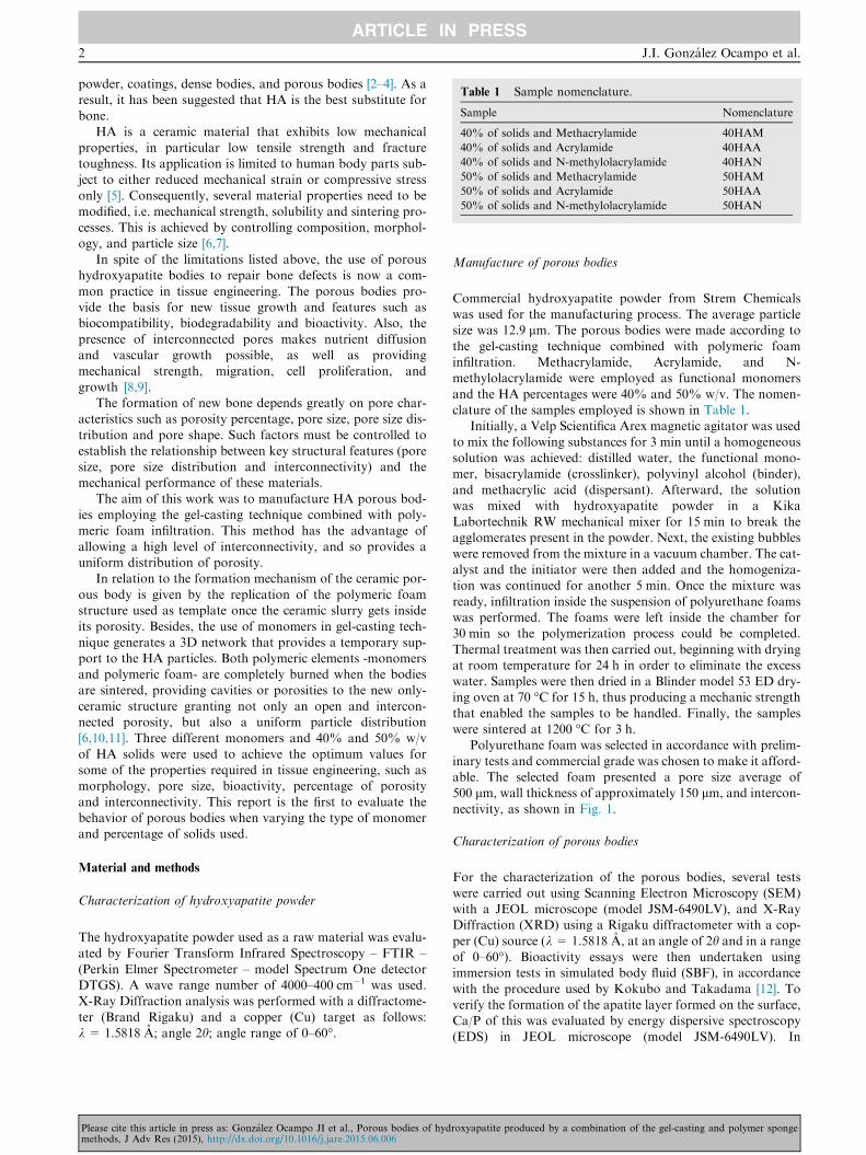
Table 1 Sample nomenclature.
Sample Nomenclature
40% of solids and Methacrylamide 40HAM
40% of solids and Acrylamide 40HAA
40% of solids and N-methylolacrylamide 40HAN
50% of solids and Methacrylamide 50HAM
50% of solids and Acrylamide 50HAA
50% of solids and N-methylolacrylamide 50HAN
2 J.I. Gonzalez Ocampo et al.
powder, coatings, dense bodies, and porous bodies [2–4]. As aresult, it has been suggested that HA is the best substitute forbone.
HA is a ceramic material that exhibits low mechanicalproperties, in particular low tensile strength and fracturetoughness. Its application is limited to human body parts sub-
ject to either reduced mechanical strain or compressive stressonly [5]. Consequently, several material properties need to bemodified, i.e. mechanical strength, solubility and sintering pro-
cesses. This is achieved by controlling composition, morphol-ogy, and particle size [6,7].
In spite of the limitations listed above, the use of poroushydroxyapatite bodies to repair bone defects is now a com-
mon practice in tissue engineering. The porous bodies pro-vide the basis for new tissue growth and features such asbiocompatibility, biodegradability and bioactivity. Also, the
presence of interconnected pores makes nutrient diffusionand vascular growth possible, as well as providingmechanical strength, migration, cell proliferation, and
growth [8,9].The formation of new bone depends greatly on pore char-
acteristics such as porosity percentage, pore size, pore size dis-
tribution and pore shape. Such factors must be controlled toestablish the relationship between key structural features (poresize, pore size distribution and interconnectivity) and themechanical performance of these materials.
The aim of this work was to manufacture HA porous bod-ies employing the gel-casting technique combined with poly-meric foam infiltration. This method has the advantage of
allowing a high level of interconnectivity, and so provides auniform distribution of porosity.
In relation to the formation mechanism of the ceramic por-
ous body is given by the replication of the polymeric foamstructure used as template once the ceramic slurry gets insideits porosity. Besides, the use of monomers in gel-casting tech-
nique generates a 3D network that provides a temporary sup-port to the HA particles. Both polymeric elements -monomersand polymeric foam- are completely burned when the bodiesare sintered, providing cavities or porosities to the new only-
ceramic structure granting not only an open and intercon-nected porosity, but also a uniform particle distribution[6,10,11]. Three different monomers and 40% and 50% w/v
of HA solids were used to achieve the optimum values forsome of the properties required in tissue engineering, such asmorphology, pore size, bioactivity, percentage of porosity
and interconnectivity. This report is the first to evaluate thebehavior of porous bodies when varying the type of monomerand percentage of solids used.
Material and methods
Characterization of hydroxyapatite powder
The hydroxyapatite powder used as a raw material was evalu-ated by Fourier Transform Infrared Spectroscopy – FTIR –
(Perkin Elmer Spectrometer – model Spectrum One detectorDTGS). A wave range number of 4000–400 cm�1 was used.X-Ray Diffraction analysis was performed with a diffractome-
ter (Brand Rigaku) and a copper (Cu) target as follows:k = 1.5818 A; angle 2h; angle range of 0–60�.
Please cite this article in press as: Gonzalez Ocampo JI et al., Porous bodies of hydmethods, J Adv Res (2015), http://dx.doi.org/10.1016/j.jare.2015.06.006
Manufacture of porous bodies
Commercial hydroxyapatite powder from Strem Chemicalswas used for the manufacturing process. The average particlesize was 12.9 lm. The porous bodies were made according to
the gel-casting technique combined with polymeric foaminfiltration. Methacrylamide, Acrylamide, and N-methylolacrylamide were employed as functional monomers
and the HA percentages were 40% and 50% w/v. The nomen-clature of the samples employed is shown in Table 1.
Initially, a Velp Scientifica Arex magnetic agitator was used
to mix the following substances for 3 min until a homogeneoussolution was achieved: distilled water, the functional mono-mer, bisacrylamide (crosslinker), polyvinyl alcohol (binder),
and methacrylic acid (dispersant). Afterward, the solutionwas mixed with hydroxyapatite powder in a KikaLabortechnik RW mechanical mixer for 15 min to break theagglomerates present in the powder. Next, the existing bubbles
were removed from the mixture in a vacuum chamber. The cat-alyst and the initiator were then added and the homogeniza-tion was continued for another 5 min. Once the mixture was
ready, infiltration inside the suspension of polyurethane foamswas performed. The foams were left inside the chamber for30 min so the polymerization process could be completed.
Thermal treatment was then carried out, beginning with dryingat room temperature for 24 h in order to eliminate the excesswater. Samples were then dried in a Blinder model 53 ED dry-ing oven at 70 �C for 15 h, thus producing a mechanic strength
that enabled the samples to be handled. Finally, the sampleswere sintered at 1200 �C for 3 h.
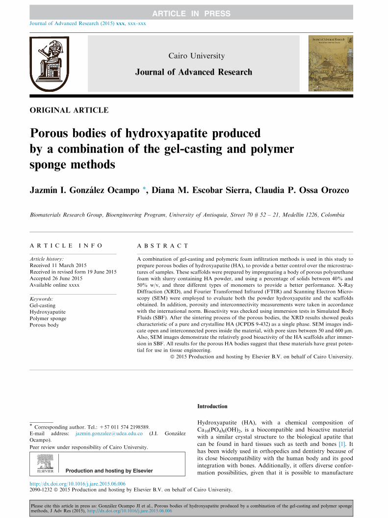
Polyurethane foam was selected in accordance with prelim-
inary tests and commercial grade was chosen to make it afford-able. The selected foam presented a pore size average of500 lm, wall thickness of approximately 150 lm, and intercon-
nectivity, as shown in Fig. 1.
Characterization of porous bodies
For the characterization of the porous bodies, several testswere carried out using Scanning Electron Microscopy (SEM)with a JEOL microscope (model JSM-6490LV), and X-RayDiffraction (XRD) using a Rigaku diffractometer with a cop-
per (Cu) source (k = 1.5818 A, at an angle of 2h and in a rangeof 0–60�). Bioactivity essays were then undertaken usingimmersion tests in simulated body fluid (SBF), in accordance
with the procedure used by Kokubo and Takadama [12]. Toverify the formation of the apatite layer formed on the surface,Ca/P of this was evaluated by energy dispersive spectroscopy
(EDS) in JEOL microscope (model JSM-6490LV). In
roxyapatite produced by a combination of the gel-casting and polymer sponge
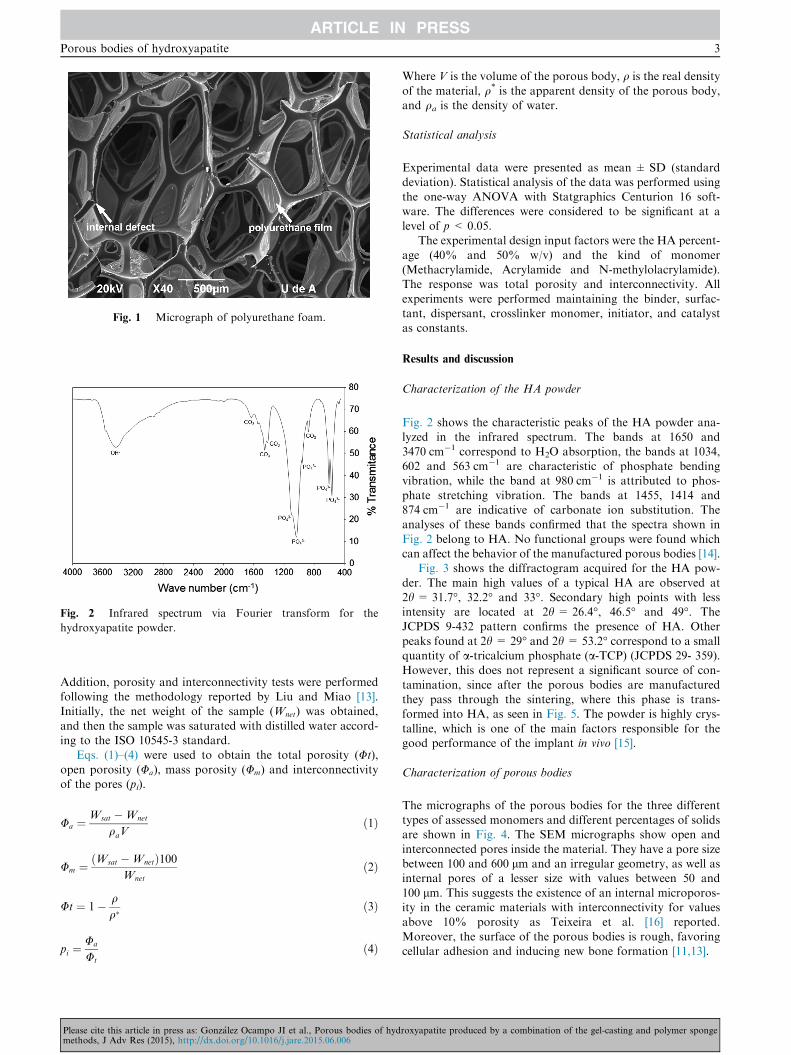
Fig. 1 Micrograph of polyurethane foam.
Fig. 2 Infrared spectrum via Fourier transform for the
hydroxyapatite powder.
Porous bodies of hydroxyapatite 3
Addition, porosity and interconnectivity tests were performedfollowing the methodology reported by Liu and Miao [13].Initially, the net weight of the sample (Wnet) was obtained,
and then the sample was saturated with distilled water accord-ing to the ISO 10545-3 standard.
Eqs. (1)–(4) were used to obtain the total porosity (Ut),
open porosity (Ua), mass porosity (Um) and interconnectivityof the pores (pi).
Ua ¼Wsat �Wnet
qaVð1Þ
Um ¼ðWsat �WnetÞ100
Wnet
ð2Þ
Ut ¼ 1� qq�
ð3Þ
pi ¼Ua
Ut
ð4Þ
Please cite this article in press as: Gonzalez Ocampo JI et al., Porous bodies of hydmethods, J Adv Res (2015), http://dx.doi.org/10.1016/j.jare.2015.06.006
Where V is the volume of the porous body, q is the real density
of the material, q* is the apparent density of the porous body,and qa is the density of water.
Statistical analysis
Experimental data were presented as mean ± SD (standarddeviation). Statistical analysis of the data was performed usingthe one-way ANOVA with Statgraphics Centurion 16 soft-
ware. The differences were considered to be significant at alevel of p < 0.05.
The experimental design input factors were the HA percent-
age (40% and 50% w/v) and the kind of monomer(Methacrylamide, Acrylamide and N-methylolacrylamide).The response was total porosity and interconnectivity. All
experiments were performed maintaining the binder, surfac-tant, dispersant, crosslinker monomer, initiator, and catalystas constants.
Results and discussion
Characterization of the HA powder
Fig. 2 shows the characteristic peaks of the HA powder ana-lyzed in the infrared spectrum. The bands at 1650 and
3470 cm�1 correspond to H2O absorption, the bands at 1034,602 and 563 cm�1 are characteristic of phosphate bendingvibration, while the band at 980 cm�1 is attributed to phos-
phate stretching vibration. The bands at 1455, 1414 and874 cm�1 are indicative of carbonate ion substitution. Theanalyses of these bands confirmed that the spectra shown in
Fig. 2 belong to HA. No functional groups were found whichcan affect the behavior of the manufactured porous bodies [14].
Fig. 3 shows the diffractogram acquired for the HA pow-
der. The main high values of a typical HA are observed at2h = 31.7�, 32.2� and 33�. Secondary high points with lessintensity are located at 2h = 26.4�, 46.5� and 49�. TheJCPDS 9-432 pattern confirms the presence of HA. Other
peaks found at 2h = 29� and 2h = 53.2� correspond to a smallquantity of a-tricalcium phosphate (a-TCP) (JCPDS 29- 359).However, this does not represent a significant source of con-
tamination, since after the porous bodies are manufacturedthey pass through the sintering, where this phase is trans-formed into HA, as seen in Fig. 5. The powder is highly crys-
talline, which is one of the main factors responsible for thegood performance of the implant in vivo [15].
Characterization of porous bodies
The micrographs of the porous bodies for the three differenttypes of assessed monomers and different percentages of solidsare shown in Fig. 4. The SEM micrographs show open and
interconnected pores inside the material. They have a pore sizebetween 100 and 600 lm and an irregular geometry, as well asinternal pores of a lesser size with values between 50 and
100 lm. This suggests the existence of an internal microporos-ity in the ceramic materials with interconnectivity for valuesabove 10% porosity as Teixeira et al. [16] reported.
Moreover, the surface of the porous bodies is rough, favoringcellular adhesion and inducing new bone formation [11,13].
roxyapatite produced by a combination of the gel-casting and polymer sponge
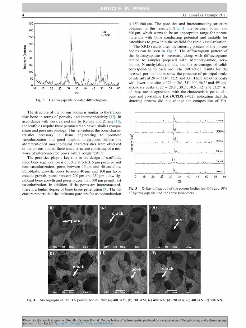
Fig. 3 Hydroxyapatite powder diffractogram.
Fig. 5 X-Ray diffraction of the porous bodies for 40% and 50%
of hydroxyapatite and the three monomers.
4 J.I. Gonzalez Ocampo et al.
The structure of the porous bodies is similar to the trabec-ular bone in terms of porosity and interconnectivity [17]. Inaccordance with work carried out by Ramay and Zhang [11],
the scaffolds require these parameters to have a similar compo-sition and pore morphology. This reproduces the bone charac-teristics necessary in tissue engineering to promote
vascularization and good implant integration. Before theaforementioned morphological characteristics were observedin the porous bodies, there was a structure consisting of a net-
work of interconnected pores with a rough texture.The pore size plays a key role in the design of scaffolds,
since bone regeneration is directly affected. 5 lm pores permit
new vascularization, pores between 15 lm and 40 lm allowfibroblastic growth, pores between 40 lm and 100 lm favorosteoid growth, pores between 200 lm and 350 lm allow sig-nificant bone growth and pores bigger than 500 lm permit fast
vascularization. In addition, if the pores are interconnected,there is a higher degree of bone tissue penetration [9]. The lit-erature reports that the optimum pore size for osteoconduction
Fig. 4 Micrographs of the HA porous bodies, 50·: (a) 40HAM,
Please cite this article in press as: Gonzalez Ocampo JI et al., Porous bodies of hydmethods, J Adv Res (2015), http://dx.doi.org/10.1016/j.jare.2015.06.006
is 150–600 lm. The pore size and interconnecting structureobtained in this research (Fig. 4) are between 50 lm and600 lm, which seems to be an appropriate range for porous
materials with bone conducting potential and suitable forosteoblasts to grow into the scaffold for rapid vascularization.
The XRD results after the sintering process of the porous
bodies can be seen in Fig. 5. The diffractogram pattern ofthe hydroxyapatite is presented along with diffractogramsrelated to samples prepared with: Methacrylamide, acry-
lamide, N-methylolacrylamide, and the percentages of solidscorresponding to each one. The diffraction results for theassessed porous bodies show the presence of principal peaksof intensity at 2h = 31.6�, 32.2� and 33�. There are other peakswith lower intensities of 2h = 26�, 34�, 40�, 46.5� and 49� andsecondary peaks at 2h = 28.8�, 39.2�, 50.5�, 52� and 53.2�. Allof these are in agreement with the characteristic peaks of a
pure and crystalline HA (JCPDS 9-432), indicating that thesintering process did not change the composition of HA.
(b) 50HAM, (c) 40HAA, (d) 50HAA, (e) 40HAN, (f) 50HAN.
roxyapatite produced by a combination of the gel-casting and polymer sponge
Porous bodies of hydroxyapatite 5
Peaks for the 50HAA and 50HAN samples have a displace-ment relative to the standard conditions (JCPDS 9-432).However, the relative intensity of peaks and crystallinity
remain. This error in the position of the diffraction peaksmay be due to movement of the sample relative to the axisof the diffractometer. Nevertheless, after thermal treatment
all samples can be considered to have HA as a single phase.The other chemical reagents used in this process were elimi-nated during firing, and the sintered material is potentially
non-toxic to living tissues. This allows the material to be usedfor biomedical applications.
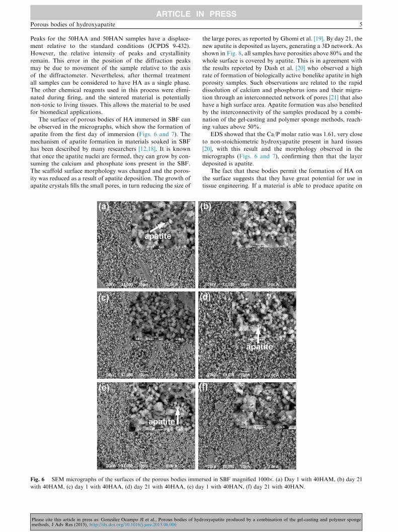
The surface of porous bodies of HA immersed in SBF canbe observed in the micrographs, which show the formation of
apatite from the first day of immersion (Figs. 6 and 7). Themechanism of apatite formation in materials soaked in SBFhas been described by many researchers [12,18]. It is known
that once the apatite nuclei are formed, they can grow by con-suming the calcium and phosphate ions present in the SBF.The scaffold surface morphology was changed and the poros-
ity was reduced as a result of apatite deposition. The growth ofapatite crystals fills the small pores, in turn reducing the size of
Fig. 6 SEM micrographs of the surfaces of the porous bodies imme
with 40HAM, (c) day 1 with 40HAA, (d) day 21 with 40HAA, (e) da
Please cite this article in press as: Gonzalez Ocampo JI et al., Porous bodies of hydmethods, J Adv Res (2015), http://dx.doi.org/10.1016/j.jare.2015.06.006
the large pores, as reported by Ghomi et al. [19]. By day 21, thenew apatite is deposited as layers, generating a 3D network. Asshown in Fig. 8, all samples have porosities above 80% and the
whole surface is covered by apatite. This is in agreement withthe results reported by Dash et al. [20] who observed a highrate of formation of biologically active bonelike apatite in high
porosity samples. Such observations are related to the rapiddissolution of calcium and phosphorus ions and their migra-tion through an interconnected network of pores [21] that also
have a high surface area. Apatite formation was also benefitedby the interconnectivity of the samples produced by a combi-nation of the gel-casting and polymer sponge methods, reach-ing values above 50%.
EDS showed that the Ca/P molar ratio was 1.61, very closeto non-stoichiometric hydroxyapatite present in hard tissues[20], with this result and the morphology observed in the
micrographs (Figs. 6 and 7), confirming then that the layerdeposited is apatite.
The fact that these bodies permit the formation of HA on
the surface suggests that they have great potential for use intissue engineering. If a material is able to produce apatite on
rsed in SBF magnified 1000·. (a) Day 1 with 40HAM, (b) day 21
y 1 with 40HAN, (f) day 21 with 40HAN.
roxyapatite produced by a combination of the gel-casting and polymer sponge

Fig. 7 SEM Micrographs of the surfaces of the porous bodies immersed in SBF magnified 1000·. (a) Day 1 with 50HAM, (b) day 21
with 50HAM, (c) day 1 with 50HAA, (d) day 21 with 50HAA, (e) day 1 with 50HAN, (f) day 21 with 50HAN.
Fig. 8 Total porosity versus the amount of hydroxyapatite for
the three types of monomers.
6 J.I. Gonzalez Ocampo et al.
Please cite this article in press as: Gonzalez Ocampo JI et al., Porous bodies of hydmethods, J Adv Res (2015), http://dx.doi.org/10.1016/j.jare.2015.06.006
the surface through SBF, it will also be possible to produceapatite on an implant inside the human body, in turn aidingthe chemical interaction between the implant and the host tis-sue [12]. Numerous studies have shown that apatite formation
on the surface of materials is indicative of a bioactivity poten-tial in vivo [22].
Some monomers could potentially irritate the human body.
Therefore, in order for stable components to be obtained, it isnecessary that the monomers undergo complete polymeriza-tion and curing [23]. In the case of foam processing, due to
low evaporation point of polyurethane foam and monomers,they should volatilize during sintering in concordance withRamay and Zhang [11], who report burn temperature for poly-
urethane foam as 500 �C and with Callcut and Knowles [24]who reported a temperature at 600 �C. XRD (Fig. 5) showsthat HA is the only phase present. The monomers were volati-lized, which suggests that there will be an appropriate response
in future biocompatibility tests performed to confirm the use ofthis material as an implant.
roxyapatite produced by a combination of the gel-casting and polymer sponge
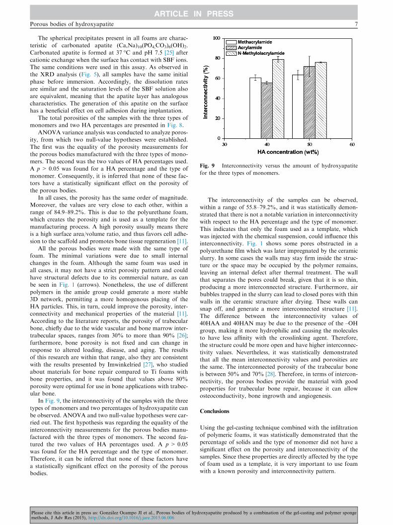
Fig. 9 Interconnectivity versus the amount of hydroxyapatite
for the three types of monomers.
Porous bodies of hydroxyapatite 7
The spherical precipitates present in all foams are charac-teristic of carbonated apatite (Ca,Na)10(PO4,CO3)6(OH)2.Carbonated apatite is formed at 37 �C and pH 7.5 [25] after
cationic exchange when the surface has contact with SBF ions.The same conditions were used in this assay. As observed inthe XRD analysis (Fig. 5), all samples have the same initial
phase before immersion. Accordingly, the dissolution ratesare similar and the saturation levels of the SBF solution alsoare equivalent, meaning that the apatite layer has analogous
characteristics. The generation of this apatite on the surfacehas a beneficial effect on cell adhesion during implantation.
The total porosities of the samples with the three types ofmonomers and two HA percentages are presented in Fig. 8.
ANOVA variance analysis was conducted to analyze poros-ity, from which two null-value hypotheses were established.The first was the equality of the porosity measurements for
the porous bodies manufactured with the three types of mono-mers. The second was the two values of HA percentages used.A p > 0.05 was found for a HA percentage and the type of
monomer. Consequently, it is inferred that none of these fac-tors have a statistically significant effect on the porosity ofthe porous bodies.
In all cases, the porosity has the same order of magnitude.Moreover, the values are very close to each other, within arange of 84.9–89.2%. This is due to the polyurethane foam,which creates the porosity and is used as a template for the
manufacturing process. A high porosity usually means thereis a high surface area/volume ratio, and thus favors cell adhe-sion to the scaffold and promotes bone tissue regeneration [11].
All the porous bodies were made with the same type offoam. The minimal variations were due to small internalchanges in the foam. Although the same foam was used in
all cases, it may not have a strict porosity pattern and couldhave structural defects due to its commercial nature, as canbe seen in Fig. 1 (arrows). Nonetheless, the use of different
polymers in the amide group could generate a more stable3D network, permitting a more homogenous placing of theHA particles. This, in turn, could improve the porosity, inter-connectivity and mechanical properties of the material [11].
According to the literature reports, the porosity of trabecularbone, chiefly due to the wide vascular and bone marrow inter-trabecular spaces, ranges from 30% to more than 90% [26];
furthermore, bone porosity is not fixed and can change inresponse to altered loading, disease, and aging. The resultsof this research are within that range, also they are consistent
with the results presented by Imwinkelried [27], who studiedabout materials for bone repair compared to Ti foams withbone properties, and it was found that values above 80%porosity were optimal for use in bone applications with trabec-
ular bone.In Fig. 9, the interconnectivity of the samples with the three
types of monomers and two percentages of hydroxyapatite can
be observed. ANOVA and two null-value hypotheses were car-ried out. The first hypothesis was regarding the equality of theinterconnectivity measurements for the porous bodies manu-
factured with the three types of monomers. The second fea-tured the two values of HA percentages used. A p > 0.05was found for the HA percentage and the type of monomer.
Therefore, it can be inferred that none of these factors havea statistically significant effect on the porosity of the porousbodies.
Please cite this article in press as: Gonzalez Ocampo JI et al., Porous bodies of hydmethods, J Adv Res (2015), http://dx.doi.org/10.1016/j.jare.2015.06.006
The interconnectivity of the samples can be observed,within a range of 55.8–79.2%, and it was statistically demon-
strated that there is not a notable variation in interconnectivitywith respect to the HA percentage and the type of monomer.This indicates that only the foam used as a template, which
was injected with the chemical suspension, could influence thisinterconnectivity. Fig. 1 shows some pores obstructed in apolyurethane film which was later impregnated by the ceramic
slurry. In some cases the walls may stay firm inside the struc-ture or the space may be occupied by the polymer remains,leaving an internal defect after thermal treatment. The wallthat separates the pores could break, given that it is so thin,
producing a more interconnected structure. Furthermore, airbubbles trapped in the slurry can lead to closed pores with thinwalls in the ceramic structure after drying. These walls can
snap off, and generate a more interconnected structure [11].The difference between the interconnectivity values of40HAA and 40HAN may be due to the presence of the –OH
group, making it more hydrophilic and causing the moleculesto have less affinity with the crosslinking agent. Therefore,the structure could be more open and have higher interconnec-
tivity values. Nevertheless, it was statistically demonstratedthat all the mean interconnectivity values and porosities arethe same. The interconnected porosity of the trabecular boneis between 50% and 70% [28]. Therefore, in terms of intercon-
nectivity, the porous bodies provide the material with goodproperties for trabecular bone repair, because it can allowosteoconductivity, bone ingrowth and angiogenesis.
Conclusions
Using the gel-casting technique combined with the infiltration
of polymeric foams, it was statistically demonstrated that thepercentage of solids and the type of monomer did not have asignificant effect on the porosity and interconnectivity of the
samples. Since these properties are directly affected by the typeof foam used as a template, it is very important to use foamwith a known porosity and interconnectivity pattern.
roxyapatite produced by a combination of the gel-casting and polymer sponge
8 J.I. Gonzalez Ocampo et al.
The porous bodies internally possess macro-pores andmicro-pores. The pore size is between 100 and 600 lm andthe internal pore size is between 50 and 100 lm. This was cor-
roborated by porosity and interconnectivity results that showedvalues from 84.9% to 89.2% and 55.8% to 79.2% respectively,which are very similar to the values of the trabecular bone.
The behavior exhibited by all of the porous bodies confirmstheir bioactivity. There were no monomers in the final foamswhich could generate sensitization and irritation reactions.
This demonstrates the great potential that these bodies havefor use in tissue engineering.
Conflict of interest
The authors have declared no conflict of interest.
Compliance with ethics requirements
This article does not contain any studies with human or animal
subjects.
Acknowledgments
The authors of this research thank the University of Antioquia-CODI- Research Committee for funding the development of
the project: ‘‘Synthesis and Characterization of PorousHydroxyapatite Bodies Obtained by Different ProductionTechniques’’. They also wish to thank the company
Colorquımica S.A. for donating the monomers.
References
[1] Villora JM, Callejas P, Barba MF. Metodos de sıntesis y
comportamiento termico del Hidroxiapatito. Boletın La Soc
Espanola Ceramica Y Vidr 2002;41:443–50.
[2] Zyman Z, Glushko V. Nonstoichiometric hydroxyapatite
granules for orthopaedic applications. Sci Mater Med
2004;15:551–8.
[3] Rodrıguez R, Gomez J, Rodrıguez R, Blardoni F. Biomaterial
de restauracion osea. Rev Cuba Investig Biomedica
1999;18:203–7.
[4] Vallecillo M, Romero N, Pardo A. La hidroxiapatita en
reconstruccion de defectos oseos de los maxilares: estudio y
seguimiento de 15 casos clınicos. Rev COE 1999;4:137–43.
[5] Black J, Hastings G. Handbook of biomaterials properties. 1st
ed. Londres: Chapman & Hall; 1998.
[6] Dhara S, Kamboj R, Pradhan M, Bhargava P. Shape forming of
ceramics via gel-casting of aqueous particulate slurries. Bull
Mater Sci 2002;25:565–8.
[7] Sepulveda P. Processing of cellular ceramics synthesized by
Gelcasting of foams. University of Nottingham; 1996.
[8] Dutta T, Simon J, Ricci J, Rekow E, Thompson V. Performance
of hydroxyapatite bone repair scaffolds created via three-
dimensional fabrication techniques. J Biomed Mater Res A
2003;67A:1228–37.
[9] Cunningham E, Dunne N, Walker G, Maggs C, Wilcox R,
Buchanan F. Hydroxyapatite bone substitutes developed via
Please cite this article in press as: Gonzalez Ocampo JI et al., Porous bodies of hydmethods, J Adv Res (2015), http://dx.doi.org/10.1016/j.jare.2015.06.006
replication of natural marine sponges. J Mater Sci Mater Med
2010;21:2255–61.
[10] Sepulveda P, Binner JG. Processing of cellular ceramics by
foaming and in situ polymerisation of organic monomers. J Eur
Ceram Soc 1999;19:2059–66.
[11] Ramay HR, Zhang M. Preparation of porous hydroxyapatite
scaffolds by combination of the gel-casting and polymer sponge
methods. Biomaterials 2003;24:3293–302.
[12] Kokubo T, Takadama H. How useful is SBF in predicting
in vivo bone bioactivity? Biomaterials 2006;27:2907–15.
[13] Liu J, Miao X. Porous alumina ceramics prepared by slurry
infiltration of expanded polystyrene beads. J Mater Sci
2005;40:6145–50.
[14] Londono E, Echavarrıa A, De La Calle F. Caracterısticas
cristaloquımicas de la hidroxiapatita sintetica tratada a
diferentes temperaturas. Rev EIA 2006:109–18.
[15] Navarro ME. Desarrollo y Caracterizacion de Materiales
Biodegradables para Regeneracion Osea. Universidad
Politecnica de Cataluna; 2005.
[16] Teixeira S, Rodriguez M a, Pena P, De Aza a H, De Aza S,
Ferraz MP, et al. Physical characterization of hydroxyapatite
porous scaffolds for tissue engineering. Mater Sci Eng C 2009;
29: 1510–4.
[17] Montufar EB. Espumas inyectables de hidroxiapatita obtenidas
por el metodo de espumado de la fase lıquida de un cemento de
fosfato tricalcico alfa. Universidad Politecnica de Cataluna;
2010.
[18] Kim H-M, Himeno T, Kokubo T, Nakamura T. Process and
kinetics of bonelike apatite formation on sintered
hydroxyapatite in a simulated body fluid. Biomaterials
2005;26:4366–73.
[19] Ghomi H, Fathi MH, Edris H. Effect of the composition of
hydroxyapatite/bioactive glass nanocomposite foams on their
bioactivity and mechanical properties. Mater Res Bull
2012;47:3523–32.
[20] Dash SR, Sarkar R, Bhattacharyya S. Gel casting of
hydroxyapatite with naphthalene as pore former. Ceram Int
2014:1–16.
[21] Swain SK, Bhattacharyya S, Sarkar D. Preparation of porous
scaffold from hydroxyapatite powders. Mater Sci Eng C
2011;31:1240–4.
[22] Ducheyne P, Qiu Q. Bioactive ceramics : the effect of surface
reactivity on bone formation and bone cell function.
Biomaterials 1999;20.
[23] Janney MA, Omatete OO, Walls CA, Nunn SD, Ogle RJ,
Westmoreland G. Development of low-toxicity gelcasting
systems. J Am Ceram Soc 1998;81:581–91.
[24] Callcut S, Knowles JC. Correlation between structure and
compressive strength in a reticulated glass-reinforced
hydroxyapatite foam 2002;3:485–9.
[25] LeGeros RZ. Calcium phosphates in oral biology and medicine.
Monogr Oral Sci 1991;15.
[26] Bonucci E. Basic structure and composition of bone. In:
Mechanical testing of bone and the bone-implant interface. In:
An YH, Draughn RA, editors. Basic compos struct bone. Boca
Raton: CRC Press; 2000. p. 3–22.
[27] Imwinkelried T. Mechanical properties of open-pore titanium
foam. J Biomed Mater Res Part A 2007;81(4):964–70.
[28] Jones JR, Hench LL. Regeneration of trabecular bone using
porous ceramics. Curr Opin Solid State Mater Sci 2003;7:
301–7.
roxyapatite produced by a combination of the gel-casting and polymer sponge
Related Documents